One stop doc renal and urinary system and electrolyte bakalis, spyros, stamoulos, panos
Bạn đang xem bản rút gọn của tài liệu. Xem và tải ngay bản đầy đủ của tài liệu tại đây (962.54 KB, 129 trang )
ONE STOP DOC
Renal and Urinary
System and
Electrolyte Balance
One Stop Doc
Titles in the series include:
Cardiovascular System – Jonathan Aron
Editorial Advisor – Jeremy Ward
Cell and Molecular Biology – Desikan Rangarajan and David Shaw
Editorial Advisor – Barbara Moreland
Endocrine and Reproductive Systems – Caroline Jewels and Alexandra Tillett
Editorial Advisor – Stuart Milligan
Gastrointestinal System – Miruna Canagaratnam
Editorial Advisor – Richard Naftalin
Musculoskeletal System – Wayne Lam, Bassel Zebian and Rishi Aggarwal
Editorial Advisor – Alistair Hunter
Nervous System – Elliott Smock
Editorial Advisor – Clive Coen
Nutrition and Metabolism – Miruna Canagaratnam and David Shaw
Editorial Advisors – Barbara Moreland and Richard Naftalin
Respiratory System – Jo Dartnell and Michelle Ramsay
Editorial Advisor – John Rees
ONE STOP DOC
Renal and Urinary
System and
Electrolyte Balance
Panos Stamoulos MBBS BSc(Hons)
Pre-Registration House Officer, Conquest Hospital, Hastings, East Sussex, UK
Spyridon Bakalis MBBS BSc(Hons)
Pre-Registration House Officer, William Harvey Hospital, Ashford, Kent, UK
Editorial Advisors
Alistair Hunter BSc(Hons) PhD
Senior Lecturer, Guy’s, King’s and St Thomas’ School of Biomedical Sciences, King’s College
London, London, UK
Richard Naftalin MB ChB MSc PhD DSc
Professor of Epithelial Physiology, King’s College, London and Guy’s Campus Centre for
Vascular Biology and Medicine, London, UK
Series Editor
Elliott Smock BSc(Hons)
Fifth year medical student, Guy’s, King’s and St Thomas’ Medical School, London, UK
Contributing Author
Megan Morris BSc(Hons)
Fifth year medical student, Guy’s, King’s and St Thomas’ Medical School, London, UK
A MEMBER OF THE HODDER HEADLINE GROUP
First published in Great Britain in 2005 by
Hodder Education, a member of the Hodder Headline Group,
338 Euston Road, London NW1 3BH
Distributed in the United States of America by
Oxford University Press Inc.,
198 Madison Avenue, New York, NY10016
Oxford is a registered trademark of Oxford University Press
© 2005 Edward Arnold (Publishers) Ltd
All rights reserved. Apart from any use permitted under UK copyright law,
this publication may only be reproduced, stored or transmitted, in any form,
or by any means with prior permission in writing of the publishers or in the
case of reprographic production in accordance with the terms of licences
issued by the Copyright Licensing Agency. In the United Kingdom such
licences are issued by the Copyright Licensing Agency: 90 Tottenham Court
Road, London W1T 4LP.
Whilst the advice and information in this book are believed to be true and
accurate at the date of going to press, neither the author[s] nor the publisher
can accept any legal responsibility or liability for any errors or omissions
that may be made. In particular, (but without limiting the generality of the
preceding disclaimer) every effort has been made to check drug dosages;
however it is still possible that errors have been missed. Furthermore,
dosage schedules are constantly being revised and new side-effects
recognized. For these reasons the reader is strongly urged to consult the
drug companies’ printed instructions before administering any of the drugs
recommended in this book.
British Library Cataloguing in Publication Data
A catalogue record for this book is available from the British Library
Library of Congress Cataloging-in-Publication Data
A catalog record for this book is available from the Library of Congress
ISBN-10: 0 340 885076
ISBN-13: 978 0 340 88507 9
1 2 3 4 5 6 7 8 9 10
Commissioning Editor: Georgina Bentliff
Project Editor: Heather Smith
Production Controller: Jane Lawrence
Cover Design: Amina Dudhia
Illustrations: Cactus Design
Typeset in 10/12pt Adobe Garamond/Akzidenz GroteskBE by Servis Filmsetting Ltd, Manchester
Printed and bound in Spain
Hodder Headline’s policy is to use papers that are natural, renewable and recyclable
Products and made from wood grown in sustainable forests. The logging and manufacturing processes are
expected to conform to the environmental regulations of the country of origin.
What do you think about this book? Or any other Hodder Arnold title?
Please visit our website at www.hoddereducation.co.uk
CONTENTS
PREFACE
vi
ABBREVIATIONS
viii
SECTION 1
THE KIDNEYS
SECTION 2
THE URINARY TRACT
49
SECTION 3
ELECTROLYTES
65
INDEX
1
109
PREFACE
From the Series Editor, Elliott Smock
From the author, Panos Stamoulos
Are you ready to face your looming exams? If you
have done loads of work, then congratulations; we
hope this opportunity to practise SAQs, EMQs,
MCQs and Problem-based Questions on every part
of the core curriculum will help you consolidate what
you’ve learnt and improve your exam technique. If
you don’t feel ready, don’t panic – the One Stop Doc
series has all the answers you need to catch up and
pass.
I decided to write this book after a colleague of mine
invited me to participate in a series of books directed
at medical students. I started writing it while I was
still a medical student, after considering the current
demands put on medical students by the current
medical curriculum. I also used my experience as a
medical tutee to tune it to a form that will be both
appealing and easily absorbed for exam purposes.
This book is not directed at replacing the standard
textbook; its purpose is to challenge students
academically and prepare them for their exams using
an integrated approach towards all the key topics
pertaining to the renal system.
There are only a limited number of questions an
examiner can throw at a beleaguered student and this
text can turn that to your advantage. By getting
straight into the heart of the core questions that come
up year after year and by giving you the model
answers you need, this book will arm you with the
knowledge to succeed in your exams. Broken down
into logical sections, you can learn all the important
facts you need to pass without having to wade
through tons of different textbooks when you simply
don’t have the time. All questions presented here are
‘core’; those of the highest importance have been
highlighted to allow even sharper focus if time for
revision is running out. In addition, to allow you to
organize your revision efficiently, questions have been
grouped by topic, with answers supported by detailed
integrated explanations.
On behalf of all the One Stop Doc authors I wish
you the very best of luck in your exams and hope
these books serve you well!
Writing a book is a long and demanding process. It
requires determination and perseverance to reach a
form that will satisfy its goals, its author and its
readers. I have watched it grow day by day and I am
pleased to say that my work has been successful as
well as fulfilling.
I thank Professor Naftalin and Dr Hunter for their
invaluable input and advice during the birth of this
book. I would also like to thank Elliott for trusting
me with this work and his patience. I would like to
dedicate this book to my parents and my godparents
as a small token of appreciation for their support and
sacrifice throughout my medical course
Preface
From the author, Spyridon Bakalis
‘Whatever does not spring from a man’s free choice,
or is only the result of instruction and guidance, does
not enter into his very being, but still remains alien
to his true nature; he does not perform it with truly
human energies, but merely with mechanical exactness’.
Karl Wilhelm Von Humboldt
vii
I would like to thank the following people for the
help, patience and advice: My family, Panos,
Katerina, Zacharoula, Maria, Eleni and Petros, my
co-author Panos and advisors Professor Richard
Naftalin and Dr Alistair Hunter. Finally my friends
who supported me throughout my medical years:
George, Neil, Asim, Thanos, Vasanthan, Alex and
Richard the house officers at WHH, and to all those
I have no space for (I know who you are). Finally, to
Heather who may have kept me out of trouble.
ABBREVIATIONS
ADP
ACE
ADH
ANP
ATP
COPD
CT
DCT
DNA
DT
ECF
ECG
ECV
adenosine diphosphate
angiotensin converting enzyme
anti-diuretic hormone
atrial natriuretic peptide
adenosine triphosphate
chronic obstructive pulmonary
disorder
collecting tubules
distal convoluted tubule
deoxyribonucleic acid
distal tubule
extracellular fluid
electrocardiogram
effective circulating volume
ECV
ENaC
GFR
ICF
ISF
JGA
MAP
1,25[OH]2D3
PAH
PCT
PTH
RBF
RNA
RPF
extracellular volume;
epithelial Na+ conduction channels
glomerular filtration rate
intracellular fluid
interstitial fluid
juxtaglomerular apparatus
mean arterial pressure
1,25-dihydroxyvitamin D3
para-aminohippurate
proximal convoluted tubule
parathyroid hormone
renal blood flow
ribonucleic acid
renal plasma flow rate
SECTION
1
THE KIDNEYS
• THE ANATOMY OF THE KIDNEY (i)
2
• THE ANATOMY OF THE KIDNEY (ii)
4
• THE RENAL MICROCIRCULATION (i)
6
• THE RENAL MICROCIRCULATION (ii)
8
• THE NEPHRON
10
• THE RENAL CORPUSCLE (i)
12
• THE RENAL CORPUSCLE (ii)
14
• THE PROXIMAL CONVOLUTED TUBULE (i)
16
• THE PROXIMAL CONVOLUTED TUBULE (ii)
18
• THE LOOP OF HENLE
20
• THE RENAL CONCENTRATION MECHANISM
22
• THE DISTAL CONVOLUTED TUBULE
24
• THE JUXTAGLOMERULAR APPARATUS AND
THE MESANGIUM
26
• THE COLLECTING TUBULES
28
• RENAL CLEARANCE
30
• RENAL BLOOD FLOW
32
• LOOP DIURETICS – RENAL ACTIONS AND
SIDE EFFECTS
34
• THIAZIDE DIURETICS – RENAL ACTIONS
AND SIDE EFFECTS
36
• K+-SPARING AND OSMOTIC DIURETICS –
RENAL ACTIONS AND SIDE EFFECTS
38
• GLOMERULONEPHRITIS
40
• ACUTE TUBULAR NEPHROSIS
42
• ACUTE RENAL FAILURE
44
• CHRONIC RENAL FAILURE
46
1
SECTION
THE KIDNEYS
1. Label the following diagram showing the gross anatomy of the kidney. Each option can
be used once, more than once or not at all
Options
1.
2.
3.
4.
5.
6.
7.
8.
9.
10.
Medulla
Cortex
Ureter
Renal pelvis
Adrenal gland
Pyramid
Major calyx
Minor calyx
Renal capsule
Renal hilum
A
I
B
C
D
G
E
F
H
2. In the anatomy of the kidney
a.
b.
c.
d.
e.
The inner part of the kidney is called the cortex
Nephrons are found in the medulla and cortex of the kidney
The pyramids are only found in the medulla
Collecting ducts are found in the pyramids only
Each kidney has four major calyces
3. Concerning the surface anatomy of the kidney
a. The subcostal plane is the surface marking used for locating the kidneys
b. The left kidney is higher than the right kidney
c. The inferior pole of the right kidney is about a fingerbreadth above the posterior iliac
crest
d. The inferior pole of the right kidney is usually palpable
e. The hilum of the left kidney lies 10 cm from the median plane
The kidneys
3
EXPLANATION: THE ANATOMY OF THE KIDNEY (i)
The kidneys are paired, retroperitoneal organs that act as filters and control H2O, electrolyte and acid–base
balance homeostasis. They also have an important endocrine role.
Each kidney is made up from an outer cortex and inner medulla. The most important structural component
of the kidney is the nephron. These are found in both the cortex and medulla; however, the renal corpuscle
component of the nephron is only found in the cortex. The medulla contains the collecting ducts, which are
concentrated in the pyramids. The pyramids are ordered so that their apical ends empty urine into the minor
calyces, which in turn drain into a major calyx. There are three major calyces in each kidney. These drain into
the renal pelvis, through the ureter, then down into the bladder.
Medulla
Adrenal gland
Cortex
Minor calyx
Renal hilum
Renal pelvis
Major calyx
Renal pyramid
Ureter
Renal capsule
The transpyloric plane is the surface marking used to locate the kidneys. It is halfway between the suprasternal notch and the pubis at the level of L1 and passes through hilum of the left kidney (which lies 5 cm
from the median plane) and the superior pole of the right kidney. The superior poles of the left and right
kidneys lie deep to the eleventh and twelfth ribs respectively (the right kidney is lower than the left kidney –
it is pushed down by the liver which sits above it). The inferior pole of the right kidney is a fingerbreadth above
the posterior iliac crest and is usually palpable except if the patient is obese.
Median plane
Left
Right
Transpyloric plane
11th rib
12th rib
Iliac crest
Posterior view
Answers
1. 1 – B, 2 – A, 3 – H, 4 – G, 5 – I, 6 – C, 7 – E, 8 – F, 9 – D, 10 – J
2. F T T T F
3. F F T T F
ONE STOP DOC
4
4. For each of the following choose the one correct answer:
Options
A.
B.
C.
D.
E.
F.
G.
H.
I.
Diaphragm
Quadratus lumborum muscle
Pancreas
Second part of the duodenum
First part of the duodenum
Third part of the duodenum
Liver
Aorta
Inferior vena cava
1.
2.
3.
4.
5.
6.
7.
8.
Lies superior to the right kidney
Lies superior to the left kidney
Lies posterior to the right kidney
Lies posterior to the left kidney
Lies anterior to the right kidney
Lies anterior to the left kidney
Lies medial to the right kidney
Lies medial to the left kidney
5. The kidneys
a.
b.
c.
d.
e.
Are 10 cm long by 5 cm wide and 2.5 cm deep
Lie between T10 and L3
Are retroperitoneal organs
Have superior poles that are both in the same transverse plane
Are positioned so their long axes are oblique
6. Concerning the kidney
a.
b.
c.
d.
e.
The kidneys move about 5 cm during respiration
The collagenous capsule around the kidney readily expands
The kidneys are in direct contact with the eleventh and twelfth ribs
The perinephric fat helps holds the kidney in place
The renal fascia is the kidney’s attachment to the diaphragm
The kidneys
5
EXPLANATION: THE ANATOMY OF THE KIDNEY (ii)
The relations to the kidneys are as follows: the diaphragm lies superior to both kidneys. The diaphragm, the
quadratus lumborum, the psoas, the transversus abdominis, the twelfth rib and the three nerves (the subcostal,
iliohypogastric and ilio-inguinal) lie posterior to the right kidney. The quadratus lumborum lies posterior to
the left kidney. The liver, second part of the duodenum and the ascending colon lie anterior to the right
kidney. The stomach, the pancreas and its vessels, the spleen, the jejunum and the descending colon lie anterior to the left kidney. The inferior vena cava lies medial to the right kidney and the aorta lies medial to the
left kidney.
On entering the hilum the renal vein lies anterior to the renal artery, which is anterior to the renal pelvis.
Renal arteries arise at L1 to L2. The right renal artery, which is longer, passes posterior to the inferior vena
cava. The renal artery splits to form an anterior and a posterior branch.
Right kidney
Diaphragm
Transverse
abdominis
Left kidney
Subcostal nerve
Iliohypogastric
ilioinguginal nerve
Spleen
Transversus abdominis
Inferior vena cava
Aorta
Quadratus
lumborum
Psoas
Each kidney is around 10 cm long, 5 cm wide and 2.5 cm deep, weighing about 150 g and lies between T12
and L3. Their long axes are oblique as the superior poles of the kidneys lie medially to the inferior poles.
The kidneys move only 3 cm on respiration; the movement comes from the superior-lying diaphragm. The
surrounding collagenous capsule does not expand, therefore inflammation of the kidney may cause an
increase in pressure within the kidney. The kidneys are separated from the eleventh and twelfth ribs by the
diaphragm, though they are both related to both ribs. The perinephric fat surrounds the kidney, and lies
outside the renal capsule, but inside the renal fascia. It provides protection from trauma. The tough renal fascia
blends with the diaphragmatic fascia.
Answers
4. 1 – A, 2 – A, 3 – B, 4 – B, 5 – D, 6 – C, 7 – I, 8 – H
5. T F T F T
6. F F F T T
ONE STOP DOC
6
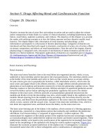
7. Label the diagram below of the renal microcirculation
7 3 4 1
6
2
8
5
Renal vein
Renal artery
Options
A.
B.
C.
D.
E.
F.
G.
H.
Afferent arteriole
Glomerulus
Arcuate artery
Efferent arteriole
Interlobar artery
Vasa recta
Peritubular capillaries
Interlobular artery
8. Concerning the renal microcirculation
a.
b.
c.
d.
The glomerular capillaries have a different structure to other capillaries
The renal capillaries are all made of four cell layers
The renal vasculature contains three different capillary systems
The glomerular capillary system of the kidneys works at a higher intraluminal pressure
than in any other organ system
e. To optimize glomerular filtration, the transluminal pressure in the glomerulus is always
the same
The kidneys
7
EXPLANATION: THE RENAL MICROCIRCULATION (i)
The renal artery flows into the kidney and immediately branches into the interlobar arteries. They then run
through the medulla, and sub-divide into the arcuate arteries, which then divide into interlobular arteries.
Efferent
arteriole
Peritubular
capillaries
Glomerulus
Afferent arteriole
Interlobular artery
Arcuate
artery
Interlobar artery
Vasa recta
Renal vein
Renal artery
The afferent arterioles branch off the interlobular arteries and run into the cortex and form the glomerulus,
the efferent arteriole, peritubular capillaries and vasa recta.
The renal capillary system in the kidneys is different and more complex from that in other organs. The differences are:
1. Capillaries are made from a single layer of endothelium, but in the kidney they are made of four layers:
endothelial cells internally, sitting on a basement membrane (not cells), surrounded by special epithelial cells
called podocytes (which have pores), which are in turn surrounded by a set of Bowman’s capsule cells.
2. The renal capillary system consists of three sub-capillary systems: the glomerulus, the cortical peritubular
capillaries, and the vasa recta.
3. The intraluminal pressure in the glomerulus is about twice as high as that in other capillaries, i.e.
50 mmHg. This can be varied and aids filtration.
Answers
7. 1 – A, 2 – C, 3 – D, 4 – B, 5 – E, 6 – H, 7 – G, 8 – F
8. T F T T F
ONE STOP DOC
8
9. Concerning the renal microcirculation
a. The glomerulus receives blood from the efferent arteriole
b. Twenty per cent of blood flowing into the glomerulus flows out through the efferent
arterioles
c. The afferent arterioles from superficial nephrons run mainly in the medulla
d. The peritubular capillaries absorb and secrete substances from the kidney tubules
e. The efferent arterioles of superficial nephrons form the vasa recta
10. Concerning the renal microcirculation
a.
b.
c.
d.
e.
The vasa recta lie in the cortex of the kidney
The vasa recta absorb substances from the loop of Henle
The descending wall of the vasa recta releases H2O into the interstitium
The descending wall of the vasa recta releases Na+ and Cl− into the interstitium
The ascending wall of the vasa recta loses Na+ and Cl− into the interstitium
The kidneys
9
EXPLANATION: THE RENAL MICROCIRCULATION (ii)
Blood flows through the afferent arteriole, into the glomerulus and then out through the efferent arteriole.
Twenty per cent of blood is filtered off by the glomerulus, whilst the remaining 80 per cent passes through
to the efferent arteriole. The efferent arterioles (which have a smaller diameter than afferent arterioles) from
different types of nephrons then form two further capillary networks.
Macula densa
Afferent arteriole
Renal cortex
Descending limb
Squamous
epithelium
Thin ascending
limb
Renal medulla
Thick ascending limb
Cuboidal
epithelium
Vasa recta
Efferent arterioles from superficial nephrons surround the tubular parts of the nephrons (hence the name
peritubular capillaries) in the cortex, and are involved in nutrient transfer, removing reabsorbed H2O and
solutes and transporting substances to the tubules for secretion.
Efferent arterioles from juxtamedullary nephrons penetrate into the medulla, perform a U-turn and then run
back up into the cortex. These arterioles form the vasa recta. Along their whole length the vasa recta are in
close proximity to the loop of Henle and reabsorb substances from the loop (countercurrent multiplier).
Permeability of the vasa recta to solutes in the loop of Henle varies: the descending arterial side excretes H2O
into the interstitium and absorbs Na+ and Cl−, whilst the ascending venous loop excretes Na+ and Cl− and
absorbs H2O.
The venous microcirculation of the kidneys mirrors that of the arterial.
Answers
9. F F F T F
10. F T T F T
ONE STOP DOC
10
11. Label the diagram below of a nephron. Each option can be used once, more than once,
or not at all
B
A
K
F
J
N
G
M
E
B C H
I
D
P
O
Options
1.
2.
3.
4.
5.
6.
7.
8.
9.
10.
11.
12.
13.
14.
15.
16.
Afferent arteriole
Bowman’s capsule
Distal convoluted tubule
Bowman’s space
Efferent arteriole
Cortex
Collecting duct
Glomerulus
Proximal convoluted tubule
Thick ascending loop of Henle
Medulla
Proximal straight tubule
Renal corpuscle
Thick ascending loop of Henle
Thin ascending loop of Henle
Thin descending loop of Henle
12. Concerning the nephron
a.
b.
c.
d.
e.
Each kidney has about half a million nephrons
Sixty per cent of the nephrons are juxtamedullary
The renal corpuscle lies in the cortex
The distal convoluted tubules lie in the medulla
The juxtamedullary nephrons have shorter loops of Henle
The kidneys
11
EXPLANATION: THE NEPHRON
The functional unit of the kidney is the nephron, and there are around one million nephrons in each kidney.
Each nephron can be sub-divided into two functional parts: the renal corpuscle (consisting of a glomerulus, a
Bowman’s capsule and a Bowman’s space), which forms the ultrafiltrate, and the tubular system.
The renal corpuscle, the proximal and distal convoluted tubules are in the cortex of the kidney. The collecting tubules are in both the cortex and the medulla. In the latter part they run through the pyramids. The
loop of Henle is also in both the cortex and medulla.
There are two types of nephrons: juxtamedullary (10–15 per cent) and superficial (85–90 per cent).
Juxtamedullary nephrons have their larger corpuscles on the border of the cortex and medulla, and their longer
loops of Henle penetrate deep into the medulla. Superficial nephrons have their corpuscles in the cortex, and
their shorter loops of Henle barely, if at all, enter the medulla. The blood supply of the two also differs, with
the efferent arteriole forming the vasa recta as well as the capillary network in juxtamedullary nephrons.
Answers
11. 1 – A, 2 – B, 3 – F, 4 – C, 5 – G, 6 – E, 7 – D, 8 – H, 9 – J, 10– M, 11 – I, 12 – K, 13 – L, 14– M, 15 – O, 16 – P
12. F F T F F
ONE STOP DOC
12
13. Label the diagram below of the renal corpuscle. Each option can be used once, more
than once or not at all
6
1
4
5
7
8
2
3
Options
A.
C.
E.
G.
Afferent arteriole
Capillary endothelial cells
Capillary basement membrane
Glomerulus
B.
D.
F.
H.
Bowman’s capsule’s cells
Bowman’s space
Efferent arteriole
Podocytes
14. Consider the renal corpuscle
a.
b.
c.
d.
e.
Blood flows into the glomerulus via the efferent arteriole
It is the site of nutrient transfer to the kidney
It is made up of a glomerulus and a Bowman’s capsule
The glomerular capillaries contain small holes that allow proteins to filter through
The Bowman’s capsule is made of a single layer of cuboidal cells
15. Regarding the renal corpuscle
a.
b.
c.
d.
e.
The filtered material is dependent on size alone
The basement membrane has a positive charge
The filtration barrier is made up only of podocytes
There is high selective permeability of negatively charged ions
Myoglobin is filtered through the endothelium
The kidneys
13
EXPLANATION: THE RENAL CORPUSCLE (i)
The renal corpuscle filters the blood of waste products, forming an ultrafiltrate, the composition of which
is adjusted by the tubular parts of the nephron to produce urine. The renal corpuscle has a glomerulus and a
Bowman’s capsule separated by a gap (Bowman’s space).
The Bowman’s capsule is made up of a single layer of squamous cells on a basement membrane.
The glomerulus is the capillary network that is fed by the afferent arteriole and drained by the efferent arteriole. The capillaries themselves are made up of endothelial cells, sitting on a basement membrane, surrounded
by special mesothelium cells called podocytes. Some have a negative charge that influences substance flow
through them. These cell layers have multiple fenestrations (small windows) that allow the blood to be filtered, with the filtrate passing into the Bowman’s space and from there on through the nephron. The remainder
of the unfiltered blood carries on through the efferent arteriole.
The fenestrated endothelium of the glomerular capillary wall acts as a sieve – H2O and small solutes (urea,
glucose, Na+ and small proteins) may pass through. Negatively charged glycoproteins on some of the components of the filtration barrier permit the passage of neutral particles but restrict those with a negative charge.
This explains the absence of albumin from the urine, but the presence of myoglobin.
Efferent arteriole
Afferent arteriole
Bowman's
capsule's cells
Capillary basement
membrane
Capillary endothelial
cells
Bowman's space
Glomerulus
Podocytes
Answers
13. 1 – A, 2 – B, 3 – D, 4 – E, 5 – C, 6 – F, 7 – G, 8 – H
14. F F T T F
15. F F F T T
ONE STOP DOC
14
16. Match the arrows with the forces affecting ultrafiltration. Each option can be used once,
more than once or not at all
Afferent
arteriole
Efferent
arteriole
Bowman's
space
A
B
Options
1.
2.
3.
4.
5.
6.
Hydrostatic pressure in the glomerulus capillary
Hydrostatic pressure in Bowman’s space
Effective hydrostatic pressure
Oncotic pressure in the glomerulus capillary
Oncotic pressure in Bowman’s space
Effective oncotic pressure
17. Concerning ultrafiltrate formation
a. The oncotic pressure is dependent on proteins only
b. The oncotic pressure difference drives substances from the glomerulus into the
Bowman’s space
c. The efferent oncotic pressure is formed by the lower concentration of proteins in the
ultrafiltrate than in the glomerulus
d. The glomerular oncotic pressure decreases along the afferent capillary
e. A balance between hydrostatic and oncotic pressure may be reached where filtration
ceases (glomerular capillary oncotic pressure decreases along the length of the
capillary)
GFR, glomerular filtration rate
The kidneys
15
EXPLANATION: THE RENAL CORPUSCLE (ii)
The ultrafiltrate is formed from water, salts and organic molecules – it contains the same salts and organic
material as plasma and in the same concentrations. The driving force for ultrafiltration are the hydrostatic and
oncotic pressures (the latter is protein dependent) within the renal corpuscle:
GFR = Kf × [(PGC – PBS) – (ϕGC – ϕBS)]
where GFR = glomerular filtration rate (mL/min), Kf = ultrafiltration coefficient, PGC = hydrostatic pressure
in the glomerulus capillary (50 mmHg), PBS = hydrostatic pressure in Bowman’s space (15 mmHg), ϕGC =
oncotic pressure in the glomerulus capillary (25 mmHg), and ϕBS = oncotic pressure in Bowman’s space
(0 mmHg).
The net driving force between hydrostatic and oncotic pressures favours ultrafiltration. The overall hydrostatic pressure forces H2O and solutes out into Bowman’s space, but the lack of protein in the ultrafiltrate
means the higher oncotic pressure in Bowman’s capsule tends to draw it back again. As the ultrafiltrate is
squeezed out, the plasma protein in the glomerular capillaries becomes more concentrated, thus the oncotic
pressure in the glomerulus increases along its length. There is a point along the glomerulus where the oncotic
pressure becomes so high that it equals the hydrostatic pressure. Beyond this point the forces are balanced and
are said to be in filtration equilibrium, and no more filtration can take place.
Answers
16. 1 – A, 2 – B, 3 – A, 4 – B, 5 –A, 6 – B
17. T F T F T
ONE STOP DOC
16
18. Label the following simplified diagram of the ion transport system at the proximal
convoluted tubule. Each option can be used once, more than once or not at all
5
ATP
1
2
ADP
6
Tubular
fluid
Interstitial
fluid
3
4
Options
A.
C.
E.
G.
Na+
Glucose
H2O
Ca2+
B.
D.
F.
H.
K+
H+
Mg2+
Cl−
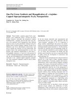
19. Cells lining the proximal convoluted tubule
a.
b.
c.
d.
e.
Form a simple squamous epithelium
Have a large surface area in contact with the lumen
Reabsorb Na+ via the Na+ solute symport system
Are involved in secretion of organic acids
Have few mitochondria
20. The proximal convoluted tubule
a.
b.
c.
d.
e.
Is the longest part of the nephron
Is the main site of solute reabsorption
Reabsorbs most of the water in the ultrafiltrate
Lies adjacent to the U-turn of the loop of Henle
Lies in the renal medulla
PCT, proximal convoluted tubule