Stimuli responsive PEGylated nano assemblies for cancer targeted drug delivery
Bạn đang xem bản rút gọn của tài liệu. Xem và tải ngay bản đầy đủ của tài liệu tại đây (3.99 MB, 140 trang )
A dissertation for the degree of doctor of philosophy
Stimuli responsive PEGylated nano-assemblies for cancertargeted drug delivery
Department of Molecular Science and Technology
The Graduate School of Ajou University
Dai Hai Nguyen
Acknowledgement
I wish to express in this part my gratitude to the scientists, technicians and other
people who were directly and indirectly involved in this work, without the help of whom
the findings of this thesis surely could not have been done.
First and foremost, I would like to extend immeasurable gratitude to Professor Ki
Dong Park, for giving me the opportunity to do my PhD thesis under his supervision. I
greatly appreciated his supervision for teaching, advising and supporting me throughout
my work. I am very grateful for his extreme patience and encouragement during the most
stressful time when my results were not good. He is a respectable mentor who has kindly
supported me in the name of family. It was an honor to work under his supervisor.
I am grateful to my thesis committee members, Professor Sung-Hwa Yoon,
Professor Won-Hee Suh at Ajou University, Professor Ji Hoon Jeong at Sungkyunkwan
University, Dr. In Kwon Jung at Genoss Company for their numerous suggestions and
helpful advice. This is a good opportunity to express my gratitude to Professors at Ajou
University whose teaching and advice helped me to complete my PhD coursework.
I would especially like to thank Dr. Yoon Ki Joung who has supported for me for
about three years. He kindly and friendly guided me from laboratory studies to routine
life in Korea. I also have deep gratitude towards Dr. Jin Woo Bae for being a great mentor.
His scientific comments are always useful in doing experiments, preparing presentation,
and writing a scientific paper.
I would like to thank my Vietnamese Professors Thi Phuong Thoa Nguyen, Thi
Kieu Xuan Huynh, and Huu Khanh Hung Nguyen for giving this opportunity to me, who
taught me fundamental knowledge of chemistry at University of Science-HCMC.
I especially appreciate all supports of my past and current members in Biomaterial
and Tissue Engineering Laboratory: Dr. Kyoung Soo Jee, Dr. Jin Woo Bae, Dr. Dong
Hyun Go, Dr. Jung Seok Lee, Dr. Kyung Min Park, Dr. Se Jin Son, Dr. Ngoc Quyen Tran,
Dr. Eugene Lih, Jong Hoon Choi, Yeo Jin Jun, In Kyu Hwang, Bae Young Kim, Ji Ho
Heo, Seung Soo You, Ki Seong Ko, Ji Hye Oh, Seung Mee Hyun, Dong Hwan Oh, Joo
Young Son, Yun Ki Lee, Ji Ho Kim, Min Yong Eom, Thi Thai Thanh Hoang, Thi Phuong
Le. I hope all members in BT Lab will obtain the outstanding achievement in your dream
and get the happiness in their life.
I appreciate all help of my Vietnamese best friends in Korea, Minh Dung Truong,
Van Thinh Nguyen, Dinh Chuong Pham, Ngoc Hoi Nguyen, Thanh Quy Nguyen, Hung
Cuong Dinh, Thi Hiep Nguyen, Chan Khon Huynh, who helped in several experiments
such as XRD, AFM, DLS, Confocal, FACS, cell culture, and animal studies. Without
them this thesis surely would not have been so multifaceted and prolific. I also would like
to be thankful to Korean friends in School of Engineering, Medicine School for your help
and support me during my stay here. Good luck to all of them.
Korean life could be some times stressful and tough, with all the competitiveness
and perfectionism. Luckily, I have had extensive care, support, and help from my family
and friends, who shared with me many wonderful and unforgettable moments throughout
my time here. I would like to devote this thesis to them with my sincere gratitude.
I would like to thank many of my best friends, Hoang Duy Nguyen, Minh Triet
Thieu, Hoang Chuong Nguyen, Nhat Nguyen Nguyen, Xuan Huong Ho… With them I
shared the first journey to Korea, as well as the sadness of leaving our lovely home and
country.
All this would not be possible without my loving immediate family. For good or
for bad, they are the ones who always stand behind me, and let me know that I am not
alone. Finally, deeply from my heart, I would like to thank my parents who believe and
support me at all time.
My best regards to all,
Dai Hai Nguyen
Stimuli responsive PEGylated nano-assemblies for cancertargeted drug delivery
Supervisor: Professor Ki Dong Park
A Dissertation
Submitted to the Graduate Faculty in Partial Fulfillment of the
Requirements for the Degree of Doctor of Philosophy
June 2013
Department of Molecular Science and Technology
The Graduate School of Ajou University
Dai Hai Nguyen
Abstract
Cancer is one of the leading causes of death worldwide and chemotherapy is a major
therapeutic approach for the treatment which may be used alone or combined with other
forms of therapy. However, conventional chemotherapy has the potential to harm healthy
cells in addition to tumor cells. Using targeted nanoparticles to deliver chemotherapeutic
agents in cancer therapy offers many advantages to improve drug delivery and to
overcome many problems associated with conventional chemotherapy. This work covers
the general areas of responsive nanocarriers and encompassed methods of fabricating
nanocarrier-based drug delivery systems for controlled and targeted therapeutic
application.
Chapter 1 provides general information of cancer and cancer treatment strategies. The
recently cancer treatment based on nanocarrier were introduced. In addition, the special
features as well as requirements of nanoparticles for targeted drug delivery were
presented. This chapter describes overall objectives of this study with the current status of
stimuli-responsive self-assembled nanocarriers for cancer chemotherapy. In chapter 2,
self-assembled nanogels based on reducible heparin-Pluronic copolymer was developed
for intracellular protein delivery. Heparin was conjugated with cystamine and the
terminal hydroxyl groups of Pluronic were activated with the VS group, followed by
coupling of VS groups of Pluronic with cystamine of heparin. The chemical structure,
heparin content and VS group content of the resulting product were determined by 1H
i
Chapter 1.
General introduction
1
Overall conclusion
Research activity aimed towards achieving specific and targeted delivery of anticancer
agents has expanded tremendously in the last 5 years or so with new avenues of directing drugs
to tumors as well as new types of drugs.
In this dissertation, we presented how nanoparticles took advantage of these special
features and how nanoparticles could act as a vehicle to specifically deliver cancer-fighting drugs
to tumors. We have developed three differ drug delivery systems using PEG and its block
copolymer for targeted drug delivery. The presence of PEG outer shell helps nanoscale carriers
to bypass the RES clearance, thereby prolonging the circulation time in the blood stream.
Another advantage that could be taken from the stability of PEG-coated nanospheres is
the possibility of attaching antibodies or a fragment of them to the surface of the
particles, without destabilizing them, in order to achieve site-specific drug delivery, a
major challenge for drug administration. Ideally, these “magic missiles” would accumulate
at the diseased tissue and locally liberate the necessary amount of drug. The drugs can be
released at the desired sites of actions by designing environment-sensitive linkers in side
structure of nanoparticles where the linkers respond to the extra/intracellular microenvironment
or external stimuli. The design of these types of nanoparticles remains a very interesting research
area. Controlled release of drug at the site of action will enhance the efficacy and reduce the side
effect of drug. The combination of the use of stimuli-responsive material and targeting moieties
will lead to nanoparticles which can be targeted to the side of action and which will deliver the
drug.
These approach should provide the creative treatment methods have made it to the clinic
and hopefully are well on their way to improving the length and quality of life for cancer
patients. However, it should be noted that extensive preclinical evaluations are required for these
types of nanoparticles before they can be considered to use in patients. Subjects which have to be
evaluated are the pharmacokinetics of drug loaded/conjugated nanoparticles, effect of the
surface-located targeting molecules on the opsonization process and blood circulation times as
well as the efficacy and toxicity of the nanoparticles in particlular after repeated administration.
Mechanistic studies of the intracellular drug release from the nanoparticles are also required to
further unravel the kinetics of intracellular nanoparticle destabilization and intracellular drug
release.
1. Cancer and strategy treatment
Cancer is one of the leading causes of death worldwide (13%). Each year 12.7 million
people worldwide are diagnosed with cancer and there are 7.6 million deaths from the
disease in 2008 (WHO).1 It is estimated that there are 24.6 million people alive who have
received a diagnosis of cancer in the last five years. By 2030, the number of new cancer
cases is expected to rise to 21.4 million, with 13.15 million cancer deaths. 2 Cancer's total
economic impact was estimated at $895 billion in 2008, or 1.5% of the world's gross
domestic product. This cost did not include direct medical costs, which could potentially
double the total economic cost, according to Atlanta-based ACS.3
The cancer treatment during the twentieth century was based on surgery, radiation
and chemotherapy. Of these modalities, surgery is most effective at an early stage of
disease progression. However, most cancer operations carry a risk of: pain, infection, loss
of organ function. Surgery can also cause cancer cells to spread to different sites.
Radiation while destroying cancer cells also burns, scars, and damages healthy cells,
tissues, and organs. Initial treatment with chemotherapy and radiation will often reduce
tumor size. Radiation can cause cancer cells to mutate and become resistant and difficult
to destroy.4 Chemotherapy is drug therapy that can kill these cells or stop them from
multiplying. However, it involves poisoning the rapidly growing cancer cells and also
destroys rapidly growing healthy cells in the bone marrow, gastro-intestinal tract, etc.,
and can cause organ damage, like liver, kidney, heart and lungs, and so on. Moreover,
when the body has too much toxic burden from chemo the immune system is either
2
compromised, hence the person can succumb to various kinds of infections and
complications.
Consequently, novel targeted drug deliveries have been extensively investigated in an
effort to improve the therapeutic effectiveness and safety profile of traditional
chemotherapeutic agents. Targeted drug delivery, sometimes called smart drug delivery,
is a method of delivering medication to a patient in a manner that increases the
concentration of the medication in some parts of the body relative to others. The goal of a
targeted drug delivery system is to prolong, localize, target and have a protected drug
interaction with the diseased tissue. The conventional drug delivery system is the
absorption of the drug across a biological membrane, whereas the targeted release system
is when the drug is released in a dosage form. The clinically most relevant drug targeting
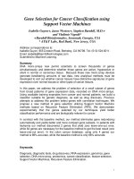
strategies were summarized in Figure 1.1. The advantages to the targeted release system
is the reduction in the frequency of the dosages taken by the patient, having a more
uniform effect of the drug, reduction of drug side effects, and reduced fluctuation in
circulating drug levels.
3
Figure 1.1 Overview of the clinically most relevant drug targeting strategies. (A)
Conventional chemotherapy (free drug) (B) passively targeted drug delivery system by
virtue of the enhanced permeability and retention (EPR) effect (C) Active drug targeting
to internalization-prone cell surface receptors (over)expressed by cancer cells generally
intends to improve the cellular uptake of the nanomedicine systems (D) Active drug
targeting to receptors (over)expressed by angiogenic endothelial cells aims to reduce
blood supply to tumours (E) Stimuli-sensitive nanomedicines (F) Local drug delivery.
4
2. Nanocarrier strategies in cancer chemotherapy
The use of nanotechnology in medicine and more specifically drug delivery is set to
spread rapidly. Currently many substances are under investigation for drug delivery and
more specifically for cancer therapy are used in the clinic. Interestingly pharmaceutical
sciences are using nanocarriers to reduce toxicity and side effects of drugs and up to
recently did not realize that carrier systems themselves may impose risks to the patient.
The kind of hazards that are introduced by using nanocarriers for drug delivery are
beyond that posed by conventional hazards imposed by chemicals in classical delivery
matrices. For nanocarriers the knowledge on particle toxicity as obtained in inhalation
toxicity shows the way how to investigate the potential hazards of nanocarriers. The
toxicology of particulate matter differs from toxicology of substances as the composing
chemical(s) may or may not be soluble in biological matrices, thus influencing greatly the
potential exposure of various internal organs.
Appropriately engineered nano-sized delivery systems can achieve finer temporal
control over drug release rates due to their large surface area. Nanocarriers can also be
inherently useful in systems that require a burst release. Nanocarriers, unlike bulk drug
delivery systems, can enter cells to deliver drugs and can be designed to respond to
intracellular cues. Further, since nanocarriers can circulate in the body after being
injected they have the ability to target diseases at the site of disorder. This feature of
nanocarriers is especially useful in cancer therapy, where the size of the delivery system
is the key to target cancers through the enhanced permeability and retention effect (EPR).
5
Diseased cells can also be targeted by attaching ligands or antibodies to the surface of
nano-drug delivery systems. Targeting allows nanocarriers to hone into diseased cells by
targeting specific features of a disease phenotype, such as an over expressed protein or
enzyme. Another important aspect of delivery that is now being given the importance it
deserves is the drug encapsulation stability in these carriers. This is especially relevant
because it is increasingly realized that the thermodynamic parameters like percent (%)
loading do not adequately describe how stable the delivery vehicle would be during
circulation in blood, since these vehicles could potentially leak out drugs into
hydrophobic sites in surrounding tissue and blood components. Delivery vehicles, based
on a single platform, which can satisfy all basic requirements of a versatile nanoscopic
delivery vehicle, are quite rare. These features however are the foundations of a good
delivery vehicle and are fundamental design requirements. Thus there are key aspects of a
delivery vehicle design that was described as the basic anatomy of a drug delivery
vehicle.
3. Self-assembled nanocarrier for drug delivery
Nanoparticles are now available that are attractive for a wide range of materials and
devices, but novel fabrication methods are also required to take full advantage of the
interesting properties of nanoparticulates. Approaches based on the self-assembly of
systems from individual components offer tremendous cost advantages and an almost a
magical "ease of manufacture" compared to lithographic methods. Self-assembled
nanoparticles also give the great opportunity in terms of diversity and functionality in the
design for defined drug delivery purposes. Self-assembled nanoparticles have many
6
advantages as highly efficient drug delivery vehicles including nanoscale size, controlled
composition and capacity to encapsulate a wide range of drug molecules. In particular, by
using advanced chemistry and precision engineering at a molecular level, these synthetic
polymers provide a wide opportunity for functionalization and versatility which impact
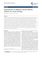
the physico-chemical properties of self-assembled systems. Examples of self-assembled
nanocarriers for targeted drug delivery are showed in Figure 1.2.
7
a. Micelles
d. Nanocapsules
b. Liposomes
e. Nanogels
c. Oil/water emulsion
f. Core-shell particles
Figure 1.2 Example of self-assembled nanocarriers for targeted drug delivery: a.
Micelles, an aggregate of surfactant molecules dispersed in a liquid colloid where drugs
are physically encapsulated in the inner core. b. Liposomes, a spherically arranged
bilayer structure with drug loaded either in the inner aqueous phase or between the lipid
bilayers. c. Oil/water emulsion, a mixture of liquids that are normally immiscible with
drug loaded in the inner oil phase. d. Nanocapsules, a polymeric membrane which
encapsulates an inner liquid core. e. Nanogels, a nanoparticle composed of a hydrogel. f.
Core-shell particles, the location of nanocrystals at the core with the polymers on the
outer layer.
8
4. PEGylated nanocarriers for systemic delivery
Clearly, particles with longer circulation times have superior ability to reach the
tumor site through passive targeting. As opsonization is an integral step in the removal of
foreign macromolecules by the RES, many efforts for increasing serum stability and
extending circulation time have focused on blocking absorption of opsonins onto the
nanoparticle surface.5 For passive targeting to be successful, the nanocarriers need to
circulate in the blood for extended times so that there will be multiple possibilities for the
nanocarriers to pass by the target site. Nanoparticulates usually have short circulation
half-lives due to natural defense mechanisms of the body to eliminate them after
opsonization by the mononuclear phagocytic system (MPS, also known as
reticuloendothelial system. Therefore, the particle needs to be extended circulation halflives.
Cellular entrapment in macrophages can be avoided by surface modification of the
nanocarriers. Among many materials used to make or modify pharmaceutical carriers
(lipids, natural and synthetic polymers, emulsions, or dendrimers) special attention was
paid to polyethylene glycol (PEG, also known as polyethylene oxide (PEO)), which was
used both for chemical modification of various drugs (peptide and protein, first of all) to
make them more stable and long-circulating and for the decoration of pharmaceutical
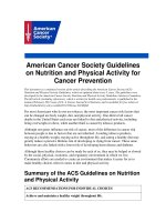
carriers to improve their pharmacokinetic properties. Figure 1.6 illustrates how opsonin
proteins associate with foreign bodies and coat its surface. As bacteria and viruses have
the same negative surface charge as phagocytic cells, opsonins are critical to reducing the
charge repulsion between the two systems. Next, phagocytic cells engulf the material and
9
transport it to the liver or spleen for degradation and excretion (Figure 1.3 a3–a4).
Additional phagocytic macrophages are permanently located in the liver. Known as
Kupffer cells, these cells serve as a major filter for many types of NPs and are a major
interference with long t½. The PEG polymer on a NP surface increases t½ by reducing
this opsonization process (Figure 1.3 b2), thus preventing recognition by monocytes and
macrophages, allowing the NPs to remain in the blood pool. Hydrophobic particles are
also more vulnerable to the RES and hydrophilic PEG reduces these complications. In
addition to NP–RES interactions, poor t½ can also result from NP–NP interactions (i.e.,
aggregation). NPs aggregate primarily because the attraction between particles is stronger
than the attraction for solvent. For spherical NPs, the interaction potential is related to the
electrostatic repulsive potential and the van der Waals attraction potential. PEG decreases
the surface energy of NPs and minimizes van der Waals attraction.6
Prior to NP applications, PEG was used as a nontoxic, water-soluble
dispersant/stabilizer. FDA approved PEG is a highly hydrophilic, flexible polymer which
has an inherent long circulating property. The array of already available versatile PEG
chemistries make it an attractive polymer to be used in modifying pharmaceuticals or
surfaces of pharmaceutical carriers to achieve the desired long-circulating property or add
convenient functional groups to conjugate ligands for active targeting. Early work with
PEGylated NPs stemmed mostly from drug delivery.7 One of the first reports on
PEGylation was described by Davis and Abuchowski,8 where they covalently attached
methoxy-PEGs (mPEGs) of 1900 and 5000 Da to bovine serum albumin and to liver
catalase. Later, acrylic microspheres functionalized with PEG-modified human serum
albumin increased t½ in vivo.9 Li and colleagues found that 75-nm latex particles
10
remained in rat circulation 40-times longer (half-life 20 min vs 13 h) when coated than
uncoated with PEG larger than 5000 kDa.7b In the mid-1990s, Doxil® (liposomal
delivery vehicle for doxorubicin) and oncospar (PEG-l-asparaginase) became the first
FDA-approved NP therapeutics.7c Doxil increases doxorubicin bioavailability nearly 90fold at 1 week from injection of PEGylated liposomes versus free drug.10 Later,
Abraxane® was introduced as an albumin-functionalized NP for delivery of taxane
without cremphor to enhance drug efficiency.11
11
Figure 1.3 (a) Nanocarriers (a1) are coated with opsonin proteins (a2) and associate with
macrophages (a3) for transit to the liver (a4). Macrophages stationary in the liver, known
as Kupffer cells, also participate in nanoparticle scavenging. (b) Nanocarriers coated with
PEG coating (b1) prevents this opsonization (b2), resulting in decreased liver
accumulation (b3) and increased availability of the NP for imaging or therapy.12
12
5. Targeted drug delivery systems for cancer therapy
Conventional cancer chemotherapies have dose-related side effects owing to
nonspecific biodistribution of drugs. Targeted nanomedicines are emerging as one of the
promising approaches in anticancer treatment and have major advantages. Targeting
active molecules to specific sites in the body had been pursued actively ever since Ehrlich
first envisaged the use of 'magic bullets' for the therapy of various diseases.13 Interest in
this concept has increased significantly in recent decades with the innovations of
nanomedicine. Cancer nanomedicines have the ability to improve the therapeutic index of
drugs by preferential localization at target sites, lower distribution in healthy tissues,
delivery of hydrophobic drugs and extended release rate. Progress in the development of
nanomedicines for targeted drug delivery has been reviewed by Moghimi and
colleagues.14 Targeted delivery can be achieved passive, active targeting, or their
combinative targeting.
13
Figure 1.4. Conceptual representation of nanoparticle tumor-targeting modalities. Passive
targeting: Unlike that found in normal tissue, tumor vasculature is leaky owing to
fenestrations and gaps between endothelial cells that result from abnormal angiogenesis.
NPs in circulation can passively extravasate through these gaps and enter the tumor
interstitium. Poor lymphatic drainage found in some tissues helps to retain particles in the
tumor space. Active targeting: Ligands (e.g. antibodies, peptides, small molecules, etc.)
targeted toward moieties overexpressed or uniquely present on the plasma membrane of
tumor cells can be used to actively enhance NP accumulation at the tumor site and can
also help to internalize particles into cells via endocytosis.15
14
5.1.
Passive targeting strategies and recent developments
Cellular barriers present formidable obstacles in the delivery of therapeutics for
cancer treatment. Fortunately, certain aspects of cancer physiology can be exploited to
achieve passive targeting to tumor sites. Rapid growth of tumors leads to aberrant
angiogenic vasculature. The newly formed blood vessels are often disorganized and
discontinuous, resulting in increased permeability to macromolecules. Moreover,
lymphatic drainage systems are often poorly developed or non-existent in tumor sites,
enabling accumulation of therapeutics.16 This phenomenon, called the enhanced
permeation and retention (EPR) effect has increased the tumor concentration of
anticancer agents up to 70-fold in some cases.17 Since the pioneering work of Couvreur et
al.,18 nanoscale systems have been aggressively investigated for their utility in drug
delivery applications (Figure 1.4).
Nanoparticle size is known to play a critical role in achieving passive targeting. The
majority of solid tumors exhibit a vascular pore cutoff size between 380 and 780 nm. 19
Therefore, particles need to be of a size much smaller than the cutoff pore diameter to
reach to the target tumor sites. By contrast, normal vasculature is impermeable to drugassociated carriers larger than 2 to 4 nm compared to free, unassociated drug molecules.20
This nanosize window offers the opportunity to increase drug accumulation and local
concentration in target sites such as tumor or inflamed sites by extravasation, and
significantly to reduce drug distribution and toxicity to normal tissues. Nanocarriers
above 10 nm in diameter are generally able to avoid filtration by the kidneys, while less
well understood, the upper size limit for passively targeted nanocarriers is thought to be
15
approximately 150 nm.21 Extravasation and diffusional barriers limit nanoparticle access
to tumors when particle size is over 200 nm.21b Additionally, previous studies have
shown that nanoparticle clearance rate increases with size.22 One such investigation
demonstrated that the blood clearance of 80 nm nanocarriers was half as fast as the
clearance of 170 and 240 nm particles. Presumably, this effect is due to non-specific
protein adsorption on the surface of larger nanocarriers, leading to opsonization and
subsequent clearance by the RES.22
5.2.
Active targeting strategies and stimuli-triggered ligand presentation
Localized diseases such as cancer or inflammation not only have leaky vasculature
but also overexpress some epitopes or receptors that can be used as targets. Therefore,
nanomedicines can also be actively targeted to these sites. Ligands that specifically bind
to surface epitopes or receptors, preferentially overexpressed at target sites, have been
coupled to the surface of long circulating nanocarriers.23 Ligand-mediated active binding
to sites and cellular uptake are particularly valuable to therapeutics that are not taken up
easily by cells and require facilitation by fusion, endocytosis, or other processes to access
their cellular active sites.24 Active targeting can also enhance the distribution of
nanomedicine within the tumor interstitium. More recently, active targeting has been
explored to deliver drugs into resistant cancer cells.25 An important consideration when
selecting the type of targeting ligand is its immunogenicity. For example, whole
antibodies that expose their constant regions on the liposomal surface are more
susceptible to Fc-receptor-mediated phagocytosis by the mononuclear phagocytic
system.26 Examples of targeting ligands and their targets are listed in Table 1.1.
16
Table 1.1
Small
Organic
Molecules
Peptides
Selected examples of ligands used in active drug targeting
Name
Folic acid
Target
Folate receptor
Methotrexate
Folate receptor
Non-peptidic RGD
mimetic
Mimetic of the sialyl
Lewisx
RGD
avβ3 integrin
Chlorotoxin
MMP-2
Synaptotagm in I, C2
domain
VHSPNKK
Phospholipids
E-selectin
avβ3 integrin
VCAM-1
Application
Breast cancer
imaging
Brain tumor imaging
and therapy
Integrin positive cell
imaging
Inflammatory
disease imaging
Breast cancer
imaging
Brain tumor imaging
and therapy
Apoptosis imaging
Cardiovascular
disease imaging
EPPT1 (YCAREPPT
Underglycosylated
Multiple tumor type
RTFAYWG)
mucin-1 antigen
imaging
Aptamers A10 RNA aptamer
Prostate-specific
Prostate cancer
membrane antigen
imaging
Thrm-A and Thrm-B
Human alphaSerum protein
DNA aptamers
thrombin protein
detection
Proteins
Annexin V
Phosphatidylserine
Apoptosis imaging
LHRH
LHRH receptor
Breast cancer
imaging
Transferrin
Transferrin receptor
Breast cancer
imaging
Antibodies Monoclonal antibody
Colorectal carcinoma
Colon cancer
A7
imaging
Herceptin
Her2/neu (Breast
Breast cancer
(Trastuzumab)
cancer)
imaging and therapy
Rituxan (Rituximab)
CD20 antigen
Lymphoma imaging
therapy
RGD, Arg-Gly-Asp tripeptide: LHRP, luteinizing hormone releasing hormone;
Endothelial vascular adhesion molecule-1, VCAM-1
17