Adaptive evolution of a duplicated pancreatic ribonuclease gene in a leafeating monkey
Bạn đang xem bản rút gọn của tài liệu. Xem và tải ngay bản đầy đủ của tài liệu tại đây (120.77 KB, 6 trang )
letter
Adaptive evolution of a duplicated pancreatic
ribonuclease gene in a leaf-eating monkey
Jianzhi Zhang1,2, Ya-ping Zhang3 & Helene F. Rosenberg1
© 2002 Nature Publishing Group
Published online: 4 March 2002, DOI: 10.1038/ng852
Although the complete genome sequences of over 50 representative species have revealed the many duplicated genes in all
three domains of life1–4, the roles of gene duplication in organismal adaptation and biodiversity are poorly understood. In
addition, the evolutionary forces behind the functional divergence of duplicated genes are often unknown, leading to disagreement on the relative importance of positive Darwinian
selection versus relaxation of functional constraints in this
process5–10. The methodology of earlier studies relied largely
on DNA sequence analysis but lacked functional assays of
duplicated genes, frequently generating contentious
results11,12. Here we use both computational and experimental
approaches to address these questions in a study of the pancreatic ribonuclease gene (RNASE1) and its duplicate gene
(RNASE1B) in a leaf-eating colobine monkey, douc langur. We
show that RNASE1B has evolved rapidly under positive selection for enhanced ribonucleolytic activity in an altered microenvironment, a response to increased demands for the enzyme
for digesting bacterial RNA. At the same time, the ability to
degrade double-stranded RNA, a non-digestive activity characteristic of primate RNASE1, has been lost in RNASE1B, indicating functional specialization and relaxation of purifying
selection. Our findings demonstrate the contribution of gene
duplication to organismal adaptation and show the power of
combining sequence analysis and functional assays in delineating the molecular basis of adaptive evolution.
A subfamily of Old World monkeys, colobines are unique primates that use leaves rather than fruits and insects as their primary food source; these leaves are then fermented by symbiotic
bacteria in the foregut13. Similar to ruminants, colobines recover
nutrients by breaking and digesting the bacteria with various
enzymes, including pancreatic ribonuclease (RNASE1), which is
secreted from the pancreas and transported into the small intestine to degrade RNA14,15. Earlier studies revealed a substantially
greater amount of ribonuclease (RNase) in the pancreas of
foregut fermenting mammals (colobines and ruminants) than in
other mammals14,15. This is believed to be related to the fact that
rapidly growing bacteria have the highest ratio of RNA-nitrogen
to total nitrogen of all cells, and high concentrations of RNase are
needed to break down bacterial RNA so that nitrogen can be
recycled efficiently14.
Using a screening method based on PCR and sequencing, we
detected one RNASE1 gene in each of the 15 non-colobine primates examined, including 5 hominoids, 5 Old World monkeys,
4 New World monkeys and 1 prosimian. We determined the
DNA sequences of these RNASE1 genes; the deduced protein
sequences are shown in Fig. 1a. The phylogenetic tree of the
RNASE1 sequences (Fig. 2a) is consistent with the known species
relationships16 at all nodes, with greater than 55% bootstrap support, suggesting that the RNASE1 genes are orthologous. By contrast, two RNASE1 genes were found in the Asian colobine, douc
langur (Pygathrix nemaeus). Phylogenetic analysis (Fig. 2a) suggests that these two genes were generated by recent duplication
postdating the separation of colobines from other Old World
monkeys (cercopithecines). The branch lengths of the gene tree
indicate that the nucleotide sequence of one daughter gene
(RNASE1) has not changed since duplication, whereas that of the
other gene (RNASE1B) has accumulated many substitutions
(Fig. 2a). Beintema15 previously purified an RNase from the pancreas of another Asian colobine, hanuman langur (Presbytis
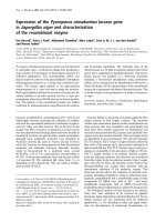
entellus), and obtained the mature peptide sequence for this protein. Our phylogenetic analysis of these protein sequences shows
that the hanuman langur pancreatic RNase clusters with douc
langur RNASE1B with 99% bootstrap support (Fig. 2b). This
result implies an orthologous relationship between these two
proteins, which suggests that the douc langur RNASE1B is also
expressed in the pancreas.
We determined the structures of RNASE1 of human, rhesus
monkey and douc langur and that of douc langur RNASE1B by
sequencing genomic regions flanking the coding sequences; we
found no variation in gene structure (Fig. 1b). The entire
RNASE1 or RNASE1B protein is encoded by exon 2, which is
separated from an upstream noncoding exon by an intron of
703–706 nt. The presence of a homologous intron (98.9%
sequence identity) in RNASE1 and RNASE1B suggests that
gene duplication was probably due to unequal crossing-over
rather than to retroposition, which usually generates intronless duplicates.
To trace the evolutionary history of RNASE1B, we inferred the
gene sequence of the most recent common ancestor of douc langur RNASE1 and RNASE1B. As the sequences involved are closely
related, the parsimony ancestral inference17 was unambiguous at
all sites and the distance-based Bayesian method18 gave the same
inference with nearly 100% probability, indicating high reliability of the ancestral inference. The coding region of the inferred
ancestral sequence is identical to that of present-day RNASE1 of
douc langur, in agreement with the zero branch length of the
douc langur RNASE1 lineage (Fig. 2a). Thus, the 12 nucleotide
differences between the coding regions of douc langur RNASE1
and RNASE1B all occurred in the RNASE1B lineage (Fig. 3). We
1Laboratory of Host Defenses, National Institute of Allergy and Infectious Diseases, National Institutes of Health, Bethesda, Maryland, USA. 2Departments of
Ecology and Evolutionary Biology and Molecular, Cellular and Developmental Biology, University of Michigan, 3003 Natural Sciences Building, 830 North
University Avenue, Ann Arbor, Michigan 48109, USA. 3Kunming Institute of Zoology, Chinese Academy of Sciences, Kunming, Yunnan, China.
Correspondence should be addressed to J.Z. (e-mail: ).
nature genetics • volume 30 • april 2002
411
© 2002 Nature Publishing Group
letter
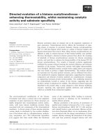
tested the molecular-clock hypothesis (that is, equal rates of
nucleotide substitution) for the two genes of douc langur using
rhesus monkey RNASE1 as an outgroup (Fig. 3); this molecularclock hypothesis is rejected (P<0.001)19. When we divided the
nucleotide substitutions into synonymous and nonsynonymous
(amino acid–altering) substitutions (Fig. 3), we found that the
synonymous substitutions passed the clock test (P>0.10),
whereas the nonsynonymous substitutions did not (P<0.005).
This suggests that the rate difference between the two genes is
due to a difference in natural selection rather than in mutation
rate. Consistent with this result, the clock hypothesis cannot be
rejected for the noncoding region of roughly 1,500 nt (P>0.1),
which is presumably free from selection (see below). In addition,
the molecular-clock hypothesis for the noncoding region cannot
be rejected between rhesus monkey RNASE1 and douc langur
RNASE1 (or RNASE1B) when human RNASE1 is used as an outgroup (P>0.2). These results allowed us to use the noncoding
regions to date the gene duplication event. Using the fossil record
of a divergence time of 15 million years (Myr) between colobines
and cercopithecines20, we estimated that the duplication of
RNASE1 to RNASE1B occurred 4.2 Myr ago, with a 95% bootstrap confidence interval of 2.4–6.4 Myr ago.
a
RNASE1
RNASE1B
RNASE1
RNASE1B
human
chimpanzee
gorilla
orangutan
gibbon
rhesus monkey
pig-tailed macaque
baboon
green monkey
talapoin monkey
squirrel monkey
tamarin
spider monkey
woolly monkey
lemur
signal peptide
MALEKSLVRL LLLVLILLVL
........L. P.........
........L. P.........
........L. P.........
........L. P.F..M....
...D..VIL. P....V....
...D..VIL. P....V....
...D..VIL. P....V....
...D..VIL. P....V....
...D..VIL. P....V....
.......AL. P....V....
.......AL. P....V....
.......AL. P....V....
.......AL. P....V....
........L. P....A....
To explore the evolutionary forces driving the accelerated evolution of RNASE1B, we compared the number of nucleotide substitutions per site at nonsynonymous sites in RNASE1B since its
origin through gene duplication, and the corresponding number
at synonymous and noncoding sites. We found that the number
of substitutions per nonsynonymous site (0.0310) is significantly
greater than that per synonymous and noncoding sites (0.0077;
P<0.002, Fisher’s exact test). Synonymous and noncoding sites
are generally not considered to be subject to purifying selection.
In the present case, the percent nucleotide difference between
humans and Old World monkeys at synonymous and noncoding
sites of the RNASE1 (or RNASE1B) locus is 6.45 ± 0.61, which is
similar to the reported average percentage difference (7.1)21
between orthologous sequences of humans and Old World monkeys at various pseudogenes and introns (P>0.20, t-test). Taken
together, these analyses suggest that the synonymous and noncoding sites at the RNASE1B locus are not subject to selective constraints and that the accelerated evolution of the coding sequence
of RNASE1B is due to positive Darwinian selection. To investigate
the nature of the amino-acid substitutions favored by selection,
we divided nonsynonymous substitutions into two groups: those
altering the amino-acid charge (radical substitutions) and those
1
GWVQPSLGKE
..........
..........
..........
..........
.----C..R.
.----C..R.
.----C..R.
.----C..R.
.----C..R.
........R.
..........
..........
..........
..I.......
SRAKKFQRQH
..........
..........
..........
..........
..........
..........
..........
..........
..........
..........
...Q......
..........
..........
...M......
MDSDSSPSSS
V.........
.........N
...G...N.N
.........N
...G.....N
.........N
...G.....N
...G.....N
...G.....N
....G....N
....G....N
....G....N
....G.L..N
..PG..S...
STYCNQMMRR
..........
..........
..........
..........
........K.
........K.
........K.
........K.
........K.
P....D....
P....N....
P....D..K.
P....N....
..........
RNMTQGRCKP
..........
..........
..........
..........
.S..H.....
.S..H.....
.S..H.....
.S........
.S........
..........
..........
..........
......W...
....N.W...
52
VNTFVHEPLV
..........
..........
..........
..........
..........
..........
..........
..........
..........
..........
..........
..........
..........
..........
douc langur
douc langur
...D..VIL. P...VV.... ..A.....R. .......... ...G...... ........K. .......... ..........
...D..VIP. P...VV.... ..A.....G. .Q.E...... ...G...... ........KL ......W..S ..........
*
* *
*
*
128 pl
human
DVQNVCFQEK VTCKNGQGNC YKSNSSMHIT DCRLTNGSRY PNCAYRTSPK ERHIIVACEG SPYVPVHFDA SVEDST 8.6
chimpanzee
.......... .......... .......R.. .......... .......... .......... .......... ...... 8.8
gorilla
.......... .......... .......... .......... .......... .......... N......... ...... 8.6
orangutan
.......... .......... .......... .....H.... ........T. .......... .......... ...... 8.8
gibbon
.......... .......A.. .......... .......... .......... .......... .......... ...... 8.6
rhesus monkey
.......... .......T.. F..K...... .......... .......... .......... ..H....... ...... 9.1
pig-tailed macaque .......... .......T.. F..K...... .......... .......... ..R....... ...M...... ...... 9.1
baboon
.......... .......T.. F..K...... .......... .......... ..R....... .......... ...... 9.5
green monkey
.......... .......T.. F..K...... .......... .......... ..R....... .......... ...... 9.5
talapoin monkey
.......... .......T.. F..K...... .......... .......... ..R....... .......... ...... 9.5
squirrel monkey
...D...... .......A.. ...S...... .......... ........Q. .......... N......... ...... 8.2
tamarin PR
...D...... .......P.. ...S...R.. .......... ........Q. .......... N......... ...... 8.4
spider monkey
...D.....N .......A.. ...S...... .......... ........Q. .......... N......... ...... 8.1
woolly monkey
...D.....N .......A.. ...S...... .....S.... .....Q..Q. .......... N......... ...... 7.8
lemur
...AI....N .......T.. .....T.... .....GS.K. ........Q. ..R....... .......... ...... 8.4
douc langur
douc langur
.......... .......T.. F....K.... ........K. .......... .......... .......... ......
.......... .......T.. F....K.... E.......K. .....Q.... .......... .........D ......
*
*
9.1
7.3
b
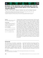
Fig. 1 Protein sequences and genomic
structures of RNASE1 and RNASE1B of
DNA sequences surveyed
primates. a, Protein sequence align471 bp
ment of RNASE1 and RNASE1B.
protein coding region
Amino
acid
substitutions
that
occurred in RNASE1B since its origin
exon 1
intron
exon 2
by duplication are underlined, with
408 bp 35 bp
191 bp
703 bp
634 bp
human RNASE1
those involving changes in charge
191 bp
705 bp
634 bp
douc langur RNASE1 441 bp 35 bp
indicated by an asterisk. pI, isoelectric
191 bp
706 bp
634 bp
douc langur RNASE1B 441 bp 35 bp
point of mature peptides. b, The conserved structure of RNASE1 and
RNASE1B. The structures of douc langur RNASE1 and RNASE1B were determined by homology to that of human RNASE1, which was determined by comparing the cDNA and genomic sequences.
Compared with douc langur RNASE1, there is a 1-nt insertion in the intron of RNASE1B. We found no other insertions or deletions between them in the
sequenced regions shown here, although there are a total of 28 nucleotide substitutions.
412
nature genetics • volume 30 • april 2002
letter
© 2002 Nature Publishing Group
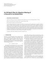
Fig. 2 Phylogenetic relationships among RNASE1 and RNASE1B of primates. a, The
gene tree of RNASE1 and RNASE1B. Kimura’s two-parameter distances are used.
Virtually identical trees are obtained when Tajima-Nei, Tamura-Nei or TamuraNei-γ distances (S. Kumar et al., MEGA2, Arizona State University) are used. The
differences only occur at some low-bootstrap (<50%) nodes of the tree shown
here. b, Phylogenetic relationship of the purified RNase from hanuman langur
and douc langur RNASE1B. Poisson distances of the amino acid sequences of the
mature peptides are used. Bootstrap percentages higher than 50 are shown on
tree branches. Branch lengths are drawn to scale, indicating the number of
nucleotide or amino acid substitutions per site.
that leave charge unaltered (conservative substitutions). Earlier
studies showed that, for most mammalian genes, the rate of radical substitution is lower than that of conservative substitution,
owing to stronger purifying selection on radical substitution22. In
RNASE1B, however, the opposite is found. The number of radical
substitutions per site since duplication (0.067) is significantly
greater than that (0.012) of conservative substitutions per site
(P<0.02; Fisher’s exact test). There are nine amino-acid substitutions in the mature peptide of RNASE1B, and seven of them
involve charge changes. Unexpectedly, all seven charge-altering
substitutions increase the negative charge of the protein (Fig. 1a).
Apparently, the amino-acid substitutions are nonrandom
(P<0.016, randomization test), with negatively charged residues
being selectively favored. Notably, the rate of radical substitution
is not statistically different from the conservative rate when amino
acid polarity or volume22 is considered (P>0.15).
The charge-altering substitutions reduced the net charge of
RNASE1B from 8.8 to 0.8 (at pH 7) and the isoelectric point
from 9.1 to 7.3 (Fig. 1a). Because RNA is negatively charged, the
net charge of RNase influences its interaction with the substrate
and its catalytic performance23. We therefore hypothesized that
the charge-altering substitutions may have changed the optimal
pH of RNASE1B in catalyzing the digestion of RNA. To test this
hypothesis, we prepared recombinant proteins from douc langur RNASE1B as well as the RNASE1 genes of human, rhesus
monkey and douc langur, and examined their ribonucleolytic
activities at different pH levels in a standard RNase assay against
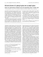
yeast tRNA. We determined that the optimal pH for human
RNASE1 is 7.4, a value that is within the pH range (7.4–8.0)
measured in the small intestine of humans24,25. The same optimal pH was observed for RNASE1 of rhesus monkey and douc
langur (Fig. 4a). Probably because of foregut fermentation and
related changes in digestive physiology, the pH in the small
intestine of colobine monkeys shifts to 6–7 (ref. 13). Notably,
the optimal pH for douc langur RNASE1B was found to be 6.3
(Fig. 4a). At pH 6.3, RNASE1B is about six times as active as
RNASE1 in digesting RNA, and the difference in their activities
is statistically significant (P<0.001, t-test). These results suggest
that the rapid amino acid substitutions in RNASE1B were driven
by selection for enhanced RNase activity at the relatively low pH
environment of the colobine small intestine.
nucleotide substitutions
coding (syn, nonsyn) noncoding
douc langur RNASE1B
douc langur RNASE1
rhesus monkey RNASE1
**
12
0
14
**
(2, 10 )
(0, 0)
(4, 10)
11
5
49
amino-acid changes
sig pep mat pep
1
0
2
9**
0
7
Fig. 3 Tests of the molecular clock hypothesis for RNASE1 and RNASE1B of
douc langur. Rhesus monkey RNASE1 is used as an outgroup. The numbers
of substitutions on each of the three branches of the tree are determined by
comparing the present-day sequences with the ancestral sequence at the
interior node of the tree. Significance level of the Tajima’s test: *, 5% ; **,
0.5%. syn, synonymous; nonsyn, nonsynonymous; sig pep, signal peptide;
mat pep, mature peptide.
nature genetics • volume 30 • april 2002
a
gene duplication
douc langur (RNASE1B)
100 douc langur (RNASE1)
colobine
green monkey
talapoin monkey
cercopithecines
baboon
pig-tailed macaque
97
78
rhesus monkey
60
human
89
chimpanzee
87
gorilla
hominoids
orangutan
97
gibbon
77
woolly monkey
55
New World
spider monkey
monkeys
tamari n
98
99
squirrel monkey
lemur
Old World
monkeys
prosimian
0.05
b
96
78
99
hanuman langur (RNase from pancreas)
douc langur (RNA SE1B)
douc langur (RNA SE1)
rhesus monkey
human
squirrel monkey
0.05
Sequence conservation of douc langur RNASE1 after gene
duplication and its unchanged optimal catalytic pH at 7.4 suggest that this protein acts in non-digestive processes. Of note,
human RNASE1 is found in many other tissues besides the pancreas26 and has enzyme activity (EAdsRNA) in degrading doublestranded (ds) RNA, although the physiological relevance of this
catalytic activity is unclear23. We found similar EAdsRNA among
RNASE1 of human, rhesus monkey and douc langur (Fig. 4b),
with that of douc langur RNASE1B reduced to approximately
0.3% (Fig. 4b). As one interpretation, RNASE1B can afford to
loose EAdsRNA function because the paralogous RNASE1 retains
it; it is likely that some of the adaptive charge-altering substitutions in RNASE1B are detrimental to EAdsRNA. To determine
which of the nine amino-acid substitutions in RNASE1B are
responsible for loss of EAdsRNA, we used site-directed mutagenesis to create mutant forms of douc langur RNASE1, each with
one substitution. We found that eight of the nine substitutions
reduce EAdsRNA substantially (P<0.005, two-tail t-test), whereas
the other (R4Q, Fig. 4b) has a mild and marginally significant
effect (P=0.069, two-tail t-test and P=0.035, one-tail test). The
detrimental effect of these substitutions on EAdsRNA might also
be predicated from the fact that seven of the nine substitutions
that occurred in RNASE1B are not found in any of the 16 primate
RNASE1 proteins examined, and that five of the substitutions
occurred in positions that are otherwise invariant (Fig. 1a). Two
of the most influential substitutions are Arg→Leu at position
32 and Asp→Glu at position 83, each reducing EAdsRNA to
approximately 3%. Both Arg32 and Asp83 are invariant among
primate RNASE1 proteins, suggesting that they are essential for
RNASE1 function and that mutations at these sites have been
subject to strong purifying selection. It should also be noted
that each of the nine RNASE1 single-substitution constructs
has a significantly higher EAdsRNA than that of RNASE1B
(P<0.005), suggesting that it is not a single substitution, but a
collective effect of multiple substitutions, that has dramatically
reduced the EAdsRNA of RNASE1B. Future analyses of mutant
RNASE1 proteins with multiple substitutions may uncover
possible interactions among these amino-acid changes.
413
letter
b
RNase activity against
yeast tRNA (sec.-1)
a
0.4
RNASE1B
RNASE1
0.3
0.2
0.1
© 2002 Nature Publishing Group
0
3
4
5
6
pH
7
8
9
human RNASE1
rhesus monkey RNASE1
douc langur RNASE1
douc langur RNASE1B 0.0066 ± 0.0013
R1G
R4Q
K6E
mutant
R32L
forms of
douc langur
R39W
RNASE1
P42S
D83E
R98Q
A122D
2.03 ± 0.50
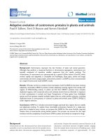
Fig. 4 Enzyme activities of recombinant RNASE1B, RNASE1 and mutant
forms of RNASE1. a, RNase activity
against yeast tRNA at different pH
levels. b, RNase activity against
dsRNA. Mutant forms of douc langur
RNASE1 are indicated by formula XyZ,
in which amino acid X is replaced by Z
at position y of the mature peptide.
Error bars indicate 1 s.e.m.
lagotricha) and lemur (Lemur
catta), with primers PR5 and PR3.
We carried out PCR with highfidelity Taq, under conditions recommended by the manufacturer
(Life Technology), cloned the products into pCR4Blunt-TOPO vector
(Invitrogen) and sequenced from both directions using the dideoxy
chain termination method with the Perkin-Elmer 377 automatic
sequencer. We sequenced several colonies for each species and found no
sequence variation within species, except for douc langur, for which we
identified two distinct sequences. Although possible, it is unlikely that
the two sequences of douc langur are derived from two alleles rather
than two genes, because of their unusually high divergence (7.8% at the
protein sequence level). If they were allelic sequences, overdominant
selection would have to be considered to explain the existence of this
trans-specific polymorphism (Fig. 2b). In addition, our preliminary
study from another Asian colobine (Presbytis francoisi) identified at least
three distinct RNASE1 sequences in one individual (data not shown),
providing definite evidence of RNASE1 gene duplication in colobines.
We also ruled out the possibility that RNASE1B exists in non-colobine
primates but was not detected because it has not diverged in sequence
from RNASE1 (homoduplication). If RNASE1-RNASE1B duplication
had occurred before the separation of colobines from other Old World
monkeys, the age of RNASE1B would be at least 15 Myr (ref. 20), which
converts to a nucleotide difference of 4.5% (2 × 15 × 106 × 1.5 × 10–9)
between the duplicates in noncoding regions, given that the nucleotide
mutation rate in higher primates is about 1.5 × 10⁻⁹ per site per year21.
Thus, the expected number of nucleotide differences between RNASE1
and RNASE1B should be 69 (1,533 × 4.5%) in the 1,533 bp of noncoding
regions we sequenced, and our experiment would have easily detected
two sequences with this level of divergence if they indeed existed in a
non-colobine primate such as the rhesus monkey. The noncoding
regions of RNASE1 and RNASE1B were amplified with primers 263 and
264 using a Platinum TaqPCRx system (Life Technology) under conditions recommended by the manufacturer, and the products were cloned
into pCR4-TOPO of Invitrogen and sequenced. PCR primers are available upon request.
0.0
0.5
1.0
1.5
2.0
2.5
RNase activity against
dsRNA (OD/min/nmol)
Using statistical analysis of nucleotide substitutions and biochemical assays of recombinant proteins, we have described
the adaptive evolution of the duplicated douc langur RNASE1B
in response to increased demands for RNase in an altered
microenvironment of the enzyme. The origin and functional
changes of RNASE1B probably made the digestive system of
these leaf-eating monkeys more efficient. Taken together, our
results provide evidence of the important contribution of gene
duplication to adaptation of organisms to their environments.
It has been debated whether positive selection or relaxation of
purifying selection drives functional divergence of duplicated
genes5–10. In the present case, had the functional constraints
for EAdsRNA not been relaxed, the mutations affecting the catalytic optimal pH for RNASE1B could not have been accepted
because, as we have shown, they result in a loss of EAdsRNA. On
the other hand, without positive selection, it is unlikely that
the net charge of RNASE1B would have undergone such a dramatic change in a short period of evolutionary time before the
gene was deactivated by random nonsense mutations. Functional relaxation clearly made these otherwise deleterious
mutations acceptable, and positive selection further enhanced
the fixation probability of the mutations. In short, the two evolutionary forces had complementary roles in the functional
divergence of RNASE1B from RNASE1. Our observation that
EAdsRNA is retained in RNASE1, with the digestive role transferred to RNASE1B, supports the proposal that gene duplication provides the opportunity for daughter genes to achieve
functional specialization27,28. Fossil records show the emergence of leaf-eating and foregut fermentation in colobines no
later than 10 Myr ago20, predating the origin of RNASE1B.
This suggests that changes in diet and digestive physiology in
colobines provided the selective forces for the evolution of a
more effective digestive RNase, whereas gene duplication provided the raw genetic material. We also note the temporal
proximity of the gene duplication and the radiation of Asian
colobines about 3.5 Myr ago20, which, together with the presence of RNASE1B in at least two genera of Asian colobines
(Pygathrix and Presbytis), suggests the possibility of a causal
link between these events.
Methods
Isolation of RNASE1 and RNASE1B. We amplified the coding region of
RNASE1 and RNASE1B from the genomic DNA of one individual each
of human (Homo sapiens), chimpanzee (Pan troglodytes), gorilla (Gorilla
gorilla), orangutan (Pongo pygmaeus), gibbon (Hylobates leucogenys),
douc langur (Pygathrix nemaeus), rhesus monkey (Macaca mulatta),
pig-tailed macaque (Macaca nemestrina), baboon (Papio hamadryas),
green monkey (Cercopithecus aethiops), talapoin monkey (Miopithecus
talapoin), squirrel monkey (Saimiri sciureus), tamarin (Saguinus oedipus), spider monkey (Ateles geoffroyi), woolly monkey (Lagothrix
414
Evolutionary analysis. Phylogenetic trees were reconstructed by the
MEGA2 program (S. Kumar et al., Arizona State Univ.) using the neighbor-joining method with 1,000 bootstrap replications. We used PHYLIP v.
3.57c (J. Felsenstein, Univ. of Washington) to confirm the MEGA2 results.
Ancestral gene sequences were inferred by the parsimony17 and distancebased Bayesian methods18. The transition/transversion mutational bias29
was estimated from the noncoding region to be 4.37. We computed the
potential numbers of noncoding (I), synonymous (S), nonsynonymous
(N), conservative nonsynonymous (C) and radical nonsynonymous (R)
sites of a sequence as well as the observed substitutions (i, s, n, c, r), at these
sites, between two sequences9,22,30. For the common ancestral gene of douc
langur RNASE1 and RNASE1B, I=1538, S=144.8, N=323.2, C=166.4
(mature peptide) and R=103.9 (mature peptide), and for the RNASE1B
lineage since gene duplication, i=11, s=2, n=10, c=2, and r=7. We used
Fisher’s exact test to compare the rates of substitutions at different types of
sites31. We tested the molecular clock hypothesis using Tajima’s method19.
The duplication event was dated using the noncoding DNA sequences of
douc langur RNASE1, RNASE1B, and rhesus monkey RNASE1, and the
bootstrap method was used to obtain the 95% confidence interval of the
time estimate. We computed isoelectric points (pI) as well as the net
charges of mature peptides with the Wisconsin GCG program.
nature genetics • volume 30 • april 2002
© 2002 Nature Publishing Group
letter
Recombinant proteins and their enzymatic activities. Human, rhesus
monkey, and douc langur RNASE1 and douc langur RNASE1B were subcloned into the bacterial expression vector pFLAG CTS (Kodak) and verified
by sequencing. The vector adds the octapeptide DYKDDDDK (FLAG) to the
recombinant protein, which facilitates its purification and detection with M2
anti-FLAG monoclonal antibody but does not affect the RNase activity32. We
used the QuikChange site-directed mutagenesis kit (Stratagene) to mutate
douc langur RNASE1 and confirmed the mutations by sequencing. Recombinant proteins were isolated, purified and quantified as described32. The
RNase activity of the recombinant proteins in digesting yeast tRNA was measured at different pHs (40 mM sodium acetate buffer with pH=4.0–5.6 and
40 mM sodium phosphate buffer with pH=6.3–8.2) at 25 °C. We added
purified RNase (0.1–1.0 pmol) into 0.8 ml of the aforementioned buffer with
1.42 nmol tRNA. The reaction was stopped by 0.5 ml of 20 mM lanthanum
nitrate with 3% perchloric acid, and insoluble tRNA was removed by centrifugation. The amount of solubilized RNA was determined from ultraviolet absorbance at 260 nm. We computed the catalytic activity of the RNase as
the nmol of RNA digested per second per nmol of RNase32. The RNase activity (EAdsRNA) against dsRNA (poly(U)•poly(A) combined from poly(U)
and poly(A); Pharmacia) was measured at 25 °C in 1 ml buffer of 0.15 M
sodium chloride and 0.015 M sodium citrate (pH 6.3–8.4) with 5 ng substrate and 10–100 pmol RNase, and was determined from ultraviolet
absorbance at 260 nm (ref. 33). EAdsRNA for douc langur RNASE1 and
RNASE1B were both found to be highest at pH 7; the results at pH 7 are
thus reported for all constructs. We carried out at least three replications of
experiments at each condition examined and computed the means and
their standard errors.
GenBank accession numbers. Human RNASE1 cDNA, W84323; Human
RNASE1 genomic sequence, AL133371. The DNA sequences reported in
this paper have been submitted to GenBank (AF449628–46).
5.
6.
7.
8.
9.
10.
11.
12.
13.
14.
15.
16.
17.
18.
19.
20.
21.
22.
23.
24.
Acknowledgments
We thank K. Dyer for technical assistance and J. Beintema, A. Rooney and three
anonymous referees for their comments on early versions of the manuscript. This
work was supported in part by a start-up fund and a Rackham grant from the
University of Michigan (to J.Z.) and grants from the Natural Science
Foundation of China and Chinese Academy of Sciences (to Y.P.Z.).
25.
26.
27.
28.
29.
Received 20 September 2001; accepted 23 January 2002.
1.
2.
3.
4.
Tomb, J.F. et al. The complete genome sequence of the gastric pathogen
Helicobacter pylori. Nature 388, 539–547 (1997).
Makarova, K.S. et al. Comparative genomics of the Archaea (Euryarchaeota):
evolution of conserved protein families, the stable core, and the variable shell.
Genome Res. 9, 608–628 (1999).
Rubin, G.M. et al. Comparative genomics of the eukaryotes. Science 287,
2204–2215 (2000).
Gaudieri, S., Kulski, J.K., Dawkins, R.L. & Gojobori, T. Different evolutionary
histories in two subgenomic regions of the major histocompatibility complex.
Genome Res. 9, 541–549 (1999).
nature genetics • volume 30 • april 2002
30.
31.
32.
33.
Ohno, S. Ancient linkage groups and frozen accidents. Nature 244, 259–262
(1973).
Goodman, M., Moore, G.W. & Matsuda, G. Darwinian evolution in the genealogy
of haemoglobin. Nature 253, 603–608 (1975).
Li, W.H. & Gojobori, T. Rapid evolution of goat and sheep globin genes following
gene duplication. Mol. Biol. Evol. 1, 94–108 (1983).
Kimura, M. The Neutral Theory of Molecular Evolution. (Cambridge University
Press, Cambridge, 1983).
Zhang, J., Rosenberg, H.F. & Nei, M. Positive Darwinian selection after gene
duplication in primate ribonuclease genes. Proc. Natl Acad. Sci. USA 95,
3708–3713 (1998).
Hughes, A.L. Adaptive Evolution of Genes and Genomes (Oxford University Press,
New York, 1999).
Brookfield, J.F. & Sharp, P.M. Neutralism and selectionism face up to DNA data.
Trends Genet. 10, 109–111 (1994).
Charlesworth, B., Charlesworth, D., Nurminsky, D.I. & Hartl, D.L. How was the Sdic
gene fixed? Nature 400, 519–520 (1999).
Kay, R.N.B. & Davies, A.G. in Colobinem Monkeys: Their Ecology, Behaviour and
Evolution (eds Davies, A.G. & Oates, J.F.) 229–250 (Cambridge University Press,
Cambridge, 1994).
Barnard, E.A. Biological function of pancreatic ribonuclease. Nature 221,
340–344 (1969).
Beintema, J.J. The primary structure of langur (Presbytis entellus) pancreatic
ribonuclease: adaptive features in digestive enzymes in mammals. Mol. Biol. Evol.
7, 470–477 (1990).
Goodman, M. et al. Toward a phylogenetic classification of primates based on
DNA evidence complemented by fossil evidence. Mol. Phylogenet. Evol. 9,
585–598 (1998).
Fitch, W.M. Toward defining the course of evolution: minimum change for a
specific tree topology. Syst. Zool. 20, 406–416 (1971).
Zhang, J. & Nei, M. Accuracies of ancestral amino acid sequences inferred by
parsimony, likelihood, and distance methods. J. Mol. Evol. 44, S139–S146 (1997).
Tajima, F. Simple methods for testing the molecular evolutionary clock
hypothesis. Genetics 135, 599–607 (1993).
Delsen, E. in Colobine Monkeys: Their Ecology, Behaviour and Evolution (eds
Davies, A.G. & Oates, J.F.) 11–44 (Cambridge University Press, Cambridge, 1994).
Li, W.H. Molecular Evolution 221–224 (Sinauer Associates, Sunderland,
Massachusetts, 1997).
Zhang, J. Rates of conservative and radical nonsynonymous nucleotide
substitutions in mammalian nuclear genes. J. Mol. Evol. 50, 56–68 (2000).
Sorrentino, S. & Libonati, M. Structure-function relationships in human
ribonucleases: main distinctive features of the major RNase types. FEBS Lett. 404,
1–5 (1997).
Guyton, A.C. & Hall, J.E. Textbook of Medical Physiology 9th edn (Saunders,
London, 1996).
Code, C.F. Handbook of Physiology Vol. III (American Physiological Association,
Washington DC, 1968).
Futami, J. et al. Tissue-specific expression of pancreatic-type RNases and RNase
inhibitor in humans. DNA Cell Biol. 16, 413–419 (1997).
Hughes, A.L. The evolution of functionally novel proteins after gene duplication.
Proc. R. Soc. Lond. B Biol. Sci. 256, 119–124 (1994).
Lynch, M. & Force, A. The probability of duplicate gene preservation by
subfunctionalization. Genetics 154, 459–473 (2000).
Kimura, M. A simple method for estimating evolutionary rates of base
substitutions through comparative studies of nucleotide sequences. J. Mol. Evol.
16, 111–120 (1980).
Rooney, A.P. & Zhang, J. Rapid evolution of a primate sperm protein: relaxation
of functional constraint or positive Darwinian selection? Mol. Biol. Evol. 16,
706–710 (1999).
Zhang, J., Kumar, S. & Nei, M. Small-sample tests of episodic adaptive evolution: a
case study of primate lysozyme genes. Mol. Biol. Evol. 14, 1335–1338 (1997).
Rosenberg, H.F. & Dyer, K.D. Eosinophil cationic protein and eosinophil-derived
neurotoxin. Evolution of novel function in a primate ribonuclease gene family. J.
Biol. Chem. 270, 21539–21544 (1995).
Libonati, M. & Floridi, A. Breakdown of double-stranded RNA by bull semen
ribonuclease. Eur. J. Biochem. 8, 81–87 (1969).
415