Diagnosis and management of spontaneous intracerebral hemorrhage
Bạn đang xem bản rút gọn của tài liệu. Xem và tải ngay bản đầy đủ của tài liệu tại đây (1.44 MB, 11 trang )
Review Article
Address correspondence to
Dr Andrew M. Naidech, 710
N Lake Shore Drive 11th floor,
Chicago, IL 60611,
Relationship Disclosure:
Dr Naidech reports no disclosure.
Unlabeled Use of
Products/Investigational
Use Disclosure:
Dr Naidech discusses the
unlabeled/investigational use
of desmopressin for the
treatment of acute
intracerebral hemorrhage.
* 2015, American Academy
of Neurology.
Diagnosis and
Management of
Spontaneous
Intracerebral
Hemorrhage
Andrew M. Naidech, MD, MSPH, FANA
ABSTRACT
Purpose of Review: This article updates neurologists on recent insights and management strategies of intracerebral hemorrhage (ICH).
Recent Findings: Blood pressure reduction likely improves outcomes in patients with
intracerebral hemorrhage, although not by the expected mechanism of reducing
hematoma growth. One formulation of prothrombin complex concentrate for reversing severe bleeding associated with warfarin is now approved by the US Food and Drug
Administration (FDA), and specific reversal therapies for the novel oral anticoagulants
are in development. Neurologic monitoring frequently detects ICH worsening that requires an intervention. Platelet transfusion and pharmacologic platelet activation are
promising and often used as part of patient management but have not yet been shown
to improve patient outcomes.
Summary: Measurable progress continues toward establishing effective therapies to
improve outcomes in patients with ICH. Blood pressure reduction and reversal of medications that exacerbate bleeding are likely to improve outcomes. Recommendations for
neuromonitoring will help clinicians at the bedside attend to the most important abnormalities and optimize later quality of life. This article reviews standards for diagnosis
and severity of ICH, monitoring and treatment of complications in the hospital, available
interventions, and the measurement of outcomes.
Continuum (Minneap Minn) 2015;21(5):1228–1298.
INTRODUCTION
Intracerebral hemorrhage (ICH) is the
most deadly form of stroke and leaves
many of its survivors with a persistent
neurologic deficit. Despite the high toll
of the disease, the field continues to
improve in diagnosis, targeted neuromonitoring, and patient management.
DIAGNOSIS
ICH is less common than acute ischemic stroke but has a substantially higher
acute mortality and a higher rate of early
1288
clinical decompensation1 and is more
likely to cause subsequent disability.2
Consequently, misdiagnosis is potentially catastrophic. The clinical presentation is often similar to ischemic stroke
in that patients usually present with a
focal neurologic deficit, but are more
likely to have very elevated blood pressure; altered consciousness; and headache, nausea, or vomiting.
The etiology of ICH depends on the
population. ICH in younger populations is more likely due to chronic
www.ContinuumJournal.com
Copyright © American Academy of Neurology. Unauthorized reproduction of this article is prohibited.
October 2015
hypertension, and the hematoma is
more likely to be in the basal ganglia
or brainstem. ICH in older populations
is more likely to be lobar. (This article
does not consider traumatic ICH.) Many
older patients with lobar hematomas
will meet criteria for probable cerebral
amyloid angiopathy (age at least 55 years,
appropriate clinical history, evidence
of multiple cerebral hemorrhages on
MRI), a condition of amyloid deposition in cerebral vessels, and these patients are more likely to be harmed by
anticoagulant medication.3 Ataxia may
be the presenting symptom in patients
with cerebellar hematomas, and these
patients should be considered for early
surgical decompression if there is concern for brainstem compression.
Imaging
The diagnosis of ICH is established by
an appropriate clinical history with corroborating imaging evidence of hemorrhage on CT or MRI scanning. MRI
scanning should be performed to help determine the etiology of ICH (Figure 2-1).
Blood vessel imaging with magnetic
resonance angiography (MRA), CT
angiography (CTA), or conventional
angiography should be considered if
there is a question of a vascular malformation such as an aneurysm or arteriovenous malformation. The yield of
angiographic studies in patients with a
history of hypertension and a typical
appearance of ICH due to hypertension
is very small.
Hematomas frequently expand after
the diagnostic CT scan, particularly in
patients who present soon after symptom onset; patients with hematoma
expansion have a substantially worse
outcome. Thus, minimizing hematoma
expansion is a primary goal of acute
ICH treatment and the driving force
behind aggressively lowering blood
pressure and reversing coagulopathy.
After the diagnostic CT scan, at least
one more brain imaging study should
be performed in symptomatic patients
to determine final hematoma size and
assess for hematoma expansion.
KEY POINT
h Obtaining an MRI is
desirable to help
determine the etiology
of intracerebral
hemorrhage and is
particularly helpful
for cerebral
amyloid angiopathy.
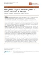
Using MRI to improve diagnosis of intracerebral hemorrhage. A, The patient presented with a small lobar
intracerebral hemorrhage, seen as hyperdensity on noncontrast CT. B, MRI revealed a second hematoma, seen
as dark (hypointense) signal on gradient echo sequence in the left temporal lobe (arrow). Given the
patient’s age, these findings made the diagnosis of amyloid angiopathy likely. C, Later that month, noncontrast CT performed
when the patient presented with right-sided weakness showed spontaneous hemorrhage in another location with
intraventricular extension.
FIGURE 2-1
Continuum (Minneap Minn) 2015;21(5):1228–1298
www.ContinuumJournal.com
Copyright © American Academy of Neurology. Unauthorized reproduction of this article is prohibited.
1289
Intracerebral Hemorrhage
KEY POINTS
h The Intracerebral
Hemorrhage Score is
required documentation
at comprehensive
stroke centers for
patients with
intracerebral hemorrhage.
h Neuromonitoring
encompasses a range of
techniques and data.
Neuromonitoring may
refer to repeated
neurological assessments
(eg, level of alertness,
orientation) over time, to
repeated noninvasive
measures (such as
processed scores from
EEG data), to invasive
monitors that
display brain-specific
measurements.
SEVERITY OF ILLNESS
The Joint Commission has adopted
the ICH Score as a standard severityof-illness scoring system for patients
with ICH, and its documentation will
be required at comprehensive stroke
centers.4 Scores range from 0 (least
severe with low expected mortality)
to 6 (the worse possible score with
death likely). Modifications to the ICH
score that take into account more
clinical or imaging variables may have
slightly better predictive value for outcomes (Table 2-1).
Do-not-resuscitate (DNR) status is a
known confounder of outcomes in patients with ICH. Unsurprisingly, in some
(but not all) centers, patients with DNR
status receive fewer interventions and
have higher mortality rates than patients
with a similar severity of injury. This is
not due to the withholding of any single
beneficial intervention for ICH but may
be owing to a pattern of less-aggressive
care. DNR status should be considered
TABLE 2-1 Intracerebral
Hemorrhage Scorea
Variable
ICH Score
Points
Hematoma volume
Q30 mL
1
Age Q80 years
1
Glasgow Coma
Scale 3 or 4
2
Glasgow Coma
Scale 5Y12
1
Infratentorial
hematoma location
1
Intraventricular
hemorrhage
1
ICH = intracerebral hemorrhage.
a
Modified with permission from Hemphill JC
3rd, et al, Stroke.4 B 2001 American Heart
Association, Inc. stroke.ahajournals.org/content/
32/4/891.full.
1290
with the patient and representatives
while being mindful of its potential impact on subsequent care and outcomes.
NEUROMONITORING
Many ‘‘neuromonitors’’ exist, ranging
from repeated examinations such as
level of consciousness2 and delirium
screening to invasive monitors.5,6 As a
general guide, all patients with acute
ICH should be admitted to an intensive care unit setting to assess for neurologic deterioration, although some
patients may be triaged to a stroke unit
or step-down intensive care unit based
on clinical severity and resource availability (Figure 2-2).
The field of neuromonitoring has recently been exhaustively reviewed by the
Neurocritical Care Society.7 Strong recommendations include the following:
& Invasive blood pressure
monitoring helps patients who
are hemodynamically unstable and
helps establish goals that take
cerebral perfusion into account.
& Oximetry and capnography
(measurement of carbon dioxide
concentration in the blood) are
helpful for mechanically ventilated
patients. There is enthusiasm,
but still only preliminary data,
for the use of brain oxygen
tension monitors.
& Electroencephalography is
recommended to detect subclinical
seizures in patients with persistently
altered consciousness (Case 2-1).
& Blood glucose levels should be
routinely measured.
& For patients whose body
temperature is being actively
managed (eg, cooling blankets,
intravascular devices), shivering
should be regularly monitored with
a standard scale.
These recommendations may be reconsidered in light of locally available
www.ContinuumJournal.com
Copyright © American Academy of Neurology. Unauthorized reproduction of this article is prohibited.
October 2015
FIGURE 2-2
Algorithm of care for intracerebral hemorrhage from presentation through
hospital discharge and follow-up.
CT = computed tomography; EEG = electroencephalogram; ICH = intracerebral
hemorrhage; ICU = intensive care unit; MRI = magnetic resonance imaging.
resources and may not be possible in
all settings. Some monitoring systems
are very resource intensive in terms of
skilled labor and equipment.
INTRAVENTRICULAR HEMORRHAGE
Intraventricular hemorrhage (IVH), the
spread of blood into the ventricular
system, is more common with hematoma locations that are closer to the
ventricular system, such as the thalamus
and caudate nuclei. IVH is a common
and serious complication of ICH that may
lead to reduced consciousness, hydrocephalus, fever, and a worse outcome.
For patients with small to moderate
Case 2-1
A 54-year-old woman presented with a new left-sided hemiparesis. Her blood
pressure was 140/80 mm Hg, and her history was significant for hypertension.
During the initial examination, the patient required stimulation to attend
to the examiner and to follow voice commands. When aroused, she was
oriented to the hospital. On physical examination, there was weakness of the
left face, arm, and leg with moderate dysarthria and neglect to sensation.
There was no aphasia or ataxia. CT scanning revealed a 15-mL right-sided
lobar hematoma. Initial laboratory studies were unremarkable.
Continued on page 1292
Continuum (Minneap Minn) 2015;21(5):1228–1298
www.ContinuumJournal.com
Copyright © American Academy of Neurology. Unauthorized reproduction of this article is prohibited.
1291
Intracerebral Hemorrhage
KEY POINTS
h Dissolving intraventricular
clots with fibrinolytics is
an attractive strategy
for the treatment of
intraventricular
hemorrhage, and a
phase 3 trial is
nearing completion.
h Anticoagulation should
be emergently reversed
in patients with
intracerebral
hemorrhage; the
optimal agent is not
clear, but most
physicians prefer
prothrombin complex
concentrates over fresh
frozen plasma at
this time.
Continued from page 1291
The patient’s mental status initially waxed and waned, and the patient
no longer followed commands the next day. EEG monitoring was initiated
due to encephalopathy, and a lateralized rhythmic pattern was seen on the
same side as the hematoma. After 6 hours of EEG monitoring, a focal seizure
on EEG without a clinical correlate was seen. Levetiracetam 1000 mg IV was
administered with resolution of electrographic seizures but intermittent
rhythmic activity on EEG was still seen. Her mental status improved, and she
resumed following commands on bedside examination.
Comment. Subclinical seizures are common after intracerebral hemorrhage
(ICH) and may be reflected as a depressed mental status or a worsening
neurologic examination. Guidelines do not support the use of prophylactic
antiepileptic drugs (AEDs), particularly phenytoin. However, AEDs are
indicated for clinical or electroencephalographic seizures. Patients with lobar
hematomas, as in this patient, are at a particularly high risk for seizures.
New-onset seizures weeks to months after ICH are also common.
intraparenchymal hematomas and substantial amounts of IVH, intraventricular
clot-busting therapy involves removing
a small volume of CSF via the external
ventricular drain with a syringe and replacing it with alteplase and sterile flush
solution. This is the rationale behind
the phase 3 Clot Lysis: Evaluating Accelerating Resolution of Intraventricular
Hemorrhage (CLEAR-IVH) trial, currently
in progress. Preliminary results have been
promising, although this therapy remains
investigational pending the outcome
of this ongoing phase 3 clinical trial.8
MEDICAL MANAGEMENT
Table 2-2 summarizes the general medical management of ICH.
Anticoagulation-Related
Intracerebral Hemorrhage
As subclinical atrial fibrillation is found
more often, more patients will be
prescribed anticoagulant medication.9 The traditional treatment for
atrial fibrillation has been warfarin,
and ICH is the most feared complication of anticoagulant treatment.
When patients taking warfarin experience severe bleeding, fresh-frozen
plasma has been typically prescribed.
Recently, prothrombin complex con-
1292
centrates have been evaluated as a
potentially more effective alternative, and
one proprietary formulation (KCentra)
has been recently approved by the US
Food and Drug Administration (FDA)
specifically for reversing bleeding related to warfarin.10 This is a general indication, and few patients with ICH were
in the study leading to this approval.
Trials of novel oral anticoagulants
(NOACs) in otherwise healthy patients
with atrial fibrillation showed NOACs
to be equivalent or superior to warfarin for stroke prevention and to be
associated with lower rates of ICH.11,12
However, NOAC-associated ICH may
be difficult to treat. This is likely to be
especially problematic for older people, who are more likely to have atrial
fibrillation and more likely to die after
ICH. How best to reverse NOACs is not
known, although a specific antidote for
dabigatran is the subject of an ongoing clinical study.
The optimal timing of restarting anticoagulant medication after ICH is controversial, and few data exist to guide
management (Case 2-2). A delay of 1 week
to 3 months is considered reasonable, with early anticoagulation favored
for patients with a high risk of thromboembolism, such as patients with
www.ContinuumJournal.com
Copyright © American Academy of Neurology. Unauthorized reproduction of this article is prohibited.
October 2015
TABLE 2-2 General Management of Intracerebral Hemorrhage
Condition
Recommendation
Anticoagulant medication
Normalization of international normalized ratio (INR)
Blood pressure
For patients with systolic blood pressure 9150 mm Hg
and e220 mm Hg, consider lowering to 140 mm Hg
For patients presenting with systolic blood pressure
9220 mm Hg, consider aggressive reduction of
blood pressure with a continuous IV infusion
of an antihypertensive and frequent blood
pressure monitoring1
Fever
Antipyretic medication; consider ice packs or
devices for temperature control (preferably
avoiding sedation, as appropriate)
Cerebral edema
Hypertonic saline and/or mannitol, usual goal
320 mOsm/L with weaning over several days
Antiplatelet medication
Consider desmopressin or platelet transfusion
Hyperglycemia
Routine protocol for glucose control
Deep venous thrombosis
prevention
Consider mechanical prophylaxis; consider
chemoprophylaxis after hematoma size stable for
2Y3 days
IV = intravenous.
mechanical heart valves or patients with
evidence of new cerebral ischemia on
MRI, and deferred for patients with
evidence of new hemorrhage on
follow-up MRI scanning. In the absence
of clear guidelines and because of
often competing therapeutic concerns
(eg, anticoagulation to prevent cardioembolism, deferred anticoagulation to
minimize the risk of recurrent ICH),
this decision is often made after discussion among the consulting physicians.
Blood Pressure Reduction
A prevailing theory has been that hematoma expansion indicates a physical
tear in an artery or arteriole and that increased blood pressure leads to greater
blood flow out of the tear into brain parenchyma. Thus, aggressively reducing
blood pressure might reduce hematoma
expansion and improve functional outcomes. The Intensive Blood Pressure
Continuum (Minneap Minn) 2015;21(5):1228–1298
Reduction in Acute Cerebral Hemorrhage (INTERACT) trial suggested this
hypothesis was valid, with less proportional hematoma growth in patients
with more aggressive blood pressure
reduction (target systolic 140 mm Hg
or less).13 This formed the basis for
INTERACT2, which enrolled nearly 2800
patients with acute ICH. INTERACT2
did not achieve the primary end point
of improved odds of ‘‘good outcome,’’
which was defined as moderately severe
disability or better at 90 days. Neither
did aggressive blood pressure reduction have an effect on hematoma expansion.14 INTERACT2 did, however,
find that aggressive blood pressure reduction was associated with: (1) improved functional outcomes when
analyzed as an ordinal shift toward
lower levels of disability; and (2) improved quality of life. This implies that
there may be another mechanism
www.ContinuumJournal.com
Copyright © American Academy of Neurology. Unauthorized reproduction of this article is prohibited.
1293
Intracerebral Hemorrhage
KEY POINT
h Fever leads to worse
outcomes in patients
with intracranial
hemorrhage; whether
aggressive measures to
abolish fever improves
outcomes in these
patients is not clear.
Case 2-2
A 75-year-old man presented with new-onset headache and right
hemiparesis with onset 45 minutes prior to presentation to the emergency
department. His history was significant for hypertension and atrial fibrillation,
for which he took warfarin. Blood pressure was 185/95 mm Hg, and his
temperature was 37.2-C (99.0-F). Physical examination confirmed the
right hemiparesis with moderate sensory loss; he followed commands,
uttered inappropriate words, and required stimulation to open his eyes
(Glasgow Coma Scale score of 11). A CT scan revealed a 34-mL left parietal
lobe hematoma with scant intraventricular hemorrhage. His Intracerebral
Hemorrhage (ICH) Score was recorded as 3 (1 point for a Glasgow Coma Scale
of 11, 1 point for hematoma volume of greater than 30 mL, 1 point for
intraventricular hemorrhage [IVH]).
The patient’s blood pressure was reduced to 140 mm Hg systolic. Warfarin
was reversed with prothrombin complex concentrate. Fever developed on
day 3 and was treated with acetaminophen. Altered mental status prompted
EEG monitoring, which was discontinued after 48 hours when no epileptiform
abnormalities were seen. Repeat CT scanning demonstrated minimal hematoma
growth, and an MRI revealed no other foci of intracerebral hemorrhage. The
patient was discharged to a rehabilitation facility. At 1 month, he was awake,
alert, and able to ambulate with a device. Plans were being made to return
home with outpatient physical and occupational therapy. Warfarin was restarted.
Comment. When to restart anticoagulation in patients with ICH is not
well defined. One month is generally considered a reasonable time frame
in patients considered to be a low risk for recurrent ICH.
accounting for a slight benefit from
aggressive blood pressure reduction
other than reduction in hematoma
growth. INTERACT2 has been influential, particularly given the lack of other
available interventions to improve outcomes after ICH.
Patients with ICH and a long history
of hypertension may have an autoregulatory curve that is shifted to the
right (ie, have cerebral vessels that effectively regulate cerebral blood flow
at hypertensive blood pressures but
autoregulate less effectively at normal
blood pressure). However, blood pressure reduction (down to a systolic blood
pressure of 140 mm Hg) does not seem
to cause perihematomal ischemia or
neurologic decline.14,15 Small areas of
ischemia distant from the hematoma
have been reported with aggressive
blood pressure reduction.16,17
1294
Fever and Temperature Control
Temperature should be routinely measured in patients with ICH. Where available, a core measure using a bladder
catheter is preferred; however, protocols to reduce the use of indwelling
bladder catheters to minimize infection
risk may make this difficult in awake
patients. Fever (ie, elevated core temperature) has been repeatedly linked
to worse outcomes in patients with
ICH.18 Fever has many deleterious effects, including increased brain and
muscle metabolism that may, in turn,
have additional adverse consequences.
Documented associations between
fever and worse outcome have led clinicians to attempt to reduce fever, which
has been more difficult and, thus far,
less rewarding than initially hoped.19
Antipyretics are routinely given but are
typically insufficient to abolish fever. A
www.ContinuumJournal.com
Copyright © American Academy of Neurology. Unauthorized reproduction of this article is prohibited.
October 2015
variety of cooling devices are available,
some external and some intravascular.
Cold saline also acutely reduces core
temperature and is a common intervention for temperature reduction after
cardiac arrest.20,21 Devices generally are
effective in reducing temperature in patients with fever that persists despite
antipyretic medication, but shivering
is a common result that may prevent
fever control. Shivering can be reliably
assessed at the bedside, and a variety
of off-label medications (eg, buspirone,
fentanyl, meperidine) and interventions
(such as counterwarming with warm air)
have been proposed to minimize shivering; none are FDA-approved for this
indication.22Y24As of yet, high-quality data
on whether interventions to reduce fever
improve outcomes after ICH are lacking,
although several studies are under way.
Cerebral Edema
Cerebral edema is common after ICH.
In general, the volume of cerebral
edema is proportional to the volume
of the hematoma, with larger hematomas leading to more edema. This is
particularly important in patients with
hematomas large enough to cause midline shift and altered consciousness. The
exact cause of cerebral edema is not
clear; ischemia around the hematoma
does not seem to be a proximate cause.
Cerebral edema is commonly visualized as hypodensity surrounding the
hematoma on CT or hyperintensity on
T2-weighted MRI, and usually peaks several days after ICH onset. Treatment
usually consists of hyperosmolar therapy with hypertonic saline, mannitol,
or both. Mannitol can be given via peripheral IV but may lead to volume depletion with repeated dosing because
it is an osmotic diuretic. Hypertonic
saline requires a central venous catheter but can be used indefinitely. A target
serum osmolality of approximately
320 mOsm/L (to avoid nephrotoxicity),
Continuum (Minneap Minn) 2015;21(5):1228–1298
or resolution of clinical symptoms, is
the usual target of therapy. Evidencebased protocols for discontinuation
of hyperosmolar therapy have not been
developed; the usual practice is to permit serum osmolality to decrease by up
to 10 mEq/L/d as long as there are no
symptoms of recurrent cerebral edema.
For more information, refer to the
article ‘‘Management of Intracranial
Pressure’’ by W. David Freeman, MD,
FSNS, FAAN, in this issue of Continuum.
KEY POINT
h Cerebral edema is
common after
intracerebral hemorrhage
and generally reflects
the volume of the
underlying hematoma.
Antiplatelet Medication and
Platelet-Activating Therapy
Clinical interventions to improve platelet activity are analogous to correcting
coagulopathy in patients with ICH. As
anticoagulants lead to reduced blood
clotting, aspirin and nonsteroidal antiinflammatory drugs (NSAIDs) lead to
platelet inhibition that reduces the formation of a platelet plug at the site of
bleeding. The use of aspirin and NSAIDs
can be detected on rapid point-of-care
testing in patients with acute ICH.25
When detected, reduced platelet activity is associated with more IVH, more
hematoma growth, increased mortality
at 14 days, and worse functional outcomes at 3-month follow-up.26,27
Interventional trials to improve platelet activity are under way. Platelet transfusion was a logical step to improve
platelet activity and improves pointof-care assay results. A prospective, randomized controlled trial of platelet
transfusion (PATCH) is under way in
Europe.28 Pending these data, platelet
transfusion for ICH has become commonplace in some centers.29 However,
platelet transfusion has potential adverse events, such as infection, volume
overload, and limited supply. Desmopressin has been prescribed for
more than 2 decades to improve platelet activity in patients known to take
aspirin. In a recent phase 2a trial, it improved platelet activity in patients with
acute ICH.30,31
www.ContinuumJournal.com
Copyright © American Academy of Neurology. Unauthorized reproduction of this article is prohibited.
1295
Intracerebral Hemorrhage
KEY POINTS
h Immediate neurosurgical
consultation is
particularly indicated for
cerebellar hemorrhage,
large lobar hemorrhage,
hydrocephalus, midline
shift, and a decrease in
consciousness on
serial neuromonitoring.
h Neuro-QOL, the
Patient-Reported
Outcomes Measurement
Information System,
and National Institutes
of Health Toolbox are
web-based outcome
measures developed by
the National Institutes
of Health.
Statins ("-Hydroxy"-Methylglutaryl-CoA
Reductase Inhibitors)
Controversy has surrounded the use
of statin drugs in patients at high risk
for ICH, particularly lobar ICH.32 This
was buttressed by data showing an association between aggressively lowering cholesterol and a higher risk of
ICH.33 More recent data suggest statins
may be associated with no harm and
perhaps better outcomes after ICH.34,35
Withholding or reducing the dose in
patients with ICH and very low density
lipoprotein cholesterol seems prudent.
Otherwise, discontinuation of statins
does not appear to be necessary in
most ICH patients.
SURGICAL MANAGEMENT
A working relationship with neurosurgical colleagues is crucial to maximizing
outcomes for patients with ICH. Particular consideration should be given to
the following:
&
Patients with cerebellar hematomas
since these patients are at a high
risk for brainstem compression
& Large lobar hematomas since these
are most accessible
& Ventricular drainage for patients
with hydrocephalus or IVH
& Patients with midline shift, as this
may be surgically correctable
& A decrease in consciousness on
serial neuromonitoring, as this
may indicate an expanding
intracranial hematoma
Other than patients who are highly
likely to clinically benefit from surgical
decompression (eg, large cerebellar
hematomas, hemispheric hematomas
causing tissue shift, and neurologic decline in patients with good rehabilitation
potential), the best way to select patients
for surgical decompression is less clear.
In patients without a clear need for
emergent surgical decompression, two
1296
clinical trials of early surgery (via craniotomy) versus expectant management
found no difference in outcomes.36 Hematoma evacuation by means of stereotaxis is currently being investigated in the
Minimally Invasive Surgery Plus rt-PA for
Intracerebral Hemorrhage Evacuation
(MISTIE) trial.37
ANALYSIS OF OUTCOMES DATA
The most common outcome metric for
ICH is the modified Rankin Scale (mRS),
a global functional scale from 0 (no symptoms) to 6 (dead). The mRS has a high
inter-rater reliability when validated questionnaires are used for its assessment, so
different raters will generally record the
same result.38
A variety of scores for health-related
quality of life are also available and
are generally correlated with the mRS.
The National Institutes of Health (NIH)
has recently released novel outcome
measures: Neuro-QOL is a series of
questionnaires specifically developed
and validated in patients with neurologic diseases.39 The Patient-Reported
Outcomes Measurement Information
System has more general instruments,
many of which ‘‘cross-walk’’ to NeuroQOL measures. The NIH Toolbox is a
set of performance measures in motor, cognitive, sensory, and self-reported
emotional health.40 These low-cost,
web-based tools make it possible for
more centers to comprehensively
obtain state-of-the-art outcomes and
examine how their processes might
maximize health-related quality of life
(www.assessmentcenter.net).
CONCLUSION
Care of patients with acute ICH has
been improved by better description of
severity and complications, evolutions
in monitoring and control of vital signs,
and measurable improvements in outcomes with specific interventions. The
www.ContinuumJournal.com
Copyright © American Academy of Neurology. Unauthorized reproduction of this article is prohibited.
October 2015
next several years are likely to see further advancements that improve functional outcomes and quality of life for
survivors of ICH.
REFERENCES
1. Hemphill JC, Greenberg SM, Anderson CS,
et al. American Heart Association Stroke
Council, Council on Cardiovascular and Stroke
Nursing, and Council on Clinical Cardiology.
Guidelines for the management of spontaneous
intracerebral hemorrhage: a guideline for
healthcare professionals from the American
Heart Association/American Stroke Association.
[published online ahead of print May 28,
2015] Stroke 2015;46.doi:10.1161/STR.
0000000000000069.
2. Maas MB, Rosenberg NF, Kosteva AR,
et al. Surveillance neuroimaging and
neurologic examinations affect care for
intracerebral hemorrhage. Neurology
2013;81(2):107Y112. doi:10.1212/WNL.
0b013e31829a33e4.
3. Knudsen KA, Rosand J, Karluk D,
Greenberg SM. Clinical diagnosis of
cerebral amyloid angiopathy: validation
of the Boston criteria. Neurology
2001;56(4):537Y539. doi:10.1212/WNL.56.4.537.
4. Hemphill J 3rd, Bonovich D, Besmertis L, et al.
The ICH score: a simple, reliable grading scale
for intracerebral hemorrhage. Stroke 2001;32(4):
891Y897. doi:10.1161/01.STR.32.4.891.
5. Naidech AM, Beaumont JL, Rosenberg NF,
et al. Intracerebral hemorrhage and delirium
symptoms. Length of stay, function, and
quality of life in a 114-patient cohort.
Am J Respir Crit Care Med 2013;188(11):
1331Y1337. doi:10.1164/rccm.201307-1256OC.
6. Claassen J, Perotte A, Albers D, et al.
Nonconvulsive seizures after subarachnoid
hemorrhage: multimodal detection and
outcomes. Ann Neurol 2013;74(1):53Y64.
doi:10.1002/ana.23859.
7. Le Roux P, Menon DK, Citerio G, et al.
Consensus summary statement of the
International Multidisciplinary Consensus
Conference on Multimodality Monitoring
in Neurocritical Care: a statement for
healthcare professionals from the Neurocritical
Care Society and the European Society of
Intensive Care Medicine. Neurocrit Care 2014;
21(suppl 2):S1YS26. doi:10.1007/s12028-014-0041-5.
8. Morgan T, Awad I, Keyl P, et al.
Preliminary report of the clot lysis evaluating
accelerated resolution of intraventricular
hemorrhage (CLEAR-IVH) clinical trial.
Acta Neurochir Suppl 2008;105:217Y220.
doi:10.1007/978-3-211-09469-3_41.
Continuum (Minneap Minn) 2015;21(5):1228–1298
9. Sanna T, Diener HC, Passman RS, et al.
Cryptogenic stroke and underlying atrial
fibrillation. N Engl J Med 2014;370(26):
2478Y2486. doi:10.1056/NEJMoa1313600.
10. Sarode R, Milling TJ Jr, Refaai MA,
et al. Efficacy and safety of a 4-factor
prothrombin complex concentrate in
patients on vitamin K antagonists
presenting with major bleeding: a randomized,
plasma-controlled, phase IIIb study. Circulation
2013;128(11):1234Y1243. doi:10.1161/
CIRCULATIONAHA.113.002283.
11. Connolly SJ, Ezekowitz MD, Yusuf S,
et al. Dabigatran versus warfarin in
patients with atrial fibrillation. N Engl
J Med 2009;361(12):1139Y1151.
doi:10.1056/NEJMoa0905561.
12. Connolly SJ, Eikelboom J, Joyner C, et al.
Apixaban in patients with atrial fibrillation.
N Engl J Med 2011;364(9):806Y817.
doi:10.1056/NEJMoa1007432.
13. Anderson CS, Huang Y, Wang JG, et al.
Intensive blood pressure reduction in acute
cerebral haemorrhage trial (INTERACT):
a randomised pilot trial. Lancet Neurol
2008;7(5):391Y399. doi:10.1016/
S1474-4422(08)70069-3.
14. Anderson CS, Heeley E, Huang Y, et al.
Rapid blood-pressure lowering in patients
with acute intracerebral hemorrhage.
N Engl J Med 2013;368(25):2355Y2365.
doi:10.1056/NEJMoa1214609.
15. Gould B, McCourt R, Gioia LC, et al. Acute
blood pressure reduction in patients with
intracerebral hemorrhage does not result in
borderzone region hypoperfusion. Stroke
2014;45(10):2894Y2899. doi:10.1161/
STROKEAHA.114.005614.
16. Prabhakaran S, Gupta R, Ouyang B, et al.
Acute brain infarcts after spontaneous
intracerebral hemorrhage: a diffusion-weighted
imaging study. Stroke 2010;41(1):89Y94.
doi:10.1161/STROKEAHA.109.566257.
17. Garg RK, Liebling SM, Maas MB, et al.
Blood pressure reduction, decreased diffusion
on MRI, and outcomes after intracerebral
hemorrhage. Stroke 2012;43(1):67Y71.
doi:10.1161/STROKEAHA.111.629493.
18. Schwarz S, Ha¨fner K, Aschoff A, Schwab S.
Incidence and prognostic significance of
fever following intracerebral hemorrhage.
Neurology 2000;54(2):354Y361. doi:10.1212/
WNL.54.2.354.
19. Lord A, Karinja S, Lantigua H, et al.
Therapeutic temperature modulation for
fever after intracerebral hemorrhage.
Neurocrit Care 2014;21(2):200Y206.
doi:10.1007/s12028-013-9948-5.
www.ContinuumJournal.com
Copyright © American Academy of Neurology. Unauthorized reproduction of this article is prohibited.
1297
Intracerebral Hemorrhage
20. Mayer SA, Kowalski RG, Presciutti M, et al.
Clinical trial of a novel surface cooling system
for fever control in neurocritical care patients.
Crit Care Med 2004;32(12):2508Y2515.
doi:10.1097/01.CCM.0000147441.39670.37.
31. Naidech AM, Maas MB, Levasseur-Franklin KE,
et al. Desmopressin improves platelet activity
in acute intracerebral hemorrhage. Stroke
2014;45(8):2451Y2453. doi:10.1161/
STROKEAHA.114.006061.
21. Diringer MN; Neurocritical Care Fever
Reduction Trial Group. Treatment of fever in
the neurologic intensive care unit with a
catheter-based heat exchange system. Crit
Care Med 2004;32(2):559Y564. doi:10.1097/
01.CCM.0000108868.97433.3F.
32. Westover M, Bianchi MT, Eckman MH,
Greenberg SM. Statin use following
intracerebral hemorrhage: a decision
analysis. Arch Neurol 2011;68(5):573Y579.
doi:10.1001/archneurol.2010.356.
22. Badjatia N, Strongilis E, Gordon E, et al.
Metabolic impact of shivering during
therapeutic temperature modulation: the
Bedside Shivering Assessment Scale. Stroke
2008;39(12):3242Y3247. doi:10.1161/
STROKEAHA.108.523654.
23. Mokhtarani M, Mahgoub AN, Morioka N,
et al. Buspirone and meperidine synergistically
reduce the shivering threshold. Anesth Analg
2001;93(5):1233Y1239. doi:10.1097/
00000539-200111000-00038.
24. Badjatia N, Strongilis E, Prescutti M, et al.
Metabolic benefits of surface counter warming
during therapeutic temperature modulation.
Crit Care Med 2009;37(6):1893Y1897.
doi:10.1097/CCM.0b013e31819fffd3.
25. Naidech AM, Bernstein RA, Levasseur K,
et al. Platelet activity and outcome
after intracerebral hemorrhage.
Ann Neurol 2009;65(3):352Y356.
doi:10.1002/ana.21618.
34. Lei C, Wu B, Liu M, Chen Y. Association
between statin use and intracerebral
hemorrhage: a systematic review and
meta-analysis. Eur J Neurol 2014;
21(2):192Y198. doi:10.1111/ene.12273.
35. Flint AC, Conell C, Rao VA, et al. Effect
of statin use during hospitalization for
intracerebral hemorrhage on mortality
and discharge disposition. JAMA Neurol
2014;71(11):1364Y1371. doi:10.1001/
jamaneurol.2014.2124.
26. Naidech AM, Bendok BR, Garg RK, et al.
Reduced platelet activity is associated with
more intraventricular hemorrhage.
Neurosurgery 2009;65(4):684Y688. doi:10.1227/
01.NEU.0000351769.39990.16.
36. Mendelow AD, Gregson BA, Rowan EN,
et al. Early surgery versus initial
conservative treatment in patients with
spontaneous supratentorial lobar intracerebral
haematomas (STICH II): a randomised trial.
Lancet 2013;382(9890):397Y408.
doi:10.1016/S0140-6736(13)60986-1.
27. Naidech AM, Jovanovic B, Liebling S, et al.
Reduced platelet activity is associated
with early clot growth and worse 3-month
outcome after intracerebral hemorrhage.
Stroke 2009;40(7):2398Y2401.
doi:10.1161/STROKEAHA.109.550939.
37. United States National Institutes of Health.
Minimally Invasive Surgery Plus Rt-PA
for ICH Evacuation Phase III. Clinical Trials
web site. www.clinicaltrials.gov/ct2/
results?term=NCT01827046&Search=Search.
Accessed July 30, 2015.
28. de Gans K, de Haan RJ, Majoie CB,
et al. PATCH: platelet transfusion in
cerebral haemorrhage: study protocol
for a multicentre, randomised, controlled
trial. BMC Neurol 2010;10:19.
doi:10.1186/1471-2377-10-19.
38. Saver JL, Filip B, Hamilton S, et al.
Improving the reliability of stroke disability
grading in clinical trials and clinical practice:
the Rankin Focused Assessment (RFA).
Stroke 2010;41(5):992Y995. doi:10.1161/
STROKEAHA.109.571364.
29. Ziai WC, Mirski MA. The slippery slope of
platelet transfusion for intracerebral
hemorrhage. Neurocrit Care 2012;17(1):
154Y155. doi:10.1007/s12028-012-9694-0.
39. Cella D, Lai JS, Nowinski CJ, et al. Neuro-QOL:
brief measures of health-related quality of
life for clinical research in neurology.
Neurology 2012;78(23):1860Y1867. doi:10.
1212/WNL.0b013e318258f744.
30. Keyl C, Kmitta E, Kueri S, et al. Effects
of aspirin and desmopressin on platelet
reactivity in patients undergoing cardiac
surgery with extracorporeal circulation.
Thromb Haemost 2010;105(1):113Y121.
doi:10.1160/TH10-07-0471.
1298
33. Goldstein LB, Amarenco P, LaMonte M,
et al. Relative effects of statin therapy on
stroke and cardiovascular events in men and
women: secondary analysis of the Stroke
Prevention by Aggressive Reduction in
Cholesterol Levels (SPARCL) Study. Stroke
2008;39(9):2444Y2448. doi:10.1161/
STROKEAHA.107.513747.
40. Gershon RC, Cella D, Fox NA, et al.
Assessment of neurological and behavioural
function: the NIH Toolbox. Lancet Neurol
2010;9(2):138Y139. doi:10.1016/
S1474-4422(09)70335-7.
www.ContinuumJournal.com
Copyright © American Academy of Neurology. Unauthorized reproduction of this article is prohibited.
October 2015