Điện tâm đồ trong nhồi máu cơ tim
Bạn đang xem bản rút gọn của tài liệu. Xem và tải ngay bản đầy đủ của tài liệu tại đây (10.85 MB, 119 trang )
Điện tâm đồ
trong nhồi
máu cơ tim
TS. Đinh Hiếu Nhân
Acute Myocardial Infarction
• ST segment elevation MI – persistent complete occlusion of an
artery supplying a significant area of myocardium without adequate
collateral circulation
• UA/NSTEMI – result from non-occlusive thrombus, small risk area,
brief occlusion, or an occlusion with adequate collaterals
I. Chẩn đoán NMCT
ECG trong NMCT
• Chẩn đoán (+) NMCT cấp có ST chênh lên.
• Chẩn đoán giai đoạn NMCT cấp.
• Chẩn đoán vùng NMCT.
• Chẩn đoán biến chứng RLNT
ECG changes in AMI
• In the early stages of AMI the ECG may be normal
• <50% of patients with AMI have clear diagnostic
changes on their first trace.
• About 10% of patients with a proved acute myocardial
infarction fail to develop ST segment elevation or
depression.
• In most cases, however, serial ECG’s show evolving
changes that tend to follow well recognised patterns.
Biến đổi ECG trong NMCT
• ST – T chênh lên.
• Sóng Q bệnh lý
J point
ST segment
Last deflection of QRS
Sự tạo thành các biến đổi của sóng
ECG trong NMCT
Tạo thành sóng Q
Tạo thành đạon ST chênh lên hay
chênh xuống
Đoạn ST
• ST segment of the cardiac cycle represents the period between
depolarization and repolarization of the left ventricle
• In normal state, ST segment is isoelectric relative to PR segment
Minnesota Code
• The Minnesota code 9-2 requires ≥1 mm ST elevation in one or
more of leads I, II, III, aVL, aVF, V5, V6, or ≥ 2 mm ST elevation in
one or more of leads V1–V4
• Menown IB, Mackenzie G, Adgey AA. Optimizing the initial 12-lead electrocardiographic
diagnosis of acute myocardial infarction. Eur Heart J 2000; 21 (4):275-83.
• Đoạn ST chênh lên ở ít nhất 2 chuyển đạo kế tiếp nhau
ST Segment
Elevation
Acute Myocardial Infarction
• Irrespective of which definition is used, ST elevation has poor
sensitivity for AMI where up to 50% of patients exhibit ‘atypical’
changes at presentation including isolated ST depression, T
inversion or even a normal ECG
• Menown IB, Mackenzie G, Adgey AA. Optimizing the initial 12-lead electrocardiographic
diagnosis of acute myocardial infarction. Eur Heart J 2000; 21 (4):275-83.
How To Differentiate STE due
to AMI from Other Causes?
• Magnitude of the elevation
• Morphology
• Distribution
• Prominent Electrical Forces (Voltage Amplitude)
• QRS width
• Other Features
Morphology of the
ST Elevation
Variable Shapes Of ST
Segment Elevations in AMI
Goldberger AL. Goldberger: Clinical Electrocardiography: A Simplified Approach. 7th
ed: Mosby Elsevier; 2006.
Morphology of STE
• Concave shape STE – non AMI causes
• AMI causes – usually demonstrate convex/straight STE
Apex of T wave
J point
Convex STE
Concave STE
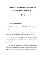
Benign Early Repolarization
Concave STE
Large amplitude T
wave
Notching or slurring of
J point
Benign Early Repolarization
•
1.
2.
3.
4.
ECG characteristics:
STE <2 mm
Concavity of initial portion of the ST segment
Notching or slurring of the terminal QRS complex
Symmetrical, concordant T wave of large
amplitude
5. Widespread or diffuse distribution of STE
o Does not demonstrate territorial distribution
6. Relative temporal stability
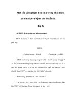
Distribution
• STE due to AMI usually demonstrate regional or territorial pattern
•
•
•
•
•
•
Examples:
Anterior MI – V3-V4
Septal MI – V2-V3
Anteroseptal MI – V1/2 – V4/5
Lateral MI – V5/V6
Inferior MI – II, III, aVF
• Diffuse STE – non AMI causes, e.g. pericarditis
Lateral Wall MI: I, aVL, V5, V6
Source: The 12-Lead ECG in Acute Coronary Syndromes, MosbyJems, 2006.
Inferior Wall MI II, III, aVF
Source: The 12-Lead ECG in Acute Coronary Syndromes, MosbyJems, 2006.