Classification of vascular anomalies
Bạn đang xem bản rút gọn của tài liệu. Xem và tải ngay bản đầy đủ của tài liệu tại đây (16.86 MB, 43 trang )
Updated ISSVA
(International Society for the Study of Vascular Anomalies)
Children’s Hospital 2
Department of diagnosis imaging
Dr Lien Bang
INTRODUCTION
Vascular anomalies are the most common skin and soft
tissue lesions observed in infants and children.
Older nomenclature continues to cause confusion,
misunderstood diagnoses, and potential mismanagement
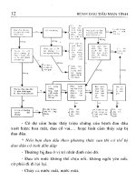
In 1982, Mulliken and Glowacki proposed a classification
system for vascular anomalies based on their clinical
behavior and endothelial cell characteristics into two groups:
hemangiomas and vascular malformations.This system,
which was adopted by the ISSVA, has since been expanded
and is now widely accepted.
Radiologists can use the ISSVA classification system by
correlating imaging findings with patient history and physical
findings. Consistent use of this system will help patients
receive the correct diagnosis and treatment.
Vascular anomalies
Hemangioma
Vascular malformation
“…Not
“…Not every skin lesion looking like a strawberry is a hemangioma;
hemangioma; not all hemangiomas look
like strawberries…
strawberries…" J.B. Mulliken,
Mulliken, MD
Traditional classification
Capillary hemangioma
Strawberry hemangioma
Strawberry nevus
Port
wine stain
Flame nevus
Cavernous hemangioma
Venous angioma
Lymphangioma
Arteriovenous malformation
Translation from old terminology into
classification of ISSVA
Hemangioma
Old Terminology
*Capillary
*Strawberry
*Port-wine
*Capillary-cavernous
*Cavernous
*Vernous
*Hemangiolymphangioma
*Lymphangioma
*Arteriovenous
Vascular
malformation
* CAPILLARY
* VENOUS
*LYMPHATIC
*ARTERIOVENOUS
Updated ISSVA classification of vascular anomalies.
Vascular tumors
Infantile hemangiomas
Congenital hemangiomas (RICH and
NICH)
Tufted angioma (with or without
KasabachKasabach-Merritt syndrome)
Kaposiform hemangioendothelioma
(with or without KasabachKasabach-Merritt
syndrome)
Spindle cell hemangioendothelioma
Other, rare hemangioendotheliomas
(epithelioid,
epithelioid, composite, retiform,
retiform,
polymorphous, Dabska tumor,
lymphangioendotheliomatosis,
lymphangioendotheliomatosis, etc.)
Dermatologic acquired vascular
tumors (pyogenic
(pyogenic granuloma,
granuloma,
targetoid hemangioma,
hemangioma, glomeruloid
hemangioma,
hemangioma, microvenular
hemangioma,
hemangioma, etc.)
Vascular malformations
1..Slow1..Slow-flow vascular malformations:
Capillary malformation (CM)
PortPort-wine stain
Telangiectasia
Angiokeratoma
Venous malformation (VM)
Common sporadic VM
Bean syndrome
Familial cutaneous and mucosal venous
malformation (VMCM)
Glomuvenous malformation
(GVM)(glomangioma)
GVM)(glomangioma)
Maffucci syndrome
Lymphatic malformation (LM)
2. FastFast-flow vascular malformations:
Arterial malformation (AM)
Arteriovenous fistula (AVF)
Arteriovenous malformation (AVM)
3.Complex3.Complex-combined vascular malformations:
CVM, CLM, LVM, CLVM,
AVMAVM-LM, CMCM-AVM
C:capillary; V:venous; L:lymphatic; AV:arteriovenous; M:malformation.
RICH:rapidly involuting congenital hemangioma; NICH:noninvoluting congenital hemangioma.
Vascular anormalies
#
Vascular tumors
Infantile
hemangioma
NICH
Congenital
hemangioma
Vascular malformations
Slow-flow
Fast-flow
RICH
•Capillary malformation(CM)
•Venous malformation(VM)
•Lymphatic malformation(LM)
•Arterial malformation(AM)
•Arteriovenous fistula(AVF)
•Arteriovenous malformation(AVM)
•Combined types
Differentiating Features
Hemangiomas
True tumors, with
Vascular Malformations
proliferation No tumor, Comprised of
of the vascular endothelium
dysplastic vessels
>3:1 female:male
1:1 female:male
Small or absent at birth
Present at birth
Rapid growth during infancy
Growth proportional to child
Self-limited
Never disappear
Diagnosis:Clinical history+
Diagnosis: MRI,Doppler
appearance
ultrasonography,angiography
HEMANGOMA
Benign endothelial
cell tumor
2 main types
1. Infantile Hemangioma
•
•
•
•
Most common tumor of infancy/childhood
Usually has overlying patch of redness
Appears weeks/months after birth
Natural course - 3 stages
1. Proliferating - first year
2. Involuting - few years
3. Involuted - most resolved by age 10
HEMANGIOMA (cont)
2. Congenital Hemangioma
•
•
•
•
Present at birth
Rare (compared to infantile)
Blue/gray hue, pale halo (skin)
2 types
Non Involuting Congenital Hemangioma (NICH) - persistent
Rapidly Involuting Congenital Hemangioma (RICH) resolved by 1-2 yrs
Growth patterns of hemangiomas
NICH
GROWTH
RICH
IH
AGE
BIRTH
1 YR
2 YRS
RICH:rapidly involuting congenital hemangioma; NICH:noninvoluting congenital hemangioma. IH: Infantile Hemangioma
HEMANGIOMA
Infantile hemangioma
congenital hemangioma
HEMANGIOMA
Infantilehemangioma in a 4-month-old female
HEMANGIOMA
Kaposiform Hemangioendothelioma with Kasabach-Merritt Phenomenon
HEMANGIOMA
3 months of age
4 years of age
after 2 months of therapy with propranolol
HEMANGIOMA
Hemangioma of the parotid
Capillary malformation
Dilated capillary channels
Present at birth as flat, red or purple patch
Can be associated with Hypertrophy of solf
tissues or facial skeleton, Sturge- Weber
syndrome
Capillary malformation
Lymphatic malformation
Collection of lymph filled channels/ cysts
Present at birth , 5-6 w GA
Most common:
Head/neck
Extremities/axilla
Trunk
2 type:
Microcystic: multiple small vesicles
Macrocystic: Few large septaled cysts
Complications:
Infection, bleeding,
obstruction/ displacement of ogans
Overgrowth of involved tissue
Lymphatic malformation
Lymphatic malformation
Neck lymphatic malformation
Venous Malformation (VM)
Thin-walled, dilated veins:Inadequate
smooth muscle layer
Present at birth
Skin discoloration, local swelling, and pain
Complications: Thrombosis, bleeding
Venous Malformation (VM)
Venous Malformation
Venous Malformation