Nearly monodisperse cu2o and cuo nanospheres
Bạn đang xem bản rút gọn của tài liệu. Xem và tải ngay bản đầy đủ của tài liệu tại đây (597.29 KB, 5 trang )
Chem. Mater. 2006, 18, 867-871
Nearly Monodisperse Cu2O and CuO Nanospheres:
Preparation and Applications for Sensitive Gas
Sensors
Jiatao Zhang, Junfeng Liu, Qing Peng, Xun Wang, and
Yadong Li*
Department of Chemistry, Tsinghua UniVersity,
Beijing 100084, People’s Republic of China
ReceiVed October 12, 2005
ReVised Manuscript ReceiVed January 13, 2006
Monodisperse nanospheres and spherical structures derived
from them, such as core-shell or hollow nanospheres, have
become a new study focus because their potential applications in optics, electrics, catalysis, sensors, and so forth.1-4
Many researchers are working on the preparation of new
monodisperse nanospheres and their functional transformation.5-14 The traditional monodisperse micro- or nanospheres
are amorphous silica and polymer colloids which were prepared by controlled hydrolyzation of tetraethyl orthosilicate
and emulsion polymerization.5-8 Using these colloidal templates, the Caruso group has prepared many kinds of porous
hollow spheres and core-shell nanospheres through the
“layer-by-layer” method, such as coating the spheres with
noble-metal nanoparticles, metal oxide nanoparticles, polyelectrolytes, or biomolecules with specific electronic, optical,
catalytic, and biological applications.2,10 In recent years, much
progress has been made on the preparation of monodisperse
inorganic nanospheres.11-13 For example, the Xia group has
developed the glycol refluxing method to synthesize monodisperse metal micro or nanospheres, such as Bi, Pb, Se,
metal alloys, and their functional core-shell structures.11 Our
group has developed the hydrothermal or solvothermal
method to synthesize monodisperse micro- and nanospheres,
such as chalcogenide, carbon, single-crystalline magnetic
* To whom all correspondence should be addressed. E-mail: ydli@
tsinghua.edu.cn. Tel.: 86-10-62772350. Fax: 86-10-62788765.
(1) Joannopoulos, J. D.; Villeneuve, P. R.; Fan, S. Nature 1997, 386, 143.
(2) Caruso, F.; Caruso, R. A.; Mohwald, H. Science 1998, 282, 1111.
(3) (a) Matijevic, E. Acc. Chem. Res. 1981, 14, 22. (b) Matijevic, E.
Langmuir 1994, 10, 8.
(4) Norris, D. J.; Vlasov, Y. A. AdV. Mater. 2001, 13, 371.
(5) Sto¨ber, W.; Fink, A. J. Colloid Interface Sci. 1968, 26, 62.
(6) Xia, Y. N.; Gates, B.; Yin, Y. D.; Lu, Y. AdV. Mater. 2000, 12, 693.
(7) Zhang, H.; Cooper, A. I. Chem. Mater. 2002, 14, 4017.
(8) Egen, M.; Zentel, R. Chem. Mater. 2002, 14, 2176.
(9) Jiang, P.; Bertone, J. F.; Colvin, V. L. Science 2001, 291, 453.
(10) (a) Caruso, F. AdV. Mater. 2001, 13, 11. (b) Wang, D. Y.; Rogach, A.
L.; Caruso, F. Nano Lett. 2002, 2, 857. (c) Wang, Y. J.; Caruso, F.
Chem. Mater. 2005, 17, 953. (d) Schuetz, P.; Caruso, F. Chem. Mater.
2004, 16, 3066. (e) Cho, J.; Caruso, F. Chem. Mater. 2005, 17, 4547.
(11) (a) Jiang, X.; Herricks, T.; Xia, Y. N. AdV. Mater. 2003, 15, 1205.
(b) Jeong, U.; Xia, Y. AdV. Mater. 2005, 17, 102. (c) Wang, Y.; Cai,
L.; Xia, Y. AdV. Mater. 2005, 17, 473. (d) Wang, Y.; Xia, Y. Nano.
Lett. 2004, 4, 2047.
(12) (a) Peng, Q.; Dong, Y. J.; Li, Y. D. Angew. Chem., Int. Ed. 2003, 42,
3027. (b) Peng, Q.; Xu, S.; Zhuang, Z. B.; Wang, X. Li, Y. D. Small
2004, 1, 216.
(13) (a) Sun, X. M.; Li, Y. D. Angew. Chem., Int. Ed. 2004, 43, 597. (b)
Sun, X. M.; Li, Y. D. Angew. Chem., Int. Ed. 2004, 43, 3827. (c) Li,
X. L.; Lou, T. J.; Sun, X. M.; Li, Y. D. Inorg. Chem. 2004, 43, 5442.
(14) (a) Deng, H.; Li, X. L.; Peng, Q.; Wang, X.; Chen, J.; Li, Y. D. Angew.
Chem., Int. Ed. 2005, 44, 2782. (b) Wang, J. W.; Wang, X.; Peng,
Q.; Li, Y. D. Inorg. Chem. 2004, 43, 7552.
867
ferrite, and so forth.12-14 By the template of carbon colloids,
many hollow spheres, such as Ga2O3, GaN, WO3, and so
forth have been prepared with special optical or sensor
properties.14 From above, it is concluded that because the
intrinsic properties of monodisperse nanospheres can be
finely tuned by changing parameters such as diameter, chemical composition, bulk structure, and crystallinity, searching
for novel methods and preparing more kinds of monodisperse
nanospheres are still required for some special applications.6
Cuprous oxide (Cu2O), a p-type semiconductor with
unique optical and magnetic properties, has potential applications in solar energy conversion, electronics, magnetic
storage, catalysis, and gas sensors. CuO is also a potential
material with many applications in catalysis, gas sensing,
and lithium-copper oxide electrochemical cells.15-18 CuO
was the first kind of humidity sensing material found by
Braver et al. in 1931. It was reported that Cu2O films had
gas sensing activity at ∼200 °C.16 Considering the potential
applications of copper-based materials, many kinds of
morphologies have been reported, such as wires, monodisperse nanocubes, octahedral nanocages, hollow nanospheres,
and so forth.15,17,18 Typically, the Zeng group used a
solvothermal method in N,N-dimethylformamide (DMF) at
150-180 °C for 20-40 h to get hollow Cu2O nanospheres.
They found the formation process of Cu2O hollow spheres
included formation of CuO nanocrystals, aggregation of
primary CuO nanocrystals, and the reductive transformation
to Cu2O.15c On the basis of the previous work on the
preparation of Cu2O nanostructures, we introduce a low
temperature solution-phase method to synthesize nearly
monodisperse Cu2O nanospheres with controlled diameter
and crystallization and then transform them to CuO nanospheres by gas-phase oxidation. Monodisperse Cu2O or CuO
nanospheres which can form three-dimensional self-assembly
patterns should have potential usage on gas sensors because
they have sufficient surface area and interspaces for gas
absorption. Thus, their sensor properties have been explored
in this communication.
Nearly Monodisperse Cu2O Nanospheres. Typically,
Cu(CH3COO)2‚H2O (2 mmol, A.R., Tianjin BODI chemical reagent Co., Ltd.) was dissolved in 25 mL of DMF (A.
R., Tianjin chemical reagent factory, containing ∼0.3%
water), followed by the addition of poly(vinyl pyrrolidone)
(PVP; 0.5-2 mmol, molecular weight ) 30 000, C.R.,
Beijing chemical reagent Co., Ltd.) and NaBH4 (0.01-0.3
(15) (a) Chang, Y.; Lye, M.; Zeng, H. Langmuir 2005, 9, 3746. (b) Liu,
B.; Zeng, H. J. Am. Chem. Soc. 2004, 126, 8124. (c) Chang, Y.; Teo,
J.; Zeng, H. Langmuir 2005, 21, 1074.
(16) Xu, J. Q.; Zhang, Q. F.; Fan, F. L. Sensors Technology; Harbin Institute
of Technology Press: Harbin, China, 2004; p 92.
(17) (a) Wen, X.; Xie, Y.; Choi, C.; Wan, K.; Li, X.; Yang, S. Langmuir
2005, 10, 4729. (b) Zhao, Y.; Zhu, J.; Hong, J.; Bian, N.; Chen, H.
Eur. J. Inorg. Chem. 2004, 4072. (c) Gou, L.; Murphy, C. J. Nano
Lett. 2003, 3, 231. (d) Wang, D.; Mo, M.; Yu, D.; Xu, L.; Li, F.;
Qian, Y. T. Cryst. Growth Des. 2003, 3, 717. (e) Lu, C.; Qi, L.; Yang,
J.; Wang, X.; Zhang, D.; Xie, J.; Ma, J. AdV. Mater. 2005, 17, 2562.
(18) (a) Muramatsu, A.; Sugimoto, T. J. Colloid Interface Sci. 1997, 189,
167. (b) Orel, Z. C.; Matijeviæ, E.; Goia, D. V. J. Mater. Res. 2003,
18, 1017.
10.1021/cm052256f CCC: $33.50 © 2006 American Chemical Society
Published on Web 01/28/2006
868 Chem. Mater., Vol. 18, No. 4, 2006
Communications
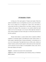
Figure 1. Flowchart of the nanospheres sample and sensor preparation
details.
g, A. R., Tianjin chemical reagent factory). After stirring
for several minutes, the mixture was heated to and maintained
at 85-95 °C, and in 2-6 min, the color of the mixture was
an orange color. The mixture was cooled to room temperature
at once and washed by alcohol several times (Figure 1).
CuO Nanospheres. The spherical colloids of Cu2O
nanospheres were deposited on a Si wafer and dried in oven
at 60 °C for 10 min. They were then transferred into the
muffle furnace and heated at 500 °C for 1-2 h.
Scanning electron microscopy (SEM) images were taken
using a field-emission microscope (Sirion FEI). Transmission
electron microscopy (TEM) images were taken by a JEM1200EX microscope operated at 120 kV. Electron diffraction
(ED) patterns were taken by a Hitachi H800 (acceleration
voltage 200 kV) and JEM-1200EX microscope operated at
120 kV. The X-ray diffraction (XRD) test was performed
with a Bruker D8 Advance X-ray diffractometer with
monochromatized Cu KR radiation (λ ) 1.5418 Å). The
UV-vis spectra were obtained by a Hitachi U-3010 spectrophotometer. The gas sensitive properties were measured
using a static test system made by Hanwei Electronics Co.,
Ltd., Henan Province, China.
In this communication, we introduce NaBH4 as the
reducing agent and DMF as the solvent. Differing from the
reported formation mechanism on the preparation of Cu2O
hollow spheres by the Zeng group,15c NaBH4, working as a
strong reducing agent, reacts with a trace amount of H2O in
DMF and then reduces Cu(CH3COO)2 to Cu2O nanocrystals.
Under the low temperature of 80-90 °C, DMF, a weak
reducing agent, acts merely as the solvent of this reaction.
The chemical reaction is as follows:
DMF
4Cu2+ + BH4- + 5H2O 9
8
PVP
2Cu2O(s) + B(OH)3 + 2H2 + 7H+ (1)
With the effect of DMF solvent and PVP, the primary Cu2O
nanocrystals prefer to aggregate into spherical nanospheres.
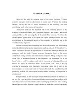
The formation process of Cu2O nanospheres can be identified
by Figure 2A-C, which are obtained from the Cu2O products
collected at different aging times. Figure 2A shows the
existence of small nanospheres during the process of aggregation to spherical morphology. With the increase of aging
time, the nearly monodisperse nanospheres gradually come
into being which can be confirmed by Figure 2B,C. The ED
pattern of one Cu2O nanosphere shown in Figure 2D
confirms that the as-prepared Cu2O nanospheres are the
regular spherical aggregation of Cu2O single-crystalline
nanoparticles. The XRD pattern shown in Figure 1 of
Figure 2. SEM images of Cu2O nanospheres at different aging time: (A)
0.5, (B) 1.5, and (C) 3 min. The inset of part C shows the clear surface
morphology of near-monodisperse Cu2O nanospheres. Part D shows TEM
and ED patterns of the nanospheres in part C. Parts E and F show the SEM,
TEM, and ED patterns of nanospheres prepared by adding more appropriate
H2O.
Supporting Information identifies the pure cubic phase of
Cu2O (JCPDF 05-0667). The narrow size distribution of
obtained nanospheres in Figure 2C can be demonstrated by
Figure 2A of Supporting Information and mostly concentrates
at the size of 200 nm. Most importantly, the properly high
concentration of reactants is necessary for the supersaturation
of uniform Cu2O single-crystalline nanoparticles which can
aggregate to be nearly monodisperse spherical colloids.11d,15c
To confirm the reaction mechanism mentioned in eq 1, we
add ∼0.05 mL of H2O additionally to the same reaction
system as above. As shown in Figure 2E,F, we gain singlecrystalline Cu2O nanospheres with improved monodispersity
and the size distribution concentrates at ∼225 nm (Figure
2B, Supporting Information). This result is reasonable
because, when adding an appropriate amount of additional
water, the reaction velocity of eq 1 is increased and leads to
the enhanced supersaturation of uniform Cu2O nanoparticles.
In the present case, Cu2O nanoparticles could aggregate
regularly to single-crystalline nanospheres with larger size.
However, the addition of H2O enables the larger nanospheres
and makes the monodispersity worse. This can be identified
by the SEM image shown in Figure 3 of Supporting
Information. Thus, the proper quantity of H2O is necessary
Communications
Chem. Mater., Vol. 18, No. 4, 2006 869
Figure 3. SEM images of two-dimensional (A) or three-dimensional (B)
self-assembly modes of as-prepared Cu2O nanospheres when spreading on
Si wafer or ITO glass.
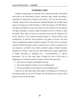
Figure 5. (A) SEM image of as-prepared CuO nanospheres with an inset
of clear morphology of several nanospheres; (B) two-dimensional selfassembly pattern of CuO nanospheres; (C) TEM image of the CuO product
with an inset to show the ED pattern of one nanosphere; and (D) XRD
pattern of prepared CuO nanospheres.
Figure 4. UV-vis absorption spectra of Cu2O primary nanoparticles and
near-monodisperse nanospheres.
for the kinetically controlled synthesis of monodisperse Cu2O
nanospheres. Of course, the optimal concentration of reagents, especially the concentration of H2O in DMF, should
be further studied. Because of the monodispersity of asprepared Cu2O nanospheres, as shown by the SEM images
of Figure 3A,B, the two-dimensional or three-dimensional
self-assembly pattern would spontaneously form when their
alcohol colloids are spread on the Si or indium tin oxide
(ITO) conductive glass substrate. This is perhaps attributed
to the attractive capillary forces among the colloidal nanospheres. When the solvent evaporates slowly, these colloidal
spheres are self-assembled into a closely packed array.6,19
As a p-type semiconductor, Cu2O has been widely
researched because it has the potential to form a solar cell
with high open-circuit voltage by combination with a suitable
n-type semiconductor. So the optical characterizations of the
as-obtained Cu2O nanosphere colloid have been carried out
by the UV-vis absorption spectrum which was demonstrated
in Figure 4. Differing from the nanoparticles which have an
obvious absorption edge at ∼550 nm consistent with the
reported band gap energy,17 the prepared nanospheres of
Cu2O have a wide absorption peak at ∼520 nm. This result
is consistent with many reported Cu2O nanostructures.20 It
is mainly because when the Cu2O nanoparticles aggregate
to be nanospheres, the size becomes larger and uniform, and
(19) Denkov, N. D.; Velev, O. D.; Kralchevsky, P. A.; Ivanov, I. B.;
Yoshimura, H.; Nagayama, K. Nature 1993, 361, 26.
(20) (a) He, P.; Shen, X.; Gao, H. J. Colloid Interface Sci. 2005, 284, 510.
(b) Borgohain, K.; Murase, N.; Mahamuni, S. J. Appl. Phys. 2002,
92, 1292.
then the scattering of visible light superimposes on the
absorption of as-prepared nanospheres.
Considering the importance of the Cu-based materials
family and the feasibility of chemical transformation between
them, the CuO nanospheres were prepared by heat treatment
of as-obtained Cu2O nanospheres in a muffle furnace at 500
°C for 1-2 h. The SEM image in Figure 5A and TEM image
in Figure 5C show that the nanospheres nearly have no shape
evolution. Figure 5B shows the two-dimensional selfassembly state of CuO nanospheres. As shown in Figure 5C,
the irregular diffraction dots in the ED pattern of one CuO
nanosphere identify that CuO nanospheres are also the
aggregation of single-crystalline nanoparticles. Clearly, the
surface of the CuO nanospheres is more rough than that of
Cu2O nanospheres which may be helpful for their catalysis
application. The XRD pattern in Figure 5D identifies that
the product is monoclinic CuO (JCPDF 48-1548, Tenorite).
In recent years, many gas sensors based on copper oxides
and n-type metal oxides have been researched, such as ZnOCuO, SnO2-CuO, and so forth. Typically, SnO2 is sensitive
to H2S gas by the addition of a small amount of CuO because
of the formation and the disruption of p-n junction.21,22
Herein, we fabricated the gas sensors based on our prepared
Cu2O and CuO nanospheres only. Interestingly, we found
they had high sensitivity to some gases, such as alcohol or
gasoline.
The Cu2O sensor was fabricated by dip-coating as-prepared
Cu2O alcohol colloids to the ceramic tube of the sensor body
without an additional annealing process except for aging in
the gas sensor system. Figure 6A shows the photograph of
(21) (a) Kong, X. H.; Li, Y. D. Sens. Actuators, B 2005, 105, 449. (b)
Choi, J. K.; Choi, G. M.; Man, G. Sens. Actuators, B 2000, 69, 120.
(22) (a) Chowdhuri, A.; Gupta, V.; Sreenivas, K.; Et al. Appl. Phys. Lett.
2004, 84, 1180. (b) Chowdhuri, A.; Gupta, V.; Sreenivas, K. Sens.
Actuators, B. 2003, 93, 572. (c) Chowdhuri, A.; Sharma, P.; Gupta,
V.; Et al. J. Appl. Phys. 2002, 92, 2172.
870 Chem. Mater., Vol. 18, No. 4, 2006
Communications
Figure 6. (A) Photograph of the gas sensor and the SEM image of thin
films coated on it. (B) Schematic diagram showing the structure of a typical
Cu2O nanosphere gas sensor by top view and sectional view.
Figure 8. (A) Typical response curves of Cu2O (black curve) and CuO
(gray curve) nanosphere gas sensors to gas oil with increasing concentrations
at 210 °C. (B) Typical response curves of Cu2O octahedral microparticle
(black curve) and nanoparticle (gray curve) gas sensors to gas oil with
increasing concentrations at 210 °C.
Figure 7. (A) Typical response curves of Cu2O (black curve) and CuO
(gray curve) nanosphere gas sensors to alcohol with increasing concentrations at 210 °C. (B) Typical response curves of Cu2O octahedral microparticle (gray curve) and nanoparticle (black curve) gas sensors to alcohol
with increasing concentrations at 210 °C.
a typical sensor and the SEM image of the Cu2O nanospheres
covering on it before the annealing process. Figure 6B shows
the schematic diagram of the Cu2O sensor structure and the
sectional view. The CuO gas sensor was made by heattreating the as-prepared Cu2O sensor in a muffle stove at
500 °C for ∼1 h. The nanosphere morphology was kept as
a rough surface as shown in Figure 5A. From the SEM image
shown in Figure 6A, it is clear that the nearly monodisperse
nanospheres in the film can provide enough surface area and
interspaces to contact the detected gas or atmosphere.
Figures 7A and 8A show the typical isothermal response
curves of Cu2O and CuO sensors when cycled by increasing
alcohol and gasoline concentrations in ambient air with the
range of 10-800 ppm, at a working temperature of 210 °C.
For comparison with the prepared Cu2O nanosphere gas
sensors, we choose Cu2O octahedral microparticle and
nanoparticle alcohol colloids with the same concentration
as the Cu2O nanosphere colloid to make gas sensors by dipcoating and the same aging process. Their sensitivities to
alcohol and gas oil have been demonstrated in Figures 7B
and 8B. From these sensor results, it is evident that our asprepared Cu2O nanosphere sensor is much more sensitive
than CuO, Cu2O octahedral microparticle, and Cu2O nanoparticle sensors. The Cu2O nanosphere sensor offers a better
response and quicker response/recovery time than many
literature reports.23 The resistance of the Cu2O nanosphere
sensor will decrease dramatically on the injection of ethanol
or gasoline and reach its initial value quickly when releasing
gases. The response time of the Cu2O sensor is only ∼15 s
to alcohol and ∼25 s to gasoline. The resume time of Cu2O
sensor also is only ∼30 s for alcohol and ∼45 s for gasoline.
The detection limit of the as-prepared Cu2O sensor can reach
as little as several parts per million when detecting certain
kinds of gas, such as alcohol or gasoline. On explaining these
results, one important reason is that, in the Cu2O nanosphere
film coated on the sensor body, the monodispersity of singlecrystalline Cu2O nanospheres and their stacking mode result
in larger surface area and much more capacious interspaces
than any other shapes, which can provide sufficient space
for the interaction between Cu2O and detected gases.
However, the Cu2O microparticle film has less surface area
and fewer active sites to contact with the gas. The SEM
(23) (a) Khatko, V.; Calderer, J.; Llobet, E.; Correig, X. Sens. Actuators,
B 2005, 109, 128. (b) Gopal Reddy, C. V.; Cao, W.; Tan, O. K.; Et
al. Sens. Actuators, B 2003, 94, 99.
Communications
Chem. Mater., Vol. 18, No. 4, 2006 871
temperature, they have bad sensitivity or even no sensitivity
possibly as a result of the less active O- and O2- ions. This
result is consistent with the theory by Gray et al. in 1948.16
Of course, the radical mechanism about the high sensitivity
of our as-prepared Cu2O nansphere sensor should be further
researched. Furthermore, as reported by much of the
literature,21-25 to enhance the selectivity of some gases and
satisfy more applications, the work of noble metal doping
on as-prepared nanospheres and preparation of core-shell
structures, such as Cu2O-SnO2, Cu2O-ZnO, CuO-SnO2,
and CuO-ZnO, should be further studied.
Figure 9. Typical response curves of Cu2O (black curve) and CuO (gray
curve) nanosphere gas sensors to H2S with increasing concentrations at
210 °C.
image of the microparticle film is shown in Figure 4A of
Supporting Information. For the Cu2O nanoparticle film, as
shown by the SEM image in Figure 4B of Supporting
Information, although the nanoparticles have a smaller size
than the nanospheres, their strong irregular aggregation also
leads to fewer opportunities to contact the detected gas.
Considering the selectivity of the as-prepared Cu2O sensor,
we have detected H2S gas with the same concentration level
at the working temperature of 210 °C, and the result is shown
in Figure 9. From the response curve, we find it is not
sensitive enough for practical usage and the voltage response
is not proportional to the increasing concentration of H2S.
The sensitivity difference of the Cu2O and CuO nanosphere films perhaps is attributed to the reaction mechanism
of Cu2O or CuO to detected gases. When in contact with
the reducing gas (electron donator), such as alcohol or
gasoline, the negative charged oxygen (O-, O2-) absorbed
on the Cu2O or CuO nanosphere surface will react.24 The
reaction between the reducing gas and O- or O2- leads to
the decrease of the carrier hole density in the surface charge
layer and the increase of the Cu2O or CuO resistance.
Specifically, under the working temperature of 210 °C, the
Cu2O nanospheres absorb the oxygen in the air easier and
then cover the surface with more active negatively charged
oxygen (O-, O2-) than as-prepared CuO nanospheres.
Different from the body formation, the surface Cu2O
formation was easily changed to be Cu2O2-x (0 < x < 1),
an active transient state, which is more favorable for the
interaction with reducing gases. When testing under lower
(24) Liu, J. F.; Wang, X.; Peng, Q.; Li, Y. D. AdV. Mater. 2005, 17, 764.
In summary, this communication shows an effective
method to prepare nearly monodisperse Cu2O and CuO
nanospheres. The rapid production of supersaturated Cu2O
single-crystalline nanoparticles and their regular spherical
aggregation lead to the monodispersity of Cu2O nanospheres.
By modulating the concentration of reactant H2O, the
diameter, crystallization, and monodispersity of Cu2O nanospheres can be kinetically controlled. Because the thin films
prepared by as-obtained nanospheres have big surface areas
and plentiful spaces to interact with gases, the gas sensors
based on as-prepared Cu2O nanospheres have high sensitivity
and good selectivity to some flammable gases. The surface
modification with noble metals or n-type metal oxides based
on obtained nanospheres will be more helpful to enrich their
sensor applications.
Acknowledgment. This work was supported by NSFC
(90406003), the Specialized Research Fund for the Doctoral
Program of Higher Education, and the Foundation for the Author
of National Excellent Doctoral Dissertation of P. R. China and
the State Key Project of Fundamental Research for Nanomaterials and Nanostructures (2003CB716901).
Supporting Information Available: Typical XRD pattern of
as-prepared nearly monodisperse Cu2O nanospheres, different size
distribution histograms of nearly monodisperse cuprous oxide
nanospheres obtained by modulating the concentration of water,
SEM image of as-obtained Cu2O nanospheres when adding excess
H2O, and SEM images of octahedral microparticles and nanoparticles used for gas sensors (PDF). This material is available free of
charge via the Internet at .
CM052256F
(25) Ivanov, P.; Llobet, E.; Vilanova, X.; Et al. Sens. Actuators, B 2004,
99, 201.