Robust optimization of radiation therapy accounting for geometric uncertainty
Bạn đang xem bản rút gọn của tài liệu. Xem và tải ngay bản đầy đủ của tài liệu tại đây (2.69 MB, 57 trang )
Robust optimization of radiation therapy
accounting for geometric uncertainty
Albin Fredriksson
Robust optimization of radiation therapy
accounting for geometric uncertainty
ALBIN FREDRIKSSON
Doctoral Thesis
Stockholm, Sweden 2013
Cover illustration copyright © 1980 by Bob Marshall. Used with permission. It
illustrates the first canon of Das Musikalische Opfer by Johann Sebastian Bach.
This movement is a canon cancrizans.
TRITA MAT 13/OS/06
ISSN 1401-2294
ISRN KTH/OPT/DA-13/06-SE
ISBN 978-91-7501-771-6
Optimization and Systems Theory
Department of Mathematics
Royal Institute of Technology
SE-100 44 Stockholm, Sweden
Akademisk avhandling som med tillstånd av Kungl Tekniska Högskolan framlägges till offentlig granskning för avläggande av teknologie doktorsexamen, onsdagen den 5 juni 2013 klockan 10.00 i sal F3, Lindstedtsvägen 26, Kungl Tekniska
Högskolan, Stockholm.
© Albin Fredriksson, April 2013
Print: Universitetsservice US-AB, Stockholm, Sweden
Till min familj
De flesta menar att skinnet skiljer dem från det som omger
dem. Men människans skinn är tunt och genomsläppligt, fullt
av hål och öppningar likt en trasig rock. Det omänskliga far in
och ut genom revorna; jord och vind blåser tvärs igenom oss.
Vår hjälplöshet är höggradig.
- Willy Kyrklund, Mästaren Ma
ix
Abstract
Geometric errors may compromise the quality of radiation therapy treatments. Optimization methods that account for errors can reduce their effects.
The first paper of this thesis introduces minimax optimization to account
for systematic range and setup errors in intensity-modulated proton therapy.
The minimax method optimizes the worst case outcome of the errors within
a given set. It is applied to three patient cases and shown to yield improved
target coverage robustness and healthy structure sparing compared to conventional methods using margins, uniform beam doses, and density override.
Information about the uncertainties enables the optimization to counterbalance the effects of errors.
In the second paper, random setup errors of uncertain distribution—in
addition to the systematic range and setup errors—are considered in a framework that enables scaling between expected value and minimax optimization.
Experiments on a phantom show that the best and mean case tradeoffs between target coverage and critical structure sparing are similar between the
methods of the framework, but that the worst case tradeoff improves with
conservativeness.
Minimax optimization only considers the worst case errors. When the
planning criteria cannot be fulfilled for all errors, this may have an adverse
effect on the plan quality. The third paper introduces a method for such cases
that modifies the set of considered errors to maximize the probability of satisfying the planning criteria. For two cases treated with intensity-modulated
photon and proton therapy, the method increased the number of satisfied criteria substantially. Grasping for a little less sometimes yields better plans.
In the fourth paper, the theory for multicriteria optimization is extended
to incorporate minimax optimization. Minimax optimization is shown to better exploit spatial information than objective-wise worst case optimization,
which has previously been used for robust multicriteria optimization.
The fifth and sixth papers introduce methods for improving treatment
plans: one for deliverable Pareto surface navigation, which improves upon
the Pareto set representations of previous methods; and one that minimizes
healthy structure doses while constraining the doses of all structures not to
deteriorate compared to a reference plan, thereby improving upon plans that
have been reached with too weak planning goals.
Keywords: Optimization, intensity-modulated proton therapy, uncertainty,
robust planning, setup error, range error, intensity-modulated radiation therapy, multicriteria optimization.
x
Sammanfattning
Geometriska fel kan försämra kvaliteten på strålbehandlingar, men optimeringsmetoder som tar hänsyn till felen kan minska deras effekt.
I denna avhandlings första artikel introduceras minimaxoptimering för
att ta hänsyn till systematiska fel på räckvidd och positionering i intensitesmodulerad protonterapi. Minimaxmetoden optimerar det värsta utfallet av
felen från en given mängd. Metoden prövas på tre patientfall. För dessa leder den till mer robust måltäckning och ökat riskorgansskydd jämfört med
konventionella metoder som använder marginaler, likformiga stråldoser och
ersatta densiteter. Information om osäkerheterna gör att optimeringen kan
motverka effekterna av fel.
I den andra artikeln betraktas slumpmässiga positioneringsfel av osäker
sannolikhetsfördelning – utöver de systematiska räckvidds- och positioneringsfelen – i ett ramverk som möjliggör skalning mellan väntevärdes- och
minimaxoptimering. Experiment på ett fantom visar att avvägningen mellan
måltäckning och riskorgansskydd i det bästa fallet och i medelfallet är likartad mellan metoderna från ramverket, men att avvägningen i värsta fallet
förbättras med graden av försiktighet.
Minimaxoptimering tar bara hänsyn till de värsta felen. Detta kan leda till att plankvaliteten blir lidande i fall där planeringsmålen inte går att
uppfylla för alla fel. I den tredje artikeln introduceras en metod för sådana
fall. Denna metod modifierar mängden av beaktade fel i syfte att maximera
sannolikheten att uppfylla planeringsmålen. För två fall behandlade med intensitetsmodulerad foton- och protonterapi ledde metoden till en avsevärd
ökning av antalet uppfyllda mål. Sänkta krav på robustheten kan ibland leda
till bättre planer.
I den fjärde artikeln utökas teorin för flermålsoptimering till att innefatta minimaxoptimering. Minimaxoptimering visas vara bättre på att utnyttja
spatiell information än målvis värsta fallet-optimering, vilket tidigare använts
för robust flermålsoptimering.
Artikel fem och sex introducerar metoder för att förbättra strålbehandlingsplaner: en för levererbar navigering av Pareto-ytor, vilken förbättrar
tidigare metoders representationer av Pareto-mängder; och en som minimerar doserna till friska strukturer under bivillkor att doserna till alla strukturer
inte försämras jämfört med en referensplan, för att på så sätt förbättra planer
som har tagits fram med för lågt satta mål.
Nyckelord: Optimering, intensitetsmodulerad protonterapi, osäkerhet, robustplanering, positioneringsfel, räckviddsfel, intensitetsmodulerad strålterapi, flermålsoptimering.
xi
Acknowledgments
First of all, many thanks to my advisor Anders Forsgren, whose encouraging
and helpful attitude and mathematical expertise have been invaluable to me
during these years. It has been a privilege to work with you.
My industrial co-advisors from RaySearch Laboratories have also been
of significant importance: Thanks to Henrik Rehbinder for guiding me during
the initial phase of this project. And thanks to Björn Hårdemark, my current
co-advisor, for sharing your abounding knowledge of radiation therapy and
just about all other topics.
I thank my academic co-advisors Johan Håstad and Krister Svanberg for
their helpful comments during reference group meetings.
The work presented in this thesis was co-funded by the Swedish Research Council (VR) and RaySearch Laboratories. I am grateful to both and
especially thank Johan Löf, the founder and CEO of RaySearch, for hosting
this project and for creating the inspiring workplace that RaySearch is.
Thanks to Rasmus Bokrantz, my academic twin, for all discussions concerning research, typography, line widths; and for co-authorship, traveling
companionship, and friendship.
I have deeply appreciated the conversations with previous and present
colleagues at RaySearch: with Kjell Eriksson and Fredrik Löfman about optimization of radiation therapy, and with Göran Sporre about optimization in
general. Thanks also to Tore Ersmark, Lars Glimelius, and Martin Janson,
who have generously shared their knowledge of proton physics.
Further, I am grateful to the faculty members and staff of the Division
of Optimization and Systems Theory at KTH. Special thanks to my fellow
students for course collaborations and for all laughs during fika: Mikael Fallgren, Johan Markdahl, Anders Möller, Hildur Æsa Oddsdóttir, Tove Odland,
Göran Svensson, Henrik Svärd, Johan Thunberg, and Yuecheng Yang.
I am grateful to my other friends for sharing my interests outside of research. A special thanks to Johan Hallgren for drawing my attention to this
PhD student position.
Thanks to my family for your unconditional love and support. I hope that
I can express how much you mean to me by stating that I cannot. Finally, my
deepest love to Sanna, who is my best friend.
Stockholm, April 2013
Albin Fredriksson
Contents
Introduction
1
Radiation therapy . . . . . . . . . . . . . . . . . . . .
1.1
Radiobiology . . . . . . . . . . . . . . . . . .
1.2
Photon therapy . . . . . . . . . . . . . . . . .
1.3
Proton therapy . . . . . . . . . . . . . . . . .
2
Treatment planning . . . . . . . . . . . . . . . . . . .
2.1
Patient geometry . . . . . . . . . . . . . . . .
2.2
Evaluation of plan quality . . . . . . . . . . .
2.3
Optimization functions . . . . . . . . . . . . .
2.4
Optimization problem . . . . . . . . . . . . .
2.5
Optimization method . . . . . . . . . . . . . .
3
Uncertainties in radiation therapy . . . . . . . . . . . .
3.1
Optimization under uncertainty . . . . . . . .
3.2
Treatment plan optimization under uncertainty
3.2.1
Robust IMRT . . . . . . . . . . . .
3.2.2
Robust IMPT . . . . . . . . . . . .
4
Multicriteria optimization of radiation therapy . . . . .
4.1
Multicriteria optimization . . . . . . . . . . .
4.2
Robust multicriteria optimization . . . . . . .
4.3
Deliverability of navigated plans . . . . . . . .
5
Summary and main contributions . . . . . . . . . . . .
5.1
Summary of the appended papers . . . . . . .
5.2
Main contributions . . . . . . . . . . . . . . .
5.3
Contributions by co-authors . . . . . . . . . .
6
Bibliography . . . . . . . . . . . . . . . . . . . . . .
xiii
.
.
.
.
.
.
.
.
.
.
.
.
.
.
.
.
.
.
.
.
.
.
.
.
.
.
.
.
.
.
.
.
.
.
.
.
.
.
.
.
.
.
.
.
.
.
.
.
.
.
.
.
.
.
.
.
.
.
.
.
.
.
.
.
.
.
.
.
.
.
.
.
.
.
.
.
.
.
.
.
.
.
.
.
.
.
.
.
.
.
.
.
.
.
.
.
.
.
.
.
.
.
.
.
.
.
.
.
.
.
.
.
.
.
.
.
.
.
.
.
.
.
.
.
.
.
.
.
.
.
.
.
.
.
.
.
.
.
.
.
.
.
.
.
1
1
2
3
4
6
7
8
10
12
14
15
16
17
18
19
21
22
22
24
25
25
29
30
31
xiv
C ONTENTS
A Minimax optimization for handling uncertainties in IMPT
A.1 Introduction . . . . . . . . . . . . . . . . . . . . . . . . . .
A.2 Methods . . . . . . . . . . . . . . . . . . . . . . . . . . . .
A.2.1 Uncertainty models . . . . . . . . . . . . . . . . . .
A.2.1.1 Range uncertainty . . . . . . . . . . . . .
A.2.1.2 Setup uncertainty . . . . . . . . . . . . .
A.2.1.3 Assessing the effects of the errors . . . . .
A.2.2 Nominal optimization formulation . . . . . . . . . .
A.2.3 Robust methods . . . . . . . . . . . . . . . . . . . .
A.2.3.1 Conventional methods . . . . . . . . . . .
A.2.3.2 Minimax optimization formulation . . . .
A.2.4 Computational study . . . . . . . . . . . . . . . . .
A.3 Results . . . . . . . . . . . . . . . . . . . . . . . . . . . . .
A.3.1 Lung case . . . . . . . . . . . . . . . . . . . . . . .
A.3.2 Paraspinal case . . . . . . . . . . . . . . . . . . . .
A.3.3 Prostate case . . . . . . . . . . . . . . . . . . . . .
A.4 Discussion . . . . . . . . . . . . . . . . . . . . . . . . . . .
A.5 Conclusion . . . . . . . . . . . . . . . . . . . . . . . . . .
A.A Minimax stochastic formulation . . . . . . . . . . . . . . .
.
.
.
.
.
.
.
.
.
.
.
.
.
.
.
.
.
.
.
.
.
.
.
.
.
.
.
.
.
.
.
.
.
.
.
.
.
.
.
.
.
.
.
.
.
.
.
.
.
.
.
.
.
.
43
44
46
46
46
46
47
47
48
48
48
51
53
53
56
60
64
65
65
B Characterization of robust radiation therapy optimization
B.1 Introduction . . . . . . . . . . . . . . . . . . . . . . . . . .
B.2 Methods . . . . . . . . . . . . . . . . . . . . . . . . . . . .
B.2.1 Uncertainties . . . . . . . . . . . . . . . . . . . . .
B.2.2 Notation . . . . . . . . . . . . . . . . . . . . . . .
B.2.3 Optimization functions . . . . . . . . . . . . . . . .
B.2.4 Accounting for systematic errors . . . . . . . . . . .
B.2.4.1 Expected value optimization . . . . . . .
B.2.4.2 Worst case optimization . . . . . . . . . .
B.2.4.3 Conditional value at risk optimization . . .
B.2.4.4 Minimax stochastic programming . . . . .
B.2.5 Accounting for random errors . . . . . . . . . . . .
B.2.6 Combining systematic and random errors . . . . . .
B.2.7 Patient geometry . . . . . . . . . . . . . . . . . . .
B.2.8 Computational study . . . . . . . . . . . . . . . . .
B.3 Results . . . . . . . . . . . . . . . . . . . . . . . . . . . . .
B.3.1 Systematic errors . . . . . . . . . . . . . . . . . . .
B.3.2 Random errors with fixed probability distribution . .
.
.
.
.
.
.
.
.
.
.
.
.
.
.
.
.
.
.
.
.
.
.
.
.
.
.
.
.
.
.
.
.
.
.
.
.
.
.
.
.
.
.
.
.
.
.
.
.
.
.
.
71
72
73
73
74
74
75
75
75
75
76
77
78
80
80
82
82
86
xv
B.3.3
B.3.4
B.4
B.5
B.A
B.B
Random errors with uncertain standard deviation . . . . .
Systematic errors and random errors with fixed probability
distribution . . . . . . . . . . . . . . . . . . . . . . . . .
B.3.5 Systematic errors and random errors with uncertain standard deviation . . . . . . . . . . . . . . . . . . . . . . . .
Discussion . . . . . . . . . . . . . . . . . . . . . . . . . . . . . .
Conclusion . . . . . . . . . . . . . . . . . . . . . . . . . . . . .
CVaR as a minimax stochastic program . . . . . . . . . . . . . .
Conventional planning . . . . . . . . . . . . . . . . . . . . . . .
C Maximizing the probability of satisfying planning criteria
C.1 Introduction . . . . . . . . . . . . . . . . . . . . . . . . . .
C.2 Method . . . . . . . . . . . . . . . . . . . . . . . . . . . .
C.2.1 Uncertainties and scenarios . . . . . . . . . . . . . .
C.2.2 Mathematical formulation . . . . . . . . . . . . . .
C.2.3 Scenario position optimization problem . . . . . . .
C.3 Probability computation . . . . . . . . . . . . . . . . . . . .
C.3.1 Optimizing margins . . . . . . . . . . . . . . . . .
C.3.2 Computational study . . . . . . . . . . . . . . . . .
C.3.2.1 Patient cases . . . . . . . . . . . . . . . .
C.3.2.2 Optimization . . . . . . . . . . . . . . . .
C.3.2.3 Scenario dose computation . . . . . . . .
C.4 Results . . . . . . . . . . . . . . . . . . . . . . . . . . . . .
C.4.1 Prostate case . . . . . . . . . . . . . . . . . . . . .
C.4.1.1 Optimization problem . . . . . . . . . . .
C.4.1.2 Optimized scenarios . . . . . . . . . . . .
C.4.1.3 Feasible scenario positions . . . . . . . .
C.4.1.4 Robust plans with selected scenarios . . .
C.4.2 Lung case . . . . . . . . . . . . . . . . . . . . . . .
C.4.2.1 Optimization problem . . . . . . . . . . .
C.4.2.2 Optimized scenarios . . . . . . . . . . . .
C.4.2.3 Feasible scenario positions . . . . . . . .
C.4.2.4 Robust plans with selected scenarios . . .
C.5 Discussion . . . . . . . . . . . . . . . . . . . . . . . . . . .
C.6 Conclusion . . . . . . . . . . . . . . . . . . . . . . . . . .
C.A Optimization functions . . . . . . . . . . . . . . . . . . . .
.
.
.
.
.
.
.
.
.
.
.
.
.
.
.
.
.
.
.
.
.
.
.
.
.
.
.
.
.
.
.
.
.
.
.
.
.
.
.
.
.
.
.
.
.
.
.
.
.
.
.
.
.
.
.
.
.
.
.
.
.
.
.
.
.
.
.
.
.
.
.
.
.
.
.
86
89
89
92
95
95
95
101
102
104
104
104
107
108
109
110
110
111
111
112
112
112
113
113
115
116
118
118
118
120
122
124
124
xvi
C ONTENTS
D Robust multicriteria optimization for proton therapy
D.1 Introduction . . . . . . . . . . . . . . . . . . . . . . . . . . . . .
D.2 Methods . . . . . . . . . . . . . . . . . . . . . . . . . . . . . . .
D.2.1 Notation . . . . . . . . . . . . . . . . . . . . . . . . . .
D.2.2 Robust optimization . . . . . . . . . . . . . . . . . . . .
D.2.2.1 Singlecriterion formulation . . . . . . . . . . .
D.2.2.2 Multicriteria formulation . . . . . . . . . . . .
D.2.2.3 Pareto optimality for deterministic programs . .
D.2.2.4 Pareto optimality for uncertain programs . . . .
D.2.3 Pareto surface-based planning . . . . . . . . . . . . . . .
D.2.3.1 Algorithmic considerations . . . . . . . . . . .
D.2.3.2 Tradeoffs with variable level of robustness . . .
D.2.4 Computational study . . . . . . . . . . . . . . . . . . . .
D.2.4.1 Patient case and dose calculation . . . . . . . .
D.2.4.2 Optimization problem formulation . . . . . . .
D.3 Results . . . . . . . . . . . . . . . . . . . . . . . . . . . . . . . .
D.3.1 Comparison between worst case and objective-wise worst
case . . . . . . . . . . . . . . . . . . . . . . . . . . . . .
D.3.2 Tradeoffs in robustness and conservativeness . . . . . . .
D.3.3 Optimal lateral dose fall-off as function of dose response .
D.4 Discussion . . . . . . . . . . . . . . . . . . . . . . . . . . . . . .
D.5 Conclusion . . . . . . . . . . . . . . . . . . . . . . . . . . . . .
D.A Theory of robust multicriteria programming . . . . . . . . . . . .
D.A.1 Scalarization for deterministic multicriteria programs . . .
D.A.2 Scalarization of uncertain multicriteria programs . . . . .
D.A.2.1 Definitions . . . . . . . . . . . . . . . . . . . .
D.A.2.2 Results . . . . . . . . . . . . . . . . . . . . . .
D.B Finding the worst case probability distribution . . . . . . . . . . .
129
130
132
132
132
132
133
134
134
136
136
138
139
139
140
141
E Deliverable navigation for multicriteria IMRT
E.1 Introduction . . . . . . . . . . . . . . . . . . . . . . . . . . . . .
E.2 Methods . . . . . . . . . . . . . . . . . . . . . . . . . . . . . . .
E.2.1 Multicriteria direct step-and-shoot optimization . . . . . .
E.2.2 Direct step-and-shoot optimization using shared apertures
E.2.3 Convergence towards the unrestricted Pareto set . . . . . .
E.2.4 Computational study . . . . . . . . . . . . . . . . . . . .
E.3 Results . . . . . . . . . . . . . . . . . . . . . . . . . . . . . . . .
E.3.1 Two-dimensional tradeoff . . . . . . . . . . . . . . . . .
169
169
172
172
173
175
176
179
179
142
144
146
148
150
151
152
152
152
153
161
xvii
E.3.2 Three-dimensional tradeoff . . . . . . . . . . . . . . . . . 180
E.4 Discussion . . . . . . . . . . . . . . . . . . . . . . . . . . . . . . 183
E.5 Conclusion . . . . . . . . . . . . . . . . . . . . . . . . . . . . . 184
F Automated improvement of radiation therapy treatment plans
F.1 Introduction . . . . . . . . . . . . . . . . . . . . . . . . . . . . .
F.2 Methods . . . . . . . . . . . . . . . . . . . . . . . . . . . . . . .
F.2.1 Optimization formulation . . . . . . . . . . . . . . . . .
F.2.2 Reference DVH constraints . . . . . . . . . . . . . . . .
F.2.3 Regularization of positive part functions . . . . . . . . . .
F.3 Results . . . . . . . . . . . . . . . . . . . . . . . . . . . . . . . .
F.3.1 Computational study . . . . . . . . . . . . . . . . . . . .
F.3.2 Plan comparison . . . . . . . . . . . . . . . . . . . . . .
F.3.3 Regularization error . . . . . . . . . . . . . . . . . . . .
F.4 Discussion . . . . . . . . . . . . . . . . . . . . . . . . . . . . . .
F.5 Conclusion . . . . . . . . . . . . . . . . . . . . . . . . . . . . .
F.A Optimization functions . . . . . . . . . . . . . . . . . . . . . . .
189
190
192
192
194
195
198
199
201
201
202
205
205
Introduction
It is estimated that every third person in Sweden will get cancer [68]. Half of
Swedish cancer patients receive radiation therapy during their illness [54]. The
quality of radiation therapy treatment is of high consequence.
This thesis concerns optimization approaches for radiation therapy in the presence of geometric uncertainty. It consists of an introduction and six appended
papers. The introduction first presents radiation therapy and relevant optimization
concepts. It then introduces optimization approaches to account for uncertainties
in radiation therapy. The considered uncertainties are mainly with respect to the
patient densities and the alignment of the patient relative to the beams. The topic of
facilitating decision making by multicriteria optimization accounting for geometric
uncertainty is also discussed, as is the problem of deliverability of plans obtained
by Pareto surface navigation. The introduction is concluded with a short summary
of the appended papers.
1
Radiation therapy
X-rays were discovered in 1895 by Wilhelm Röntgen. A few months after the
announcement of the discovery, x-rays were used to treat skin disease, and within
a year, the first accounts of using x-rays to treat cancer were reported. Thus was
the birth of radiation therapy.
Radiation therapy is the medical use of ionizing radiation. It is primarily used
to treat cancer. In most cases, radiation therapy is given with curative intent. It may
also be used in palliative care in cases where the cancer is too advanced for a curative treatment to be possible, but for which symptoms such as pain may be relieved.
Radiation therapy is used both as a stand-alone treatment and in combination with
other cancer treatments such as surgery and chemotherapy.
1
2
I NTRODUCTION
Radiation therapy is delivered either by internal radiation sources (brachytherapy), which are placed inside or close to the region to be treated, or by external
sources (external beam radiation therapy). The latter is the most common form of
radiation therapy and is the form that this thesis concerns. In external beam radiation therapy, the patient is irradiated by an external radiation source that directs the
radiation towards the region requiring treatment. The radiation is commonly delivered in the form of high-energy photon (x-ray), electron, or proton beams, but other
particles may also be used. In this thesis, photon and proton beams are considered.
Blocking material is used to shape the beams in order to conform the radiation to
the target. The superposition of radiation of several beams from different directions
enables high doses of radiation to the target while the doses to surrounding healthy
tissues can be kept low. Greater amounts of radiation delivered to the target than
to healthy tissues increases the probability of eradicating the tumor while sparing
critical organs and avoiding radiation-induced second cancers.
1.1
Radiobiology
For curative radiation therapy, the clonogenic cancer cells must be killed to an
extent that results in permanent tumor control. Radiation kills cells by damaging
the cellular DNA. Sufficient damage to the DNA of a cell disables the ability of the
cell to proliferate, ultimately leading to its death.
The cellular DNA is damaged by interaction with ionizing particles. As x-ray
photons pass through tissue, they interact with free electrons or electrons with negligible binding energy compared to the photon energy. In the interaction between
a photon and an electron, part of the photon energy is given to the electron in the
form of kinetic energy. The resulting fast-moving electron may damage the DNA
directly or indirectly. In direct action, the electron interacts with the DNA to produce damage. In indirect action, the electron interacts with other molecules, such
as water, to produce free radicals, which in turn damage the DNA. Since photon radiation ionizes the absorber (in this case the DNA) via recoil electrons, it is said to
be indirectly ionizing. Proton radiation is directly ionizing; it has sufficient energy
to ionize the absorber directly.
Cancer cells generally have reduced ability to repair DNA damages compared
to healthy cells, and sublethal DNA damages that accumulate over time may eventually lead to lethal damages. Radiation therapy treatment is therefore typically
divided into a number (∼30) of treatment fractions that are delivered with daily
intervals (with breaks for the weekends). Between fractions, the DNA molecules
of healthy cells are repaired to a higher degree than those of cancer cells, and the
ROBUST OPTIMIZATION OF RADIATION THERAPY
3
healthy tissues often repopulate faster than the tumor tissue. Moreover, fractionation allows the tumor cells to reassort into more radiosensitive phases of the cell
cycle, and allows for the reoxygenization of hypoxic tumor regions, which are resistant to indirect action of radiation.
For more details concerning radiobiology, see, e.g., Hall and Giaccia [41].
1.2
Photon therapy
A photon therapy treatment is typically delivered by a gantry-mounted linear accelerator, which accelerates electrons onto a high-density bremsstrahlung target. This
results in the scattering of high-energy photons. The photons are filtered to produce
a uniform intensity distribution and leave the gantry through a gantry head. The
output of the accelerator is measured in monitor units (MUs), which are calibrated
such that 1 MU yields an absorbed dose of 1 cGy at a specific depth in water for
a standardized field. The gantry can be rotated about the patient, which enables
the delivery of photon fields from different directions. Photon fields from several
directions are combined to yield higher dose to the target than to the surrounding
tissues.
Collimating blocks made out of a shielding material such as tungsten are used
to shape the beam. Mounted on the gantry head are one or two pairs of opposing
blocks called jaws, which can create rectangular beam shapes. The gantry head
may further be equipped with a multileaf collimator (MLC), a device consisting of
two opposing rows of shielding leaves, which may individually move in and out
of the field to shape the beam. Figure 1 includes illustrations of MLCs. The jaws
and the MLC are used to conform the beam to the projection of the target volume
onto the beam plane. An arrangement of the jaws and the MLC leaves is called an
aperture.
Before the invention of three-dimensional (3D) imaging techniques such as
computed tomography (CT), two-dimensional (2D) x-ray images were used to plan
radiation therapy treatments. The beam setups were typically simple, consisting of
one to four beams. With 3D information, it has become practicable to use beams
from multiple angles, each shaped as its corresponding target projection. This type
of treatment is called 3D conformal radiation therapy (3DCRT), and enables more
conformal dose distributions than 2D planning.
An improvement over 3DCRT came with the introduction of varying fluence
over the cross-section of each beam. Such treatment is called intensity-modulated
radiation therapy (IMRT). In IMRT, the superposition of the fluences transmitted
through a succession of apertures forms a field of modulated fluence. Delivery
4
I NTRODUCTION
where the beam is switched off while the MLC leaves move is called step-andshoot delivery. The combination of an aperture and a weight specifying the fraction
of the beam MU delivered through the aperture is called a segment, and the weight
is called the segment weight. Delivery where the leaves move during irradiation is
called dynamic MLC delivery. In this thesis, step-and-shoot delivery is considered.
Figure 1 illustrates an IMRT plan for a head and neck case treated with seven
equispaced beams.
Figure 1. An IMRT plan for a head and neck case. The MLCs shape the beams;
a series of MLC apertures for each beam yield the beam fluence distributions; and
the fluences from all beams result in the indicated dose distribution in the patient.
It was shown by Brahme et al. [15] and Brahme [14] that modulation of the
fluence within each field can yield dose distributions that conform closer to the
target than when only uniform beam fluences are used. This enables lower doses to
sensitive structures adjacent to the target. Clinical trials show that IMRT reduces
acute and late toxicity of healthy structures compared to 3DCRT; for a review, see
Staffurth et al. [88]. A drawback of IMRT is that larger volumes are exposed to low
doses, which may increase the risk of radiation-induced second malignancies [42,
64]. It also leads to prolonged treatment times and higher beam dose gradients,
which increase respectively the risk and the impact of geometric errors [64].
The evolution of photon therapy has been reviewed by Bucci et al. [18]. For
reviews of IMRT, see Bortfeld [12] and Ahnesjö et al. [2].
1.3
Proton therapy
A proton therapy treatment is delivered by a narrow proton beam extracted from a
particle accelerator. In pencil beam scanning of protons, steering magnets are used
to scan the narrow proton beam over the treatment volume. Pencil beam scanning
ROBUST OPTIMIZATION OF RADIATION THERAPY
5
may be contrasted to passive scattering techniques, in which the proton beam is
broadened by a scattering foil. In this thesis, pencil beam scanning, or intensitymodulated proton therapy (IMPT), is considered.
Protons exhibit two key advantages over photons with respect to therapeutic
properties. First, a broad proton beam shows a significant increase in dose deposition at the end of the proton range. The region of increased dose is called the Bragg
peak. Beyond the Bragg peak, the dose deposition is negligible, which enables improved sparing of healthy tissues behind the target compared to when photon beams
are used. Second, the depth of the Bragg peak can be controlled by alteration of the
energy of the incident protons. This amounts to an additional degree of freedom as
compared to photon therapy. The superposition of pencil beams of different energies allows for spread-out Bragg peaks that cover the full target volume in depth.
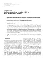
Depth-dose curves of proton pencil beams, a spread-out Bragg peak, and a photon
beam are illustrated in Figure 2. That the doses increase with depth until the end of
Normalized dose [−]
1
6 MV photons
Spread−out Bragg peak
0.8
0.6
0.4
0.2
0
0
135−200 MeV protons
5
10
15
20
25
30
Depth [cm]
Figure 2. Depth-dose curves of a 6 MV photon beam (red), a proton spread-out
Bragg peak (blue, thick), and the 135–200 MeV proton pencil beams constituting
the spread-out Bragg peak (blue, thin).
the proton range makes proton treatments feasible within fewer beams than photon
treatments. Figure 3 illustrates an IMPT plan for a head and neck case treated with
two beams.
The location of the Bragg peak is highly affected by the proton stopping power
of the traversed medium, which is the average energy loss of the protons per unit
6
I NTRODUCTION
Figure 3. An IMPT plan for a head and neck case. For each beam, the fluence
distribution resulting from a specific energy is illustrated. The fluences from all
energies result in the indicated dose distribution in the patient.
path length. This amounts to a disadvantage of scanned proton treatments because
it makes them much more affected by geometric errors than photon treatments.
A scanned proton beam is represented by a number of spots. A spot is defined
by a lateral position in the fluence plane through which the narrow proton beam
should pass, i.e., a point determining how the scanning magnets should direct the
beam, and an energy level determining the depth of the Bragg peak. The fraction of
the beam MU delivered by a given spot is controlled via the spot weight. Individual
spot weights allow for modulated dose distributions in three dimensions from a
single beam direction.
Pencil beam scanning results in 2–3 times less dose to uninvolved normal tissues as compared to IMRT [40]. Although there is yet little clinical evidence that
proton therapy leads to improved outcomes [66], it has been argued that the high
rates of local tumor control after 15 years that proton treatment has yielded would
be unlikely to achieve with any other treatment technique [39].
For a historical review of proton therapy, see Smith [85], and for a review of
proton therapy treatment planning, see Schwarz [82].
2
Treatment planning
A radiation therapy treatment plan is a specification of the number of beams and
the settings that determine the manner in which the beams are delivered to the patient. The goal of treatment planning is to find a plan that yields a high probability
of a curative treatment without complications. Since this probability cannot be de-
ROBUST OPTIMIZATION OF RADIATION THERAPY
7
termined precisely without further assumptions, a plan with high such probability
is often approximated as one with an appropriate balance between high target dose
and low doses to healthy structures.
Planning based on 2D images, as well as 3DCRT treatment planning, is often
performed by forward planning, which means that the treatment planner specifies
the directions, shapes, and intensities of the beams, calculates and evaluates the
resulting dose, and—if the dose is unsatisfactory—determines desirable parameter
changes. The process is repeated until a satisfactory dose distribution is obtained.
The large number of parameters (e.g., aperture shapes, segment weights, spot
weights) of IMRT and IMPT makes forward planning of all parameters practically
impossible. Instead, computerized automated search methods are required. To this
end, the treatment planner specifies desired qualities of the treatment plan, such as
high target dose and low doses to healthy structures, and an optimization algorithm
determines parameters with the aim to achieve these qualities as well as possible.
This type of treatment planning is called inverse planning.
In this thesis, it is assumed that the treatment plan is identical over the course
of the treatment. This is the standard practice of treatment planning today. In
adaptive radiation therapy, the treatment is modified as new information becomes
available [57, 99]. This has the possibility of increasing the probability of tumor
control and reducing the doses to healthy structures.
2.1
Patient geometry
Images of the patient geometry guide the treatment planning process and help the
treatment planner determine where the tumor and the healthy organs are located
and hence which regions to treat and which to avoid.
Tomographic imaging techniques are used to generate 3D representations of the
patient geometry. These techniques use 2D projections of the patient from multiple
directions to compute cross-sectional images (or “slices”) of the patient. The slices
can be stacked to reconstruct a 3D representation of the patient. The most common
imaging technique in treatment planning is CT, which provides a 3D representation
of the patient tissue densities. The densities not only show where the organs are
located, but are also required for accurate dose calculation. Other tomographic
imaging techniques used in treatment planning are magnetic resonance imaging
and positron emission tomography.
The image data is commonly visualized as 2D slices normal to the anteroposterior, superoinferior, or sinistrodextral axis of the patient. The data is used to specify
the regions of interest (ROIs) of the treatment volume. The ROIs are regions of im-