Hopkins general surgery review manual
Bạn đang xem bản rút gọn của tài liệu. Xem và tải ngay bản đầy đủ của tài liệu tại đây (5.77 MB, 158 trang )
Hopkins
General
Surgery
Review Manual
Hopkins General Surgery Manual
1
Introduction
This “manual” is a compilation of study notes I have made over the past 5 years based on a
number of sources, including those listed here:
Text books (see reference list)
Review books (see reference list)
Didactic lectures and conferences at both Johns Hopkins and the NCI Surgery Branch
Presentations I gave during weekly conferences at NCI
Primary and review articles
Points made by attendings and other residents on rounds or in the OR
Intern Sunday morning lecture with Dr. Cameron (2001 – 2002)
Halsted quizzes
SESAP questions
UpToDate®
Disclaimer: Individual illustrations and material may belong to a third party.
Unless otherwise stated all figures and tables by Peter Attia
When I began putting my notes together on random pieces of paper and my Palm Pilot, I did not
intend to do much else with them. However, in time, they became so numerous that I needed to
organize them in a better way. A resident from the Brigham whom I worked with in the lab at
NIH encouraged me to put them together in what he jokingly referred to as an “Attia Bible” of
surgical wisdom, something he had done with his own notes. The intent of these notes was not as
much to be a review for a specific test per se, as it was an “all‐purpose” compilation of salient
points to consider as I go through residency.
Of course, these notes come with the standard disclaimer that they are not meant to replace
reading from primary sources, rather to supplement it. In addition, while I have tried to be as
accurate as possible, during my readings I encountered several “facts” that were either
contradictory to “facts” I had been taught as a resident or read in other sources. For this reason I
can make no guarantees about the validity of each statement made here. I have tried my best to
amalgamate each set of facts into a somewhat concise, yet accurate document.
Hopefully, these notes will provide you with some benefit as well. I welcome all criticism and
correction and look forward to supplementing and augmenting this first edition many times over.
Peter Attia, MD
Surgical Resident
The Johns Hopkins Hospital
Copyright, Peter Attia, 2005. All rights reserved.
Hopkins General Surgery Manual
2
Reference List
1. Vascular Surgery 3rd Ed. House Officer Series. Faust GR, Cohen JR., 1998.
2. ABSITE Killer. Lipkin AP, 2000
3. Rush University Review of Surgery 3rd Ed. Deziel, Witt, Bines, et al., 2000.
4. Current Surgical Therapy 6th Ed. Cameron JL, 1998.
5. Current Therapy of Trauma 4th Ed. Trunkey DD and Lewis FR, 1999.
6. Surgery: Scientific Principles and Practice 3rd Ed. Greenfield LJ, et al., 2001.
7. Shackelford’s Surgery of the Alimentary Tract 5th Ed. Yeo CJ and Zuidema GD. Volumes I – V, 2001.
8. Atlas of Human Anatomy 8th Ed. Netter FH, 1995.
9. Atlas of Surgical Operations 7th Ed. Zollinger & Zollinger, 1993.
10. General Surgery Board Review 3rd Ed. Gold MS, Scher LA, and Weinberg G, 1999.
11. General Surgery Review. Makary MA, 2004.
12. Advances in Surgery® Vol 33. Ed. Cameron JL, et al., 1999.
13. Pitfalls of Data Analysis. Clay Helberg, 1995.
14. Principles of Biostatistics. 2nd Ed. Pagano M and Gauvreau K. 2000.
Editors
The following individuals have been generous with their time and thoughts, and have made several changes and
additions to my original “manual”.
H. Richard Alexander
William A. Baumgartner
John L. Cameron
Michael A. Choti
Peter L. Choyke
Paul M. Colombani
Matthew Cooper
Edward E. Cornwell, III
Todd Dorman
Frederic E. Eckhauser
David T. Efron
Anne C. Fischer
Julie A. Freischlag
Susan L. Gearhart
Vincent L. Gott
McDonald Horne
Udai S. Kammula
Herbert Kotz
Steven K. Libutti
Pamela A. Lipsett
Martin A. Makary
Bruce J. Perler
Peter A. Pinto
Jorge D. Salazar
Anthony P. Tufaro
Peter S. Walinsky
Stephen S. Yang
Charles J. Yeo
Martha A. Zeiger
Hopkins General Surgery Manual
Surgery Branch, National Cancer Institute
The Johns Hopkins Hospital
The Johns Hopkins Hospital
The Johns Hopkins Hospital
Department of Radiology, National Institutes of Health
The Johns Hopkins Hospital
The University of Maryland
The Johns Hopkins Hospital
The Johns Hopkins Hospital
The Johns Hopkins Hospital
The Johns Hopkins Hospital
The Johns Hopkins Hospital
The Johns Hopkins Hospital
The Johns Hopkins Hospital
The Johns Hopkins Hospital
Department of Hematology, National Institutes of Health
Surgery Branch, National Cancer Institute
Department of Gynecology, National Cancer Institute
Surgery Branch, National Cancer Institute
The Johns Hopkins Hospital
The Johns Hopkins Hospital
The Johns Hopkins Hospital
Urology Branch, National Cancer Institute
University of Texas, San Antonio, TX
The Johns Hopkins Hospital
Presbyterian Heart Group, Albuquerque, NM
The Johns Hopkins Hospital
Thomas Jefferson University
The Johns Hopkins Hospital
3
Table of Contents
Breast Disease ................................................................................................................................................................................... 5
Head & Neck Disease...................................................................................................................................................................... 9
Thyroid Gland and Disease ......................................................................................................................................................... 12
Parathyroid Gland and Disease................................................................................................................................................... 15
Multiple Endocrine Neoplasia (MEN) ....................................................................................................................................... 18
Gastrinoma ...................................................................................................................................................................................... 21
Glucagonoma .................................................................................................................................................................................. 22
Insulinoma ...................................................................................................................................................................................... 23
Adrenal Gland ................................................................................................................................................................................ 24
Pheochromocytoma........................................................................................................................................................................ 27
Pituitary Gland ............................................................................................................................................................................... 28
Thoracic Surgery ............................................................................................................................................................................ 29
Mediastinal Disease ...................................................................................................................................................................... 32
Cardiac Surgery: Congenital Defects ......................................................................................................................................... 34
Cardiac Surgery: Acquired Defects............................................................................................................................................. 36
Vascular Surgery ............................................................................................................................................................................ 42
Urology............................................................................................................................................................................................. 54
Orthopedic Surgery ....................................................................................................................................................................... 55
Gynecologic Pathology ................................................................................................................................................................. 56
Neurosurgery .................................................................................................................................................................................. 57
Cancer Epidemiology .................................................................................................................................................................... 58
Esophageal Disease........................................................................................................................................................................ 59
Stomach & Gut Physiology and Disease ................................................................................................................................... 63
Small Bowel Physiology and Disease ........................................................................................................................................ 67
Colorectal Disease.......................................................................................................................................................................... 71
Pediatric Surgery ............................................................................................................................................................................ 79
Spleen and Splenectomy .............................................................................................................................................................. 84
Hepatobiliary Anatomy, Physiology, and Disease .................................................................................................................. 86
Pancreas............................................................................................................................................................................................ 97
Sarcoma .......................................................................................................................................................................................... 102
Melanoma ...................................................................................................................................................................................... 103
Hernia & Abdominal Wall ......................................................................................................................................................... 105
Trauma Principles ........................................................................................................................................................................ 106
Critical Care................................................................................................................................................................................... 117
Hemostasis & Transfusion ......................................................................................................................................................... 126
Metabolism.................................................................................................................................................................................... 130
Transplant Surgery ...................................................................................................................................................................... 131
Nutrition ........................................................................................................................................................................................ 134
Fluids & Electrolytes ................................................................................................................................................................... 136
Renal Physiology.......................................................................................................................................................................... 137
Immunology/Infections .............................................................................................................................................................. 139
Burns............................................................................................................................................................................................... 140
Skin & Wound Healing............................................................................................................................................................... 141
Pharmacology................................................................................................................................................................................ 142
Radiology....................................................................................................................................................................................... 143
Statistics in Medicine .................................................................................................................................................................. 149
Notes............................................................................................................................................................................................... 154
Hopkins General Surgery Manual
4
Breast Disease
Surgical Anatomy:
Intercostobrachial nerve (off 2nd intercostal nerve) sensation to medial arm Æ can sacrifice
Long thoracic nerve: to serratus anterior Æ winged scapula
Thoracodorsal nerve: to latissimus dorsi Æ weak arm adduction
Medial pectoral nerve to pec minor and major; lateral pectoral nerve to pec minor only
Batson’s plexus: valveless vertebral veins Æ allow direct metastases to spine
Poland syndrome: amastia, hypoplastic shoulder, no pecs
Mastodynia: Rx with danazol, OCP
Mondor’s disease: thrombophlebitis of superficial vein of breast Æ Rx with NSAID
DCIS
•
Highly curable with survival of 94 – 100%
•
50% of recurrences are invasive
•
Excision and radiotherapy OR mastectomy; axillary lymph node dissection (ALND) not required
(only 1% have positive nodes). NSABP‐17 showed that lumpectomy alone had 13.4% recurrent
DCIS and 13.4% recurrent invasive cancer vs. 8.2% and 3.9%, respectively for lumpectomy +
radiation.
•
Tamoxifen decreases rate of ipsilateral and contralateral breast cancer in ER positive women, role in
ER negative women, if any, unknown; but must be balanced against risk factors (1 – 2% DVT, PE;
Endometrial cancer). Tamoxifen has NOT been shown to increase survival, only to decrease rate
of recurrence (DCIS and ipsilateral/contralateral invasive breast cancer). Several large studies
have been done (NSABP‐24, 1800 patients, [Fisher B, et al. Lancet 1999;353:1993]) and failed to identify a
survival advantage, despite adequate power.
•
Ongoing research to identify subset of patients who could be treated without radiation
•
Role of Sentinel Lymph is undefined. NO evidence to support use as of 2004.
•
Van Nuys classification MAY identify patients who can benefit from lumpectomy alone (low‐grade,
without necrosis; margin > 1 cm; lesion < 1.5 cm)
LCIS
1. Aka Lobular Neoplasia, encompasses LCIS (> 50% lobular involvement) and Atypical Lobular
Hyperplasia (ALH, < 50% lobular involvement)
2. Not clinically, radiographically, grossly detectable
3. 7 – 10 x increased risk of invasive cancer in either breast (especially in young women with a family
history)
4. 17% risk at 15 years, 5.6% at 5 years; 20% lifetime risk (70% of which will be ductal invasive, 30%
will be lobular invasive); ≈ 1% per year
5. Margins are irrelevant, disease is diffuse (unlike DCIS)
6. LCIS is not itself pre‐cancerous, it is simply a marker of a susceptible field
Phyllodes tumor: 10% malignant; large; rare nodes (spread, if any, hematogenous): Rx Æ WLE, mastectomy
not necessary; NO ALND
Intraductal papilloma: No risk of cancer; #1 cause of bloody nipple discharge
Hopkins General Surgery Manual
5
Comedo breast cancer: Likely multicentric; do mastectomy; poor prognosis
Paget’s disease of the breast: Eczematous lesion on nipple Æ underlying DCIS or ductal CA
Most recent screening recommendations: First at 40; q 1 – 2 years until 50; yearly thereafter
Radial Scar: associated with carcinoma anywhere in the scar; do not stereotactically biopsy (↑ chance of
sampling error), instead Æ excisional biopsy
Staging:
T1: < 2 cm T2: 2.1 – 5 cm T3: > 5 cm T4: skin involvement (inflammatory Æ dermal lymphatic invasion)
N1: + ax nodes
N2: matted/fixed
N3: internal mammary nodes
Stage I: T1
Stage II: up to T2N1 or T3N0
Stage III: T4 or N3 Stage IV: any M
Survival by stage (5 years):
I: 90 – 95%
II: 50 – 80% III: 30 – 50% IV: 15 – 20%
Note: FNA cannot distinguish between DCIS and invasive
Who gets chemotherapy?
1. Pre‐menopausal:
ER/PR‐
T > 1 cm
Any N, including micro (SN+)
2. Post‐menopausal (up to 90% are ER/PR+ Æ get tamoxifen):
ER/PR‐ & T > 2 cm
≥ 4 nodes OR matted nodes (regardless of ER/PR)
(Hence, ER/PR+, ≤ 3 unmatted nodes Æ no chemo)
Who gets axillary radiation? (In general, want to avoid axillary radiation following dissection)
+ supraclavicular node
matted nodes (extracapsular extension)
≥ 4 nodes
Who gets breast irradiation?
any segmental resection for invasive or DCIS
inflammatory disease (T4/skin involvement); some T3
Major studies evaluating role of adjuvant radiation* therapy:
1. The addition of post‐op irradiation to chemotherapy (CMF) for women with stage II or III breast
cancer following mastectomy increased overall survival and reduced locoregional recurrence.
[Postoperative radiotherapy in high‐risk premenopausal women with breast cancer who receive adjuvant chemotherapy.
Danish Breast Cancer Cooperative Group 82b Trial. Overgaard M, et al. NEJM 1997;337:949].
2. Radiotherapy combined with chemotherapy (CMF) after modified radical mastectomy decreases
rates of locoregional and systemic relapse and reduces mortality from breast cancer. [Adjuvant
radiotherapy and chemotherapy in node‐positive premenopausal women with breast cancer. Ragaz J, et al. NEJM
1997;337:956].
*Trental is very effective in treating radiation mastitis
Hopkins General Surgery Manual
6
Locally Advanced Breast Cancer
•
Locally Advanced Breast Cancer (LABC) & Inflammatory Breast Cancer (IBC) sometimes
(incorrectly) used interchangeably
•
Strictly speaking, LABC includes: T3+N1 – 3 or T4+N0 – 3 or any T+N2 – 3 (i.e. Stage III A/B disease)
•
Term IBC first used in 1924 by Lee and Tannenbaum at Memorial Hospital to describe clinical
presentation of 28 patients with: “…breast of affected side usually increased in size…skin becomes
deep red or reddish purple…to the touch brawny and infiltrated…after the fashion of erysipelas…”
•
Accounts for 1 – 6% of all breast carcinomas (IBC)
•
50 – 75% axillary involvement at diagnosis
Overall prognosis median survival: 2 years
•
Diagnosis based on histology of invasive carcinoma PLUS
1. Erythema
2. Edema, or peau d’orange
3. Wheals, or ridging of the skin secondary to dermal lymphatic invasion (although tumor
invasion only seen in 30%)
•
Neoadjuvant treatment and early diagnosis crucial for successful treatment
•
Approximately 75% undergo CR or PR to induction therapy Æ response predicts outcome
Effectiveness of mastectomy by response to induction chemotherapy for control of Inflammatory Breast
Cancer [Fleming R, et al. Ann Surg Onc 1997 4:452]
Initial Response to Induction therapy:
•
CR Æ median survival: 120 months (12%)
•
PR Æ median survival: 48 months (62%)
•
NR Æ median survival: < 24 months (26%)
Further Breakdown:
•
If > 1 cm3 residual tumor Æ median survival: 36 months
•
If < 1 cm3 residual tumor Æ 70% alive at 5 years
Role of Mastectomy:
•
If CR or PR Æ Chemo + RT + Mastectomy increased median survival from 48 to 120 months (vs.
Chemo + RT)
•
If NR Æ Chemo + RT + Mastectomy did not influence median survival (< 24 months), or disease‐
free interval
Summary for Treatment for Inflammatory breast cancer:
1. Neoadjuvant chemo (cytoxan/adriamycin); response to this predicts survival (10% CR, 80%PR)
2. MRM (if PR or CR)
3. Adjuvant chemo (taxane based)
4. Radiation to chest wall
Hopkins General Surgery Manual
7
Chemotherapy/Hormonal* Treatment:
Premenopausal
Postmenopausal
chemo for almost any tumor > 1 cm (regardless of
nodal status)
cytoxan & adriamycin
add taxane if node positive
tamoxifen if ER/PR positive
arimidex and aromatase inhibitors not effective in
premenopausal since can’t compete with
estrogen produced
tamoxifen or arimidex if node negative and ER/PR+
Chemo if poorly differentiated and > 1 cm (even if node
negative)
cytoxan & adriamycin ± taxane if node positive
tamoxifen or adriamycin if elderly, node positive, and
ER/PR+
*Responses to hormonal therapy by marker:
ER/PR+
80%
ER‐/PR+
45%
ER+/PR‐
35%
ER/PR‐
10%
Inherited Breast Cancer Syndromes: 4 appear to be important
1. Li‐Fraumeni Syndrome Æ mutation of p53
2. Mutation of bcl‐2 (18q21) Æ ↑ expression of bcl‐2, which is anti‐apoptotic
3. BRCA‐1 Æ on long arm of 17
4. BRCA‐2 Æ on short region of 13q12‐13
BRCA 1
Ch 17q21; reported 1990, positionally cloned 1994
↑ Risk of breast cancer (85%) and ovarian cancer (40 – 50%)
BRCA 2
Ch 13q12‐13; reported 1994; positionally cloned 1995
↑ Risk of breast cancer (85%) and ovarian cancer (10%)
↑ Risk of male breast cancer (6%)
Risks of Tamoxifen use
↑Uterine adenocarcinoma, sarcoma
↑Cataracts
↑DVT, PE
↓osteoporosis
No change in incidence of heart disease
Hopkins General Surgery Manual
8
Head & Neck Disease
Parotiditis: Usually caused staph spp; seen in elderly, dehydrated; Rx: antibiotics Æ drainage of abscess if
not improving
“Ludwig’s angina”: Sublingual space infection (severe deep soft tissue infection of neck involving the floor
of the mouth); if airway compromise Æ perform awake tracheostomy under local anesthetic Æ operative
debridement
Leukoplakia can be premalignant; erythroplakia is premalignant (and of much more concern)
Head & Neck SCC: Stage I, II (up to 4 cm, no nodes) Æ single modality treatment (surgery or RT)
Stage III, IV Æ combined modality
Perform FNA, not excisional biopsy for suspicious masses
Nasopharyngeal SCC: associated with EBV; 50% present late as neck mass; drainage to posterior neck
nodes; most common nasopharyngeal cancer in adults (lymphoma is most common in kids). Often see in
Asian population
Glottic Cancer: if cords not fixed Æ RT; if fixed Æ surgery + RT. Chemo + RT used more often for organ
preservation
Lip Cancer (99% epidermoid [i.e. squamous] carcinoma): Lower > upper lip (because of sun exposure) Æ
resect with primary closure if < ½ lip; otherwise flap
Tongue Cancer: usually surgery + RT; seen ↑ in Plummer Vinson (dysphagia, spoon fingers, anemia). More
commonly seen in smokers/drinkers
As salivary gland size ↑ [sublingual (60%), submandibular (50%), parotid (20%)] Æ incidence of malignant
disease ↓
Pharyngeal cancers have worse prognosis than oral cancers
Mucoepidermoid carcinoma: #1 malignant salivary tumor overall
Adenoid cystic carcinoma: #1 malignant salivary tumor of submandibular/minor glands. Overall: poor
prognosis
Pleomorphic adenoma ≡ mixed parotid tumor = #1 benign tumor (40 – 70% of all salivary gland tumors)
Do NOT enucleate (or will recur) Æ needs superficial parotidectomy (spare CN VII).
If malignant Æ take whole gland + CN VII;
If high grade (anaplastic) Æ need neck dissection
Warthin’s tumor (adenolymphoma) #2 benign salivary tumor; male predominance; 10% bilateral; 70% of
bilateral parotid tumors are Warthin’s tumor; Rx Æ superficial parotidectomy
Frey’s Syndrome: late complication of parotidectomy (occurs ≈ 50% when facial nerve is preserved); perfuse
perspiration over cheek following salivary stimulation. Intracutaneous injection of Botox A ≈ 100% effective
in treatment, but responses may be short‐lived (can be repeated). Usually self‐limiting.
Ipsilateral drooling following submandibular gland resection: likely injury to marginal mandibular nerve
Radical neck dissection: takes CN XI, SCM, IJ, submandibular gland; most morbid is CN XI
Hopkins General Surgery Manual
9
Classification of Cervical Lymph Nodes
[ACS Surgery Principles and Practice, 2004]
Level
I
II
III
IV
V
VI
Nodes
Submental, submandibular nodes
Upper IJ nodes
Middle IJ nodes
Lower IJ nodes
Spinal accessory nodes, Transverse cervical nodes
Treacheoesophageal grove nodes
Hopkins General Surgery Manual
10
Cancers of the oral cavity usually metastasize to the nodes in levels I – III.
Laryngeal cancers typically metastasize to the nodes in levels II – IV.
Presence of Horner Syndrome (paralysis of the vagus nerve, phrenic nerve, invasion of brachial plexus,
and/or paravertebral musculature) generally indicates tumor unresectability
Tracheo‐innominate fistula
Massive bleeding from trachea is innominate artery until proven otherwise; avoid by making
tracheostomy no lower than 3rd ring
Usually occurs 2 – 3 weeks post tracheostomy; poor nutrition and steroids use may contribute
Mortality ≈ 80%
Sentinel bleed Æ to OR for bronchoscopy
Temporary control (on route to OR) via cuff hyperinflation or finger compression of innominate
artery (anterior pressure)
Treatment is ligation of innominate artery
Most common locations for mandibular fractures: angle (25%) and subcondyl (30%); the most common
long‐term complication of mandibular fracture is malocclusion
Carotid body: chemoreceptor within the adventitia of the CCA (posteromedial side); responds to ↓O2
tension, ↑CO2 tension, ↑blood acidity, and ↑blood temperature by ↑HR, ↑BP, and ↑rate & depth of
respiration in an attempt to overcome the above stimuli
Carotid sinus: pressure sensor within wall of proximal ICA; responds to ↑BP by ↓HR and ↓BP
Hopkins General Surgery Manual
11
Thyroid Gland and Disease
from the Greek work “Theros” (shield) and “eidos” (form)
secretes hormones (T4, T3, calcitonin) from basal membrane side (into bloodstream)
antithyroid agents impair (i) iodination and (ii) coupling of DIT/MIT
T4 Æ T3 peripherally (kidney, liver) (T3; 10 x more active than T4). Propothiouracil (PTU) blocks
peripheral conversion of T4 Æ T3
Note: Suppression of iodine uptake in patients with increased T3 and T4 levels is pathognomonic for
subacute thyroiditis
Usual Causes of Hyperthyroidism:
1. Toxic nodule
2. Toxic multinodular goiter
3. Graves’ disease
4. Early subacute thyroiditis
Ways to Treat Hyperthyroidism:
1. Medical (PTU, methimazole): interfere with iodine conversion; up to 60% recur
2. Radioiodine Ablation (I131): weeks to months; 1st choice by many except in pregnancy
3. Surgery: risks of surgery
Thyroid Storm: untreated hyperthyroidism + stress (trauma, infection, pregnancy, DKA, etc)
Rx: fluids, O2, glucose, anti‐thyroid drugs, but first Æ treat underlying cause;
NB: do not use ASA, as it displaces T4 from thyroglobulin
(Differentiated) Thyroid Cancer
15,000 – 20,000 cases/yr US
15,000,000 nodules/yr (5 – 10% harbor cancer)
mortality < 1%
Risks
age < 14, > 65
previous thyroid cancer
family history
enlarging nodule on thyroid hormone suppression
exposure to low‐dose radiation
Graves’ disease or thyroiditis
syndromes (MEN II, Carney’s)
Cancer Histology
Papillary (60%)
Follicular variant of papillary (20%)
Follicular (< 5%)*
Hürthle cell carcinoma (< 5%)
Medullary (5%)
Anaplastic (1%)
Other (1%)
*difficult on FNA to differentiate follicular adenoma from carcinoma
Hopkins General Surgery Manual
12
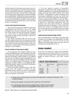
Of FNA’ed lesions in adults*
Inadequate,
15%
Carcinoma,
5%
Suspicious,
10%
Benign, 70%
*Children have higher incidence of carcinoma: 20 – 50%
One option for lesions deemed benign on FNA is hormone suppression: if regresses Æ follow;
If growsÆ remove; if sameÆ repeat FNA
Surgical management
Lobectomy: unclear path (go back for completion, if necessary)
Lobectomy + isthmusectomy: papillary < 1 cm, benign unilateral lesions or suspicious lesions
Total thyroidectomy (followed by RAI): papillary ≥ 1 cm, follicular, Hürthle, medullary
IF planning post‐op RAI Æ must do total thyroidectomy, regardless of size (RAI only useful in well‐
differentiated cancers‐not MTC)
Medullary is the only histology where you do central dissection (level VI and VII) prophylactically (in
addition to total thyroidectomy) and modified radical neck dissection (levels II – V) on affected side
*Performing a total thyroidectomy allows use of thyroglobulin for recurrence monitoring and use of RAI
for microscopic disease
[Figure taken from talk given by H.R. Alexander, NCI, 2003]
Hopkins General Surgery Manual
13
Medullary Thyroid Cancer: 20% of those with MTC have MEN II (100% of those with MEN II have MTC).
MEN II associated MTC tends to be bilateral, younger, worse prognosis, RET‐proto‐oncogene;
aggressiveness as follows: MEN IIB [perform thyroidectomy by 6‐months‐old] > MEN IIA [perform
thyroidectomy by 5‐years‐old] > FMTC
•
May see amyloid on pathology
•
↑ serum calcitonin (can use serum calcitonin levels to monitor for recurrence)
•
Originates from parafollicular C cells, which produce calcitonin and hence do not concentrate
iodine.
Anaplastic: Only operation that should be considered is tracheostomy. Minimal role for palliative resection
Medical management
Thyroid hormone suppression
Radioactive iodine ablation (RIA)
Cytomel (T3) [half‐life 3 – 4 days] vs. Synthroid (T4) [half‐life 4 weeks]
*Hence use T3 replacement post‐op before RIA
Thyroglobulin can only serve as a tumor marker when the following 2 conditions are met:
1. The tumor is well differentiated (since it’s produced by follicular cells)
2. The patient has had a total thyroidectomy
Lymph nodes
For differentiated cancer: no role for prophylactic LND – only for palpable or FNA+ nodes Æ “regional
dissection” (Radical takes levels I – VI + jugular + CNXI; Modified takes levels II – VII, spares IJV, SCM,
spinal accessory nerve XI). Levels most at risk are II – VI
Prognosis (for well differentiated thyroid cancer):
AGES/AMES: age, grade/mets, extent, size; TNM;
However, age, grade (histology), size most important
Age (> 45, or < 14) is single greatest factor
Superior laryngeal nerve (both sensory and motor), External branch: motor to cricothyroid; injury Æ lose
projection, high pitch tone; provides sensory to supraglottis
Recurrent laryngeal nerve: innervates all of larynx except cricothyroid; bilateral injury Æ airway occlusion
Note: Always assess cord function before any operation on thyroid to document RLN function
Hopkins General Surgery Manual
14
Parathyroid Gland and Disease
Superior parathyroid glands from 4th pharyngeal pouch; Inferior (and thymus) from 3rd pharyngeal pouch
Æ more variable position (since longer distance traveled)
All parathyroid glands generally receive blood supply from the inferior thyroid artery
If only 3 glands found at surgery, fourth may be in:
•
Thymus, anterior mediastinum
•
Thyroid
•
Carotid sheath
•
Tracheoesophageal groove*, posterior mediastinum
•
Behind esophagus
*Most common ectopic site
PTH produced by Chief cells Æ increases Ca++ via bone breakdown, GI absorption, increased kidney re‐
absorption, excretion of phosphate by kidney
Hyperparathyroidism
1. Primary: ↑ PTH secretion by parathyroid (high Ca++, low PO4; look for Cl‐/PO4 > 33, even with
normal Ca++)
2. Secondary: ↑ PTH secretion due to renal failure or decreased GI Ca++ abs (Ca++ low or normal)
3. Tertiary: ↑ PTH after correction of 2° hyperparathyroidism (high Ca++)
4. Familial Hypercalcemia Hypocaluria (FHH): see ↑ serum Ca++, PTH, but ↓ urine Ca++ (defect in set‐
point for “normal” Ca++ levels; patients do not experience the sequelae of elevated Ca++); No surgery
Parathyroid Imagining:
•
Sestamibi scan
•
U/S
201Technetium‐thallium subtraction scan
•
•
CT/MRI
Primary Hyperparathyroidism
Incidence: 1/4000
Risks: MEN I, IIa, irradiation, family history (autosomal dominant)
Adenoma > 85% [1], Hyperplasia ≈ 10% [4], Carcinoma ≈ 1% [1], [# glands typically involved]
Typically: [Cl‐]/[PO4] > 33
Initial medical treatment: IV fluids, lasix, NOT thiazides
Treatment
•
1° Adenoma: Surgically remove adenoma (± biopsy all enlarged glands)
•
1° Hyperplasia: Bilateral neck exploration and intraoperative PTH. Subtotal parathyroidectomy
(leave ½ lower gland in situ) or total parathyroidectomy with autotransplantation
•
1° Carcinoma: WLE with ipsilateral thyroidectomy and lymph node dissection
•
2°: Correct Ca++ and PO4, perform renal transplant (no parathyroid surgery)
•
3°: Correct Ca++ and PO4, perform renal transplant, remove parathyroid glands and re‐implant 30 to
40 mg in forearm
Hopkins General Surgery Manual
15
Parathyroid Carcinoma
Signs/Sx: HyperCa++, elevated PTH, palpable gland (50%), neck pain, recurrent laryngeal nerve paralysis
HCG is a marker
Treatment: En bloc resection including ipsilateral thyroid lobe + associated lymph nodes
Post op Complications:
•
Recurrent laryngeal nerve injury
•
Neck hematoma (open at bedside if breathing compromised)
•
HypoCa++
Parathyroid Pearls
•
90% of primary hyperparathyroidism due to a single adenoma Æ unilateral exposure is ok (with
intraop PTH)
•
MUST exclude familial/MEN disease (a different entity altogether which requires subtotal
parathyroidectomy Æ leave ½ of a lower gland in situ)
•
Nuclear medicine expertise is crucial: if possible, subtraction of Tc99m pertechnetate (potassium
analog specific for thyroid) from Tc99m Sestamibi (taken up by both thyroid and parathyroid)
•
For intra‐op PTH to be valid must have >50% drop in baseline PTH within 10 minutes
•
Intra‐op PTH must be used if doing single gland exploration (MIP), else Æ must do 4 gland
exploration
If disease recurs, MUST distinguish between persistent and recurrent:
•
Persistent: Only transient “cure”. Almost always implies missed adenoma. #1 place is TE groove on
right side; also consider ectopic glands
•
Recurrent (> 6 months normocalcemia): Implies hyperplasia with re‐growth (e.g. familial, possibly
cancer)
•
10 x increase in RLN injury during re‐do surgery. Hence, first step in re‐do is confirm diagnosis
with 24 hour urinary Ca++ (if normal Æ no disease). Second, check for family history of MEN I
manifestations
•
Localization with Sestamibi and U/S. Consider CT/MRI (very bright on T2 to differentiate from
LNs)
Hopkins General Surgery Manual
16
Ultimate pearls:
“Superior glands” is really a misnomer, they should be called “Posterior glands”, since they are virtually
always posterior and cephalad to the RLN. Ectopic sites are generally posterior in TE grove
“Inferior glands” should be called “Anterior glands” since they are virtually always anterior and caudal to
the RLN. Ectopic glands are usually anterior/mediastinal
The figure below shows in dotted lines the possible locations for the parathyroid glands in relation to the
RLN. There is significant vertical overlap, such that superior glands can actually be below inferior glands,
and vice versa.
Hopkins General Surgery Manual
17
Multiple Endocrine Neoplasia (MEN)
*inherited autosomal dominant (with variable penetrance)
MEN Type I
aka Wermer’s Syndrome (“PPP”)
•Parathyroid Hyperplasia (≈ 90%)
HyperCa++ Æ usually first
•Pancreatic islet cell tumors (≈ 67%)
Gastrinoma (ZES) (≈ 50%)
Insulinoma (≈ 20%)
•Pituitary Tumor (≈ 67%) most often PL‐secreting tumor
MEN Type IIA
aka Sipple’s Syndrome (“MPP”)
•Medullary Thyroid Carcinoma (100%) Æ 2nd to 3rd decade
calcitonin secreting
usually quite indolent
•Pheochromocytoma (> 33%)
catechol excess
usually benign, bilateral, adrenal
•Parathyroid Hyperplasia (≈ 50%)
hyperCa++
MEN Type IIB
ʺMMMPʺ
•Mucosal Neuromas (100%)
naso, oropharynx, larynx, conjunctiva
•Medullary Thyroid Carcinoma (≈ 85%)
more aggressive than IIA
•Marfanoid body habitus
•Pheochromocytoma (≈ 50%)
often bilateral (70%)
MEN1 Consensus Summary Statements (loss of function)
Diverse array of defects (missense, nonsense, frameshift, mRNA splicing); hence difficult to
screen because of so many possible mutations
1997: gene Menin found of Ch 11. Exact function unknown, but it is a tumor suppressor gene
The MEN1 germline mutation test is recommended for MEN1 carrier identification.
All kindred with MEN1 are likely to have a mutation in the MEN1 gene.
However, MEN1 germline mutation tests fail to detect 10 – 20% of mutations. If a family lacks an
identifiable MEN1 mutation, 11q13 haplotype testing about the MEN1 locus or genetic linkage
analysis can identify MEN1 carriers. Periodic biochemical testing is an alternative when DNA‐based
tests are not possible.
Hopkins General Surgery Manual
18
The main candidates for MEN1 mutation analysis include index cases with MEN1, their unaffected
relatives, and some cases with features atypical for MEN1.
MEN1 carrier analysis should be used mainly for information. It should rarely determine a major
intervention.
MEN1 tumor patterns in families do not have clear variants or specific correlations with an MEN1
germline mutation pattern. Thus, the MEN1 carriers in a family with either typical or atypical
expression of MEN1 should be monitored similarly for typical expressions of MEN1 tumors.
MEN1 tumors cause morbidity through hormone excess (PTH, gastrin, PRL, etc.) and through
malignancies (gastrinoma/islet cell or foregut carcinoid).
Medications control most features of hormone excess (gastrin, PRL, etc.). Surgery should control
features of excess of some other hormones (PTH and insulin). Surgery has not been shown to
prevent or cure MEN1‐related cancers.
Hyperparathyroidism develops in over 90% of MEN1 carriers. There is controversy over indications
for parathyroid surgery in MEN1 patients.
The preferred parathyroid operation in the HPT of MEN1 is subtotal parathyroidectomy (without
autograft); transcervical near‐total thymectomy is also simultaneously. Parathyroid tissue should be
cryopreserved.
Curative surgery for gastrinoma in MEN1 is rare. There is controversy over the indications for
surgery for gastrinomas in MEN1.
Surgery in MEN1 is indicated and is usually successful for insulinoma. For most other pancreatic
islet tumors, except gastrinomas, surgery is also indicated; however, there is no consensus over
tumor criteria for the latter operations.
The management of pituitary tumor in MEN1 should be similar to that in sporadic cases.
MEN2 Consensus Summary Statements (gain of function)
1995: RET‐proto‐oncogene (responsible for tyrosine kinase activity) identified on Ch 10
Fewer possible mutations (codons 609, 611, 618, 620, 634; involve replacement of a cystine residue)
The main morbidity from MEN2 is MTC. MEN2 variants differ in aggressiveness of MTC, in
decreasing order as follows: MEN2B > MEN2A > FMTC.
MEN2 carrier detection should be the basis for recommending thyroidectomy to prevent or cure
MTC. This carrier testing is mandatory in all children at 50% risk.
Compared with RET mutation testing, immunoassay of basal or stimulated CT results in more
frequent false positive diagnoses and delays of the true positive diagnosis of the MEN2 carrier state.
However, the CT test still should be used to monitor the tumor status of MTC.
RET germline mutation (10q11 – 12) testing has replaced CT testing as the basis for carrier diagnosis
in MEN2 families. It reveals a RET mutation in over 95% of MEN2 index cases.
The RET codon mutations can be stratified into three levels of risk from MTC. These three categories
predict the MEN2 syndromic variant, the age of onset of MTC, and the aggressiveness of MTC.
Hopkins General Surgery Manual
19
Thyroidectomy should be performed before age 6 months in MEN2B, perhaps much earlier, and
before age 5 yr in MEN2A. Policies about central lymph node dissection at initial thyroidectomy are
controversial and may differ among the MEN2 variants.
MEN2 has distinctive variants. MEN2A and MEN2B are the MEN2 variants with the greatest
syndromic consistency.
FMTC is the mildest variant of MEN2. To avoid missing a diagnosis of MEN2A with its risk of
pheochromocytoma, physicians should diagnose FMTC only from rigorous criteria.
Morbidity from pheochromocytoma in MEN2 has been markedly decreased by improved
recognition and management. The preferred treatment for unilateral pheochromocytoma in MEN2 is
laparoscopic adrenalectomy.
HPT is less intense in MEN2 than in MEN1. Parathyroidectomy should be the same as in other
disorders with multiple parathyroid tumors.
Hopkins General Surgery Manual
20
Gastrinoma
•
•
•
•
•
•
50% of patients with MEN I have a gastrinoma
33% of patients with ZES have MEN I (Clinical Triad: 1. PUD 2. gastric acid hypersecretion 3. Islet
cell tumor)
In MEN I associated ZES: Most common site is duodenum (2nd is pancreas)
⅔ are malignant
25% of patients with pheochromocytoma have ZES
Risk of metastatic behavior from gastrinoma rises sharply at 3 cm (< 3 cm Æ < 2% chance of mets)
The sporadic disease is a different one from the MEN‐associated one
•
Sporadic ZES occurs outside of the duodenum 30 – 60% of the time; MEN version is virtually always
in the duodenum (with or without pancreatic involvement)
•
Sporadic ZES is much more likely to be malignant (the extrapancreatic primaries are less
biologically aggressive in both diseases)
•
Sporadic ZES is more often solitary and larger at the time of detection, usually > 2 cm
•
MEN ZES is a diffuse disease, rarely amenable to true “cure”
“Gastrinoma Triangle” joins:
1. junction of cystic duct & CBD
2. junction of 2nd and 3rd portion duodenum
3. junction of neck & body of pancreas
(90% found in this triangle) [Am J Surg 1984 147:25 Stabile, Morrow, Passaro]
Ddx for increased gastrin:
Gastrinoma
Retained excluded antrum (a surgical mistake)
High acid
Gastric outlet obstruction
Antral G‐cell hyperplasia/hyperfunction
Postvagotomy
Pernicious anemia
Atrophic gastritis
Low/minimal acid
Short gut syndrome
Renal failure
H2 blocker, proton pump inhibitor*
*pH < 2 inhibits gastrin secretion in normal patients
Check gastrin levels in patients with:
1. recurrent ulcers
2. ulcers in unusual places (e.g. jejunum),
3. refractory to medical management
4. prior to any elective operation for an ulcer
5. unexplained or persistent diarrhea
6. peptic ulcer and any endocrinopathy
7. family history of PUD
8. family history of MEN I
Check: 1. fasting gastrin level
2. post‐secretin challenge gastrin level (synthetic secretin – 2 units/kg IV bolus)
3. Ca++ (MEN I screen)
4. Chemistry panel
Hopkins General Surgery Manual
21
Look for:
•
ZES fasting: 200 – 1000 pg/mL (normal < 100 pg/mL)
•
Basal acid secretion: ZES > 15 mEq/hr (normal < 10 mEq/hr)
Note: generally will see failure of feedback: fasting gastrin ≈ 1000 with gastric pH < 2.5
Secretin Stimulation Test:
•
IV secretin administered, gastrin measured
•
ZES: increased gastrin (by > 200 pg/mL) within 10 to 20 minutes (normal response is decrease in
gastrin)
Management issues:
•
Some believe distal pancreatectomy should be done in any patient with MEN I with either hormonal
syndrome or a neuroendocrine tumor regardless of location in pancreas or duodenum. Virtually all
patients with MEN I ZES have concomitant neuroendocrine tumors in neck, body, or tail.
•
If patient has gastrinoma and hyperparathyroidism Æ remove parathyroid first to normalize Ca++
levels (since hypercalcemia is more dangerous than hypergastrinemia)
•
Higher than “normal” doses of PPI are needed for achlorhydria
•
Minimal role for CT scan/octreoscan
•
Imamura Test: intra‐arterial secretin into visceral arteries to measure hepatic vein gastrin levels
(look for step up) is good for localization
•
No role for debulking functional gastrinoma mets, since patients can be managed medically in this
setting with PPIs
Post‐Op
Must stay on acid suppression for 3 – 4 months because even after resection acid secretion high for some
time
Glucagonoma
The 4D Syndrome: diabetes, diarrhea, dementia, dermatitis (patients look cachectic)
Many patients also have normochromic normocytic anemia, hypoalbuminemia, weight loss, beefy red
tongue (glossitis), stomatitis, angular chelosis (i.e. signs of malnutrition)
Usually found in tail of pancreas, but can be anywhere. Usually > 3 cm at time of diagnosis; 70% malignant
Diagnosis simple by measuring serum glucagon level, although most patients with elevated glucagons do
not have gastrinoma [see review: Wermers RA, et al. Medicine (Baltimore). 1996;75:53]
Sx: Necrotizing Migratory Erythema‐NME (usually below waist), glossitis, stomatitis, diabetes
•
IV Tolbutamide results in elevated glucagon
•
Medical treatment for Necrotizing migratory erythema: Somatostatin/octreotide, IV Amino acids
(TPN)
•
Aggressive surgical resections are indicated, even if metastatic
Hopkins General Surgery Manual
22
Insulinoma
Number 1 islet cell neoplasm; associated with MEN I
•
•
•
•
•
80 – 90% are benign solitary adenomas cured by surgical resection
30% < 1 cm
10% multiple
10 – 15% malignant
10% hyperplasia or nesidioblastosis
Presents with sympathetic nervous system symptoms due to hypoglycemia (patients look like “Pillsbury
Dough Boy”)
Whipple’s Triad:
1. Hypoglycemia < 50 mg/dl
2. CNS symptoms.
3. Reversal of CNS symptoms. with glucose admin.
Ddx for hyperinsulinemia:
•
Reactive hypoglycemia (very common),
•
Functional hypoglycemia with gastrectomy,
•
Adrenal insufficiency, hypopituitarism, hepatic insufficiency,
•
Munchausen’s syndrome (self injection)
•
Tumors secreting insulin‐like molecule (sarcoma, mesothelioma, etc.)
First, check for proinsulin, then:
Get 72 hour fasting levels with q6 hour checks until patient becomes symptomatic
Insulin:Glucose ratio = insulin (uU/ml)/glucose (mg/dl) > 0.3 found in almost all patients with insulinoma.
Accuracy increased by “Amended ratio” = insulin (uU/ml)/ [glucose (mg/dl) – 30] > 0.3
Localizing Tests:
•
CT, A‐gram, endoscopic ultrasound, venous catheterization (sample blood along portal and splenic
veins)
•
Calcium angiogram: Ca++ causes insulin secretion Æ localize to artery (e.g. splenic for tail) where
tumor nearest
•
Intraoperative U/S is probably the best test for localization
Medical Treatment:
•
Diazoxide to suppress insulin levels (until resection). Diazoxide inhibits the sulfonylurea receptor 1
(SUR1) on the beta cell, which is a component of the K+ATPase responsible for insulin secretion
•
Octreotide, IV glucose
Hopkins General Surgery Manual
23
Adrenal Gland
Embryology:
Cortex: mesoderm (4th to 6th week)
Medulla: ectoderm/neural crest (sympathetic NS and ganglion); migrates along sympathetic chain
Ectopic location: IMA, Organ of Zuckerkandl
Right gland: drains into IVC; Left gland: drains into renal vein
[Figure adapted from RUSH review manual, 2000]
Aldosterone is produced exclusively in the ZG because of the presence of corticosterone
methyloxidase (“anatomically specific” enzyme location)
PNMT converts norepinephrine Æ epinephrine. Found only in adrenal medulla.
Rate limiting step is Tyrosine Æ DOPA via Tyrosinase hydroxylase
11β‐hydroxysteroid dehydrogenase type I is required to convert inactive prednisone to active
prednisolone; its activity varies markedly from person to person
Cortisol is not a “storage” hormone; however, it does promote gluconeogenesis to preserve hepatic
reserve
Regardless of size, an adrenal mass should be removed if it is:
1. Growing,
2. Functioning, or
3. Suspicious on T2 MRI (the brighter it is, the more suspicious it is)
If none of the above, remove lesions greater than 4 – 6 cm (since 15% chance of malignancy if greater than 6
cm Æ risk increases with size)
Ask: is it Functional? Malignant tumor of adrenal gland? Likely metastatic to adrenal gland?
Hopkins General Surgery Manual
24
Syndromes:
I.
Conn’s (↑ aldosterone): 75% unilateral adenoma Æ Rx: with resection
25% bilateral hyperplasia Æ Rx: spironolactone + C++ channel blocker
II.
Addison’s (↓ aldosterone and cortisol): low Na+, high K+, hypoglycemia; can present in crisis with
hypotension
III. Waterhouse Friedrickson: adrenal hemorrhage with meningococcal sepsis
IV. Nelson: post adrenalectomy (10%) Æ ↑ ACTH, pigmentation, change in vision from ↑ pituitary
response
V. Cushing’s Disease (pituitary): 80% of non‐iatrogenic causes; pituitary microadenoma Æ ↑ ACTH
(will also see urine 17‐OH progesterone)
Adrenal Cushing’s Syndrome: (aka ACTH‐independent Cushing’s) 15% of non‐iatrogenic causes Æ
10% adrenal adenoma, 5% adrenal carcinoma; bilateral hyperplasia is very rare; will see ↓ ACTH
Ectopic Cushing’s Syndrome: 20% of ACTH‐dependent; sources of ectopic ACTH including:
Pulmonary (SCLC, bronchial, thymic carcinoids), Neuroendocrine tumors, Pheochromocytoma,
MTC
Diagnosis:
1. Start with 24 hour urine free cortisol and plasma ACTH
2. Low dose dexamethasone suppression Æ will suppress causes of hypercortisolism such as obesity
and excess ethanol ingestion, but not others (confirms dx)
3. High dose dexamethasone suppression Æ will suppress pituitary adenoma, but not ectopic sources
(locates cause)
4. MRI, CT, and/or petrosal venous sampling
Treatment:
•
“Medical Adrenalectomy” = metyrapone and aminoglutethimide
•
Surgical removal of all functional adrenal masses is indicated, including bilateral adrenalectomy for
diffuse disease in patients recalcitrant to medical management
Stress Dose Steroids
[Chernow et al. Ann Surg 1994, 219:416]
• Under normal conditions, body produces 30 mg hydrocortisone equivalent (solucortef)/day
• Under extreme stress Æ up to 300 mg/day
• Prednisone is 4:1 (to solucortef)
• Solu‐Medrol is 5:1
• Decadron is 25:1
Normal adrenal secretion is 25 – 30 mg cortisol/24h
Appropriate stress test: 250 mcg cosyntropin Æ
1. ↑ in plasma cortisol by ≥ 7 mcg/dL, or
2. Absolute level > 20 mcg/dL
Hopkins General Surgery Manual
25