Colonic and anorectal dysfunction associated with multiple sclerosis
Bạn đang xem bản rút gọn của tài liệu. Xem và tải ngay bản đầy đủ của tài liệu tại đây (4.21 MB, 10 trang )
0002-9270/89/8406-0587
Vol.84. No. 6. 1989
Printed in U.S.A.
THE AMERICAN JOURNAL or GASTROENTEROLOGY
Copyright© I989by Am.Coll. of Gaslrocnterology
Clinical review
Colonic and Anorectal Dysfunction Associated
with Multiple Sclerosis
John P, Hinds, M.B.Bch., and Arnold Wald, M.D.
Gastroenterology Unit. Montefiore Hospital. University of Pittsburgh School of Medicine. Pittsburgh. Pennsylvania
In this review, we outline the clinical features and
pathophysiology of constipation and fecal incontinence
in multiple sclerosis. We also present various treatment
options and suggestions for further studies of colorectal
dysfunction in this disease.
Gastrointestinal symptoms are common in patients
with multiple sclerosis. In a recent survey of 280 unseIcctcd patients with multiple sclerosis, 68% reported
constipation and/or fecal incontinence. In contrast to
bladder dysfunction which has been extensively studied,
bowel dysfunction in tbis disease bas received relatively
little attention. Tbis review outlines tbe clinical features
and pathophysiology of constipation and fecal incontinence in multiple sclerosis and presents treatment options and suggestions for investigation of colonic and
anorectai dysfunction in tbis population.
CONSTIPATION
Constipation is a common complaint in patients witb
multiple sclerosis and was present in 43% of our survey
population. We defined constipation as infrequent
bowel movements (<3/wk) and/or rectal manipulation
to facilitate defecation {during >50% of bowel movements) and/or frequent laxative, enema, or suppository
use (>l/wk). Constipation was more common in the
moderately (48%) and severely (56%) disabled groups
compared to the mildly disabled (24%). The high prevalence even in the latter group su^ests that factors
other than lack of mobility are important.
INTRODUCTION
Multiple sclerosis is a common neurological disease
aflecting approximately one-quarter million Americans
(1). The onset of the disease is usually during the third
and fourth decades: 60% of those affected are women.
The characteristic pathological feature in multiple sclerosis is focal demyelination of axons leading to plaque
formation which can occur within any area of the white
matter of the brain and spinal cord. The result is an
intermittent partial or complete block in nerve conduction: the clinical picture that ensues depends on the
location of the foci of demyelination within the central
nervous system (2).
Gastrointestinal symptoms are common in patients
with multiple sclerosis. In a recent survey of 280 unselected subjects with multiple sclerosis (3), 68% reported
constipation and/or fecal incontinence, complaints that
were common even in mildly disabled subjects (Table
1). Upper gastrointestinal symptoms (4, 5), especially
difficulty with swallowing {6. 7). are also features of the
disease, although these complaints were less prominent
in our survey population. In contrast to bladder dysfunction, which is also common and has been extensively studied (8-10), bowel dysfunction has received
relatively little attention.
Normal colonic motility
The two major functions of the colon are to retard
tbe fecal stream, ensuring the absorption of fluid and
electrolytes, and to act as a storage organ so that defecation can occur at a socially convenient time.
The colon consists of an outer longitudinal and an
inner circular smooth muscle layer between which lies
the intrinsic nervous system (myenteric plexus). Tbe
extrinsic nerve supply is via the autonomic nervous
system. The parasympathetic nerve supply to the ascending colon and proximal one-half of the transverse
colon is via the vagus nerve. The remainder of the colon
is supplied by parasympathetic fibers from sacral nerves
2, 3, and 4 via the nervi erigentes. These nerves synapse
with intrinsic neurons, among them cholinergic nerves
which release acetylcholine to muscarinic receptors on
the muscle cells.
The sympathetic innervation of the colon is supplied
by the lower six thoracic and first three lumbar seg-
Received Nov. W. 1988: accepted Nov. 10. 1988.
587
588
Vol. 84, No. 6. 1989
HINDS AND WALD
Splk»
TARt_h I
Lower Bowel Sympioms in Muhiple Sclerosis (n = 280)
Symptoms
Prevalence
Constipation
Fecal incontinence
Constipation and fecal incontinence
Constipation and/or fecal incontinence
43%
51%
25%
68%
ments of the spinal cord via the inferior and superior
mesenteric ganglia. For the most part sympathetic
nerves are in contact with cholinergic nerves: they
inhibit release of acetylcholine rather than act on the
muscle cells directly. There is also evidence for nonadrenergic inhibitory' nerves in the colon. Generally
SF>eaking, stimulation of the sympathetic nervous system inhibits colonic motility, wbereas stimulation of
the parasympathetic nervous system enhances motility
(11.12).
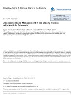
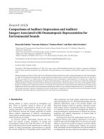
Two types of electrical activity are generated by muscle cells of the colon, the slow waves and the calciumdependent spike potentials. The slow waves are of variable frequency (3-12 cycies/min) and amplitude. Muscle contraction is generated by the spike potentials,
which are rapid depolarizations that last a few milliseconds. Spike potentials of short duration occur only
on the peak depolarization of the slow waves, suggesting
that these slow waves act as the pacemaker of the colon
(Fig. 1)- Stretch, neural, and hormonal stimulation are
all able to generate spike potential activity (12. 13).
Three main types of muscular contractions occur in
the colon. Segmental contractions are nonpropulsive
events caused by spike activity of short duration (<5 s):
their main function is to retard the fecal stream. Propulsive contractions occur in association with 15- to
30-s bursts of electrical activity, whereas mass movements propagate over the entire colon after spike bursts
of about 30 s. and may result in defecation. Administration of atropine decreases the frequency of the slow
wave-spike complexes (13). Control of the electrical
events in the colon, and, therefore, propulsion, appears
to be largely a function of the cholinergic system (13,
14), although noncholinergic excitatory nerves also
bave been identified in the human colon (12, 13).
For about 40 min after a meal is eaten, there is an
increase in colonic spike activity that is usually associated with propulsive contractions or mass movements.
This gastrocolonic response is initiated by the fat component of the meal (15) and is mediated in part by
cbolecystokinin. although other gastrointestinal peptides such as gastrin (16), neurotensin (17), and substance-P (18) may also play a role. Secretin inhibits this
choleeystokinin-induced motor activity (19). This gastrocolonic response persists after vagotomy (20), and
seems to be mediated via the spinal cord, as it is absent
after thoracic cord transection (21). The response also
polmnllml
>.
Slow w»¥»
TRANSUEyBRANE
COLONIC
PRESSURE
/
\
FIG. I. Transmcmbranc potential in the colon (top) and colonic
motor activity {bottom). Spike potentials occur only on ihc peak
depolarization of the slow waves and produce myogcnic contractions
which can be measured using pressure Iransduccr^.
SAGITTAL
SPMItlCT£ft
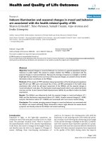
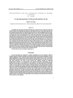
Kui. 1. Sagittal views of the anorectum illustrating the important
structural components. Inset illustrates ihe anterior pull of the anorectum and closure of the anal canal during simultaneous contraction
of the puborectalis muscle and external anal sphincter. Adapted from
Wald (23V
requires muscarinic and opioid receptors as it is blocked
by alropine and naloxone (22).
Anorectal anatomy
Tbe anal canal is an anteroposterior slit, 3 to 4 cm
long, which is kept closed by the tonic activity of the
smooth internal and striated external anal sphincter
muscles. The rectum is a compliant reservoir which at
rest is kept at an approximately 90° angle to the anal
canal by the tonic contraction of the puborectalis muscle (Fig. 2). This muscle is part of the pelvic floor, and
forms a "sling" around the anorectal junction with its
insertions attached anteriorly to the pubis (23).
The anal canal has a dense network of nerve fibers
which can discriminate between solid, liquid, and gas
(24, 25). This area is therefore felt to be important in
the maintenance of continence. The rectum possesses
stretch receptors oniy (26).
Normal defecation
At the initiation of defecation, closure of the glottis
and contraction of the abdominal wall muscles increase
June 1989
COLONIC AND ANORECTAL DYSFUNCTION IN MS
intraabdominal pressure, thus propelling feces forward.
When the fecal bolus reaches the upper rectum, the
pelvic floor and puborectalis muscle relax simultaneously, resulting in straightening ofthe anorectal angle.
Distension of the rectum by stool results in reflex
relaxation ofthe internal anal sphincter (RISR) and a
transient contraction of the external anal sphincter.
When a critical pressure is reached in the rectum, the
external sphincter is inhibited and the fecal bolus expelled. After cessation of straining, the pelvic floor
ascends, the anorectal angle is restored to normal, and
a rebound contraction of the external anal sphincter
occurs.
The higher control of defecation is incompletely
understood. There is evidence to suggest that a defecation center exists in the pons, which is under cortical
control (27. 28). Other areas of the brain may also be
important: stimulation of the hypothalamus may inhibit or enhance colonic motility, depending on the
area stimulated (28). Another defecation center is probably present in the sacral cord. In patients with high
spinal cord transection. rectal sensation is lost, but
defecation can proceed in a regular fashion if an appropriate stimulus (e.g., digital stimulation) is applied to
the rectum. This has been termed "automatic defecation," as it occurs without the influence of higher
cortical control. In patients with low spinal injury, rapid
distension of the rectum does not produce a rectal
contraction as in normal individuals and patients with
high spinal cord injury. Therefore, this contraction
presumably is mediated by a spinal reflex (29).
Colorectal dysfunction in multiple .sclerosis
Studies of colonic dysfijnction in multiple sclerosis
were performed by Glick et al. (30), who evaluated
seven men with severe spastic quadriparesis who also
were severely constipated and had bladder dysfunction.
Somatosensory-evoked potentials, cystometrograms,
colonmetrograms. and colonic motor and myoelectric
activity were carried out. All seven patients exhibited
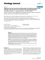
"hyperreflexic" or high pressure/volume colonmetrograms {Fig. 3); the authors felt that these were analogous
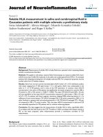
to the hyperreflexic cystometrograms which were present in most of these patients. Colonic motor and myoelectrical activity were also abnormal in all seven patients. The baseline mean amplitude of colonic motor
activity in the multiple sclerosis patients was significantly lower than in normal controls. In addition, patients had no demonstrable postprandial increase in
colonic motility as was observed in the controls (Fig.
4). All patients had abnormal cortical somatosensoryevoked responses demonstrating lesions in the central
neural pathways. Based on these results, the authors
proposed the existence of a "visceral neuropathy" as
the cause of severe constipation in this group of patients.
589
JE1^^-L^,^JL>JJ^^X/->^AA>-^
oio
mo
MOO woo
voo
jooo
2300
I"
Fici. 3. Colonomelrograms in which water is infused into Ihe
rectum while intracolonic pressures are monitored continuously, A
normal colonometrogram {ahove) is compared with a hyperreflexic
study in a patient wiih multiple sclerosis {hvlow). "C" denotes colonic
contractions and "R" indicates respiratory excursions. From Glick ei
al. (30).
Q Conlral»(n= HI
O Mul1lcWScttfOI»in
I .OH
o
I "
B-
t
<
c
4-
3
?.
Tima PsriM (tnimilM)
Fi(i. 4. Colonic myoelectrical spike potential frequency recorded
from the rectosigmoid colon of seven patients wiih multiple sclerosis
and 11 control subjects. From Glick et at. (30).
Absence of the gastrocolonic response has also been
seen in chronic idiopathic intestinal pseudoobstruction
(31) and in diabetics who are severely constipated (32).
The colonic muscle responds normally to the cholinesterase inhibitor, neostigmine, in these patients, suggesting that the cholinergic postganglionic neurons are
intact. This response to neostigmine has not been investigated in patients with multiple sclerosis. Nevertheless, it appears that all three groups of patients have a
disorder ofthe neurohormonal control of colonic motility. Further assessment ofthe gastrocolonic response
in multiple sclerosis patients should include response
to neostigmine and evaluation of patients with constipation who are less disabled than Glick's subjects.
High pressure/volume colon metrograms have been
reported in constipated subjects who have other neurological diseases (33). This response is thought to result
from interruption of the normal cortical inhibition of
colonic motor activity. The net result may be a functional obstruction to the passage of colonic contents. In
contrast, low pressure/volume colonmetrograms have
been reix)rted in patients with sacral cord and sacral
590
Vol. 84, No. 6. 1989
HINDS AND WALD
plexus lesions (33) and in multiple sclerosis patients
with demyelination ofthe conus medullaris (34). This
may result in a highly compliant rectum (megarectum)
which may lead to difficulty with defecation through
several mechanisms. There may be failure ofthe rectum
to contract around the distending stool, or the ability
to perceive rectal distension may be impaired, resulting
in the accumulation of a large fecal bolus that is too
large or painful to expel (35).
Forty-eight percent of the constipated subjects with
multiple sclerosis in our survey population reported a
need to digitally manipulate the rectum to facilitate
defecation. This may reflect poor expulsion effort due
to generalized muscle weakness or megarectum as already mentioned. However, the possibility of a functional obstruction to defecation also must be considered.
In a recent study, Weber et al. (36) evaluated 16
patients with multiple sclerosis and urinary bladder
dysfunction, using anorectal manometry, colonic
transit studies, and urodynamic testing. Fifteen described themselves as constipated, and six also complained of fecal incontinence. Prolonged colonic transit
was demonstrated in 14 patients, of whom seven had
left-sided delay only, whereas seven had a more generalized slowing. Five patients had decreased perception
of rectal sensation, and 10 exhibited manometric criteria suggestive of so-called "outlet obstruction." These
criteria were defined as hypertonia in the upF>er anal
canal, with or without ultraslow waves greater than 20
cm H:O; the presence of an "overshoot" contraction
after the rectoanal inhibitory reflex (RISR); and RISR
absent or the amplitude insufficient. Twelve patients
had a hyperreflexic urinary bladder on cystometrogram,
and detrusor urethral dyssnergia was seen in 14 cases.
On the basis of these results, the authors suggest that
the prolonged colonic transit and anorectal dysfunction
are secondary to tbe neurological disorder, and hypothesize the existence of "rectoanai dyssnergia" causing
problems of rectal evacuation in the same way that
detrusor urethral dyssnergia causes abnormal bladder
emptying.
These findings need confirmation and further clarification. As the authors point out, the manometric
abnormalities seen in outlet obstruction are also seen
in other disorders (37). Second, compliance measurements of the rectum were not performed. This is an
important parameter to measure if abnormalities of
internal sphincter relaxation are to be interpreted accurately. Furthermore, it is possible that some of the
anorectal abnormalities described are a consequence,
rather than a cause, ofthe constipation. These questions
notwithstanding, it is likely that neurogenic abnormalities of colonic and/or anorectal function are the cause
of constipation and defecatory difficulty in this disease.
Evaluation
When evaluating constipated patients with multiple
sclerosis, one should establish the duration of constipation and its relationship to the onset and other manifestations ofthe disease. Stool frequency alone may be
a poor parameter to follow, as laxative use and digital
evacuation of the rectum are very common (3). The
degree of straining and the presence or absence of a
defecatory urge should be ascertained. The presence of
other medical conditions associated with constipation
should be established. A drug (Table 2) and diet history
should be taken and the degree of immobility assessed.
Patients with genitourinary symptoms should be asked
about fluid intake, since they commonly restrict fiuids
in order to avoid urinary incontinence: unfortunately,
constipation often follows.
In addition to a detailed physical examination, a
rectal and pelvic examination should be done to look
for prolapse, rectocele, fissures, and fecal impaction.
Resting anal tone and external sphincter pressures
should be assessed digitally. Puborectalis function can
be evaluated by hooking the examining finger posteriorly onto the puborectalis "bar" and feeling the muscle contract when the patient squeezes and relax when
asked to strain. Initial investigation should include
blood glucose, electrolytes, calcium, and thyroid function tests if necessary. Flexible sigmoidoscopy and barium enema should be done to look for structural abnormalities if discontinuation of ofFending drugs and/
or addition of fiber supplementation have not resolved
the problem. If easily remediable causes of constipation
are not found, more specialized studies are indicated
(38). Such studies have been found useful in patients
with idiopathic constipation and are presumed to be
useful in constipated patients with multiple sclerosis.
If the complaint is decreased stool frequency, a colonic transit study using radiopaque markers may be
helpful, and allows estimation of segmental and total
colonic transit times. Slow transit may have several
patterns: transit may be slow through all segments
TABLE 2
Drugs That May Cause Constipation
Antacids (aluminum, calcium)
Anticholinergics
Anticonvulsants
Antidepressanls
Antihypertensives
Anti-Parkinsonian
Bismulh compounds
Laxatives (long-term)
Muscle relaxants
Diuretics
Iron
Opiates
Psychotherapeutic drugs
June 1989
COLONIC AND ANORECTAL DYSFUNCTION IN MS
(colonic inertia); slow through the left colon only (hindgut dysfunction), or through the rectosigmoid only
(outlet obstruction or functional rectosigmoid obstruction). Anorectal manometry allows the evaluation of
internal and external sphincter pressures, internal
sphincter relaxation, rectal sensation, and rectal compliance, as well as indirect assessment of defecatory
technique and effort on attempted expulsion of the
manometry apparatus (38, 39).
Defecography is a dynamic study of defecation (40)
which may be helpful in evaluating patients who have
difficulty with expulsion of stool. Puborectalis relaxation, pelvic floor descent, and completeness of evacuation of a "barium stool" can be assessed. In addition,
anatomic abnormalities that may interfere with defecation, such as an intussusception or a rectocele, may
be revealed by this technique. A recently described
modification of this technique (41) would be potentially
helpful in studying patients with multiple sclerosis. It
employs a pressure-sensitive radiotelemetry capsule
which is placed in the rectum and permits assessment
of defecatory effort, a factor that is crucial for normal
defecation and that may be significantly impaired in
this population. Abnormalities or lack of coordination
of puborectalis and external sphincter function can be
assessed by simultaneous needle electromyogram. This
technique, although complicated, may provide the most
scientific means of assessing the complex problems with
defecation that exist in this population. However, the
utility of defecography in evaluating patients with constipation is uncertain at the present time.
Colonic transit studies, anorectai manometry, and
defecography, therefore, may allow the separation of
patients into categories of constipation which may require different therapeutic approaches (Table 3).
Treatment
Treatment of constipation in patients with multiple
sclerosis should be undertaken with the recognition that
TABLE 3
Possible Mechanisms of Fecal Incontinence in Multiple Sclerosis
Mechanism
Sensory
Abnormal
Megarectum
Reservoir
Decreased compliance, ui^ency
Sphincteric
Anal sphincter
Puborectalis
Other
Immobility
Motivation
Fecal impaction with overflow
Reference
36
34, 35. 60
30.33
10.36
Not studied
23
42. 43
591
one may be treading a fine line between treating constipation and precipitating fecal incontinence (see below). Coexistence of constipation and fecal incontinence is common, as evidenced by our survey, which
revealed that 70 (59%) constipated subjects had experienced fecal incontinence at least once in the preceding
3 months. Indeed, it is our experience that certain
patients prefer to remain constipated so as to avoid
soiling episodes, regarding this as the lesser of two evils.
However, treatment is important, in that a distended
rectum may exacerbate bladder symptoms from a local
pressure effect as well as by causing increased afferent
impulses to the spinal cord (42). Constipation has also
been reported to cause increased limb spasticity (42,
43).
In the absence of a clearly defined physiological
abnormality, treatment is largely empirical. An adequate fluid intake and high-fiber diet should be encouraged. Certain individuals may tolerate only small
amounts of fiber initially, due to abdominal bloating,
necessitating a very gradual increase in dosage over
several weeks. Exercise, preferably walking, should be
encouraged; if this is not possible, wheelchair exercises
or swimming are alternatives (42, 43). Generally speaking, it is helpful to establish a regular schedule for
defecation. Patients should be encouraged to sit on the
commode for 5 or 10 min after a chosen meal to take
advantage ofthe gastrocolonic reflex, although this may
be absent in the most disabled patients (30). If these
measures are unsuccessful, a glycerin suppository or, if
ineffective, a bisacodyl suppository or an enema may
be given on a twice-per-week schedule. Digital stimulation ofthe rectum may provoke defecation in subjects
with intact sacral reflexes; rarely is manual disimpaction required. Stimulant laxatives should be avoided if
possible; not only are these agents potentially addictive,
but they have been shown to damage the enteric nervous system (44). Oral agents often loosen the stool and
have an unpredictable onset of action, thus exposing
susceptible individuals to fecal incontinence. Stool softeners are widely used, but recent evidence suggests that
some of these agents are ineffective (45).
For patients with colonic inertia, cholinergic agents
such as bethanechol and neostigmine (46) may be tried,
but experience with these agents in idiopathic constipation has been disappointing. Cisapride, a new prokinetic agent which facilitates peripheral acetylcholine
release, may be more helpful (47), although experience
with this drug has been limited. However, the use of
these drugs in multiple sclerosis carries the risk of
exacerbating genitourinary symptoms in patients with
uninhibited bladders. Alternatively, it is possible that
anticholinergic drugs may be helpful in patients with
constipation associated with hyper-reflexic colonometrograms, just as they are helpful in managing patients
592
Vol. 84. No. 6. 1989
HINDS AND WALD
with hyper-reflexic cystometrograms. This area needs
further investigation.
Abnormalities of puborectalis function and rectoanal
dyssnergia, if they exist, may cause obstruction to defecation. Further evaluation of these issues is necessary.
FECAL INCONTINENCE
Fecal incontinence occurred at least once in the
preceding 3 months in 51% of our survey population
(3). Fecal incontinence was common even in mildly
disabled subjects, increased with decreasing mobility,
and correlated strongly with the presence of genitourinary symptoms. This is not unexpected, since the corticospinal and reticulospinal tracts which serve motor,
sphincter, and bladder functions lie in close proximity
to each other and are frequent sites of demyelination
in this disease (48).
Normal anorectal continence mechanisms
At rest, the internal anal sphincter (IAS) contributes
up to 85% of the pressure in the anal canal (49) and
provides a passive barrier to leakage of stool. Stimulation of stretch receptors in the rectal wall by arrival of
stool causes a transient reflex relaxation ofthe IAS and
a phasic contraction of the external anal sphincter
(EAS). This contraction of the EAS is an important
continence mechanism. Since it is present in spinal
cord-injured patients (50, 51). it has been termed a
reflex, although it is subject to modiflcation by cortical
input (52). The amplitude and duration of the phasic
EAS response increase with increasing rectal distension
up to a volume of approximately 150 ml (5!) or a
pressure of 50 mm Hg (53), at which point it is inhibited. If defecation is to be postponed, continence is
maintained by simultaneously increasing pressure in
the anal canal (by contracting the EAS) and narrowing
the anorectal angle (by contracting the puborectalis
muscle). The rectum stretches to accommodate the
stool, intrarectal pressure decreases, and the urge to
defecate subsides. The requirements for normal continence therefore are the ability to sense the arrival of
stool in the rectum, the ability of the rectum to act as
a compliant reservoir, the effective contraction of the
EAS and puborectalis consciously and subconsciously,
and motivation to make the appropriate responses (23).
Synchrony of these events requires a normal intrinsic
and extrinsic nervous system, as well as uninterrupted
pathways to and from the cerebral cortex. IAS resting
tone is dependent on input from the sympathetic (54)
and parasympathetic nervous systems (55), whereas
relaxation of the IAS is mediated via the myenteric
plexus and persists after section of the pelvic nerves
(55). cord transection (50), and high and low spinal
anesthesia (56). The nerve supply ofthe EAS is from
sacral nerves 2,3, and 4 via the pudendal nerve, whereas
that of the puborectalis arises from branches of Si and
S4 which lie above the pelvic floor (57). Rectal sensation
is transmitted by parasympathetic flbers of S2, S3, and
S4 (26). Bilateral damage to the nerve supply of the
anorectum is probably required before fecal incontinence ensues (55), although incontinence has been
reported with unilateral paralysis of the puborectalis
(57).
Normally, the muscles of the pelvic floor are kept in
a state of constant activity by a spinal reflex. The
afferent limb of this reflex is via the posterior columns
ofthe spinal cord. Evidence for this comes from EMG
studies ofthe pelvic floor of patients with tabes dorsalis.
No activity is seen in the pelvic floor muscles at rest,
but voluntary effort produces a normal pattern of electrical activity indicating intact efferent pathways (51).
In multiple sclerosis, bilateral disease of the posterior
columns is frequently present (48).
Fecal incontinence
There is a paucity of information available concerning this problem in multiple sclerosis; accordingly, the
proposed mechanisms are largely speculative (Table 4).
ln multiple sclerosis, resting anal pressures may be low.
Arrival of stool in the rectum may trigger IAS relaxation
which is poorly antagonized by weak puborectalis and
EAS musculature (36). Furthermore, in the presence of
EAS denervation, relaxation ofthe IAS has been shown
to be more pronounced and of longer duration than
normal, thereby adding lo the risk of incontinence (49.
50). We have noted an inability of incontinent patients
with multiple sclerosis to keep an inflated balloon in
the rectum (unpublished observation). This has been
previously reported in subjects with spinal cord transection (50, 51), and probably reflects impairment of
striated muscle function.
Abnormalities of rectal sensation reported in patients
with multiple sclerosis (36) may predispose to incontinence as they do in other disorders (26. 58. 59). Demyelinating plaques in the sensory and motor pathways
TABLE 4
Po.isihle Mechanisms of Constipation in Multiple Sclerosis
Mechanism
Abnormal colonic motiiity
Colonic inertia
Absent gastrocolonic reflux
Laxative abuse
Anorectal dysfunction
High rectal compliance
Rectoanai dyssynergia
Decreased sensation
Others
Poor expulsion efTort
Immobility
Drugs
Inadequate fluid intake
Reference
36
30
44
34
36
36
June 1989
COLONIC AND ANORECTAL DYSFUNCTION IN MS
in patients with multiple sclerosis may result in loss of
or delay in perception of rectal distension with inability
to respond as quickly or as effectively as needed. The
presence of a megarectum is also associated with decreased rectal sensation (60); it therefore is important
to measure rectal compliance so that the significance
of abnormal rectal sensation can be interpreted appropriately.
Urgency of defecation is a frequent accompaniment
to incontinence in patients with multiple sclerosis (3,
61). This may be due, in part, to low colonic/rectal
compliance as demonstrated by colonometrogram in
patients with multiple sclerosis who have lesions ofthe
motor fibers of the brain or descending spinal tracts
(38). It has been suggested that this high pressure/
volume response to colonic filling is due to interruption
ofthe normal cortical inhibition of colonic motor activity (30. 33). In contrast, low pressure/volume colonometrograms have been described in patients with demyelination of the conus meduUaris (34). These patients also have fecal incontinence, but it appears to be
primarily sphincteric in origin. More manometric studies are needed to clarify these issues.
Evaluation
When evaluating bowel dysfunction in a patient with
multiple sclerosis, it is helpful to establish the time of
onset of symptoms in relation to the initial diagnosis of
the disease, as well as an association, if any. with the
appearance or worsening of urinary symptoms and long
tract signs. The frequence of incontinence episodes, the
quantity and consistency of the stool, the presence of
urgency or lack of warning, and the impact of the
problem on the patient's social life should be assessed.
Potential explanations of incontinence other than those
secondary to multiple sclerosis need to be explored,
such as history of anorectal surgery, multiple childbirths
(61, 62), back injuries, or a previous history of constipation with severe straining (63). As constipation coexisted in 49% ofthe incontinent subjects in our survey,
it is important to take a history concerning laxative use;
these agents may predispose susceptible individuals to
soiling. In addition, the possibility of fecal impaction
with overflow incontinence must be considered in severely disabled patients who complain of soiling.
On examination, signs of denervation such as atrophy
of gluteal muscles, diminished perianal sensation (S3,
S4, S5), and an absent anocutaneous reflex or anal
"wink" may be apparent. Resting anal tone and external
sphincter pressures may be diminished. After posterior
traction of the puborectalis or on withdrawal of the
examining finger, anal "gaping," a sign of neurogenic
sphincter denervation, may be seen (64). The presence
or absence of fecal impaction should also be noted.
Patients who experience troublesome fecal inconfinence may benefit from a detailed physiological assess-
593
ment. Manometric assessment of sphincter pressures
and rectal compliance measurement should be performed. Single-fiber EMG (65) ofthe EAS and puborectalis may reveal evidence of denervation. but this
test is available in only a few specialized centers. A
proctogram (66) is helpful in evaluating the anorecta!
angle at rest and on straining. Abnormalities of pelvic
floor descent may also be assessed. Paralysis of the
puborectalis has been noted in other neurological disorders (67, 68), but it is not known if this occurs in
multiple sclerosis.
Treatment of fecal incontinence
As with incontinence due to other neurological diseases, treatment of fecal incontinence in multiple sclerosis patients can be frustrating, and often is only partly
successful. Establishing a daily time for defecation and
a routine schedule of enemas or suppositories to keep
the rectum empty may reduce the frequency of unexpected soiling episodes. If stools are loose, loperamide
or diphenoxylate may be helpful in improving stool
consistency and reducing stool frequency. Loperamide
has been shown to improve anal sphincter pressures
and rectal compliance in subjects with idiopathic fecal
incontinence (69); its efficacy in patients with multiple
sclerosis is unknown. If frequent incontinence of solid
stool occurs, stool frequency and bulk can be decreased
by reducing fiber in the diet. The patient is given a
weekly enema followed by a bisacodyl suppository to
empty the colon in order to prevent fecal impaction.
There are no published trials of biofeedback therapy
for fecal incontinence in patients with multiple sclerosis. Such treatment aims to coordinate EAS contraction with rectal distension (simulating a stool in the
rectum) using a three-balloon probe (70, 71). To be
eligible, subjects must have the ability to contract the
EAS and sense appropriate volumes of air when a
balloon is distended in the rectum. Previous studies of
biofeedback in children with meningomyelocele (59)
demonstrated that abnormal rectal sensation is a predictor of therapeutic failure. Accordingly, it is likely
that only mildly disabled subjects would benefit from
this treatment. In addition, studies may be difficult to
interpret, due to the waxing and waning of neurological
signs and symptoms, characteristic of the disease.
SUMMARY
Bowel dysfunction in patients with multiple sclerosis
is common and to date has not been satisfactorily
characterized. The problem is somewhat diflRcuIt to
analyze since constipation and fecal incontinence often
coexist; furthermore, more than one causative neurological lesion may be present, thus adding to the complexity of evaluating bowel dysfunction in this disease.
In addition, the fluctuating pattern ofthe neurological
594
HINDS AND WALD
manifestations in multiple sclerosis may result in symptoms of bowel dysfunction that are acute, chronic, or
intermittent. The higher prevalence of multiple sclerosis
in women further complicates the issue, as both idiopathic constipation and fecal incontinence are more
common in women than men.
Comprehensive studies of colorectai function are
needed in multiple sclerosis patients with and without
bowel dysfunction in order to characterize more fully
the abnormalities that exist in this population. In addition, the relationship between genitourinary and anorectal dysfunction in this disease needs further clarification. Hopefully, increasing our understanding ofthe
pathophysiology of bowel dysfunction in multiple sclerosis will lead to more effective therapeutic interventions for this often socially disabling problem.
ACKNOWLEDGMENT
The authors thank Mrs. Loretta Malley for expert
secretarial assistance in the preparation of this manuscript.
Reprint requests; Arnold Wald. M,D.. Gastroenterology Unit.
Monlefiorc Hospital, 3459 Fifth Avenue. Pittsburgh. PA 15213.
REFERENCES
1. Waksman BH. Multiple sclerosis, the mystery disease. Infect Dis
1981:10:4.
2. Antel JP. .Aranson BGW. Demyelinating diseases. In: Braunwald
E, Isselbacher KJ. Petersdorf RG. et al.. eds. Harrison's principles
of internal medicine. 11th ed. New York: McGraw-Hill.
1987:1995-9.
3. Hinds JP. Wald A. Eideiman BH. Bowel dysfunction in a multiple sclerosis population. Gastroenterology 1988:94: A187.
4. Gupta YK. Gastroparesis with multiple sclerosis. JAMA
1984:252:42 (letter).
5. Graves MC. Gastric outlet obstruction in a patient with multiple
sclerosis. Ann Neurol 1981:10:397-8,
6. Fischer RA. Ellison GW. Thayer WR. et al. Esophageal motility
in neuromuscular disorders. Ann Intern Med 1965:63:229-48.
7. Daly DD, Code CF, Anderson HA. Disturbances of swallowing
and esophagea! motilily in patients with multiple sclerosis. Neurology 1962:12:250-6.
8. Goldstein I, Siroay MB. Sax DS. et al. Neurological abnormalities
in multiple sclerosis. J Urol 1982:128:541-5.
9. Andersen JT, Bradley WE. Bladder and urethral innervation in
multiple sclerosis. Br J Urol 1976:48:239-43.
10. Andersen JT. Bradley WE. Abnormalities of detrusor and sphincter function in multiple sclerosis. Br J Urol 1976:48:193-8.
11. Roman C. Gonella J, Extrinsic control of digestive tract motility.
In: Johason LR. ed. Physiology ofthe gastrointestinal tract. 2nd
ed. New York: Raven Press. 1987:507-53.
12. Christensen J. Motiiity of ihe colon. In: Johnson LR, ed. Physiology of the gastrointestinal tract. 2nd ed. New York: Raven
Press. 1987:665-93.
13. Huizinga JD. Daniel EE. Control of human colonic motor function. Dig Dis Sci 1986:31:865-77.
14. Rostad H. Colonic motility in the cat. II. Extrinsic nerve control.
Acta Physiol Scand 1973:89:91-103.
15. Renny A. Snapc WJ, Sun EA, et al. Role of cholecystokinin in
the gastrocolonic response to a fat meal. Gastroenterology
1983:85:17-21.
16. Connell AM, Logan DJH. The role of gastrin in gastroileocolic
responses. Am J Dig Dis 1967:12:277-84.
Vol. 84. No. 6. 1989
17. Calam J. Unwin R. Peast WS, Neurotensin stimulates defecation.
Uncet 1983:1:737^8.
18. Huizinga JD, Chang G. Diamant NE. et al. The electrophysiological basis of excitation of canine colonic circular muscle by
cholinergic agents and substance P. J Pharmacol Exp Ther
1986:231:692-9.
19. Dinoso VP, Meshkinpour H, Lorber SH, et al. Motor responses
of the sigmoid colon and rectum to exogenous cholecystokinin
and secretin. Gastroenterology 1973:65:438-44.
20. Connell AM, McKelvey STD. The influence of vagolomy on the
colon, Proc R Soc Med 1970:63 (suppl):7.
21. Glick ME, Meshkinpour H. Haldeman S, et al. Colonic dysfunction in patients with thoracic spinal cord injury. Gastroenterology
1984:86:287-94.
22. Sun EA. Snape WJ. Cohen S, et al. The rote of opiate receptors
and cholinergic neurons in the gastrocolonic response. Gastrocnterology 1982:82:689-93.
23. Wald A. Abnormalities of anorectal function. In: Cohen S,
Soloway RD. eds. Functional disorders of the gastrointestinal
tract. New York: Churchill Livingstone. 1987:121-38.
24. Duthic HL, Gairns FW. Sensory nerve endings and sensation in
the anal region in man. Br J Surg 1960:47:585-95.
25. Duthic HL, Bennett RC. The relation of sensation In the anal
canal to the functional sphincter length: A possible factor in anal
continence. Gut 1963:4:179-82.
26. Goligher JC. Hughes ESR. Sensibility of the rectum and colon:
Its role in the mechanism of anal continence. Lancet 1951:1:5438.
27. Weber J. Denis PL. Mihout B, et al. Effect of brain-stem lesion
on colonic and anorecta! motility: Study of three patients. Dig
Dis Sci 1985:30:419-25,
28. Rostad H. Colonic motility in the cat: III. Influence of hypothalamic and mesencephalic stimulation. Acta Physiol Scand
1973:89:104-15,
29. Dcnney-Brown D, Robertson EG. An investigation ofthe nervous control of defecation. Brain 1935:58:256-309.
30. Glick M. Meshkinpour H. Haldeman S. et al. Colonic dysfunction in multiple sclerosis. Gastroenterology 1982:83:1002-7.
31. Snape WJ. Sullivan MA. Cohen S. Abnormal gastrocolic response
in patients with intestinal pseudo-obstruction. Arch Intern Med
1980:140:386-7.
32. Battle WM. Snape WJ, Alavi A. et al. Colonic dysfunction in
diabetes mellitus. Gastroenterology 198O;79:1217-21,
33. White JC, Verlot MG. Ehrentheil D. Neurogenic disturbances of
the colon and their investigation by the colonmetrogram. Ann
Surg 1940:112:1042-57.
34. Taylor MC. Bradley WE. Bhatia N, et al. The conns demyelination syndrome in multiple sclerosis. Acta Neurol Scand
1984:69:80-9.
35. Whilehead WE, Schuster MM. Anorectal physiology and pathophysiology. Am J Gastroenterol 1987:82:487-97.
36. Weber J. Grise P. Roquebcret M, ct al, Radiopaque markers
transit and anorectal manometry in 16 patients with multiple
sclerosis and urinary bladder dysfunction. Dis Colon Rectum
1986:30:95-100,
37. Marteili H. Devroede G, Arhan P. et al. Mechanisms of idiopathic
constipation: Outlet obstruction, Gastroenterology 1978:75:62331.
38. Wald A. Colonic transit and anorectal manometry in chronic
idiopathic constipation. Arch Intern Med 1986:146:1713-6,
39. Wald A. Chandra R, Chiponis D, et al. Anorectal function and
continence mechanisms in childhood encopresis. J Pediatr Gastroentcrol Nutr 1986:5:346-51.
40. Mahieu P. Pringot J, Bodart P, Defecography: II. Contribution
to diagnosis of defecation disorders. Gastrointest Radiol
1984:9:253-61.
41. Womack NR. Williams NS, Holmfield JHM. et al. New method
for the dynamic assessment of anorectal function in constir)ation.
BrJ Surg 1985:72:994-8.
42. Holland NJ. Abramson AS. Bladder and bowel management. In:
Scheinberg LC. ed. Multiple sclerosis. New York: Raven Press,
1983:129-53.
43. Poser S. Management of patients with multiple sclerosis. In:
Koetsicr JC, ed. Handbook of clinical neurology, vol 3. Demye-
June 1989
44.
45.
46.
47.
48.
49.
50.
51.
52.
53.
54.
55.
56.
57.
58.
COLONIC AND ANORECTAL DYSFUNCTION IN MS
linating diseases. New York: Elsevier Science Publishers,
1985:167.
Smith B. Effect of irritant pui^tives on the myenteric plexus in
man and the mouse. Gut 1968:9:139-43.
Chapman RW. Sillery J, Fontana DD. et al. Effect of oral dioctyl
sodium sulfosuccinate on intake-output studies of human small
and large intestine. Gastroenterology 1985:89:489-93.
Burks TF. Actions of drugs on gastrointestinal motility. In:
Johnson LR. ed. Physiology of the gastrointestinal tract. 2nd ed.
New York: Raven Press. 1987:723-43.
First International Cisapride Investigators Meeting. Digestion
1986:34:137-60,
Fog T. Topographic distribution of plaques in the spinal cord in
multiple sclerosis. Arch Neurol Psychiatry 195O;63:382-4I4.
Frcnckner B. Euler CV, Inlluence of pudendal block on the
function of the anal sphincters. Gut 1975:16:482-9,
Frenckner B, Function of the anal sphincters in spinal man. Gut
1975:16:638-46.
Parks AG. Porter NH, Melzak J. Experimental study of the reilex
mechanism controlling the muscles of the pelvic floor. Dis Colon
Rectum 1962:5:407-14.
Whitehead WE. Orr WC. Engel BT, et al. External anal sphincter
response to rectal distension: Learned response or reflex. Psychophysiology 1981:19:57-62.
Schuster MM. The riddle of the sphincters. Gastroenterology
1975:69:249-62.
Garrett JR. Howard ER. Jones W. The internal anal sphincter in
the cat: A study of nervous mechanisms affecting tone and reflex
activity, J Physiol (Lond) 1974:243:153-66.
Gunterberg B, Kewenter J, Petersen I, et al. Anorectal function
after major resection of the sacrum with bilateral or unilateral
sacrifice of sacral nerves. Br J Surg 1976:63:546-54.
Frenckner B, Ihre T. influence of autonomic nerves on the
internal anal sphincter in man. Gut 1976;17:306-12.
Percy JP. Swash M. Neill ME. et al. Electrophysiological study
of motor nerve supply of pelvic floor. Lancet 1981:1:16-7.
Wald A, Tunuguntla AK, Anorectai sensorimotor dysfunction
in fecal incontinence and diabetes mellitus. N Engl J Med
595
1984:310:1282-7.
59. Wald A. Biofeedback for neurogenic fecal incontinence: Rectal
sensation is the determinant of outcome. J Pediatr Gastroenterol
Nutr 1983:2:302-6.
60. Meunier P, Mollard P. Marechal J-M. Physiology of megarectum:
The association of megarectum with encopresis. Gut
1976:17:224-7,
61. Swash M. Snooks SJ. Chalmers DHK. Parity as a factor in
incontinence in multiple sclerosis. Arch Neuroi 1987:44:504-8,
62. Snooks SJ. Swash M, Setchell MM, et al. Injury to innervation
of pelvic floor sphincter musculature in childbirth. Lancet
1984:2:546-50.
63. Snooks SJ. Barnes PRH, Swash M. et al. Damage to the innervation of the pelvic musculature in constipation. Gastroenterology 1985:89:971-81.
64. Corman ML, Devroede G. Rudd WWH. et al. Anal incontinence.
Dis Colon Rectum 1982:25:90-107 (symposium).
65. Neill ME, Swash M. Increased motor unit fibre density in the
external anal sphincter muscle in anorectal incontinence: A single
fibre EMG study. J Neurol Neurosurg Psychiatry 1980:43:3437.
66. Bartolo DCC, Read NW. Jarratt JA. et al. Differences in anal
sphincter function and clinical presentation in patient with pelvic
floor descent. Gastroenterology 1983:85:68-75.
67. Tsuchida Y. Sato T. Ishida M. Radiographic anorectal function:
Study in myelomeningocele. J Pediatr Surg 1972:7:50-4.
68. Whitehead WE. Biofeedback and habit training for fecal incontinence associated with myelomeningocele. J Pediatr Neurosci
1986:2:27-36.
69. Read M. Read NW. Barber DC. et al. Effects of loperamide on
anal sphincter function in patients complaining of chronic diarrhea with incontinence and urgency. Dig Dis Sci 1982:27:80714.
70. Wald A. Biofeedback therapy for fecal incontinence. Ann Intern
Med 1981:95:146-9.
71. Engei BT. Nikoomanesh P. Schuster MM. Operant conditioning
of rectosphineteric responses in the treatment of fecal incontinence. N Engl J Med 1974:290:646-9.