Chapter 2 intestinal absorption and bioavailability of vitamins
Bạn đang xem bản rút gọn của tài liệu. Xem và tải ngay bản đầy đủ của tài liệu tại đây (177.73 KB, 16 trang )
2
Intestinal Absorption and Bioavailability
of Vitamins: Introduction
2.1
General Principles of Solute Translocation
Mammalian epithelia are enveloped by a plasma membrane composed of
a phospholipid bilayer interspersed frequently with cholesterol molecules. Integral transmembrane proteins span the lipid bilayer in a
weaving fashion and account for most membrane-associated receptors
and transporters and certain enzymes. Tight junctions prevent the
passage of water and molecular solutes between adjacent epithelial cells.
The plasma membrane constitutes a selective barrier to the transcellular
movement of molecules and ions between the extracellular and intracellular fluid compartments. Fat-soluble substances, water, and small
uncharged polar solutes can simply diffuse through the membrane, but
ions and water-soluble molecules having five or more carbon atoms
cannot do so. Most biologically important water-soluble substances
(e.g., glucose, amino acids, water-soluble vitamins, and certain inorganic
ions) are translocated across the plasma membrane by means of
protein transporters, which exert their effect through a change in their
three-dimensional shape. Specific transporters are responsible for the
translocation of a specific molecule or a group of closely related molecules. Specificity is imparted by the tertiary and quaternary structures
of the transporter molecule — only if a solute’s spatial configuration
fits into the protein, will the solute be transferred across the membrane.
Transporters fall into two main classes: carriers and ion channels. Ion
pumps are a type of carrier protein, which is also an enzyme.
At physiological concentrations, the translocation of several watersoluble vitamins (thiamin, riboflavin, pantothenic acid, biotin, and
vitamin C) across cell membranes is mediated by carrier proteins. The
term “transport” implies a carrier-mediated translocation. The interaction
of a transportable substrate with its carrier is characterized by saturation at
high substrate concentration, stereospecificity, and competition with
structural analogs. These properties are shared by the interaction of a
substrate and an enzyme, and therefore, the terms Vmax and Km can be
© 2006 by Taylor & Francis Group, LLC
23
Intestinal Absorption and Bioavailability of Vitamins
24
used to describe the kinetics of transport. The maximum rate of transport
(Vmax) is the point at which all of the available binding sites on the carrier
are occupied by substrate — a further increase in the substrate concentration has no effect on the transport rate. Vmax values are expressed in
picomoles of substrate per milligram protein during a specified period
of minutes. Each carrier protein has a characteristic binding constant
(Km) for its substrate. Km is defined as the concentration of substrate
(expressed in units of molarity, mM) at which half of the available
carrier sites are occupied and is determined experimentally as Vmax/2.
Km describes the affinity of the carrier for its substrate in a reciprocal
manner and is independent of the amount of carrier. The lower the
value of Km, the greater the affinity of the carrier for its substrate and the
greater the transport rate.
The downhill movement of a substance from a region of higher concentration to one of lower concentration is a passive process driven by the concentration gradient. There are two types of passive movement: facilitated
diffusion, which is carrier mediated, and simple diffusion, which is not.
The uphill movement of a substance is referred to as active transport,
either primary or secondary, and requires the expenditure of metabolic
energy. Primary active transport is driven directly by metabolic energy and
is carried out exclusively by ion pumps, such as the calcium pumps, the
sodium pump, and the proton pumps. Ion pumps are ATPases, which
utilize the energy released by the hydrolysis of ATP. Secondary active
transport is indirectly linked to metabolic energy through a coupling of the
solute to the pump-driven movement of an inorganic ion (usually Naþ).
At many places in the body, substances must be translocated all the way
through an epithelium, instead of simply through the plasma membrane.
Movement of this type occurs, for example, through the epithelia of the
intestine and renal tubules. The vectorial nature of such movement is
made possible by the polarity of the cell surface, whereby distinct sets
of surface components (carriers, ion channels, and ion pumps) are localized to separate plasma membrane domains. Transepithelial movement
may involve concentrative active transport through the apical membrane
domain, and facilitated diffusion for the downhill exit through the basolateral membrane domain.
2.2
2.2.1
Intestinal Absorption
The Villus
The functional absorptive unit of the small intestine is the villus, a finger-like
projection of the mucosa. Contained within the lamina propria core of each
© 2006 by Taylor & Francis Group, LLC
Vitamins in Foods: Analysis, Bioavailability, and Stability
25
villus is a capillary network with a supplying arteriole and draining venule.
A blind-ending lymphatic vessel (lacteal) in the center of each villus drains
into a plexus of collecting vesicles in the submucosa. Each villus is covered
by an epithelium composed of a single layer of columnar absorptive cells
(enterocytes) interspersed occasionally with mucus-secreting goblet cells.
The enterocyte constitutes the only anatomical barrier of physiological significance controlling the absorption of nutrients. The apical membrane of
the enterocyte (i.e., the membrane facing the intestinal lumen) is covered
with microvilli, which are minute projections of the plasma membrane.
Because of its brush-like appearance under the microcope, the apical membrane is also known as the brush-border membrane.
2.2.2
The Luminal Environment
Bulk contents of the intestinal lumen are mixed by segmentation and peristalsis, and water and solutes are brought to the surface of the mucosa by
convection. However, the luminal environment immediately adjacent to
the brush-border membrane is stationary and unaffected by gut motility.
The lack of convective mixing in this region creates a series of thin
layers, each progressively more stirred, extending from the surface of
the enterocyte to the bulk phase of the lumen. These thin layers constitute
the so-called “unstirred layer,” whose effective thickness has been
calculated to be 35 mm [1].
Solute movement within an unstirred layer takes place by diffusion,
which is slow compared with the convective movement in the bulk
luminal phase. The pH at the luminal surface is approximately two
units lower than that of the bulk phase and varies less than +0.5 units,
despite large pH variations in the intestinal chyme. It has been suggested
that the formation of the low-pH microclimate is due to the presence
of mucin, which covers the entire surface of the epithelium [2,3].
Mucopolysaccharides possess a wide range of ionizable groups and hence
mucin is an ampholyte. If the luminal chyme is of low pH, the ampholyte
is positively charged, and so it repels additional hydrogen ions entering
the microclimate. If, on the other hand, the chyme is alkaline, the ampholyte
becomes negatively charged, and retains hydrogen ions within the microclimate. In this manner, the mucin layer functions as a restrictive barrier
for hydrogen ions diffusing in and out of the microclimate.
2.2.3
2.2.3.1
Adaptive Regulation of Intestinal Nutrient Transport
Nonspecific Anatomical Adaptations to Changing Metabolic
Requirements and Food Deprivation
Increases in metabolic requirements, such as arise during pregnancy,
lactation, growth, exercise, and cold stress, are met by an increased
© 2006 by Taylor & Francis Group, LLC
26
Intestinal Absorption and Bioavailability of Vitamins
absorption of all available nutrients, mediated at least in part by an
induced increase in food intake. The increased absorption is due to an
increase in mucosal mass per unit length of intestine and a consequent
increase in absorptive surface area. Not only is there an increase in the
total number of cells, but the villi become taller.
The mammalian intestine adapts to prolonged food deprivation by
dramatically slowing the rate of epithelial cell production in the crypts in
order to conserve proteins and biosynthetic energy. This effect on mitosis
and enterocyte renewal leads to markedly shortened villi. Because cell
migration along the crypt-villus unit is also slowed, more cells lining the
villi are functionally mature. Therefore, food deprivation, by reducing
mucosal mass and increasing the ratio of transporting to nontransporting
cells, effectively increases solute transport per unit mass of intestine.
2.2.3.2 Dietary Regulation of Intestinal Nutrient Carriers
It is well established that certain intestinal nutrient carriers (e.g., those
transporting glucose and amino acids) are adaptively regulated by their
substrates. In response to a signal for regulation of transport, the
number of carriers at both the apical and basolateral membranes of enterocytes is increased or decreased as appropriate. According to Karasov’s
adaptive modulation hypothesis [4], a carrier should be repressed when
its biosynthetic and maintenance costs exceed the benefits it provides.
The benefits can be provision of either metabolizable calories or an
“essential” nutrient, that is, a nutrient which cannot be synthesized
by the body and must be obtained from the diet. Glucose carriers are
up-regulated when the dietary supply of glucose is adequate or high
because glucose provides valuable calories. The down-regulation of
glucose carriers during a deficiency of glucose can be explained
by the biosynthetic and maintenance costs outweighing the benefits of
transporting this “nonessential” nutrient.
One might expect carriers for water-soluble vitamins to be downregulated by their substrates and up-regulated in deficiency of the vitamins. The rationale in this case is that carriers for these essential nutrients
are most needed at low dietary substrate levels; at high levels, the
required amount of the vitamin could be extracted from the lumen
by fewer carriers, or even cross the enterocyte by simple diffusion. As
vitamins do not provide metabolizable energy, there is nothing to gain
from the cost of synthesizing and maintaining carriers when the
vitamin supply is adequate or in excess.
The prediction of suppressed transport of vitamins at high dietary
intakes has proved to be true for ascorbic acid, biotin, and thiamin, but
not for pantothenic acid, for which carrier activity is independent of
dietary levels [5]. It appears that intestinal carriers are regulated only if
© 2006 by Taylor & Francis Group, LLC
Vitamins in Foods: Analysis, Bioavailability, and Stability
27
they make the dominant contribution to uptake, as is the case for the three
regulated vitamins. It can also be reasoned that carriers for ascorbic acid,
biotin, and thiamin would need to be regulated, because nutritional
deficiencies of these vitamins can and do occur. In contrast, there is no
need to regulate pantothenic acid carriers, because this vitamin is found
naturally in almost all foods, and cases of deficiency are very rare.
2.2.4
Digestion, Absorption, and Transport of Dietary Fat
Absorption of the fat-soluble vitamins takes place mainly in the proximal
jejunum and depends on the proper functioning of the digestion and
absorption of dietary fat. The stomach is the major site for emulsification
of fat. The coarse lipid emulsion, on entering the duodenum, is emulsified
into smaller globules by the detergent action of bile. Pancreatic lipase
hydrolyses triglycerides at the 1 and 3 positions, yielding 2-monoglycerides and free fatty acids. During their detergent action, bile salts exist as
individual molecules. Above a critical concentration of bile salts, the
bile constituents (bile salts, phospholipids, and cholesterol) form aggregates called micelles, in which the polar ends of the molecules are orientated toward the surface and the nonpolar portion forms the interior.
The 2-monoglycerides and free fatty acids are sufficiently polar to
combine with the micelles to form mixed micelles. These are stable
water-soluble structures, which can dissolve fat-soluble vitamins and
other hydrophobic compounds in their oily interior.
Mixed micelles do not cross the brush-border membrane of enterocytes
as intact structures: the products of lipolysis must dissociate from these
structures before they can be absorbed. Shiau and Levine [6] showed
that a low-pH microclimate, representing the unstirred layer lining the
luminal surface of the jejunum, facilitates micellar dissociation. Presumably, the fatty acid components of the mixed micelles become protonated
when the mixed micelles enter the unstirred layer. This protonation
reduces fatty acid solubility in the mixed micelles, allowing release of
the fatty acids together with other lipid constituents. Individual lipids,
including fat-soluble vitamins, can then be passively absorbed across
the brush-border membrane. The bile salts are left behind to be actively
reabsorbed in the distal ileum, whence they return to the liver to be
recycled via the gall-bladder.
After the lipolytic micellar products enter the enterocytes, a cytosolic
fatty acid-binding protein (FABP) facilitates intracellular transport of
fatty acids by directing them from the cell membrane to the smooth endoplasmic reticulum, where triglyceride synthesis takes place. The triglycerides are packaged into chylomicrons, together with free and esterified
cholesterol, phospholipids, apolipoproteins, fat-soluble vitamins, and
© 2006 by Taylor & Francis Group, LLC
Intestinal Absorption and Bioavailability of Vitamins
28
carotenoids. After further processing, the chylomicrons are discharged
from the enterocyte by exocytosis across the basolateral membrane and
enter the central lacteal of the villus. From there, they pass into the
larger lymphatic channels draining the intestine, into the thoracic duct,
and ultimately into the systemic circulation.
Medium-chain triglycerides, which contain fatty acids with a chain
length of 6– 12 carbon atoms, are not found in appreciable amounts in
the normal diet. However, they deserve mention because they are
included in specialized diets for patients who have fat malabsorption.
Medium-chain triglycerides are absorbed in a more efficient manner to
that described above for the longer-chain triglycerides. Being watersoluble, they can be absorbed directly as intact triglycerides. Once inside
the enterocyte, they are hydrolyzed to medium-chain fatty acids by specific
cellular lipases. Medium-chain fatty acids do not bind to FABP, are
not reesterified to triglycerides, and are not packaged in chylomicrons.
After leaving the enterocyte, medium-chain fatty acids enter the
portal vein where they are bound to albumin and transported to the
liver [7].
The chylomicrons are carried by the blood to all the tissues. Associated with the endothelium of blood capillaries in most tissues is the
enzyme lipoprotein lipase, which attacks circulating chylomicrons
and converts them into much smaller triglyceride-depleted particles
known as chylomicron remnants. These particles contain apolipoprotein E (apoE) acquired from other circulating lipoproteins. The
released free fatty acids and diglycerides can then be absorbed by the
tissue cells.
The liver has the capacity to rapidly remove chylomicron remnants
from the circulation, the apoE on the remnants serving as the ligand for
receptors present on the surface of hepatocytes. The fates of individual
fat-soluble vitamins after liver uptake of chylomicron remnants are discussed in their respective chapters (3– 6).
2.2.5
Transport of Glucose and Fructose: A Model for the Absorption
of Some Water-Soluble Vitamins
Glucose and fructose transport have been well studied [8], and the
experimental techniques and postulated mechanisms help toward understanding the absorption of water-soluble vitamins.
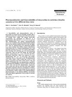
Figure 2.1 shows how physiological amounts of glucose and fructose
are absorbed by the small intestine. Luminal glucose crosses the epithelial
brush border and accumulates in the enterocyte by means of secondary
active transport. Transport is mediated by a sodium– glucose cotransporting carrier (SGLT1), which binds the substrates at a stoichiometric ratio of
© 2006 by Taylor & Francis Group, LLC
Vitamins in Foods: Analysis, Bioavailability, and Stability
29
two sodium ions to one glucose molecule. The immediate driving force
for the sodium-coupled entry of glucose is the electrochemical gradient
for sodium. This has two components: an electrical potential difference
of about 40 mV across the brush-border membrane (cell interior negative)
and a sodium concentration gradient. Both the electrical and chemical
brush-border
membrane
LUMEN
basolateral
membrane
tight
junction
SEROSA
Na+
ATP
intercellular
space
K+
ADP + Pi
Na+
Na+
Na+
K+
GLUT2
glucose
glucose
SGLT1
GLUT2
fructose
fructose
GLUT5
FIGURE 2.1
The carrier-mediated transport of D -glucose and D -fructose across the apical membrane and
basolateral membrane of an enterocyte. Naþ extruded into the intercellular space by the
basolateral Naþ –Kþ-ATPase (sodium pump) is able to equilibrate with Naþ on the
luminal side of the enterocyte by permeation through the tight junction. ATP, adenosine
triphosphate; ADP, adenosine diphosphate; Pi, inorganic phosphate. (From Ball, G.F.M.,
Vitamins. Their Role in the Human Body, Blackwell Publishing Limited, Oxford, 2004, p. 12.
With permission.)
© 2006 by Taylor & Francis Group, LLC
Intestinal Absorption and Bioavailability of Vitamins
30
components are established by the constant extrusion of sodium out of the
enterocyte by the action of the basolateral sodium pump. Fructose crosses
the brush border by facilitated diffusion mediated by the glucose transporter GLUT5. Exit of both glucose and fructose from the enterocyte to
the serosa takes place by facilitated diffusion at the basolateral membrane
and is mediated by GLUT2.
2.2.6
Effects of Dietary Fiber on Absorption of Nutrients
Dietary fiber consists of plant material that cannot be digested by the
endogenous secretions of the human digestive tract. From an analytical
standpoint, dietary fiber can be divided into insoluble fibers and
soluble fibers. The insoluble fibers include cellulose, lignin, and many
hemicelluloses; the soluble fibers include pectins, some hemicelluloses,
gums, and mucilages. The various gums and mucilages are widely
used in the food and pharmaceutical industries as emulsifiers, thickeners,
and stabilizers. The nature and physical properties of the main fiber
components are summarized in Table 2.1 [9].
Vahouny and Cassidy [10] discussed potential mechanisms by which
dietary fiber can modify nutrient absorption. Intestinal absorption of
nutrients can be influenced by modifying the rates at which food enters
or leaves the stomach. Bulky, high fiber foods may require longer
periods for ingestion, and therefore modify rates of gastric filling.
Viscous fiber components slow stomach emptying. The delayed release
of gastric emptying and modified intestinal pH might alter the regulation
of pancreatic and biliary secretions. Insoluble fibers accelerate small intestinal transit, allowing less time for nutrient absorption; in contrast,
viscous fibers slow transit. Many fiber components can alter the activities
of pancreatic enzymes by affecting viscosity and pH, and by adsorption.
Dietary fiber impairs lipid absorption by interfering with micelle formation. Evidence is the in vitro binding of bile salts and other micellar
components by lignin and guar gum, and the increase in fecal bile salts
in response to ingestion of dietary fiber. Viscous fibers can influence nutrient absorption by interfering with bulk phase diffusion of nutrients in the
intestinal lumen. The mucin layer covering the mucosal surface has been
suggested to be an important diffusion barrier to absorption. Reported
changes in mucin content or turnover in response to various fiber types
is a possible mechanism by which dietary fiber alters the transport characteristics of nutrients at the mucosal surface. Prolonged feeding of diets
supplemented with cellulose or pectin significantly increased villus
height and thickness, thereby increasing the absorptive surface area.
The dietary supplements also improved nutrient uptake by the small
intestine in vitro.
© 2006 by Taylor & Francis Group, LLC
Dietary Fiber Components
Fiber
Chemistry
Solubility in Water
Natural Source
Cellulose
Linear polymer of glucose with beta
1– 4 linkages
Insoluble
Lignin
Highly complex nonpolysaccharide
polymer derived from phenolics
Insoluble
Hemicelluloses
Heterogeneous group of
polysaccharides which contain a
variety of different sugars in the
polymeric backbone and side
chains
Polymer composed primarily of
galacturonic acid and rhamnose
with a variable degree of methyl
esterification
Complex group of highly branched
polysaccharides (e.g., gum acacia)
Polysaccharides resembling
hemicelluloses (e.g., guar gum)
Many insoluble, some soluble
Matrix of plant cell wall
Soluble, capable of forming
gels with sugar and acid
Matrix of plant cell
wall, ripe fruits
Soluble to give very viscous
colloidal solutions
Soluble to give slimy, colloidal
solutions
Extruded at site of
injury to plants
Mixed with starch in
endosperm
Pectins
Gums
Mucilages
Main structural
component of plant
cell wall
Structural component
of woody plants
Physical Properties
Binding of water
Binding of bile salts and
other organic
material
Binding of water and
cations
Formation of gels,
binding of bile salts
and other organic
material
Similar to pectins
Binding of water,
formation of gels,
binding of bile salts
and other organic
material
Vitamins in Foods: Analysis, Bioavailability, and Stability
© 2006 by Taylor & Francis Group, LLC
TABLE 2.1
Source: From Anderson, J.W. and Chen, W.-J.L., Am. J. Clin. Nutr., 32, 346, 1979. With permission.
31
© 2006 by Taylor & Francis Group, LLC
Intestinal Absorption and Bioavailability of Vitamins
32
2.3
2.3.1
Bioavailability
General Concepts
The term “bioavailability,” as applied to food-borne vitamins in human
nutrition, refers to the proportion of the quantity of vitamin ingested
that undergoes intestinal absorption and utilization by the body.
Utilization encompasses transport of the absorbed vitamin to the
tissues, cellular uptake, and ultimate fate of the vitamin. The latter can
be conversion to a form that can fulfill some biochemical or physiological
function, conversion to a nonfunctional form for subsequent excretion,
and storage. The major component of bioavailability and the rate-limiting
factor is absorption. Many ways of determining vitamin bioavailability
have been reported, most of which give an estimate of relative rather
than absolute bioavailability. Relative bioavailability is commonly
expressed as a percentage of the response obtained with a reference
material of high bioavailability. Bioavailability is an operational term
defined by the method used to determine it. Different values will be
obtained within a given study if different endpoints are used.
Intestinal absorption, and therefore bioavailability, of a vitamin
depends on the chemical form and physical state in which the vitamin
exists within the food matrix. These properties may be influenced by
the effects of food processing and cooking, particularly in the case of provitamin A carotenoids, niacin, vitamin B6, and folate. The food matrix
enhances vitamin absorption by stimulating the secretion of digestive
enzymes and bile salts. Bile salts inhibit gastric emptying and proximal
intestinal transit, resulting in an increased residence time at the absorption sites. Thus, absorption of a riboflavin supplement taken with a
meal was about 60%, as compared to 15% on an empty stomach [11].
In foods derived from animal and plant tissues, the B-group vitamins
occur as their coenzyme derivatives, usually associated with their
protein apoenzyme. In addition, niacin in cereals and vitamin B6 in
certain fruits and vegetables occur largely as bound storage forms. In
milk and eggs, which are derived from animal secretions, the B-group
vitamins occur, at least to some extent, in the underivatized form, a proportion of which may be associated with specific binding proteins. Vitamins that exist as chemically bound complexes with some other
material in the food matrix exhibit lower efficiencies of digestion and
absorption compared with the free (unbound) vitamin ingested, for
example, in tablet form.
Certain dietary components can retard or enhance a vitamin’s absorption, therefore the composition of the diet is an important factor in
bioavailability. For example, the presence of adequate amounts of
© 2006 by Taylor & Francis Group, LLC
Vitamins in Foods: Analysis, Bioavailability, and Stability
33
dietary fat is essential for the absorption of fat-soluble vitamins. Fibrous
plant material can interfere with the physiological mechanisms of absorption, as is evident by the poor bioavailability of b-carotene in a raw carrot
compared with that in a cooked carrot [12]. The binding of bile salts to
various wood fiber sources and isolated fiber components has been
demonstrated [13]. Furthermore, certain types of dietary fiber may
either interfere with the formation of mixed micelles in the intestinal
lumen or effectively alter the normal diffusion and accessibility of micellar
lipids to the absorptive surface of the intestinal mucosa. Such events
could compromise the absorption of lipids, including the fat-soluble
vitamins. The absorption of folate is impaired by ethanol in cases of
chronic alcoholism.
2.3.2
Methods for Estimating Vitamin Bioavailability in Human Subjects
There are two main experimental approaches for estimating vitamin bioavailability in human subjects: (i) determining the extent of vitamin
absorption by measuring the concentration of vitamin in the plasma,
the chylomicron fraction of plasma, or urine and (ii) comparing the
mass of vitamin consumed with the mass excreted in the feces. A difficulty arises in the plasma sampling method when newly ingested
vitamin mixes with endogenous circulating vitamin, but this can be overcome by the use of stable isotopes as tracers. A difficulty also arises in the
oral – fecal balance method because colonic flora can utilize unabsorbed
vitamin and also synthesize new vitamin. Surgical bypassing of the
colon using pigs or human ileostomy subjects overcomes this problem
to a large extent.
The application of stable isotope techniques has given much needed
impetus to the study of vitamin bioavailability, particularly the provitamin A carotenoids and folate.
2.3.2.1
Plasma Response
This method involves measuring the increase in plasma vitamin concentration over baseline level at several time intervals after ingestion of the
test meal, and plotting these values against time. The procedure is
repeated after oral dosing with a reference vitamin standard. The area
under the curve (AUC) obtained for the test meal, expressed as a percentage of the AUC for the reference dose, gives the relative bioavailability of
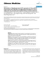
the vitamin in the meal. The post-absorption positive AUC (AUCþ)
might be followed by a negative AUC (AUC2), depending on the
degree of diurnal fluctuation (Figure 2.2). In practice, AURþ is calculated.
Only in the case of fortified foodstuffs or foods with naturally very
© 2006 by Taylor & Francis Group, LLC
Intestinal Absorption and Bioavailability of Vitamins
34
conc.
80% of total AUC+
Cmax
AUC+
baseline
AUC–
tmax
time
FIGURE 2.2
Parameters of bioavailability. AUCþ, post-absorption area under curve; AUC2, negative
area under curve; Cmax, maximal concentration; tmax, time for maximal concentration to be
reached. (From Pietrzik, K., Hages, M., and Remer, T., J. Micronutr. Anal., 7, 207–222, 1990.
With permission.)
high vitamin contents can a significant increase in vitamin blood level be
expected and AUC to be measurable [14]. The validity of the AUC
depends on the in vivo handling of the reference dose and test dose
being equivalent [15].
2.3.2.2 Urinary Excretion
Urinary excretion can be used to measure relative bioavailability because
it is proportional to the plasma concentration if urinary clearance is constant. Subjects are preloaded with synthetic vitamin in order to saturate
the tissues and ensure that the additionally absorbed vitamin will be
excreted.
2.3.2.3
Oral-Fecal Balance Studies and the Determination
of Prececal Digestibility
In a classical balance study, human subjects are fed a diet containing a
known amount of the test vitamin, and the difference between what is
ingested and what is recovered in the feces is considered to be apparent
absorption. Absolute absorption is not measured because not all of the
vitamin in the feces will originate from the ingested food: some will
© 2006 by Taylor & Francis Group, LLC
Vitamins in Foods: Analysis, Bioavailability, and Stability
35
originate from sloughed mucosal cells. There is no information about the
utilization of the vitamin. The natural presence of microflora in the colon
creates a possible bias in the balance method. Unabsorbed vitamin, on
reaching the colon, can become metabolized by the intestinal flora,
leading to overestimation of bioavailability. The gut microflora can also
synthesize B-vitamins and vitamin K, leading to underestimation of
bioavailability.
An effective and practical way of circumventing the problem of intestinal microflora is to surgically bypass the colon, causing the digesta
to move straight from the ileum to the rectum. This allows the in vivo
determination of prececal digestibility, and the calculation of absolute
absorption. The domestic pig is chosen as the animal model, because
the digestive physiology of this species resembles that of the human.
The surgical technique is an end-to-end ileo-rectal anastomosis [16,17].
The technique has been used successfully for determining the prececal
digestibility of thiamin [18], niacin [19], vitamin B6 [20], and pantothenic
acid [19]. Human patients with ileostomies fulfill a similar function to
the pig model in allowing the assessment of absolute absorption, and
have been used to determine the bioavailability of b-carotene [21] and
folate [22] from food. The body conserves folate and vitamin B12
through their excretion in bile and subsequent reabsorption by the
small intestine. This enterohepatic circulation complicates interpretation
of the results for these vitamins.
2.3.2.4
Use of Stable Isotopes
Isotopes of a particular atom contain a different number of neutrons and
can be either stable or radioactive. Unlike radioactive isotopes, stable isotopes emit no radiation and can therefore be used safely as tracers in
human studies of nutrient metabolism. Stable isotopic methods using
deuterium (2H) and 13C are being used to estimate body stores of
vitamin A, and to study the bioavailability and bioefficacy of dietary
carotenoids [23]. They are also being used to assess folate bioavailability
[24,25]. The use of stable isotopes allows differentiation between isotopically labeled vitamin from the dose and unlabeled endogenous
vitamin from body stores. The labeled vitamin or its metabolites can be
specifically determined in blood, urine, and feces, allowing the detailed
study of absorption, metabolism, and excretion. Detection methods for
stable isotopes are less sensitive than those for radioisotopes, thus the
dose administered needs to be relatively high to reach measurable
levels. Owing to complicated methodology and expensive instrumentation, stable isotopic procedures are confined to specialized laboratories.
Isotopic labeling of the vitamin can be performed intrinsically or extrinsically. Intrinsic labeling involves biological incorporation of isotope into
© 2006 by Taylor & Francis Group, LLC
36
Intestinal Absorption and Bioavailability of Vitamins
the tissues of the plant or animal food source during growth and development, so that the labeled vitamin is in the same matrix as the food consumed. For example, kale was labeled intrinsically with 13C by growing
plants continuously in an atmosphere containing 13CO2 , starting approximately 8 days after sowing [26]. Broccoli was labeled intrinsically with
deuterium by adding deuterium oxide (heavy water) to the nutrient
solution of hydroponically grown plants [27]. Extrinsic labeling refers to
the chemical incorporation of isotope into the vitamin molecule of interest. Multiple labeling is desirable to increase the molecular mass and
improve the sensitivity of detection by mass spectrometry. The labeled
vitamin is mixed with the food just before consumption. Interpretation
depends on the assumption that the labeled vitamin behaves in a
manner similar to the naturally occurring vitamin in the diet.
Mass spectrometry is an analytical technique that measures the masses
of individual molecules and atoms. During the initial conversion of
analyte molecules into gas-phase ionic species (ionization), the excess
energy transferred to the molecules leads to fragmentation. A mass
analyzer separates these molecular ions and their charged fragments
according to their m/z (mass/charge) ratio. The ion current due to these
mass-separated ions is detected by a suitable detector and displayed in
the form of a mass spectrum. The mass spectrum is a plot of m/z values
of all ions that reach the detector versus their abundance. For quantitative
analysis, the ions are usually detected by selected-ion monitoring, in
which selected m/z values are exclusively monitored [28].
Mass spectrometers designed for the analysis of organic molecules
include gas chromatography – mass spectrometry (GC –MS), highperformance liquid chromatography – mass spectrometry (LC –MS),
tandem mass spectrometry (MS – MS), and LC – MS – MS instruments.
LC –MS methods have the advantage over GC – MS methods in that
they do not require such labor-intensive sample preparation. Tandem
mass spectrometry refers to the coupling of two mass spectrometers
(MS-1 and MS-2) in series. MS-1 mass-selects a specified ion, which
undergoes fragmentation in the intermediate region, and MS-2 massanalyzes the ionic fragments. Molecular specificity is guaranteed
because the product ions are derived exclusively from the preselected
precursor.
References
1. Levitt, M.D., Strocchi, A., and Levitt, D.G., Human jejunal unstirred layer:
evidence for extremely efficient luminal stirring, Am. J. Physiol., 262, G593,
1992.
© 2006 by Taylor & Francis Group, LLC
Vitamins in Foods: Analysis, Bioavailability, and Stability
37
2. Nimmerfall, F. and Rosenthaler, J., Significance of the goblet-cell mucin layer,
the outermost luminal barrier to passage through the gut wall, Biochem.
Biophys. Res. Commun., 94, 960, 1980.
3. Shiau, Y.-F., Fernandez, P., Jackson, M.J., and McMonagle, S., Mechanisms
maintaining a low-pH microclimate in the intestine, Am. J. Physiol., 248,
G608, 1985.
4. Karasov, W.H., Tests of the adaptive modulation hypothesis for dietary control
of intestinal nutrient transport, Am. J. Physiol., 263, R496, 1992.
5. Ferraris, R.P. and Diamond, J.M., Specific regulation of intestinal nutrient
transporters by their dietary substrates, Annu. Rev. Physiol., 51, 125, 1989.
6. Shiau, Y.-F. and Levine, G.M., pH dependence of micellar diffusion and
dissociation, Am. J. Physiol., 239, G177, 1980.
7. Klein, S., Cohn, S.M., and Alpers, D.H., The alimentary tract in nutrition: a
tutorial, in Modern Nutrition in Health and Disease, 9th ed., Shils, M.E., Olson,
J.A., Shike, M., and Ross, A.C., Eds., Lippincott Williams and Wilkins,
New York, 1999, p. 605.
8. Ball, G.F.M., Vitamins: Their Role in the Human Body, Blackwell Publishing Ltd.,
Oxford, 2004, p. 41.
9. Anderson, J.W. and Chen, W.-J.L., Plant fiber: carbohydrate and lipid metabolism, Am. J. Clin. Nutr., 32, 346, 1979.
10. Vahouny, G.V. and Cassidy, M.M., Dietary fibers and absorption of nutrients,
Proc. Soc. Exp. Biol. Med., 180, 432, 1985.
11. van den Berg, H., General aspects of bioavailability of vitamins, in Bioavailability ’93. Nutritional, Chemical and Food Processing Implications of Nutrient Availability, conference proceedings, part 1, May 9 –12, 1993, Schlemmer, U., Ed.,
Bundesforschungsanstalt fu¨r Erna¨hrung, Ettlingen, 1993, p. 267.
12. Erdman, J.W., Jr., Poor, C.L., and Dietz, J.M., Factors affecting the bioavailability of vitamin A, carotenoids, and vitamin E, Food Technol., 42 (10), 214,
1988.
13. Vahouny, G.V., Dietary fibers and intestinal absorption of lipids, in Dietary
Fiber in Health and Disease, Vahouny, G.V. and Kritchevsky, D.S., Eds.,
Plenum Press, New York, 1982, p. 203.
14. Pietrzik, K., Hages, M., and Remer, T., Methodological aspects in vitamin
bioavailability testing, J. Micronutr. Anal., 7, 207, 1990.
15. Gregory, J.F., III, Recent developments in methods for the assessment of
vitamin bioavailability, Food Technol., 42 (10), 230, 1988.
16. Roth-Maier, D.A., Kirchgessner, M., Erhardt, W., Henke, J., and Hennig, U.,
Comparative studies for the determination of precaecal digestibility as a
measure for the availability of B-vitamins, J. Anim. Physiol. Anim. Nutr., 79,
198, 1998.
17. Wauer, A., Stangl, G.I., Kirchgessner, M., Erhardt, W., Henke, J., Hennig, U.,
and Roth-Maier, D.A., A comparative evaluation of ileo-rectal anastomosis
techniques for the measurement of apparent precaecal digestibilities of
folate, niacin and pantothenic acid, J. Anim. Physiol. Anim. Nutr., 82, 80, 1999.
18. Roth-Maier, D.A., Wild, S.I., Erhardt, W., Henke, J., and Kirchgessner, M.,
Investigations on the intestinal availability of native thiamin in selected
foods and feedstuffs, Eur. J. Nutr., 38, 241, 1999.
© 2006 by Taylor & Francis Group, LLC
38
Intestinal Absorption and Bioavailability of Vitamins
19. Roth-Maier, D.A., Wauer, A., Stangl, G.I., and Kirchgessner. M., Precaecal
digestibility of niacin and pantothenic acid from different foods,
Int. J. Vitam. Nutr. Res., 70, 8, 2000.
20. Roth-Maier, D.A., Kettler, S.I., and Kirchgessner, M., Availability of vitamin B6
from different food sources, Int. J. Food Sci. Nutr., 53, 171, 2002.
21. Livny, O., Reifen, R., Levy, I., Madar, Z., Faulks, R., Southon, S., and
Schwartz, B., b-Carotene bioavailability from differently processed carrot
meals in human ileostomy volunteers, Eur. J. Nutr., 42, 338, 2003.
22. Wittho¨ft, C.M., Stra˚lsjo¨, L., Berglund, G., and Lundin, E., A human model to
determine folate bioavailability from food: a pilot study for evaluation,
Scand. J. Nutr., 47, 6, 2003.
23. van Lieshout, M., West, C.E., and van Breemen, R.B., Isotopic tracer techniques
for studying the bioavailability and bioefficacy of dietary carotenoids, particularly b-carotene, in humans: a review, Am. J. Clin, Nutr., 77, 12, 2003.
24. Finglas, P.M., Hart, D., Wolfe, C, Wright, A.J.A., Southon, S., Mellon, F.,
van den Akker, H., and de Meer, K., Validity of dual-label stable isotopic
protocols and urinary excretion ratios to determine folate bioavailability
from food, Food Nutr. Bull., 23, (Suppl. 3), 107, 2002.
25. Rychlik, M., Netzel, M., Pfannbecker, I., Frank, T., and Bitsch, I., Application of
stable isotope dilution assays based on liquid chromatography — tandem
mass spectrometry for the assessment of folate bioavailability, J. Chromatogr.
B., 792, 167, 2003.
26. Kurilich, A.C., Britz, S.J., Clevidence, B.A., and Novotny, J.A., Isotopic labeling
and LC-APCI-MS quantification for investigating absorption of carotenoids
and phylloquinone from kale (Brassica oleracea), J. Agric. Food Chem., 51,
4877, 2003.
27. Dolnikowski, G.G., Sun, Z., Grusak, M.A., Peterson, J.W., and Booth, S.L.,
HPLC and GC/MS determination of deuterated vitamin K (phylloquinone)
in human serum after ingestion of deuterium-labeled broccoli, J. Nutr.
Biochem., 13, 168, 2002.
28. Dass, C., Principles and Practice of Biological Mass Spectrometry, WileyInterscience, New York, 2001.
© 2006 by Taylor & Francis Group, LLC