Overview of ELISA thermo fisher scientific
Bạn đang xem bản rút gọn của tài liệu. Xem và tải ngay bản đầy đủ của tài liệu tại đây (280.92 KB, 6 trang )
Overview of ELISA
ELISA (enzymelinked immunosorbent assay) is a platebased assay technique designed for detecting and quantifying
substances such as peptides, proteins, antibodies, and hormones. Other names, such as enzyme immunoassay (EIA), are also
used to describe the same technology. In an ELISA, an antigen must be immobilized to a solid surface and then complexed with
an antibody that is linked to an enzyme. Detection is accomplished by assessing the conjugated enzyme activity via incubation
with a substrate to produce a product that can be measured. The most crucial element of the detection strategy is a highly
specific antibodyantigen interaction.
Page contents
Introduction
ELISA formats (direct, sandwich, etc.)
Direct vs. indirect detection ELISA strategies
Other ELISA formats (competitive, ELISPOT, etc.)
Complete, readytouse ELISA kits
Selecting and coating ELISA plates
Precoated ELISA plates
Antibodies and probes for ELISA
Blocking buffers and wash buffers
Detection strategies for ELISA
View products
Browse ELISA Products
Find ELISA Kits by Target
Introduction
ELISAs are typically performed in 96well (or 384well) polystyrene plates, which will passively bind antibodies and proteins. It is this binding and immobilization of reagents that makes ELISAs so easy
to design and perform. Having the reactants of the ELISA immobilized to the microplate surface makes it easy to separate bound from nonbound material during the assay. This ability to wash away
nonspecifically bound materials makes the ELISA a powerful tool for measuring specific analytes within a crude preparation.
A detection enzyme or other tag can be linked directly to the primary antibody or introduced through a secondary antibody that recognizes the primary antibody. It also can be linked to a protein such
as streptavidin if the primary antibody is biotin labeled. The most commonly used enzyme labels are horseradish peroxidase (HRP) and alkaline phosphatase (AP). Other enzymes have been used
as well, but they have not gained widespread acceptance because of limited substrate options. These include βgalactosidase, acetylcholinesterase, and catalase. A large selection of substrates is
available for performing the ELISA with an HRP or AP conjugate. The choice of substrate depends upon the required assay sensitivity and the instrumentation available for signal detection
(spectrophotometer, fluorometer, or luminometer).
ELISA formats
ELISAs can be performed with a number of modifications to the basic procedure. The key step, immobilization of the antigen of interest, can be accomplished by direct adsorption to the assay plate or
indirectly via a capture antibody that has been attached to the plate. The antigen is then detected either directly (labeled primary antibody) or indirectly (labeled secondary antibody). The most
powerful ELISA assay format is the sandwich assay. This type of capture assay is called a “sandwich” assay because the analyte to be measured is bound between two primary antibodies—the
capture antibody and the detection antibody. The sandwich format is used because it is sensitive and robust.
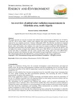
Diagram of common ELISA formats (direct vs. sandwich assays)
Common ELISA formats. In the assay, the antigen of interest is immobilized by direct adsorption to the assay plate or by first attaching a capture antibody to the plate surface. Detection of the antigen can then be performed using
an enzymeconjugated primary antibody (direct detection) or a matched set of unlabeled primary and conjugated secondary antibodies (indirect detection).
Direct vs. indirect detection ELISA strategies
Among the standard assay formats discussed and illustrated above, where differences in both capture and detection were the concern, it is important to differentiate between the particular strategies
that exist specifically for the detection step. However an antigen is captured to the plate (by direct adsorption to the surface or through a precoated "capture" antibody, as in a sandwich ELISA), it is
the detection step (as either direct or indirect detection) that largely determines the sensitivity of an ELISA.
Watch this video about ELISA detection and signal amplification strategies
0:00
/
2:31
The direct detection method uses a labeled primary antibody that reacts directly with the antigen. Direct detection can be performed with antigen that is directly immobilized on the assay plate or with
the capture assay format. Direct detection is not widely used in ELISA but is quite common for immunohistochemical staining of tissues and cells.
The indirect detection method uses a labeled secondary antibody for detection and is the most popular format for ELISA. The secondary antibody has specificity for the primary antibody. In a
sandwich ELISA, it is critical that the secondary antibody be specific for the detection primary antibody only (and not the capture antibody) or the assay will not be specific for the antigen. Generally,
this is achieved by using capture and primary antibodies from different host species (e.g., mouse IgG and rabbit IgG, respectively). For sandwich assays, it is beneficial to use secondary antibodies
that have been crossadsorbed to remove any antibodies that have affinity for the capture antibody.
Comparison of direct and indirect ELISA detection methods
Direct ELISA detection
Advantages
Quick because only one antibody and fewer steps are used.
Crossreactivity of secondary antibody is eliminated.
Disadvantages
Immunoreactivity of the primary antibody might be adversely affected by labeling with enzymes or tags.
Labeling primary antibodies for each specific ELISA system is time consuming and expensive.
No flexibility in choice of primary antibody label from one experiment to another.
Minimal signal amplification.
Indirect ELISA detection
Advantages
A wide variety of labeled secondary antibodies are available commercially.
Versatile because many primary antibodies can be made in one species and the same labeled secondary antibody can be used for detection.
Maximum immunoreactivity of the primary antibody is retained because it is not labeled.
Sensitivity is increased because each primary antibody contains several epitopes that can be bound by the labeled secondary antibody, allowing for signal amplification.
Different visualization markers can be used with the same primary antibody.
Disadvantages
Crossreactivity might occur with the secondary antibody, resulting in nonspecific signal.
An extra incubation step is required in the procedure.
Fluorescent tags and other alternatives to enzymebased detection can be used for platebased assays. Despite not involving reporterenzymes, these methods are also generally referred to as a
type of ELISA. Likewise, wherever detectable probes and specific protein binding interactions can be used in a platebased method, these assays are often called ELISAs despite not involving
antibodies.
Other ELISA formats
Besides the standard direct and sandwich formats described above, several other styles of ELISA exist:
Competitive ELISA is a strategy that is commonly used when the antigen is small and has only one epitope, or antibody binding site. One variation of this method consists of labeling purified antigen
instead of the antibody. Unlabeled antigen from samples and the labeled antigen compete for binding to the capture antibody. A decrease in signal from purified antigen indicates the presence of the
antigen in samples when compared to assay wells with labeled antigen alone.
Watch this video about competitive ELISA methods
0:00
/
1:13
ELISPOT (enzymelinked immunospot assay) refers to ELISAlike capture and measurement of proteins secreted by cells that are plated in PVDFmembranebacked microplate wells. It is a
"sandwich" assay in which the proteins are captured locally as they are secreted by the plated cells, and detection is with a precipitating substrate. ELISPOT is like a western blot in that the result is
spots on a membrane surface.
Incell ELISA is performed with cells that are plated and cultured overnight in standard microplates. After the cultured cells are fixed, permeabilized, and blocked, target proteins are detected with
antibodies. This is an indirect assay, not a sandwich assay. The secondary antibodies are either fluorescent (for direct measurement by a fluorescent plate reader or microscope) or enzyme
conjugated (for detection with a soluble substrate using a plate reader).
ELISA is nearly always performed using 96well or 384well polystyrene plates and samples in solution (i.e., biological fluids, culture media, or cell lysates). This is the platform discussed in the
remainder of this article.
Complete, readytouse ELISA kits
In addition to the individual components and general principles of ELISA discussed in the remainder of this article, readytouse sandwich ELISA kits are commercially available for detection of
hundreds of specific cytokines, neurobiology analytes, and phosphorylated proteins that are common targets of research interest.
For many targets, two kit types are available:
ELISA Kits contain precoated antibodyplates, detection antibodies, buffers, diluents, standards, and substrates.
Antibody Pair Kits contain only matched antibodies and standard (no plates or detection reagents).
Learn more
View products
ELISA Development and Optimization
Search All ELISA Kits by Target
Performing and Evaluating Spike and Recovery and Linearity of Dilution for ELISA
ReadytoUse ELISA Kits
Factors Affecting Signal Generation in ELISA
Antibody Pair Kits
ELISA Protocols
Guide to ELISA Kit Package Formats
ELISA Reagents and Accessories
Assay Development Technical Handbook
The revised Assay Development Technical Handbook is an essential resource for any laboratory using enzymelinked immunosorbent assay
(ELISA) and related platebased assay methods. The handbook describes the essential techniques and tools for designing and optimizing ELISA
Assays. Featured products include coated microplates, standards, blockers, buffers, probelabeling reagents, secondary antibodies and
detection substrates.
Contents include: Introduction to ELISA, Selecting an ELISA Plate, Thermo Scientific Pierce Microplates, Thermo Scientific Pierce Coated
Microplates, Blocking and Washing, Blocking and Washing Reagents, Detection Probes, Antibody Labeling, Choosing a Substrate, Bulk and
Custom Offerings, and Recommended Reading.
Download the Assay Development Technical Handbook
Selecting and coating ELISA plates
When developing a new ELISA for a specific antigen, the first step is to optimize the platecoating conditions for the antigen or capture antibody. Begin by choosing an assay microplate (not tissue
culture treated plates) with a minimum proteinbinding capacity of 400 ng/cm². It is also important that the CV value (coefficient of variation) of the protein binding be low (<5% is preferred) so that there
is limited deviation in values that should be identical in the assay results between wells and plates. The choice of plate color depends upon the signal being detected. Clear polystyrene flat bottom
plates are used for colorimetric signals while black or white opaque plates are used for fluorescent and chemiluminescent signals. Visually inspect plates before use as imperfections or scratches in
the plastic will cause aberrations when acquiring data from the developed assay.
Plate coating is achieved through passive adsorption of the protein to the plastic of the assay microplate. This process occurs though hydrophobic interactions between the plastic and nonpolar
protein residues. Although individual proteins may require specific conditions or pretreatment for optimal binding, the most common method for coating plates involves adding a 2–10 μg/mL solution of
protein dissolved in an alkaline buffer such as phosphatebuffered saline (pH 7.4) or carbonatebicarbonate buffer (pH 9.4). The plate is left to incubate for several hours to overnight at 4–37°C.
Typically, after removing the coating solution, blocking buffer is added to ensure that all remaining available binding surfaces of the plastic well are covered (see subsequent discussion). Coated plates
can be used immediately or dried and stored at 4°C for later use, depending on the stability of the coated protein.
It is important to note that optimal coating conditions can vary with each protein. With the exception of competition ELISAs, the plates are coated with more capture protein than can actually be bound
during the assay in order to facilitate the largest working range of detection possible. Some proteins, especially antibodies, are best coated on plates at a concentration lower than the maximum
binding capacity in order to prevent nonspecific binding in later steps by a phenomenon called "hooking". Hooking results from proteins getting trapped between the coating proteins which prevents
effective washing and removal of nonbound proteins. When hooking nonspecifically traps detection primary and secondary antibodies, high background signal results lowering the signal to noise ratio
and thus sensitivity of an assay.
View products
ELISA Labware and Accessories
BupH™ CarbonateBicarbonate Buffer Packs (pH 9.4)
Precoated ELISA plates
For antibodies and proteins, coating plates by passive adsorption usually works well. However, problems can arise from passive adsorption, including improper orientation, denaturation, poor
immobilization efficiency, and binding of contaminants along with the target molecule. Antibodies can be attached to a microplate through the Fc region using Protein A, G, or A/G coated plates, which
orients them properly and preserves their antigen binding capability. Fusion proteins can be attached to a microplate in the proper orientation using glutathione, metalchelate, or captureantibody
coated plates. Peptides and other small molecules, which typically do not bind effectively by passive adsorption, can be biotinylated and attached with high efficiency to a streptavidin or Pierce™
NeutrAvidin™ protein coated plate. Biotinylated antibodies also can be immobilized on plates precoated with biotinbinding proteins. Using precoated plates in this manner physically separates the
antigen or capture antibody from the surface of the plate as protection from its denaturing effects.
View products
Browse all Coated Plates
Antibodies and probes for ELISA
Either monoclonal or polyclonal antibodies can be used as the capture and detection antibodies in sandwich ELISA systems. Monoclonals have an inherent monospecificity toward a single epitope that
allows fine detection and quantitation of small differences in antigen. A polyclonal is often used as the capture antibody to pull down as much of the antigen as possible. Then a monoclonal is used as
the detecting antibody in the sandwich assay to provide improved specificity.
An important consideration in designing a sandwich ELISA is that the capture and detection antibodies must recognize two different nonoverlapping epitopes. When the antigen binds to the capture
antibody, the epitope recognized by the detection antibody must not be obscured or altered. Capture and detection antibodies that do not interfere with one another and can bind simultaneously are
called "matched pairs" and are suitable for developing a sandwich ELISA. Many primary antibody suppliers provide information about epitopes and indicate pairs of antibodies that have been validated
in ELISA as matched pairs.
Another design consideration in choosing antibodies is cost. A polyclonal antibody is generally less expensive (~5 fold) to produce than a monoclonal. The specificity gained by using monoclonals for
both the capture and detecting antibody must be weighed against the cost and time required for producing two monoclonal antibodies. Preparing a “selfsandwich” ELISA assay, where the same
antibody is used for the capture and detection, can limit the dynamic range and sensitivity of the final ELISA.
Learn more
Overview of Detection Probes
View products
Primary Antibodies
Secondary Antibodies and Conjugates
Secondary Antibody Selection Guide
Blocking buffers and wash buffers
The binding capacity of microplate wells is typically higher than the amount of protein coated in each well. The remaining surface area must be blocked to prevent antibodies or other proteins from
adsorbing to the plate during subsequent steps. A blocking buffer is a solution of irrelevant protein, mixture of proteins, or other compound that passively adsorbs to all remaining binding surfaces of
the plate. The blocking buffer is effective if it improves the sensitivity of an assay by reducing background signal and improving the signaltonoise ratio. The ideal blocking buffer will bind to all potential
sites of nonspecific interaction, eliminating background altogether, without altering or obscuring the epitope for antibody binding.
When developing any new ELISA, it is important to test several different blockers for the highest signal:noise ratio in the assay. Many factors can influence nonspecific binding, including various
protein:protein interactions unique to the samples and antibodies involved. The most important parameter when selecting a blocker is the signal:noise ratio, which is measured as the signal obtained
with a sample containing the target analyte as compared to that obtained with a sample without the target analyte. Using inadequate amounts of blocker will result in excessive background and a
reduced signal:noise ratio. Using excessive concentrations of blocker may mask antibodyantigen interactions or inhibit the enzyme, again causing a reduction of the signal:noise ratio. No single
blocking agent is ideal for every occasion and empirical testing is essential for true optimization of the blocking step.
In addition to blocking, it is essential to perform thorough washes between each step of the ELISA. Washing steps are necessary to remove nonbound reagents and decrease background, thereby
increasing the signal:noise ratio. Insufficient washing will allow high background, while excessive washing might result in decreased sensitivity caused by elution of the antibody and/or antigen from the
well. Washing is performed in a physiologic buffer such as Trisbuffered saline (TBS) or phosphatebuffered saline (PBS) without any additives. Usually, a detergent such as 0.05% Tween20 is added
to the buffer to help remove nonspecifically bound material. Another common technique is to use a dilute solution of the blocking buffer along with some added detergent. Including the blocking agent
and adding a detergent in wash buffers helps to minimize background in the assay. For best results, use highpurity detergents to prevent introduction of impurities that will interfere with the assay,
such as enzyme inhibitors or peroxides.
Learn more
Blocking Buffers
View products
Blocking Buffers for ELISA
Detection strategies for ELISA
The final stage in all ELISA systems is a detection step. Unless a radioactive or fluorescent tag was used, this involves the introduction of an enzyme substrate.The enzyme converts the substrate to
a detectable product. If an ELISA has been constructed and developed properly, then the intensity of signal produced when the substrate is added will by directly proportional to the amount of antigen
captured in the plate and bound by the detection reagents. Enzymeconjugated antibodies (especially those involving horseradish peroxidase, HRP) offer the most flexibility in detection and
documentation methods for ELISA because of the variety of substrates available for chromogenic, chemifluorescent, and chemiluminescent imaging.
Though not as sensitive as fluorescent or chemiluminescent substrates, chromogenic ELISA substrates allow direct visualization and enable kinetic studies to be performed. Furthermore,
chromogenic ELISA substrates are detected with standard absorbance plate readers common to many laboratories. Fluorescent ELISA substrates are not as common and require a fluorometer that
produces the correct excitation beam to cause signal emission to be generated from the fluorescent tag. Though best used with a luminometer plate reader, chemiluminescent substrates can be
detected by various means including digital camera systems. One drawback of using chemiluminescent substrates for ELISA is the signal intensity can vary more with than other substrates. For
assays requiring many plates to be read, this can present a problem if the signal begins to decay before plates are read. For this reason, it is important to make sure the assay has been optimized
with the substrate in order to ovoid misinterpreting signalfade in a sample as low antigen abundance.
Learn more
ELISA Substrate Guide
ELISA Development and Optimization
Factors Affecting Signal Generation in ELISA
ELISA Troubleshooting Guide
For Research Use Only. Not for use in diagnostic procedures.