Ebook Medical pharmacology at a glance (7th edition) Part 2
Bạn đang xem bản rút gọn của tài liệu. Xem và tải ngay bản đầy đủ của tài liệu tại đây (14.37 MB, 75 trang )
20
Lipid-lowering drugs
Endocytosis
HMG CoA
inhibitors
Lysis
Anionic exchange
resins
CE
colestyramine
colestipol
LDL
atorvastatin
simvastatin
pravastatin
others
Cholesterol
LDL-R
nicotinic acid
Increase
A
Fibrates
Inhibit
A
VLDL
BA
Bile duct
A BA
BA
HMG CoA HMG CoA
reductase
mevalonate
–
CE
Cholesterol + TG
Portal vein
BA
TG
HDL
A BA
LDL receptor
bezafibrate
fenofibrate
others
Activate
Lipoprotein lipase
(in muscle and adipose
tissue capillaries)
chol
CE
A BA
Bile acid
excretion
INHIBITOR of
cholesterol absorption
ezetimibe
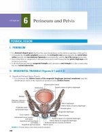
Lipids, such as triglycerides and cholesterylesters, are insoluble in
water and are transported in plasma in the core of particles (lipoproteins) that have a hydrophilic shell of phospholipids and free cholesterol. This surface layer is stabilized by one or more apolipoproteins,
which also act as ligands for cell surface receptors. About two-thirds
of plasma lipoproteins are synthesized in the liver (middle, shaded
(yellow)). Triglycerides (TG) are secreted into the blood as very-low). In muscle and adipose tissue, the
density lipoproteins (VLDL,
capillaries (right) possess an enzyme, lipoprotein lipase ( ), that
hydrolyses the triglycerides to fatty acids; these then enter the muscle
cells (for energy) and adipocytes (for storage). The residual particles
containing a core rich in cholesterylester (CE) are called low-density
lipoprotein (LDL) particles. The liver and other cells possess LDL
) that remove LDL from the plasma by endocytosis
receptors (
(top figure shaded orange). The hepatic receptor-mediated removal of
LDL is the main mechanism for controlling plasma LDL levels.
Fatty acids and cholesterol from ingested dietary fat are re-esterified
in mucosal cells of the intestine and form the core of chylomicrons,
which enter the plasma via the thoracic duct. Fatty acids are
Fatty acids
LDL
hydrolysed from the chylomicrons by lipoprotein lipase, and the residual triglyceride-depleted remnants are removed by the liver.
There is a strong positive correlation between the plasma concentration of LDL cholesterol and the development of atherosclerosis in
medium and large arteries. Therapy that lowers LDL and raises highdensity lipoprotein (HDL) has been shown to reduce the progression
of coronary atherosclerosis. Lipid-lowering drugs are indicated most
strongly in patients with coronary artery disease, or those with a high
risk of coronary artery disease because of multiple risk factors, and in
patients with familial hypercholesterolaemia. Anion exchange resins
(top left, A ) bind bile acids ( BA ) and, because they are not absorbed,
cholesterol excretion is increased. The statins, 3-hydroxy-3-methylglutaryl coenzyme A (HMG CoA) reductase inhibitors (top right),
decrease hepatic cholesterol synthesis. The fall in hepatocyte cholesterol caused by resins and statins induces a compensatory increase in
hepatic LDL receptors and consequently a fall in plasma cholesterol.
Nicotinic acid (centre right) reduces the release of VLDL by the liver,
whereas the fibrates (bottom right), which mainly lower triglyceride
levels, probably act chiefly by stimulating lipoprotein lipase. Ezetimibe
46 Medical Pharmacology at a Glance, Seventh Edition. Michael J. Neal. © 2012 John Wiley & Sons, Ltd. Published 2012 by John Wiley & Sons, Ltd.
is the first of a new class of drugs that selectively inhibits the intestinal
absorption of cholesterol.
Lipoproteins
These are classified according to their density on equilibrium ultracentrifugation. The larger particles (chylomicrons, remnants and
VLDL) are the least dense and are not atherogenic because their
greater size (diameter 30–500 nm) prevents them from passing into
blood vessel walls. LDL particles (diameter 18–25 nm) can easily
penetrate damaged arteries and are mainly responsible for the development of atherosclerosis. HDL particles are the smallest (diameter
5–12 nm), and epidemiological studies have revealed that high levels
of HDL are associated with a lower incidence of atheroma. HDL
accept excess (unesterified) cholesterol from cells and also from lipoproteins that have lost their triglycerides and therefore have an excess
of surface components, including cholesterol. The cholesterol is made
less polar by re-esterification, causing it to move into the hydrophobic
core and leaving the surface available to accept more cholesterol. The
cholesterylesters are then returned to the liver. The removal of cholesterol from artery walls by HDL is thought to be the basis of its antiatherogenic action.
Hyperlipidaemias
Primary lipoprotein disorders may involve cholesterol, triglycerides,
or both. Secondary hyperlipidaemias are the result of another illness,
e.g. diabetes mellitus or hypothyroidism. Hypercholesterolaemia is
the most common disorder. About 5% of cases are familial but, in most
cases, the cause is unknown. The main therapy for hyperlipidaemias,
except for severe and hereditary types, is dietary modification (i.e. low
fat and dietary restriction to obtain ideal body weight).
Atherosclerosis
It is not fully understood how atheromatous plaques develop in arteries, but turbulent flow is thought to initiate the process by causing
focal damage to the intima. The plaques, which protrude into the
lumen, are rich in cholesterol and have a lipid core covered by a
fibrous cap. If the cap ruptures, the subintima acts as a focus for
thrombosis, and occlusion of the artery may cause unstable angina,
myocardial infarction or stroke. Epidemiological studies have shown
a strong positive correlation between plasma cholesterol concentration
(LDL) and coronary atherosclerosis, the incidence and severity of
which is greatly increased by other risk factors, including cigarette
smoking, hypertension, diabetes, family or personal history of premature heart disease, and left ventricular hypertrophy.
Lipid-lowering drugs
HMG CoA reductase inhibitors (statins) are the most important
lipid-lowering drugs. They are very effective in lowering total and
LDL cholesterol and have been shown to reduce coronary events and
total mortality. They have few side-effects and are now usually the
drugs of first choice. HMG CoA reductase inhibitors block the synthesis of cholesterol in the liver (which takes up most of the drug).
This stimulates the expression of more enzyme, tending to restore
cholesterol synthesis to normal even in the presence of the drug.
However, this compensatory effect is incomplete and the reduction
of cholesterol in the hepatocytes leads to an increased expression of
LDL receptors, which increases the clearance of cholesterol from the
plasma. Strong evidence that the statins lower plasma cholesterol,
mainly by increasing the number of LDL receptors, is provided by the
failure of the drugs to work in patients with homozygous familial
hypercholesterolaemia (who have no LDL receptors).
Adverse effects are rare, the main one being myopathy. The incidence of myopathy is increased in patients given combined therapy
with nicotinic acid or fibrates. Statins should not be given during
pregnancy because cholesterol is essential for normal fetal
development.
Anion exchange resins. Colestyramine and colestipol are powders
taken with liquid. They increase the excretion of bile acids, causing
more cholesterol to be converted to bile acids. The fall in hepatocyte
cholesterol concentration causes compensatory increases in HMG
CoA reductase activity and the number of LDL receptors. Because
anion exchange resins do not work in patients with homozygous familial hypercholesterolaemia, it seems that increased expression of
hepatic LDL receptors is the main mechanism by which resins lower
plasma cholesterol.
Adverse effects are confined to the gut, because the resins are not
absorbed; these effects include bloating, abdominal discomfort, diarrhoea and constipation.
Nicotinic acid reduces the release of VLDL and therefore lowers
plasma triglycerides (by 30–50%). It also lowers cholesterol (by 10–
20%) and increases HDL. Nicotinic acid was the first lipid-lowering
drug to reduce overall mortality in patients with coronary artery
disease, but its use is limited by unwanted effects, which include
prostaglandin-mediated flushing, dizziness and palpitations. Nicotinic
acid is now almost never used.
Fibrates (e.g. gemfibrozil, bezafibrate) produce a modest decrease
in LDL (about 10%) and increase in HDL (about 10%). Moreover,
they cause a marked fall in plasma triglycerides (about 30%). The
fibrates act as ligands for the nuclear transcription receptor, peroxisome proliferator-activated receptor alpha (PPAR-α), and stimulate
lipoprotein lipase activity. Fibrates are first-line drugs in patients with
very high plasma triglyceride levels who are at risk of pancreatitis.
Adverse effects. All the fibrates can cause a myositis-like syndrome.
The incidence of myositis is increased by concurrent use of HMG CoA
inhibitors, and such combinations should be used with caution.
Inhibitors of intestinal cholesterol absorption. Ezetimibe reduces
cholesterol (and phytosterol) absorption and decreases LDL cholesterol by about 18% with little change in HDL cholesterol. It may be
synergistic with statins and is therefore a good choice for combination
therapy.
Drug combinations
Severe hyperlipidaemia cannot always be controlled with a single
drug, and combination therapy is increasingly being used to achieve
target lipid levels. Combinations should involve drugs with different
mechanisms of action, e.g. a statin with a fibrate. Although the combination of statins with fibrates (and nicotinic acid) may increase the
incidence of myopathy, it is increasingly believed that the benefit of
lowering LDL cholesterol in these patients outweighs the small
increase in the risk of adverse effects. Interest in fibrates has been
increased by a recent trial showing that gemfibrozil reduced myocardial infarction, stroke and overall mortality in men with coronary
artery disease associated with low HDL cholesterol. The drug increased
HDL cholesterol without decreasing LDL cholesterol.
Lipid-lowering drugs 47
21
Agents used in anaemias
CNS cell membranes
Iron preparations
ORAL
Abnormal fatty acids
CH3
Subacute combined degeneration
Methylmalonyl-CoA mutase
CHCO ~ CoA
CH3
Deoxyadenosyl
cobalamin
COOH
CH2CO
CoA
ferrous sulphate
ferrous gluconate
ferrous fumarate
PARENTERAL
COOH
Methylmalonyl-CoA
Succinyl-CoA
iron dextran
iron sucrose
Vitamin B12
hydroxocobalamin
5-CH3-H4 Folate
5-CH3-H4 folate-homocysteine
methyltransferase
H4 Folate
Cobalamin
Methylcobalamin
Folate cofactors
(Essential for DNA synthesis)
Dietary form of folate
(DR)
Methionine
Homocysteine
Dihydrofolic acid
Dihydrofolate reductase (DR)
Folic acid
Folic acid
Normal erythropoiesis requires iron, vitamin B12 and folic acid. A
deficiency of any of these causes anaemia. Erythropoietic activity is
regulated by erythropoietin, a hormone released mainly by the
kidneys. In chronic renal failure, anaemia often occurs because of a
fall in erythropoietin production.
Iron is necessary for haemoglobin production, and iron deficiency
results in small red blood cells with insufficient haemoglobin (microcytic hypochromic anaemia). The administration of iron preparations
(top right) is needed in iron deficiency, which may be because of
chronic blood loss (e.g. menorrhagia), pregnancy (the fetus takes iron
from the mother), various abnormalities of the gut, e.g. coeliac disease
(iron absorption may be reduced) or premature birth (such babies are
born with very low iron stores).
The main problem with oral iron preparations is that they frequently
cause gastrointestinal upsets. Oral therapy is continued until haemoglobin is normal and the body stores of iron are built up by several
months of lower iron doses. Children are very sensitive to iron toxicity
and can be killed by as little as 1 g of ferrous sulphate. Overdosage of
iron is treated with oral and parenteral desferrioxamine, a potent
iron-chelating agent.
Vitamin B12 and folic acid are essential for several reactions necessary for normal DNA synthesis. A deficiency of either vitamin causes
impaired production and abnormal maturation of erythroid precursor
cells (megaloblastic anaemia). In addition to anaemia, vitamin B12
deficiency causes central nervous system degeneration (subacute
combined degeneration), which may result in psychiatric or physical
symptoms. The anaemia is caused by a block of H4 folate synthesis
) and the nervous degeneration is caused by an
(lower figure,
accumulation of methylmalonyl-CoA (upper figure,
).
Vitamin B12 deficiency occurs when there is malabsorption because
of a lack of intrinsic factor (pernicious anaemia), following gastrectomy (no intrinsic factor), or in various small bowel diseases in which
absorption is impaired. Because the disease is nearly always caused
by malabsorption, oral vitamin administration is of little value, and
replacement therapy, usually for life, involves injections of vitamin
B12 (left). Hydroxocobalamin is the form of choice for therapy
because it is retained in the body longer than cyanocobalamin (cyanocobalamin is bound less to plasma proteins and is more rapidly
excreted in urine).
Folic acid deficiency leading to a megaloblastic anaemia, which
requires oral folic acid (bottom right), may occur in pregnancy (folate
requirement is increased) and in malabsorption syndromes (e.g. steatorrhoea and sprue).
Neutropenia caused by anticancer drugs can be shortened in duration by treatment with recombinant human granulocyte colonystimulating factor (lenograstim). Although the incidence of sepsis
may be reduced, there is no evidence that the drug improves overall
survival.
48 Medical Pharmacology at a Glance, Seventh Edition. Michael J. Neal. © 2012 John Wiley & Sons, Ltd. Published 2012 by John Wiley & Sons, Ltd.
Iron
The nucleus of haem is formed by iron, which, in combination with
the appropriate globin chains, forms the protein haemoglobin. Over
90% of the non-storage iron in the body is in haemoglobin (about
2.3 g). Some iron (about 1 g) is stored as ferritin and haemosiderin in
macrophages in the spleen, liver and bone marrow.
Absorption
Iron is normally absorbed in the duodenum and proximal jejunum.
Normally 5–10% of dietary iron is absorbed (about 0.5–1 mg day−1),
but this can be increased if iron stores are low. Iron must be in the
ferrous form for absorption, which occurs by active transport. In the
plasma, iron is transported bound to transferrin, a β-globulin. There is
no mechanism for the excretion of iron, and the regulation of iron
balance is achieved by appropriate changes in iron absorption.
Iron preparations
For oral therapy, iron preparations contain ferrous salts because
these are absorbed most efficiently. In iron-deficient patients, about
50–100 mg of iron can be incorporated into haemoglobin daily.
Because about 25% of oral ferrous salts can be absorbed, 100–200 mg
of iron should be given daily for the fastest possible correction
of deficiency. If this causes intolerable gastrointestinal irritation
(nausea, epigastric pain, diarrhoea, constipation), lower doses can be
given; these will completely correct the iron deficiency, but more
slowly.
Parenteral iron does not hasten the haemoglobin response and
should only be used if oral therapy has failed as a result of continuing
severe blood loss, malabsorption or lack of patient cooperation.
Iron dextran is a complex of ferric hydroxide with dextrans. Iron
sucrose is a complex of ferric hydroxide with sucrose. These drugs
are given by slow intravenous injection or infusion. Severe reactions
may occur, and drugs for resuscitation and anaphylaxis should be
available.
Iron toxicity
Acute toxicity occurs most commonly in young children who have
ingested iron tablets. These cause necrotizing gastroenteritis with
abdominal pain, vomiting, bloody diarrhoea and, later, shock. This
may be followed, even after apparent improvement, by acidosis, coma
and death.
Vitamin B12
In megaloblastic anaemias, the underlying defect is impaired DNA
synthesis. Cell division is decreased but RNA and protein synthesis
continue. This results in large (macrocytic), fragile red cells. The
cobalt atom at the centre of the vitamin B12 molecule covalently binds
different ligands, forming various cobalamins. Methylcobalamin and
deoxyadenosylcobalamin are the active forms of the vitamin, and other
cobalamins must be converted to these active forms.
Vitamin B12 (extrinsic factor) is absorbed only when complexed
with intrinsic factor, a glycoprotein secreted by the parietal cells of
the gastric mucosa. Absorption occurs in the distal ileum by a highly
specific transport process, and the vitamin is then transported bound
to transcobalamin II (a plasma glycoprotein). Pernicious anaemia
results from a deficiency in intrinsic factor caused by autoantibodies,
either to the factor itself or to the gastric parietal cells (atrophic
gastritis).
Methylmalonyl-CoA mutase
This enzyme requires deoxyadenosylcobalamin for the conversion of
methylmalonyl-CoA to succinyl-CoA. In the absence of vitamin B12,
this reaction cannot take place and there is accumulation of methylmalonyl-CoA. This results in the synthesis of abnormal fatty acids,
which become incorporated in neuronal membranes and may cause
the neurological defects seen in vitamin B12 deficiency. However, it is
also possible that the disruption of methionine synthesis may be
involved in the neuronal damage.
5-CH3-H4 folate-homocysteine methyltransferase converts
5-CH3-H4 folate and homocysteine to H4 folate and methionine. In this
reaction, cobalamin is converted to methylcobalamin. When vitamin
B12 deficiency prevents this reaction, the conversion of the major
dietary and storage folate (5-CH3-H4 folate) to the precursor of folate
cofactors (H4 folate) cannot occur and a deficiency in the folate cofactors necessary for DNA synthesis develops. This reaction links folic
acid and vitamin B12 metabolism and explains why high doses of folic
acid can improve the anaemia, but not the nervous degeneration,
caused by vitamin B12 deficiency.
Folic acid
The body stores of folates are relatively low (5–20 mg) and, as daily
requirements are high, folic acid deficiency and megaloblastic anaemia
can quickly develop (1–6 months) if the intake of folic acid stops.
Folic acid itself is completely absorbed in the proximal jejunum, but
dietary folates are mainly polyglutamate forms of 5-CH3-H4 folate. All
but one of the glutamyl residues are hydrolysed off before the absorption of monoglutamate 5-CH3-H4 folate. In contrast to vitamin B12
deficiency, folic acid deficiency is often caused by inadequate dietary
intake of folate. Some drugs (e.g. phenytoin, oral contraceptives,
isoniazid) can cause folic acid deficiency by reducing its absorption.
Folic acid and vitamin B12 have no known toxic effects. However,
it is important not to give folic acid alone in vitamin B12 deficiency
states because, although the anaemia may improve, the neurological
degeneration progresses and may become irreversible.
Erythropoietin
Hypoxia, or loss of blood, results in increased haemoglobin synthesis
and the release of erythrocytes. These changes are mediated by an
increase in circulating erythropoietin (a glycoprotein), 90% of which
is produced by the kidneys. Erythropoietin binds to receptors on erythroid cell precursors in the bone marrow and increases the transcription
of enzymes involved in haem synthesis. Recombinant human erythropoietin is available as epoetin alfa and epoetin beta, the two forms
being clinically indistinguishable. Darbepoetin alfa is a glycosylated
derivative of epoetin alfa and, because it has a longer half-life, it can
be given less frequently than epoetin alfa. These recombinant erythropoietins are given by intravenous or subcutaneous injection to
correct anaemia in chronic renal failure disease – such anaemia is
caused largely by a deficiency of the hormone. Epoetin is also used to
treat anaemia caused by platinum-containing anticancer drugs.
Agents used in anaemias 49
22
Central transmitter substances
Fast point-to-point
signalling
Excitatory postsynaptic potential
(EPSP)
acetylcholine
(nicotinic effects)
AMINO ACIDS
glutamate
aspartate
GABA
glycine
at e
Excitatory nerve
terminal
t am
G
A
GA B A –
B
B
+
A
G lu
+
Na+
GABA
+
Presynaptic
inhibitory
terminal
+
Recording pipette
Glutamate
receptor
B
r
α1 /α2 /β
D
rece 1 /D2
ptor
s
A
Re
ce
pt
o
CI–
G
GA ABA
BA
Inhibitory nerve
terminal
α2
+
Inhibitory postsynaptic
potential (IPSP)
Drugs acting on the central nervous system are used more than
any other type of agent. In addition to their therapeutic uses, drugs
such as caffeine, alcohol and nicotine are used socially to provide a
sense of well-being. Central drugs often produce dependence with
continued use (Chapter 31) and many are subject to strict legal
controls.
The mechanisms by which central drugs produce their therapeutic
effects are usually unknown, reflecting our lack of understanding of
neurological and psychiatric disease. Knowledge of central transmitter
substances is important because virtually all drugs acting on the brain
produce their effects by modifying synaptic transmission.
The transmitters used in fast point-to-point neural circuits are
amino acids (left), except for a few cholinergic synapses with nicotinic receptors. Glutamate is the main central excitatory transmitter.
It depolarizes neurones by triggering an increase in membrane
Na+ conductance. γ-Aminobutyric acid (GABA) is the main inhibitory transmitter, perhaps being released at one-third of all central
synapses. It hyperpolarizes neurones by increasing their membrane
Axon
NEUROPEPTIDES
substance P
met-enkephalin
leu-enkephalin
angiotensin
somatostatin
luteinizing hormone
releasing
hormone (LHRH)
others
MONOAMINES
Central neurone
–
Slow regulatory
signalling
Varicosities
'Cloud' of
transmitter
dopamine
norepinephrine
epinephrine
serotonin (5HT)
acetylcholine
(muscarinic effects)
OTHERS
histamine
nitric oxide
anandamide
Monoaminergic
axon
Cl− conductance and stabilizes the resting membrane potential near the
Cl− equilibrium potential. Glycine is also an inhibitory transmitter,
mainly in the spinal cord.
In addition to fast point-to-point signalling, the brain possesses
more diffuse regulatory systems, which use monoamines as their
transmitters (bottom right). The cell bodies of these branched axons
project to many areas of the brain. Transmitter release occurs diffusely
from many points along varicose terminal networks of monoaminergic
neurones, affecting very large numbers of target cells. The functions
of the central monoaminergic pathways are not fully understood, but
they are involved in disorders such as Parkinson’s disease, depression,
migraine and schizophrenia.
More than 40 peptides (top right) have been found in central neurones and nerve terminals. They form another group of diffusely acting
regulatory transmitters, but as yet, remarkably few clinically useful
drugs have been found to involve neuropeptides.
Other substances that are thought to be central transmitters include
nitric oxide, histamine and anandamide (bottom right).
50 Medical Pharmacology at a Glance, Seventh Edition. Michael J. Neal. © 2012 John Wiley & Sons, Ltd. Published 2012 by John Wiley & Sons, Ltd.
Amino acids
γ-Aminobutyric acid is present in all areas of the central nervous
system, mainly in local inhibitory interneurones. It rapidly inhibits
central neurones, the response being mediated by postsynaptic GABAA
receptors, which are blocked by the convulsant drug bicuculline. Some
GABA receptors (GABAB) are not blocked by bicuculline, but are
selectively activated by baclofen (p-chlorophenyl-GABA). Many
GABAB receptors are located on presynaptic nerve terminals and their
activation results in a reduction in transmitter release (e.g. of glutamate
and GABA itself). Baclofen reduces glutamate release in the spinal
cord and produces an antispastic effect, which is useful in controll
ing the muscular spasms that occur in diseases such as multiple
sclerosis.
Following release from presynaptic nerve terminals, amino acid
transmitters are inactivated by reuptake systems.
Drugs that are thought to act by modifying GABAergic synaptic
transmission include the benzodiazepines, barbiturates (Chapter
24) and the anticonvulsants vigabatrin and perhaps valproate
(Chapter 25).
Glycine is an inhibitory transmitter in spinal interneurones. It is
antagonized by strychnine and its release is prevented by tetanus toxin,
both substances causing convulsions.
Glutamate excites virtually all central neurones by activating
several types of excitatory amino acid receptor. These receptors are
classified into (ligand-gated) kainate, AMPA (α-amino-3-hydroxy-5methyl-4-isoxazolepropionic acid) and NMDA (N-methyl-d-aspartate)
receptors, depending on whether or not they are selectively activated
by these glutamate analogues. A family of metabotropic (G-protein
coupled) receptors also exists. NMDA-receptor antagonists (e.g.
2-aminophosphonovalerate) have been shown to have anticonvulsant
activity in many experimental animal models of epilepsy and they may
prove to be beneficial in stroke, where at least some of the neuronal
damage is thought to result from an excessive release of glutamate.
Lamotrigine is an antiepileptic drug (Chapter 25) that is thought to act
partly by reducing presynaptic glutamate release.
Monoamines
Acetylcholine is mainly excitatory in the brain. It is the transmitter
released from motorneurone nerve endings at the neuromuscular junction and at collateral axon synapses with Renshaw cells in the spinal
cord. The excitatory effects of acetylcholine on central neurones are
usually mediated via muscarinic receptors, predominantly of the M1
subtype. Nicotinic receptors are also present in the brain. They have
a different subunit construction (e.g. α4β2) from peripheral receptors
and a different pharmacology. Most central nicotinic receptors are
presynaptic and increase the release of many other transmitters.
However, their only known clinical importance is in nicotine dependence (Chapter 31).
Cholinergic neurones are particularly abundant in the basal ganglia
and others seem to be involved in cortical arousal responses and in
memory. Atropine-like drugs can impair memory and the amnesic
action of hyoscine is made use of in anaesthetic premedication
(Chapter 23). They are also used for their central actions in motion
sickness and Parkinson’s disease (Chapter 26). Loss of cholinergic
neurones and memory are prominent features of Alzheimer’s disease,
for which there is no effective treatment at present. Donepezil, galantamine and rivastigmine are anticholinesterases of modest benefit in
up to 50% of patients with Alzheimer’s disease.
Dopamine generally inhibits central neurones by opening K+ channels. Dopaminergic pathways project from the substantia nigra in the
midbrain to the basal ganglia and from the midbrain to the limbic
cortex and other limbic structures. A third (tuberoinfundibular)
pathway is involved in regulating prolactin release. The nigrostriatal
pathway is concerned with modulating the control of voluntary movement and its degeneration results in Parkinson’s disease. The mesolimbic pathway is ‘overactive’ in schizophrenia, but it is not known
why. Dopamine agonists are used in the treatment of Parkinson’s
disease (Chapter 26) and antagonists (neuroleptics) are used in schizophrenia (Chapter 27). The chemoreceptor trigger zone (CTZ) has
dopamine receptors, and dopamine antagonists have antiemetic effects
(Chapter 30).
Norepinephrine both inhibits and excites central neurones by activating α2 and α1/β receptors, respectively. Norepinephrine-containing
cell bodies occur in several groups in the brainstem. The largest of
these nuclei is the locus coeruleus in the pons, which projects to the
entire dorsal forebrain, especially the cerebral cortex and hippocampus. The hypothalamus also possesses a high density of noradrenergic
fibres. Norepinephrine and dopamine in limbic forebrain structures
(especially the nucleus accumbens) are involved in an ascending
‘reward’ system, which has been implicated in drug dependence
(Chapter 31). Ascending noradrenergic pathways are also involved in
arousal, especially in response to unfamiliar or threatening stimuli.
Depressed patients are often unresponsive to external stimuli (low
arousal) and impairment of noradrenergic function may be associated
with depression (Chapter 28). Norepinephrine in the medulla is
involved in blood pressure regulation (Chapter 15).
Serotonin (5-hydroxytryptamine, 5HT) occurs in cell bodies in the
raphe nucleus of the brainstem that projects to many forebrain areas
and to the ventral and dorsal horns of the spinal cord. The latter
descending projection modulates pain inputs (Chapter 29). 5HT pathways are involved in feeding behaviour, sleep and mood. 5HT may,
like norepinephrine, be involved in depression. 5HT3 receptors occur
in the CTZ and antagonists have antiemetic effects. 5HT1D receptors
occur in cranial blood vessels and the agonist sumatriptan relieves
migraine by constricting the vessels that are abnormally dilated during
the attack. 5HT is involved in the control of sensory transmission and
5HT2 agonists (e.g. LSD) cause hallucinations (Chapter 31).
Other transmitters/modulaters
Histamine is a relatively minor transmitter in the brain, but H1 antagonists cause sedation and have antiemetic actions (Chapter 30).
Neuropeptides form the most numerous group of central transmitters. Substance P and the enkephalins are involved in pain path
ways (Chapter 29). Opioids are agonists at enkephalin receptors.
Nitric oxide (NO). Nitric oxide synthase (NOS) is present in about
1–2% of neurones in many areas of the brain, e.g. cerebral cortex,
hippocampus, striatum. NO has been shown to have many actions in
the brain and it is believed to have a modulatory role. It affects
the release of other transmitters and there is evidence that it may
be involved in synaptic plasticity, e.g. long-term potentiation. No
therapeutic agents are known to involve central NO, but important
drugs acting via NO are organic vasodilators used in angina and
phosphodiesterase-5 inhibitors used in erectile dysfunction.
Anandamide acts at cannabinoid CB1 receptors and is termed an
endocannabinoid. The role of anandamide is unknown. However,
CB1 receptors are involved in the actions of Δ′-tetrahydrocannabinol
(THC), the active constituent of cannabis (Chapter 31).
Central transmitter substances 51
23
General anaesthetics
Premedication
RELIEF FROM ANXIETY
Diffuse projection
benzodiazepines
REDUCTION IN SECRETIONS
AND VAGAL REFLEXES
antimuscarinics
ing
Thalamic
nuclei
POSTOPERATIVE
ANTIEMESIS
antiemetics
PAIN RELIEF
opioid analgesics
NSAIDs
Reticular
activating system
(RAS)
+
s
au
n c II)
o
i
I
ss e
mi ag
ns (st
a
r
ia
t
es
al hes
on
ron est
ur
u
e
e
a
ern nt
s n l an
int eme
res gica
y
p
t
r
to ci
De sur
ibi II ex
h
n
e
s i tag
es
pr ing s
e
D us
ca
Redistribution causes
short duration of action
80
Isoflurane (1.4*)
60
40
–
+
*)
rane (1.8
Enflu
Halothane (2.3*)
10
Time (min)
*( )= Blood : gas coefficient. Larger
numbers indicate higher solubility in
blood and are associated with longer
induction and recovery times
Intravenous agents
BARBITURATES
Blood
thiopental
Brain
viscera
20
0
nitrous oxide
halothane
isoflurane
enflurane
desflurane
sevoflurane
+
Nitrous oxide (0.47*)
20
Spinal cord
General anaesthesia is the absence of sensation associated with a
reversible loss of consciousness. Numerous agents ranging from inert
gases to steroids produce anaesthesia in animals, but only a few are
used clinically (right). Historical anaesthetics include ether, chloroform, cyclopropane, ethylchloride and trichlorethylene.
Anaesthetics depress all excitable tissues, including central neurones, cardiac muscle, and smooth and striatal muscle. However, these
tissues have different sensitivities to anaesthetics, and the areas of the
brain responsible for consciousness (middle, ) are among the most
sensitive. Thus, it is possible to administer anaesthetic agents at concentrations that produce unconsciousness without unduly depressing
the cardiovascular and respiratory centres or the myocardium.
However, for most anaesthetics, the margin of safety is small.
General anaesthesia usually involves the administration of different
drugs for:
• premedication (top left)
• induction of anaesthesia (bottom right)
• maintenance of anaesthesia (top right).
Premedication has two main aims:
1 the prevention of the parasympathomimetic effects of anaesthesia
(bradycardia, bronchial secretion)
2 the reduction of anxiety or pain.
0.12
Intravenous
injection
NON-BARBITURATES
Less wellperfused
tissues
Fat
% of dose
Arterial anaesthetic tension
% inspired tension
100
Inhalation
anaesthetics
Cortex
1
Time (min)
15
propofol
etomidate
ketamine
30
Premedication is often omitted for minor operations. If necessary, the
appropriate drugs (e.g. hyoscine) are given intravenously at
induction.
Induction is most commonly achieved by the intravenous injection
of propofol or thiopental. Unconsciousness occurs within seconds
and is maintained by the administration of an inhalation anaesthetic.
Halothane was the first fluorinated volatile anaesthetic and was
widely used in the UK. However, it is associated with a very low
incidence of potentially fatal hepatotoxicity and has largely been
replaced with newer, less toxic agents, e.g. sevoflurane and isoflurane. Nitrous oxide at concentrations of up to 70% in oxygen is the
most widely used anaesthetic agent. It is used with oxygen as a carrier
gas for the volatile agents, or together with opioid analgesics (e.g.
fentanyl). Nitrous oxide causes sedation and analgesia, but it is not
sufficient alone to maintain anaesthesia.
During the induction of anaesthesia, distinct ‘stages’ occur
with some agents, especially ether. First, analgesia is produced
(stage I), followed by excitement (stage II) caused by inhibition of
). Then surgical anaesthesia (stage
inhibitory reticular neurones (
III) develops, the depth of which depends on the amount of drug
administered. These stages are not obvious with currently used
anaesthetics.
52 Medical Pharmacology at a Glance, Seventh Edition. Michael J. Neal. © 2012 John Wiley & Sons, Ltd. Published 2012 by John Wiley & Sons, Ltd.
Reticular activating system (RAS)
This is a complex polysynaptic pathway in the brainstem reticular
formation that projects diffusely to the cortex. Activity in the RAS is
concerned with maintaining consciousness and, because it is especially sensitive to the depressant action of anaesthetics, it is thought
to be their primary site of action.
Mechanism of action of anaesthetics
It is not known how anaesthetics produce their effects. Because anaesthetic potency correlates well with lipid solubility it was thought that
anaesthetics might dissolve in the lipid bilayer of the cell membrane
and somehow produce anaesthesia by expanding the membrane or
increasing its fluidity. It is now believed that anaesthetics bind to a
hydrophobic area of a protein (e.g. ion channel, receptor) and inhibit
its normal function. In support of this idea, anaesthetics have been
shown to inhibit the function of glutamate receptors and to enhance
γ-aminobutyric acid (GABA)ergic transmission.
Premedication
Relief from anxiety (Chapter 24)
Benzodiazepines such as temazapam produce anxiolysis and amnesia
and are used in particularly anxious patients.
Reduction of secretions and vagal reflexes
Antimuscarinics, usually hyoscine, are no longer used routinely for
premedication. They prevent salivation and bronchial secretions and,
more importantly, protect the heart from arrhythmias, particularly
bradycardia caused by halothane, propofol, suxamethonium and
neostigmine. Hyoscine is also antiemetic and produces some amnesia.
Analgesics
Opioid analgesics, e.g. morphine (Chapter 29), are rarely given before
an operation unless the patient is in pain. Fentanyl and related drugs
(e.g. alfentanyl) are used intravenously to supplement nitrous oxide
anaesthesia. These opioids are highly lipid soluble and have a rapid
onset of action. They have a short duration of action because of redistribution. Non-steroidal anti-inflammatory drugs (NSAIDs) (e.g.
diclofenac) may provide sufficient postoperative analgesia and do not
cause respiratory depression. They can be given orally or by injection.
Postoperative antiemesis
Nausea and vomiting are very common after anaesthesia. Often,
opioid drugs given during and after the operation are responsible.
Sometimes antiemetic drugs are given with the premedication, but
they are more effective if administered intravenously during anaesthesia. The dopamine antagonist droperidol is widely used for this
purpose and is effective against opioid-induced emesis.
Intravenous agents
These are used mainly for the induction of anaesthesia. Some agents,
particularly propofol, are used alone (by continuous infusion) for short
surgical procedures.
Thiopental injected intravenously induces anaesthesia in less than
30 s because the very lipid-soluble drug quickly dissolves in the
rapidly perfused brain. Recovery from a single dose of thiopental is
rapid because of redistribution into less-perfused tissues (bottom right
figure). The liver subsequently metabolizes thiopental. Doses of thiopental only slightly above the ‘sleep dose’ depress the myocardium
and the respiratory centre. Very occasionally anaphylaxis may occur.
Propofol (2,6-diisopropylphenol) induces anaesthesia within 30 s and
is smooth and pleasant. Recovery from propofol is rapid, without
nausea or hangover and, for this reason, it has largely replaced thiopental. Propofol is inactivated by redistribution and rapid metabolism,
and in contrast to thiopental, recovery from continuous infusion is
relatively fast. Etomidate is an unpleasant anaesthetic that is sometimes used in emergency anaesthesia because it causes less cardiovascular depression and hypotension than other agents. Ketamine may
be given by intramuscular or intravenous injection. It is analgesic in
subanaesthetic doses, but often causes hallucinations. Its main use is
in paediatric anaesthesia.
Inhalation agents
Uptake and distribution (bottom left figure)
The speed at which induction of anaesthesia occurs depends mainly
on the solubility of gas in blood and the inspired concentration of
gas. When agents of low solubility (nitrous oxide) diffuse from the
lungs into arterial blood, relatively small amounts are required to saturate the blood, and so the arterial tension (and hence brain tension)
rises quickly. More soluble agents (halothane) require the solution of
much more anaesthetic before the arterial anaesthetic tension
approaches that of the inspired gas, and so induction is slower.
Recovery from anaesthesia is also slower with increasing anaesthetic
solubility.
Nitrous oxide is not potent enough to use as a sole anaesthetic
agent, but it is commonly used as a non-flammable carrier gas for
volatile agents, allowing their concentration to be significantly
reduced. It is a good analgesic and a 50% mixture in oxygen (Entonox)
is used when analgesia is required (e.g. in childbirth, road traffic
accidents). Nitrous oxide has little effect on the cardiovascular or
respiratory systems.
Halothane is a potent agent and, as the vapour is non-irritant, induction is smooth and pleasant. It causes a concentration-dependent hypotension, largely by myocardial depression. Halothane often causes
arrhythmias and, because the myocardium is sensitized to catecholamines, infiltration of epinephrine (adrenaline) may cause cardiac
arrest. Like most volatile anaesthetics, halothane depresses the respiratory centre. More than 20% of the administered halothane is biotransformed by the liver to metabolites (e.g. trifluoroacetic acid) that may
cause severe hepatotoxicity with a high mortality. Hepatotoxicity is
more likely after repeated exposure to halothane, which should be
avoided.
Isoflurane has similar actions to halothane but is less cardiodepressant and does not sensitize the heart to epinephrine. It causes doserelated hypotension by decreasing systemic vascular resistance. Only
0.2% of the absorbed dose is metabolized and none of the metabolites
has been associated with hepatotoxicity.
Sevoflurane has a low blood:gas coefficient (0.6), and emergence
and recovery from anaesthesia are rapid. This may necessitate early
postoperative pain relief. It is very pleasant to breathe, and is a good
choice if an inhalation agent is required for induction, e.g. in children.
Enflurane is similar in action to halothane. It undergoes much less
metabolism (2%) than halothane and is unlikely to cause hepatotoxicity. The disadvantage of enflurane is that it may cause seizure activity
and, occasionally, muscle twitching.
Desflurane is similar to isoflurane, but less potent. Because higher
concentrations must be inhaled, it may cause respiratory tract irritation
(cough, breath-holding). Desflurane has low blood solubility (blood:gas
ratio = 0.4) and so recovery is rapid.
General anaesthetics 53
24
Anxiolytics and hypnotics
GABAergic nerve terminal
Anxiolytics
Hypnotics
BDZs
BDZs
temazepam* (6)
lormetazepam (10)
nitrazepam (24)
Succinic
semialdehyde
'Z-DRUGS'
ANTIDEPRESSANTS
T
A-
imipramine
paroxetine
escitalopram
ventafaxine
GAD
OTHER DRUGS
GABA
GABA
chloral hydrate (10)
chlomethiazole (6)
(barbiturates)
buspirone
β -BLOCKER
increase
affinity
GABA
( )= Approximate
elimination half life (hours)
* No active metabolites
γ2
GA
B
Glu
zopiclone (4.4)
zolpidem (1.9)
zaleplon (1.0)
α1
diazepam (32)
lorazepam* (12)
alprazolam
Reuptake
propranolol
+
β2
CI
α1
α1
GABA
+ BDZ
'Z-drug'
γ2
β2
α1
β2
GABA
CI
α1
β2
γ2
β2
CI
α1
β2
BDZs increase probability
of channel opening
Sleep disorders are treated with benzodiazepines (BDZs) or by other
drugs that act at the BDZ receptor (hypnotics, left). BDZs are now
less used in anxiety states (anxiolytics, right).
BDZs have anxiolytic, hypnotic, muscle relaxant, anticonvulsant
(Chapter 25) and amnesic actions, which are thought to be caused
mainly by the enhancement of γ-aminobutyric acid (GABA)-mediated
inhibition in the central nervous system. GABA ( ) released from
nerve terminals (top middle, shaded) binds to GABAA receptors
( ); the activation of these receptors increases the Cl− conductance
of the neurone (bottom right). The GABAA–Cl− channel complex also
has a BDZ modulatory receptor site ( ). Occupation of the BDZ
sites by BDZ receptor agonists ( ) causes a conformational change
in the GABA receptor. This increases the affinity of GABA binding
and enhances the actions of GABA on the Cl− conductance of the
neuronal membrane (bottom left). The barbiturates act at another
binding site and similarly enhance the action of GABA (not illustrated). In the absence of GABA, BDZs and low doses of barbiturates
do not affect Cl− conductance.
The popularity of BDZs arose from their apparently low toxicity,
but it is now realized that chronic BDZ treatment may cause cognitive
impairment, tolerance and dependence. For these reasons, BDZs
should only be used for 2–4 weeks to treat severe anxiety and insomnia.
Many antidepressants (right) are also anxiolytic and because they
do not cause sedation and dependence they have become the first-line
drugs in the treatment of chronic anxiety states. Buspirone is a nonsedative anxiolytic that acts at 5-hydroxytryptamine (5HT) synapses.
β-Blockers can be useful in anxiety where autonomic symptoms predominate (e.g. tremor, tachycardia, sweating).
Different BDZs are marketed as hypnotics (top left) and anxiolytics
(top right). It is mainly the duration of action that determines
the choice of drug. Many BDZs are metabolized in the liver to active
metabolites, which may have longer elimination half-lives (t1/2)
than the parent drug. For example, diazepam (t1/2 ≈ 20–80 h) has an
active N-desmethyl metabolite that has an elimination half-life of up
to 200 h.
BDZs used as hypnotics (top left) can be divided into shortacting and longer-acting. A rapidly eliminated drug (e.g. temazepam)
is usually preferred to avoid daytime sedation. A longer-acting drug
(e.g. lormetazepam) may be preferred where early morning waking
is a problem and where a daytime anxiolytic effect is needed.
Zopiclone, zolpidem and zaleplon are not BZDs but act at BDZ
receptors. They have short durations of action and because they are
likely to cause less daytime sedation are increasingly popular as
hypnotics.
54 Medical Pharmacology at a Glance, Seventh Edition. Michael J. Neal. © 2012 John Wiley & Sons, Ltd. Published 2012 by John Wiley & Sons, Ltd.
GABA receptors
GABA receptors (Chapter 22) of the GABAA type are involved in the
actions of hypnotics/anxiolytics. The GABAA receptor belongs to the
superfamily of ligand-gated ion channels (other examples are the nicotinic, glycine and 5HT3 receptors). The GABAA receptor consists of
five subunits (bottom figure). Variants of each of these subunits have
been cloned (six α-, four β-, three γ- and one δ-subunit). Several other
subunits exist, but it seems that most GABAA receptors comprise two
α-, two β- and one γ-subunit. A major type is probably 2α1, 2β2, γ2,
because mRNAs encoding these subunits are often co-localized in the
brain. Electrophysiological experiments on toad oocytes possessing
various combinations of GABAA subunits (produced by injecting their
mRNA into the oocyte) have revealed that receptors constructed from
α- and β-subunits respond to GABA (i.e. the Cl− conductance
increases), but for a receptor to respond fully to a BDZ, a γ2-subunit
is required. In mice, it seems that the α1-subunit is involved, particularly in the sedative action of BDZs, because a point mutation in the
α1-subunit (arginine replaces histidine at position 101) results in transgenic mice that are resistant to the sedative (and amnesic) effect of
diazepam without affecting its anxiolytic action. In contrast, similar
mutations in the α2-subunit of GABA receptors result in mice that are
resistant to the anxiolytic effect of BDZs. These studies suggest that
GABAA receptors containing the α2-subunit are involved in the anxiolytic action of BDZs, whereas receptors containing the α1-subunit are
involved in the sedative actions of BDZs. However, it remains to be
seen whether a non-sedative, subunit-selective drug can be found to
reduce anxiety in humans.
Some drugs that bind to the BDZ receptor actually increase anxiety
and are called inverse agonists. In the absence of ligand, most receptors are believed to be in a resting state (Chapter 2), but BDZ receptors
are appreciably activated, even when no ligand is present. Inverse
agonists are anxiogenic because they convert activated BDZ receptors
to the resting state. Antagonists do the same thing, and this may
explain why BDZ antagonists (e.g. flumazenil) are sometimes anxiogenic and very rarely cause convulsions, particularly in epileptics.
Flumazenil is a competitive BDZ antagonist that has a short duration of action and is given intravenously. It can be used to reverse the
sedative effects of BDZs in anaesthesia, intensive care, diagnostic
procedures and in overdoses.
Barbiturate receptor
Barbiturates (and chloral hydrate and chlormethiazole) are far more
depressant than BDZs, because at higher doses they increase the Cl−
conductance directly and decrease the sensitivity of the neuronal postsynaptic membrane to excitatory transmitters.
Barbiturates readily lead to dependence and relatively small overdosages may be fatal. Barbiturates (e.g. thiopental, Chapter 23) retain
a role in anaesthesia and are still used as anticonvulsants (e.g. phenobarbital, Chapter 25).
Benzodiazepines (BDZs)
These are active orally and, although most are metabolized by oxidation in the liver, they do not induce hepatic enzyme systems. They are
central depressants but, in contrast to other hypnotics and anxiolytics,
their maximum effect when given orally does not normally cause fatal,
or even severe, respiratory depression. However, respiratory depression may occur in patients with bronchopulmonary disease or with
intravenous administration. Adverse effects include drowsiness,
impaired alertness, agitation and ataxia, especially in the elderly.
Dependence. A physical withdrawal syndrome may occur in
patients given BDZs for even short periods. The symptoms, which
may persist for weeks or months, include anxiety, insomnia, depression, nausea and perceptual changes.
Drug interactions. BDZs have additive or synergistic effects
with other central depressants such as alcohol, barbiturates and
antihistamines.
Intravenous BDZs (e.g. diazepam, lorazepam) are used in status
epilepticus (Chapter 25) and very occasionally in panic attacks
(however, oral alprazolam is probably more effective for this latter
purpose and is safer). Midazolam, unlike other BDZs, forms watersoluble salts and is used as an intravenous sedative during endoscopic
and dental procedures. When given intravenously, BDZs have an
impressive amnesic action and patients may remember nothing of
unpleasant procedures. Intravenous BDZs may cause respiratory
depression, and assisted ventilation may be required.
Zopiclone, zolpidem and zaleplon, so called Z-drugs, have shorter
half-lives than the BDZs. Mouse mutation studies have shown that
zolpidem and zaleplon have a selective action on the α1-subunit. They
all have reduced propensity to tolerance and have less abuse liability.
Zaleplon has such a short half-life that it can be used to treat middleof-night insomnia as long as a 5-h period elapses before driving, etc.
Antidepressants
Antidepressants, especially specific serotonin reuptake inhibitors
(SSRIs) (Chapter 28), are used in the treatment of most types of
chronic anxiety disorders. Antidepressants have a slow onset and may
increase anxiety for several weeks before beneficial effects are seen.
Where a rapid effect is required, e.g. in panic disorder, a BDZ may be
given for a short period. Mild anxiety may only require simple supportive psychotherapy, but because of the chronic nature and disability
that often occurs in anxiety disorders, many patients will benefit from
treatment with drugs. Behavioural cognitive therapy is as effective as
drugs in most types of anxiety but is not always available.
Drugs acting at serotonergic (5HT)
receptors
Serotonergic (5HT) cell bodies are located in the raphe nuclei of the
midbrain and project to many areas of the brain, including those
thought to be important in anxiety (hippocampus, amygdala, frontal
cortex). In rats, lesions of the raphe nuclei produce anxiolytic effects,
and BDZs microinjected into the dorsal raphe nucleus reduce the rate
of neuronal firing and produce an anxiolytic effect. These experiments
suggested that 5HT antagonists might be useful anxiolytic drugs.
Buspirone, a 5HT1A partial agonist, has anxiolytic actions in humans,
perhaps by acting as an antagonist at postsynaptic 5HT1A sites in the
hippocampus (where there is little receptor reserve). Buspirone is not
sedative and does not cause dependence. Unfortunately, it is only
anxiolytic after 2 weeks of administration, and the indications for
buspirone are unclear.
Chloral hydrate is converted in the body to trichloroethanol, which
is an effective hypnotic. It may cause tolerance and dependence.
Chloral hydrate can cause gastric irritation, but it is less likely to
accumulate than the BDZs. It is little used nowadays.
Clomethiazole has no advantage over short-acting BDZs, except in
the elderly, where it may cause less hangover. It is given by intravenous infusion in cases of acute alcohol withdrawal and in status epilepticus. Chlomethiazole causes dependence and should be used only
for a limited period.
Anxiolytics and hypnotics 55
25
Antiepileptic drugs
Focus
?
Glu
+
GAD
?
Succinic
semialdehyde
BA-T
GA
–
–
GABA
GABA
BD
site Z
GABA
+
carbamazepine
valproate
phenytoin
lamotrigine
topiramate
vigabatrin
phenobarbital
gabapentin
tiagabine
Seizu re
spread
Na+
–
Glu
Blocks GABA
uptake
CI–
GAB
rece AA
ptor
Drugs used in
generalized (tonic–clonic)
and partial seizures
Glu
Glu r
pto
e
r ce
BARB
site
+
CI–
I.V. drugs used in
status epilepticus
Low threshold
Ca2+ spike
(thalamic neurones)
lorazepam
diazepam
phenytoin
propofol
thiopental
Epilepsy is a chronic disease in which seizures result from the abnormal discharge of cerebral neurones. The seizures are classified
empirically.
Partial (focal) seizures begin at a specific locus (upper right figure)
in the brain and may be limited to clonic jerking of an extremity.
) and become generalized
However, the discharge may spread (
(secondarily generalized seizure). Primarily generalized seizures
are those in which there is no evidence of localized onset, both cerebral
hemispheres being involved from the onset. They include tonic–clonic
attacks (grand mal – periods of tonic rigidity followed later by massive
jerking of the body) and absences (petit mal – changes in consciousness usually lasting less than 10 s).
Generalized tonic–clonic seizures and partial seizures are treated
mainly with oral carbamazepine (top middle), valproate, lamotrigine or topiramate. These drugs are of similar effectiveness, and a
single drug will control the fits in 70–80% of patients with tonic–
clonic seizures, but in only 30–40% of patients with partial seizures.
In these poorly controlled patients, combinations of the above drugs
or the addition of second-line drugs, e.g., levetiracetam, clobazam or
T-type
Ca2+ channel
Drugs used in
absences
–
ethosuximide
valproate
Ca2+
gabapentin may reduce the incidence of seizures, but only about 7%
of these refractory patients become totally seizure free.
Absence seizures are treated with ethosuximide (bottom right) or
valproate. Lamotrigine is also effective. Absence epilepsy only occasionally continues into adult life, but at least 10% of children will later
develop tonic–clonic seizures.
Status epilepticus is defined as continuous seizures lasting at
least 30 min or a state in which fits follow each other without consciousness being fully regained. Urgent treatment with intravenous
agents (bottom left) is necessary to stop the fits, which, if unchecked,
result in exhaustion and cerebral damage. Lorazepam or diazepam is
used initially followed by phenytoin if necessary. If the fits are
not controlled, the patient is anaesthetized with propofol or
thiopental.
Antiepileptic drugs control seizures by mechanisms that usually
involve either the enhancement of γ-aminobutyric acid (GABA)mediated inhibition (left of figure) or a reduction of Na+ fluxes (right
of figure). Ethosuximide and valproate inhibit a spike-generating Ca2+
current in thalamic neurones (bottom right).
56 Medical Pharmacology at a Glance, Seventh Edition. Michael J. Neal. © 2012 John Wiley & Sons, Ltd. Published 2012 by John Wiley & Sons, Ltd.
Causes of epilepsy
The aetiology is unknown in 60–70% of cases, but heredity is an
important factor. Damage to the brain (e.g. tumours, asphyxia, infections or head injury) may subsequently cause epilepsy. Convulsions
may be precipitated in epileptics by several groups of drugs, including
phenothiazines, tricyclic antidepressants and many antihistamines.
Mechanisms of action of anticonvulsants
Inhibition of sodium channels
Carbamazepine, lamotrigine, valproate, phenytoin and probably
topiramate act by producing a use-dependent block of neuronal Na+
channels. Their anticonvulsant action is a result of their ability to
prevent high-frequency repetitive activity. The drugs bind preferentially to inactivated (closed) Na+ channels, stabilizing them in the
inactivated state and preventing them from returning to the resting
(closed) state, which they must re-enter before they can again open
(see Chapter 5). High-frequency repetitive depolarization increases the
proportion of Na+ channels in the inactivated state and, because these
are susceptible to blockade by the antiepileptics, the Na+ current is
progressively reduced until it is eventually insufficient to evoke an
action potential. Neuronal transmission at normal frequencies is relatively unaffected because a much smaller proportion of the Na+ channels are in the inactivated state.
Enhancement of GABA action
Vigabatrin is an irreversible inhibitor of GABA-transaminase, which
increases brain GABA levels and central GABA release. Tiagabine
inhibits the reuptake of GABA, and by increasing the amount of
GABA in the synaptic cleft, increases central inhibition. The benzodiazepines (e.g. clobazam, clonazepam) and phenobarbital also
increase central inhibition, by enhancing the action of synaptically
released GABA at the GABAA receptor–Cl− channel complex (Chapter
24). Phenobarbital may also reduce the effects of glutamate at excitatory synapses. Valproate also seems to increase GABAergic central
inhibition by mechanisms that may involve stimulation of glutamic
acid decarboxylase activity and/or inhibition of GABA-T.
Inhibition of calcium channels
Absence seizures involve oscillatory neuronal activity between the
thalamus and cerebral cortex. This oscillation involves (T-type) Ca2+
channels in the thalamic neurones, which produce low threshold spikes
and allow the cells to fire in bursts. Drugs that control absences (ethosuximide, valproate and lamotrigine) reduce this Ca2+ current, dampening the thalamocortical oscillations that are critical in the generation
of absence seizures.
Drugs used in partial and generalized
tonic–clonic (grand mal) seizures
Treatment with a single drug is preferred because this reduces adverse
effects and drug interactions. Furthermore, most patients obtain no
extra benefit from multiple drug regimens. Carbamazepine and valproate are the first-line drugs in epilepsy because they cause relatively
few adverse effects and seem to have least detrimental effects on
cognitive function and behaviour. Some anticonvulsants, especially
phenytoin, phenobarbital and carbamazepine, are potent liver enzyme
inducers and stimulate the metabolism of many drugs, e.g. oral contraceptives, warfarin, theophylline.
Carbamazepine is metabolized in the liver to carbamazepine10,11-epoxide, an active metabolite that partly contributes to both its
anticonvulsant action and neurotoxicity. Mild neurotoxic effects are
common (nausea, dizziness, drowsiness, blurred vision and ataxia) and
often determine the limit of dosage. Agranulocytosis is a rarer idiosyncratic reaction to carbamazepine.
Phenytoin is hydroxylated in the liver by a saturable enzyme
system. Measurement of serum drug levels is extremely valuable
because, once the metabolizing enzymes are saturated, a small increase
in dose may produce toxic blood levels of the drug. Adverse effects
include ataxia, nystagmus gum hypertrophy, acne, greasy skin, coarsening of the facial features and hirsutism.
Topiramate blocks sodium channels in cultured neurones. It also
enhances the effects of GABA and blocks α-amino-3-hydroxy-5methyl-4-isoxazolepropionic acid (AMPA) receptors. Adverse effects
include nausea, abdominal pain and anorexia. Topiramate has been
associated with acute myopia and secondary closed-angle glaucoma.
Phenobarbital is probably as effective as carbamazepine and
phenytoin in the treatment of tonic–clonic and partial seizures, but it
is much more sedative. Tolerance occurs with prolonged use and
sudden withdrawal may precipitate status epilepticus.
Vigabatrin, gabapentin, levetiracetam, pregabalin and tiagabine
are used as ‘add-on’ drugs in patients in whom epilepsy is not satisfactorily controlled by other antiepileptics. Gabapentin (and carbamazepine) are also used to relieve shooting and stabbing neuropathic
pain that responds poorly to conventional analgesics.
Drugs used to treat absences (petit mal)
Ethosuximide is only effective in the treatment of absences and myoclonic seizures (brief jerky movements without loss of consciousness).
It is widely used as an anti-absence drug because it has relatively mild
adverse effects (e.g. nausea, vomiting).
Drugs effective in tonic–clonic (grand mal)
and absence (petit mal) seizures
Valproate. The advantages of valproate are its relative lack of sedative
effects, its wide spectrum of activity and the mild nature of most of
its adverse effects (nausea, weight gain, bleeding tendencies and transient hair loss). The main disadvantage is that occasional idiosyncratic
responses cause severe or fatal hepatic toxicity.
Lamotrigine is used alone or in combination with other agents.
Adverse effects include blurred vision, dizziness and drowsiness.
Serious skin reactions may occur, especially in children. These include
Stevens–Johnson syndrome and toxic epidermal necrolysis.
Benzodiazepines. Clonazepam is a potent anticonvulsant but is
very sedative and tolerance occurs with prolonged oral administration.
Drug withdrawal
Abrupt withdrawal of antiepileptic drugs can cause rebound seizures.
It is difficult to know when to withdraw antiepileptics but, if a patient
has been seizure-free for 3 or 4 years, gradual withdrawal may be tried.
Pregnancy
Anticonvulsant therapy in pregnancy requires care because of the teratogenic potential of many of these drugs, especially valproate and
phenytoin. Also there is concern that in utero exposure to valproate
may damage neuropsychological development even in the absence of
physical malformation.
Antiepileptic drugs 57
26
Drugs used in Parkinson’s disease
Aetiology
MAOB Inhibitor
Degeneration of
nigrostriatal neurones
MPTP
carbon monoxide
manganese
selegiline
3-0-methyl-dopa
mostly unknown
TOXIN INDUCED
Metabolites
DRUG INDUCED
neuroleptics
(DA antagonists)
MA
O
L-dopa
Antimuscarinic
drugs
Dopa
decarboxylase
MUSCARINIC
ANTAGONISTS
DA
DA
+
ACh
–
M us
ca
r e c e rinic
p t or
+
DA
entacapone
–
Dopaminergic
drugs
DOPAMINE PRECURSOR
levodopa
(+ carbidopa or
benserazide)
RELEASES DOPAMINE
DA
benzatropine
procyclidine
orphenadrine
COMT
B
–
COMT Inhibitor
amantadine
DOPAMINE AGONISTS
ERGOT DERIVATIVES
bromocriptine
cabergoline
pergolide
NON-ERGOT DERIVATIVES
ropinirole
pramipexole
D2 receptor
Excitation
Inhibition
Parkinson’s disease is a disease of the basal ganglia and is char
acterized by a poverty of movement, rigidity and tremor. It is progres
sive and leads to increasing disability unless effective treatment is
given.
In the early 1960s, analysis of brains of patients dying with
Parkinson’s disease revealed greatly decreased levels of dopamine
(DA) in the basal ganglia (caudate nucleus, putamen, globus pal
lidus). Parkinson’s disease thus became the first disease to be associ
ated with a specific transmitter abnormality in the brain. The main
pathology in Parkinson’s disease is extensive degeneration of the
), but the cause of the degen
dopaminergic nigrostriatal tract (
eration is usually unknown (top left). The cell bodies of this tract are
localized in the substantia nigra in the midbrain, and it seems that frank
symptoms of Parkinson’s disease appear only when more than 80% of
these neurones have degenerated. About one-third of patients with
Parkinson’s disease eventually develop dementia.
Replacement therapy with dopamine itself is not possible in
Parkinson’s disease because dopamine does not pass the blood–brain
barrier. However, its precursor, levodopa (l-dopa), does penetrate the
brain, where it is decarboxylated to dopamine (right figure). When
orally administered, levodopa is largely metabolized outside the brain,
and so it is given with a selective extracerebral decarboxylase
inhibitor (carbidopa or benserazide). This greatly decreases the
effective dose by reducing peripheral metabolites and reduces peri
pheral adverse effects (nausea, postural hypotension). Levodopa,
together with a peripheral decarboxylase inhibitor, is the mainstay of
treatment. Other dopaminergic drugs used in Parkinson’s disease
(bottom right) are directly acting dopamine agonists and amantadine, which causes dopamine release. Some of the peripheral sideeffects of dopaminergic drugs can be reduced with domperidone,
a dopamine antagonist that does not penetrate the brain. Inhibition
of monoamine oxidase B (MAOB) with selegiline (top right) poten
tiates the actions of levodopa. Entacapone inhibits catechol-Omethyltransferase (COMT) and prevents the peripheral conversion
of levodopa to (inactive) 3-O-methyldopa. It increases the plasma
half-life of levodopa and increases its action.
As the nigrostriatal neurones progressively degenerate in Parkinson’s
disease, the release of (inhibitory) dopamine declines and the excita
tory cholinergic interneurones in the striatum become relatively ‘over
). This simple idea provides the rationale for treatment
active’ (left,
with antimuscarinic agents (bottom left). They are most useful in
controlling the tremor that is usually the presenting feature in
Parkinson’s disease. Withdrawal of antimuscarinic drugs may worsen
symptoms.
58 Medical Pharmacology at a Glance, Seventh Edition. Michael J. Neal. © 2012 John Wiley & Sons, Ltd. Published 2012 by John Wiley & Sons, Ltd.
Aetiology
Dopamine agonists
The cause of Parkinson’s disease is unknown and no endogenous or
environmental neurotoxin has been discovered. However, the possibil
ity that such a chemical exists has been suggested dramatically by
the discovery in Californian drug addicts (who were trying to make
pethidine) that 1-methyl-4-phenyl-1,2,3,6-tetrahydropyridine (MPTP)
causes degeneration of the nigrostriatal tract and Parkinson’s disease.
MPTP acts indirectly via a metabolite, 1-methyl-4-phenylpyridine
(MPP+), which is formed by the action of MAOB. It is not certain how
MPP+ kills dopaminergic nerve cells, but free radicals generated
during its formation by MAOB may poison mitochondria and/or
damage the cell membrane by peroxidation.
Antipsychotic drugs (Chapter 27) block dopamine receptors and
often produce a Parkinson’s disease-like syndrome.
These include ergot derivatives, e.g. bromocriptine, and newer nonergot drugs, e.g. ropinirole. The ergot derivatives may cause fibrotic
changes leading to restrictive valvular heart disease. This was thought
to be rare, but in one study, pergolide was associated with valvular
effects in 30% of patients. Dopamine agonists have no advantage over
levodopa and the adverse effects are similar (nausea, psychiatric
symptoms, postural hypotension). Most patients benefit initially from
levodopa therapy, but views differ as to whether the later development
of dyskinesias and unpredictable ‘on–off’ effects are caused by the
cumulative dose of levodopa or just reflect progression of the disease.
For this reason, younger patients in particular are often given a
dopamine agonist as initial therapy (sometimes together with sele
giline). This strategy may slow the development of dyskinesias, but
only about 50% of patients show any beneficial response to mono
therapy with dopamine agonists.
When patients on levodopa therapy start to show deterioration,
dopamine agonists are often added to try to reduce the ‘off’ periods.
In late disease, it seems that progressive neuronal degeneration reduces
the capacity of the striatum to buffer fluctuating levodopa levels,
because continuous dopaminergic stimulation produced by the intra
venous infusion of levodopa, or subcutaneous infusion of apomor
phine, controls the dyskinesias. Unfortunately, this form of treatment
is not generally practical, but a simpler strategy of combining oral
levodopa with single subcutaneous injections of apomorphine given
during the ‘off’ periods helps many advanced fluctuating parkinsonian
patients to have a more stable day.
Dopaminergic drugs
Levodopa with a selective extracerebral decarboxylase inhibitor is the
most effective treatment for most patients with Parkinson’s disease.
Mechanism of action
Levodopa is the immediate precursor of dopamine and is able to pen
etrate the brain, where it is converted to dopamine. The site of this
decarboxylation in the parkinsonian brain is uncertain, but as dopa
decarboxylase is not rate limiting, there may be sufficient enzyme in
the remaining dopaminergic nerve terminals. Another possibility is
that the conversion occurs in noradrenergic or serotonergic terminals,
because the decarboxylase activity in these neurones is not specific.
In any event, the release of dopamine replaced in the brain by levodopa
therapy must be very abnormal, and it is remarkable that most
patients with Parkinson’s disease benefit, often dramatically, from
its administration.
Adverse effects
Adverse effects are frequent, and mainly result from widespread stim
ulation of dopamine receptors. Nausea and vomiting are caused by
stimulation of the chemoreceptor trigger zone (CTZ) in the area pos
trema, which lies outside the blood–brain barrier. This can be reduced
by the peripherally acting dopamine antagonist domperidone.
Psychiatric side-effects are the most common limiting factor in levo
dopa treatment and include vivid dreams, hallucinations, psychotic
states and confusion. These effects are probably caused by stimulation
of mesolimbic or mesocortical dopamine receptors (remember over
activity in these systems is associated with schizophrenia). Postural
hypotension is common, but often asymptomatic. Dyskinesias are an
important adverse effect that, in the early stages of Parkinson’s disease,
usually reflect overtreatment and respond to simple dose reduction (or
fractionation).
Problems with long-term treatment
After 5 years’ treatment, about 50% of patients will have lost ground.
In some there is a gradual recurrence of parkinsonian akinesia. A
second form of deterioration is the shortening of duration of action of
each dose of levodopa (‘end-of-dose deterioration’). Various dyskine
sias may appear and, with time, many patients start to experience
increasingly severe and rapid oscillations in mobility and dyskinesias
– the ‘on–off effect’. These fluctuations in response are related to the
peaks and troughs of plasma levodopa levels.
Drugs causing dopamine release
Amantadine has muscarinic blocking actions and probably increases
dopamine release. It has modest antiparkinsonian effects in a few
patients, but tolerance soon occurs.
MAOB and COMT inhibitors
Selegiline selectively inhibits MAOB present in the brain, for which
dopamine, but neither norepinephrine nor serotonin, is a substrate. It
reduces the metabolism of dopamine in the brain and potentiates the
actions of levodopa, the dose of which can be reduced by up to onethird. Because selegiline protects animals from the effects of MPTP,
it was hoped that the drug might slow the progression of Parkinson’s
disease in patients. However, it seems that selegiline actually increases
mortality, and although it has a mild antiparkinsonian action when
used alone and can delay the need for levodopa, its use seems unwise.
Entacapone inhibits COMT. It slows the elimination of levodopa
and prolongs the duration of a single dose. It has no antiparkinsonian
action alone, but initial studies suggest that it augments the action of
levodopa and reduces the ‘off’ time in late disease.
Antimuscarinics
Muscarinic antagonists produce a modest improvement in the early
stages of Parkinson’s disease, but the bradykinesia that is responsible
for most of the functional disability responds least well. Furthermore,
adverse effects are common and include dry mouth, urinary retention
and constipation. More seriously, antimuscarinics can affect memory
and concentration and precipitate an organic confusional state with
visual hallucinations, especially in elderly or dementing patients. The
main use of these drugs is in the treatment of drug-induced parkinson
ism (Chapter 27).
Drugs used in Parkinson’s disease 59
27
Antipsychotic drugs (neuroleptics)
CHEMICAL CLASSIFICATION
RECEPTOR BLOCKADE
D2 -DOPAMINE RECEPTOR BLOCKADE
Phenothiazines
S
R1
N
Especially
R2
Type 1
Dry mouth
Blurred vision
Difficulty with micturition
Constipation
Psychological
effects
Antipsychotic
Impaired
performance
Sedation
Cortex
Limbic system
Type 2
Type 3
chlorpromazine pericyazine
fluphenazine
Very
sedative
Moderate
anticholinergic
and
extrapyramidal
effects
Less
sedative
Less
anticholinergic
more
pronouced
extrapyramidal
effects
Moderately
sedative
Very
anticholinergic
fewer
extrapyramidal
effects
Muscarinic
receptor
blockade
Mesolimbic system
Neuroleptic
Tuberoinfundibular
Pituitary gland
Postural hypotension
Hypothermia
Atypical drugs
clozapine
risperidone
olanzapine
quetiapine
amisulpride
THIOXANTHENES
D2-dopamine
receptor
blockade
Sedation
flupenthixol
BUTYROPHENONES
haloperidol
SUBSTITUTED
BENZAMIDES
sulpiride
Schizophrenia is a syndrome characterized by specific psychological
manifestations. These include auditory hallucinations, delusions,
thought disorders and behavioural disturbances. Recent evidence sug
gests that schizophrenia is caused by developmental abnormalities
involving the medial temporal lobe (parahippocampal gyrus, hippoc
ampus and amygdala), temporal and frontal lobe cortex. Schizophrenia
can be a genetically determined illness, but there is also evidence
implicating intrauterine events and obstetric complications. Neuroleptic
drugs control many of the symptoms of schizophrenia. They have most
effect on the positive symptoms, such as hallucinations and delusion.
Negative symptoms, such as social withdrawal and emotional apathy,
are less affected by neuroleptic drugs. About 30% of patients show
only limited improvement, and 7% show no improvement even with
prolonged treatment. The neuroleptics are all antagonists at dopamine
receptors, suggesting that schizophrenia is associated with increased
activity in the dopaminergic mesolimbic and/or mesocortical pathway
(top right). In agreement with this idea, amfetamine (which causes
dopamine release) can produce a psychotic state in normal subjects.
Recent experiments using single photon emission computed tomo
graphy (SPECT) have shown that, in schizophrenics, there is a
greater occupancy of D2-receptors, implying greater dopaminergic
stimulation.
Histamine
and
serotonin
receptor
blockade
Prolactin
Endocrine
effects
Nigrostriatal
Basal ganglia
(striatum)
Movement
disorders
parkinsonism
akathisia
dystonia
dyskinesia
tardive
dyskinesia
Gynaecomastia
Galactorrhoea
Menstrual irregularities
Impotence
Weight gain
Neuroleptic drugs require several weeks to control the symptoms
of schizophrenia and most patients will require maintenance treatment
for many years. Relapses are common even in drug-maintained
patients and more than two-thirds of patients relapse within 1 year if
they stop drug treatment. Unfortunately, neuroleptics also block
dopamine receptors in the basal ganglia and this frequently results in
distressing and disabling movement disorders (extrapyramidal
effects, right). These include parkinsonism, acute dystonic reactions
(which may require treatment with antimuscarinic drugs), akathisia
(motor restlessness) and tardive dyskinesia (orofacial and trunk move
ments), which may be irreversible. It is not known what causes tardive
dyskinesia but, because it may be made worse by removing the drug,
it has been suggested that the striatal dopamine receptors become
supersensitive. Some ‘atypical’ drugs (bottom left) are free or rela
tively free of extrapyramidal side-effects at low doses.
In the pituitary gland, dopamine acting on D2-dopamine receptors
inhibits prolactin release. This effect is blocked by neuroleptics, and
the resulting increase in prolactin release often causes endocrine sideeffects (bottom right).
Many neuroleptics have muscarinic receptor and α-adrenoceptor
blocking actions and cause autonomic side-effects (middle), includ
ing postural hypotension, dry mouth and constipation. The potency of
60 Medical Pharmacology at a Glance, Seventh Edition. Michael J. Neal. © 2012 John Wiley & Sons, Ltd. Published 2012 by John Wiley & Sons, Ltd.
individual drugs in blocking autonomic receptors, and therefore their
predominant peripheral side-effects, depends on the chemical class to
which they belong (left). Up to 1% of patients using antipsychotics
develop neuroleptic malignant syndrome, a rare but potentially fatal
idiosyncratic reaction that involves hyperthermia and muscle rigidity.
Antipsychotic therapy is stopped immediately but there is no proven
effective treatment. Cooling, dopaminergic agonists (e.g. bromocrip
tine) and dantroline may be helpful, but the syndrome is fatal in 12–
15% of cases.
Dopamine receptors
because of its potent antimuscarinic effects. Unfortunately, thiori
dazine was associated with ventricular arrthythmias, conduction block
and sudden death, and has been withdrawn.
Dopamine receptors were originally subdivided into two types (D1 and
D2). Currently, there are five cloned dopamine receptors that fall into
these two classes. The D1-like receptors include D1 and D5, while the
D2-like receptors include D2, D3 and D4. The dopamine receptors all
display the seven transmembrane-spanning domains characteristic of
G-protein-linked receptors and are linked to adenylyl cyclase stimula
tion (D1) or inhibition (D2).
D1-like dopamine receptors (subtypes D1, D5) are involved mainly
in postsynaptic inhibition. Most neuroleptic drugs block D1-receptors,
but this action does not correlate with their antipsychotic activity. In
particular, the butyrophenones are potent neuroleptics, but are weak
D1-receptor antagonists.
D2-like dopamine receptors (subtypes D2, D3, D4) are involved in
presynaptic and postsynaptic inhibition. The D2-receptor is the pre
dominant subtype in the brain and is involved in most of the known
functions of dopamine. D2-receptors occur in the limbic system, which
is concerned with mood and emotional stability, and in the basal
ganglia, where they are involved in the control of movement. There
are far fewer D3- and D4-receptors in the brain and they are located
mainly in the limbic areas, where they may be involved in cognition
and emotion.
Mechanism of action of neuroleptics
The affinity of neuroleptic drugs for the D2-receptor correlates closely
with their antipsychotic potency, and the blockade of D2-receptors in
the forebrain is believed to underlie their therapeutic actions.
Unfortunately, blockade of D2-receptors in the basal ganglia usually
results in movement disorders. Some neuroleptics, in addition to
blocking D2-receptors, are also antagonists at 5HT2 receptors, and it
is thought by some that this may somehow reduce the movement
disorders caused by D2-antagonism.
Chemical classification
Drugs with a wide variety of structures have antipsychotic activity,
but they all have in common the ability to block dopamine
receptors.
Phenothiazines
Phenothiazines are subdivided according to the type of side-chain
attached to the N-atom of the phenothiazine ring.
Type 1: Propylamine side-chain
Phenothiazines with an aliphatic side-chain have relatively low
potency and produce nearly all of the side-effects shown in the figure.
Chlorpromazine was the first phenothiazine used in schizophrenia
and is widely used, although it produces more adverse effects than
newer drugs. It is very sedative and is particularly useful in treating
violent patients. Adverse effects include sensitivity reactions, such as
agranulocytosis, haemolytic anaemia, rashes, cholestatic jaundice and
photosensitization.
Type 2: Piperidine side-chain
The main drug in this group was thioridazine. It was the first drug to
be relatively rarely associated with movement disorders, perhaps
Type 3: Piperazine side-chain
Drugs in this group include fluphenazine, perphenazine and trifluoperazine. They are less sedative and less anticholinergic than chlo
rpromazine, but are particularly likely to cause movement disorders,
especially in the elderly.
Other chemical classes
Butyrophenones. Haloperidol has little anticholinergic action and is
less sedative and hypotensive than chlorpromazine. However, there is
a high incidence of movement disorders.
Atypical drugs are so called because they are associated with a
lower incidence of movement disorders and are better tolerated than
other antipsychotics.
Clozapine is regarded by some as the only truly atypical neuroleptic
because it is sometimes effective in patients refractory to other neu
roleptic drugs. The drug is restricted to this group of refractory patients
because it causes neutropenia in about 3%, and potentially fatal agranu
locytosis in about 1% of patients (blood samples are required regularly
to monitor white cells). Clozapine may be atypical because, at clini
cally effective doses, it blocks D4-receptors (present mainly in limbic
areas) with relatively little effect on striatal D2-receptors. However, a
specific D4-antagonist was completely devoid of antipsychotic activity.
Clozapine blocks many other receptors (centre figure) including mus
carinic and 5HT2 receptors. Because antimuscarinic drugs abort neu
roleptic-induced movement disorders, it is possible that blockade of
muscarinic receptors accounts for the atypical action of clozapine.
Another suggestion is that the atypical action of clozapine is because
of its potent block of 5HT2 receptors. This idea is supported by an initial
clinical trial in which ritanserin (a 5HT2 antagonist) apparently reduced
the movement disorders caused by classical neuroleptics.
Risperidone is a newer drug that is non-sedative and lacks anti
cholinergic and α-blocking actions. It blocks 5HT2 receptors, but is a
more potent antagonist than clozapine at D2-receptors. At low doses,
it does not cause extrapyramidal effects, but this advantage is lost with
higher doses.
Sulpiride is a very specific D2-blocker that is widely used because
it has a low liability for extrapyramidal effects and, although quite
sedating, can be well tolerated. It has been suggested that sulpiride
has a higher affinity for mesolimbic D2-receptors than striatal
D2-receptors.
Depot preparations
Schizophrenic patients are now treated mainly in the community. This
has led to an increased use of long-acting depot injections
for maintenance therapy. Oily injections of the decanoate derivatives
of flupenthixol, haloperidol, risperidone and fluphenazine may
be given by deep intramuscular injection at intervals of 1–4
weeks, but these preparations increase the incidence of movement
disorders.
Antipsychotic drugs (neuroleptics) 61
28
Drugs used in affective disorders: antidepressants
Norepinephrine
and/or 5HT reuptake
inhibitors
Noradrenergic
terminal
–
Metabolites
TRICYCLICS
O
MA
amitriptyline*
imipramine
dosulepin*
lofepramine
Mitochondrion
reboxetine
venlafaxine
Selective serotonin
reuptake inhibitors
Vesicle
moclobemide
IRREVERSIBLE
phenelzine
isocarboxazid
+
NE
+
Release
–
α2
–
fluoxetine
citalopram
paroxetine
others
Reuptake
Release
+
Feedback
inhibition
of release
In rats, chronic
treatment alters
receptor sensitivity
NE
β
5HT2
Receptor Blockers
* Sedative properties
REVERSIBLE SELECTIVE
FOR MAOA
–
NE
OTHERS
mirtazapine*
trazodone*
Monoamine oxidase
inhibitors (MAOIs)
Less
Little
Most
Receptor regulation?
Receptor Muscarinic
blockade α -Adrenoceptors
1
Affective disorders are characterized by a disturbance of mood associated with alterations in behaviour, energy, appetite, sleep and weight.
The extremes range from intense excitement and elation (mania) to
severe depressive states. In depression, which is much more common
than mania, a person becomes persistently sad and unhappy. Depression
is common and, although it can cause people to kill themselves, in
general the prognosis is good.
Most of the drugs used in the treatment of depression inhibit the
reuptake of norepinephrine (NE) and/or serotonin (5HT) (top left). The
tricyclics are older drugs with proven efficacy, but are often sedative
) that may limit their use. The
and have autonomic side-effects (
tricyclics are the most dangerous in overdosage, mainly because of
cardiotoxicity, but convulsions are common. Selective serotonin
reuptake inhibitors (SSRIs) are newer drugs that have a wide margin
of safety and a different spectrum of side-effects (mainly gastrointestinal). Monoamine oxidase inhibitors (MAOIs, top right) are used
less often than other antidepressants because of dangerous interactions
with some foods and drugs. A few antidepressants are receptor blockers and do not inhibit MAO or monoamine uptake (bottom left).
All antidepressants may provoke seizures and no particular drug is
safe for the depressed epileptic patient. A striking characteristic of
antidepressant treatment with drugs is that the benefit does not become
apparent for 2–3 weeks. The reason for this is unknown, but may be
related to gradual changes in the sensitivity of central 5HT and/or
). About 70% of patients respond satisfactorily to
adrenoceptors (
Atropine-like effects
Blurred vision
Dry mouth
Constipation
Difficulty in
micturition
Postural hypotension
Tachycardia
treatment with antidepressant drugs. If after trying single drugs from
different classes no response is obtained, a second augmenting drug
can be added, usually lithium. Other possibilities include tryptophan
(the precursor of 5HT) and electoconvulsive therapy. Following a
response, antidepressant drugs should be continued for 4–6 months
because this reduces the incidence of relapse. Abrupt withdrawal of
antidepressant drugs, especially MAOIs, may cause nausea, vomiting,
panic, anxiety and motor restlessness.
The cause of depression and the mechanism of action of antidepressants are unknown. The monoamine theory was based on the idea
that depression resulted from a decrease in the activity of central
noradrenergic and/or serotonergic systems. There are problems with
this theory, but it has not been replaced with a better one. More
recently, interest has focused on the mechanism of action of
antidepressants.
In mania and in bipolar affective disorders (where mania alternates
with depression), lithium has a mood-stabilizing action. Lithium salts
have a low therapeutic/toxic ratio and adverse effects are common.
Carbamazepine and valproate also have mood-stabilizing properties
and can be used in cases of non-response or intolerance to lithium.
Monoamine theory of depression
Reserpine, which depletes the brain of norepinephrine and serotonin,
often causes depression. In contrast, the tricyclics and related compounds block the reuptake of norepinephrine and/or serotonin and the
62 Medical Pharmacology at a Glance, Seventh Edition. Michael J. Neal. © 2012 John Wiley & Sons, Ltd. Published 2012 by John Wiley & Sons, Ltd.
MAOIs increase their concentration in the brain. Both of these actions
increase the amounts of norepinephrine and/or serotonin available in
the synaptic cleft. These drug effects suggest that depression might be
associated with a decrease in brain norepinephrine and/or serotonin
function, but it has proved difficult to find the expected defects in
central noradrenergic and serotonergic systems in depressed patients.
There are several problems with the monoamine theory of depression.
In particular, it has been difficult to understand why the tricyclic drugs
rapidly block norepinephrine/serotonin uptake but require weeks of
administration to achieve an antidepressant effect. Recent evidence
suggests that hippocampal neurodegeneration may be involved in
depression.
Mechanism of action of antidepressants
The mechanisms involved in antidepressant action are poorly understood. It is thought that SSRIs cause an increase in extracellular serotonin that initially activates autoreceptors, an action that inhibits
serotonin release and reduces extracellular serotonin to its previous
level. However, with chronic treatment, the inhibitory autoreceptors
desensitize and there is then a maintained increase in forebrain serotonin release that causes the therapeutic effects. Drugs that inhibit
norepinephrine uptake probably act indirectly, either by stimulating
the serotonergic neurones (that have an excitatory noradrenergic input)
or by desensitizing inhibitory presynaptic α2-receptors in the forebrain. In addition to α2-adrenoceptors, the chronic administration of
antidepressants to rodents also gradually decreases the sensitivity of
central 5HT2 and β1-adrenoceptors, but the significance of these
changes is unknown. It is also unknown whether changes in receptor
sensitivity are involved in the antidepressant action of drugs in humans,
but chronic antidepressant treatment has been shown to lower the
sensitivity of clonidine (an α2-adrenoceptor agonist).
Drugs that inhibit amine uptake
The term ‘tricyclic drug’ refers to compounds based on the dibenzazepine (e.g. imipramine) and dibenzocycloheptadiene (e.g.
amitriptyline) ring structures. No individual tricyclic drug has superior antidepressant activity and the choice of drug is determined by
the most acceptable or desired side-effects. Thus, drugs with sedative
actions, such as amitriptyline and dosulepin, are more suitable
for agitated and anxious patients and, if given at bedtime, will also
act as a hypnotic. The tricyclics resemble the phenothiazines in structure and have similar blocking actions at cholinergic muscarinic receptors, α-adrenoreceptors and histamine receptors. These actions
frequently cause dry mouth, blurred vision, constipation, urinary
retention, tachycardia and postural hypotension. In overdosage, the
anticholinergic activity and quinidine-like action of the tricyclics
on the heart may cause arrhythmias and sudden death. They are
contraindicated after myocardial infarction. Amitriptyline and
dosulepin are particularly toxic in overdosage. Lofepramine is probably the least dangerous tricyclic but is occasionally associated with
hepatotoxicity.
The SSRIs do not have the troublesome autonomic side-effects or
appetite-stimulating effects of the tricyclics, but do have different
ones, the most common being nausea, vomiting, diarrhoea and constipation. They may also cause sexual dysfunction. The SSRIs are now
generally accepted as first-line drugs, especially in patients with cardiovascular disease, those in whom any sedation must be avoided, or
for those who cannot tolerate the anticholinergic effects of the tricyclics. SSRIs should not be given to patients under 18 years of age
because they may increase the risk of suicidal behaviour. Venlafaxine
inhibits the reuptake of both 5HT and (at higher doses) norepinephrine. It may have higher efficacy than other antidepressants. Its
adverse effects generally resemble those of the SSRIs.
Receptor blockers
These drugs have little or no activity on amine uptake. They generally
cause fewer autonomic side-effects and, because they are less cardiotoxic, they are less dangerous in overdosage. Mirtazapine and trazodone are sedative antidepressants. Mirtazapine has α2-adrenoceptor
blocking activity and, by blocking inhibitory α2-autoreceptors on
central noradrenergic nerve endings, it may increase the amount of
norepinephrine in the synaptic cleft. Trazadone blocks 5HT2 receptors
and increases 5HT release.
Monoamine oxidase inhibitors
The older MAOIs (e.g. phenelzine) are irreversible non-selective
inhibitors of monoamine oxidase. They are rarely used now because
of their adverse effects (postural hypotension, dizziness, anticholinergic effects and liver damage) and interactions with sympathomimetic
amines (e.g. ephedrine, often present in cough mixtures and decongestive preparations) or foods containing tyramine (e.g. cheese, game,
alcoholic drinks), which may result in severe hypertension. Ingested
tyramine is normally metabolized by monoamine oxidase in the gut
wall and liver, but when the enzyme is inhibited, tyramine reaches the
circulation and causes the release of norepinephrine from sympathetic
nerve endings (indirect sympathomimetic action). MAOIs are not specific and reduce the metabolism of barbiturates, opioid analgesics and
alcohol. Pethidine is especially dangerous in patients taking MAOIs,
causing – by an unknown mechanism – hyperpyrexia, hypotension and
coma. Moclobemide is a reversible inhibitor that selectively inhibits
monoamine oxidase A (cf. selegiline, Chapter 26). It is well tolerated,
the main side-effects being dizziness, insomnia and nausea.
Moclobemide interacts with the same drugs as other MAOIs but,
because it is reversible, the effects of the interaction rapidly diminish
when the drug is discontinued. Moclobemide is a second-line drug
used in depression after tricyclics and SSRIs.
Lithium is used for prophylaxis in manic/depressive illness. It is
also used in treatment of acute mania but, because it may take several
days for the antimanic effect to develop, an antipsychotic drug is
usually preferred for acutely disturbed patients. Lithium is used as an
antidepressant in combination with tricyclics in refractory patients.
Lithium is rapidly absorbed from the gut. The therapeutic and toxic
doses are similar and serum lithium concentrations must be measured
regularly (therapeutic range, 0.4–1.0 mM). Adverse effects include
nausea, vomiting, anorexia, diarrhoea, tremor of the hands, polydipsia
and polyuria (a few patients develop nephrogenic diabetes insipidus),
hypothyroidism and weight gain. Signs of lithium toxicity include
drowsiness, ataxia and confusion, and, at serum levels above 2–3 mM,
life-threatening seizures and coma may occur.
Mechanism of action
This is unknown, but probably involves interactions with second messenger systems. In particular, lithium at concentrations of less than
1 mM blocks the phosphatidylinositol (PI) pathway at the point where
inositol-1-phosphate is hydrolysed to inositol. This causes depletion of
membrane PIP2 (see Chapter 1) and may reduce the actions of trans
mitters acting at receptors that involve inositol-1,4,5-trisphosphate/
diacylglycerol (InsP3/DG) as their second messengers.
Drugs used in affective disorders: antidepressants 63
29
Opioid analgesics
Endogenous
peptides
Pons/midbrain
Periaqueductal
grey matter
endorphins
dynorphins
enkephalins
+
Opioid
analgesics
OPIOIDS
STRONG
morphine
diamorphine (heroin)
oxycodone
pentazocine+
methadone
pethidine
buprenorphine*
fentanyl
Opioid receptors
+
Nucleus raphe
magnus
+
Enkephalinergic
neurones
MODERATE/WEAK
codeine
dihydrocodeine
dextropropoxyphene
Locus
coeruleus
Norepinephrine
Serotonin
(5HT)
–
Primary afferent
neurone
–
–
–
Relay
neurone
To relay neurones mainly
in the thalamus
Damage to tissue causes the release of chemicals (e.g. bradykinin,
prostaglandins, adenosine triphosphate [ATP], protons) that stimulate
pain receptors (bottom, right) and initiate firing in primary afferent
fibres that synapse in lamina I and II of the dorsal horn of the spinal
cord. The relay neurones ( ) in the dorsal horn transmit pain information to the sensory cortex via neurones in the thalamus. Little is known
about the transmitter substances utilized in the ascending pain pathways, but primary afferent fibres release glutamate and peptides (e.g.
substance P, calcitonin gene-related peptide) (lower figure, shaded).
Neuropathic pain (shooting, burning sensation) is caused by damage
to neurones in the pain pathway and often does not respond to opioids.
The activity of the dorsal horn relay neurones is modulated by
several inhibitory inputs. These include local interneurones, which
release opioid peptides (mainly dynorphin), and descending enkephalinergic, noradrenergic and serotonergic fibres, which originate in the
brainstem (top left shaded orange) and are themselves activated by
opioid peptides. Thus, opioid peptide release in both the brainstem and
the spinal cord can reduce the activity of the dorsal horn relay neurones and can cause analgesia. The effects of opioid peptides are
mediated by specific opioid receptors.
Opioid analgesics (right) are drugs that mimic endogenous opioid
peptides by causing a prolonged activation of opioid receptors (usually
+
* Partial agonist
mixed agonist/antagonist
+
Sub P
Glu
Opioid
receptor
Dorsal horn of
spinal cord
C-polymodal nociceptors
Aδ mechanoreceptors
μ-receptors). This produces analgesia, respiratory depression, euphoria and sedation. Pain acts as an antagonist of respiratory depression,
which may become a problem if the pain is removed, e.g. with a local
anaesthetic. Opioids often cause nausea and vomiting, and antiemetics
may be required. Effects on the nerve plexuses in the gut, which also
possess opioid peptides and receptors, cause constipation, and laxatives are usually required (Chapter 13). Continuous treatment with
opioid analgesics results in tolerance and dependence in addicts.
However, in terminally ill patients, a steady increase in morphine
dosage is not automatic and, where it does occur, is more likely to
result from progressively increasing pain rather than tolerance.
Similarly, in the clinical context, dependence is unimportant.
Unfortunately, overcaution in the use of opioid analgesics frequently
results in unnecessarily poor pain control in patients.
Some analgesics, such as codeine and dihydrocodeine, are less
potent than morphine and cannot be given in equianalgesic doses
because of the onset of adverse effects. As a result of this restriction
in dosage, they are less likely, in practice, to produce respiratory
depression and dependence. They are useful in controlling mild to
moderate pain.
Naloxone is a specific antagonist at opioid receptors and reverses
respiratory depression caused by morphine-like drugs. It also
64 Medical Pharmacology at a Glance, Seventh Edition. Michael J. Neal. © 2012 John Wiley & Sons, Ltd. Published 2012 by John Wiley & Sons, Ltd.
precipitates a withdrawal syndrome when dependence has occurred.
Electro-acupuncture analgesia, transcutaneous nerve stimulationinduced analgesia and placebo effects can sometimes be partially
blocked by naloxone, suggesting the involvement of the endogenous
opioid peptides.
Opioids are defined as compounds with effects that are antagonized
by naloxone. There are three families of opioid peptides, which are
derived from large precursor molecules, encoded by separate genes.
Pro-opiomelanocortin (POMC) gives rise to the opioid peptide βendorphin and a number of other non-opioid peptides, including
adrenocorticotrophic hormone (ACTH). Proenkephalin gives rise to
leu-enkephalin and met-enkephalin. Prodynorphin gives rise to a
number of opioid peptides, which contain leu-enkephalin at their
amino terminal (e.g. dynorphin A). The peptides derived from each of
these three precursor molecules have a distinct anatomical distribution
in the central nervous system and have varying affinity for the different
types of opioid receptors. The precise function of these opioid peptides
in the brain and elsewhere is still unclear.
Opioid receptors are widely distributed throughout the central
nervous system and have been classified into three main types. The
μ-receptors are most highly concentrated in brain areas involved in
nociception and are the receptors with which most opioid analgesics
interact to produce analgesia (transgenic mice lacking μ-receptors are
unresponsive to morphine). The δ- and κ-receptors display selectivity
for the enkephalins and the dynorphins, respectively. Activation of
κ-receptors also produces analgesia but, in contrast to μ-agonists (e.g.
morphine), which cause euphoria, κ-agonists (e.g. pentazocine, nalbuphine) are associated with dysphoria. Some opioid analgesics (e.g.
pentazocine) produce stimulant and psychotomimetic effects by acting
on σ-receptors (phencyclidine, a psychotomimetic drug, binds to these
receptors). Because these effects are not blocked by naloxone, σreceptors are not opioid receptors. The opioid peptides have inhibitory
actions on synapses in the central nervous system and gut. Opioid
receptors are linked to G-proteins that open K+ channels (causing
hyperpolarization) and close Ca2+ channels (inhibiting transmitter
release). Excitatory effects of opioids, e.g. in the pons/midbrain, are
indirect, resulting from the inhibition of γ-aminobutyric acid (GABA)
release.
Strong opioid analgesics
These are used particularly in the treatment of dull, poorly localized
(visceral) pain. Somatic pain is sharply defined and may be relieved
by a weak opioid analgesic or by a non-steroidal anti-inflammatory
drug (NSAID, Chapter 32). Parenteral morphine is widely used to
treat severe pain, whereas oral morphine is the drug of choice in
terminal care.
Morphine and other opioid analgesics produce a range of central
effects that include analgesia, euphoria, sedation, respiratory depression, depression of the vasomotor centre (causing postural hypotension), miosis because of IIIrd nerve nucleus stimulation (except
pethidine, which has weak atropine-like activity), and nausea and
vomiting caused by stimulation of the chemoreceptor trigger zone.
They also cause cough suppression, but this is not correlated with their
opioid activity. Peripheral effects, which include constipation, biliary
spasm and constriction of the sphincter of Oddi, may occur. Morphine
may cause histamine release with vasodilatation and itching. Morphine
is metabolized in the liver by conjugation with glucuronic acid to
form morphine-3-glucuronide, which is inactive, and morphine-6glucuronide, which is a more potent analgesic than morphine itself,
especially when given intrathecally.
Tolerance (i.e. decreased responsiveness) to many of the effects of
opioid analgesics occurs with continuous administration. Miosis and
constipation are effects to which little tolerance develops.
Both physical and psychological dependence on opioid analgesics
gradually develops, and sudden termination of drug administration
precipitates a withdrawal syndrome (Chapter 31).
Diamorphine (heroin, diacetylmorphine) is more lipid soluble than
morphine and therefore has a more rapid onset of action when given
by injection. The higher peak levels result in more sedation than that
caused by morphine. Increasingly, small epidural doses of diamorphine are being used to control severe pain.
Fentanyl, alfentanil and remifentanil (Chapter 23) are potent,
highly lipid-soluble, rapidly acting, μ-agonists. They are given
intravenously to provide analgesia during maintenance anaesthesia.
Low doses of fentanyl and alfentanil are short-acting due to rapid
redistribution, but higher doses saturate the tissues and their actions
are more prolonged. In contrast to fentanyl and alfentanil, which are
metabolized by the liver, remifentanil is metabolized by tissue
and blood esterases and has a constant t1/2, even after prolonged infusion. Fentanyl may be given transdermally in patients with chronic
stabilized pain, especially if oral opioids cause intractable nausea
or vomiting. The fentanyl patches are not suitable for treating acute
pain.
Methadone has a long duration of action and is less sedating than
morphine. It is used orally for maintenance treatment of heroin or
morphine addicts, in whom it prevents the ‘buzz’ of intravenous drugs
(see also Chapter 31).
Pethidine has a rapid onset of action, but its short duration (3 h)
makes it unsuitable for the control of prolonged pain. Pethidine is
metabolized in the liver and, at high doses, a toxic metabolite (norpethidine) can accumulate and cause convulsions. Pethidine interacts
seriously with monoamine oxidase inhibitors (MAOIs) (Chapter 28)
causing delirium, hyperpyrexia and convulsions or respiratory
depression.
Buprenorphine is a partial agonist at μ-receptors. It has a slow
onset of action, but is an effective analgesic following sublingual
administration. It has a much longer duration of action (6–8 h) than
morphine, but may cause prolonged vomiting. Respiratory depression
is rare but, if it occurs, is difficult to reverse with naloxone, because
buprenorphine dissociates very slowly from the receptors.
Weak opioid analgesics
Weak opioid analgesics are used in ‘mild-to-moderate’ pain. They may
cause dependence and are subject to abuse. However, they are less
attractive to addicts because they do not give a good ‘buzz’.
Codeine (methylmorphine) is well absorbed orally, but has a very
low affinity for opioid receptors. About 10% of the drug is demethylated
in the liver to morphine, which is responsible for the analgesic effects
of codeine. Side-effects (constipation, vomiting, sedation) limit the
possible dosage to levels that produce much less analgesia than morphine. Codeine is also used as an antitussive and antidiarrhoeal agent.
Opioid analgesics 65
30
Drugs used in nausea and vertigo (antiemetics)
Cannabinoids
nabilone
Circulating emetic agents
e.g. toxins, opioids, apomorphine
Dopamine
antagonists
+
–
prochlorperazine
metoclopramide
domperidone
Substance P
antagonist
aprepitant
–
CTZ
(D2, 5HT3
receptors)
Antimuscarinic
drugs
hyoscine
Antihistamines
Vomiting centre
(M, H, receptors)
5HT3
antagonists
X
ondansetron
granisetron
cinnarizine
promethazine
cyclizine
X
Irritants
e.g. ipecacuanha
5HT
–
Nausea and vomiting have many causes, including drugs (e.g. cytotoxic agents, opioids, anaesthetics, digoxin), vestibular disease, provocative movement (e.g. seasickness), migraine and pregnancy.
Vomiting is much easier to prevent than to stop once it has started.
Therefore, if possible, antiemetics should be given well before the
emetic stimulus is expected. Antiemetics should not be given before
the diagnosis is known because identification of the underlying cause
may be delayed.
Emesis is coordinated by the vomiting centre ( ) in the medulla
(upper figure). An important source of stimulation of the vomiting
centre is the chemoreceptor trigger zone (CTZ, ) in the area postrema. Because the CTZ is not protected by the blood–brain barrier (it
is part of the circumventricular system), it can be stimulated by circulating toxins or drugs (top). The CTZ possesses many dopamine (D2)
receptors, which explains why dopaminergic drugs used in the treatment of Parkinson’s disease frequently cause nausea and vomiting.
However, dopamine receptor antagonists are antiemetics (upper
left) and are used to reduce nausea and vomiting associated with the
administration of emetogenic drugs (e.g. many cytotoxic anticancer
agents).
The CTZ also possesses 5HT3 receptors, and 5HT3 antagonists
(e.g. ondansetron, left lower) are effective antiemetics. Because
they have fewer unwanted actions, they are widely used to prevent
or reduce the nausea and vomiting associated with cancer
chemotherapy and general anaesthesia. In some cases, it is uncertain
how 5HT3 antagonists produce their antiemetic effects. There is a
high concentration of 5HT3 receptors in the CTZ, but a peripheral
action may also be important. Many cytotoxic drugs (and Xrays) cause the release of 5HT from enterochromaffin cells ( ) in the
gut, and this activates 5HT3 receptors on vagal sensory fibres
( ) (lower figure). Stimulation of sensory fibres in the stomach by
irritants (e.g. ipecacuanha, bacterial toxins) causes ‘reflex’ nausea and
vomiting.
Dopamine antagonists and 5HT3 antagonists are ineffective in
reducing the nausea and vomiting of motion sickness. Antimuscarinic
drugs or antihistamines (right), which act directly on the vomiting
centre, may be effective, although side-effects are common. Vertigo
and vomiting associated with vestibular disease are treated with antihistamines (e.g. promethazine, cinnarizine), phenothiazines or
betahistine.
66 Medical Pharmacology at a Glance, Seventh Edition. Michael J. Neal. © 2012 John Wiley & Sons, Ltd. Published 2012 by John Wiley & Sons, Ltd.
Substance P given intravenously causes vomiting. Therefore, it was
reasoned, antagonists of substance P might have an antiemetic action.
This idea led to the introduction of aprepitant, a neurokinin-1 receptor antagonist.
The vomiting centre is in the lateral reticular formation of the medulla
at the level of the olivary nuclei. It receives afferents from the
following:
1 Limbic cortex. These afferents presumably account for the
nausea associated with unpleasant odours and sights. Cortical afferents
are also involved in the conditioned vomiting reflex that may occur
when patients see or smell the cytotoxic drugs they are about to
receive.
2 CTZ.
3 Nucleus solitarius. These afferents complete the arc for the gag
reflex (i.e. the reflex caused by poking a finger in the mouth).
4 Spinal cord (spinoreticular fibres). These are involved in the nausea
that accompanies physical injury.
5 Vestibular system. These are involved in the nausea and vomiting
associated with vestibular disease and motion sickness.
The transmitters involved in the pathways concerned with emesis
are not fully known. However, the CTZ is rich in D2 dopamine and
5HT3 receptors. Cholinergic and histaminergic synapses are involved
in transmission from the vestibular apparatus to the vomiting centre.
The vomiting centre projects to the vagus nerve and to the spinal
motor neurones supplying the abdominal muscles. It is responsible for
coordinating the complex events underlying emesis. Reverse peristalsis transfers the contents of the upper intestine into the stomach. The
glottis closes, the breath is held, the oesophagus and gastric sphincter
relax, and finally the abdominal muscles contract, ejecting the gastric
contents.
Drug-induced vomiting
Cytotoxic drugs vary in their emetic potential, but some, e.g. cisplatin,
cause severe vomiting in most patients. The emetic action of these
drugs seems to involve the CTZ, and the dopamine antagonists are
often effective antiemetics. Prochlorperazine is a phenothiazine that
has been widely used as an antiemetic. It is less sedative than chlorpromazine, but may cause severe dystonic reactions (like all typical
neuroleptics, Chapter 27). Metoclopramide is a D2 antagonist, but
also has a prokinetic action on the gut and increases the absorption of
many drugs (Chapter 13). This can be an advantage, e.g. in migraine,
where the absorption of analgesics is enhanced. Adverse effects are
usually mild, but severe dystonic reactions may occur (more commonly in the young and in females). Domperidone is similar to
metoclopramide, but does not cross the blood–brain barrier and rarely
causes sedation or extrapyramidal effects. The 5HT3 antagonists, e.g.
ondansetron, lack the adverse effects of dopamine antagonists, but
may cause constipation or headaches. It has been shown in clinical
trials that the severe vomiting caused by highly emetic cytotoxic drugs
is controlled better by combinations of intravenous antiemetic drugs,
e.g. metoclopramide and dexamethasone. A combination of
ondansetron and dexamethasone will prevent cisplatin-induced
emesis in most patients. It is not known why dexamethasone is
antiemetic.
Aprepitant is a neurokinin-1 receptor antagonist that blocks the
action of substance P in the CTZ. It is used as an adjunct to dexametha-
sone and a 5HT3 antagonist to prevent vomiting caused by cytotoxic
chemotherapy. Nabilone, a synthetic cannabinoid, decreases vomiting
caused by agents that stimulate the CTZ. The mechanism of action is
unknown but may involve opioid receptors because its antiemetic
action is blocked by naloxone. It is used in cytotoxic chemotherapy
when other antiemetics have been ineffective. Unwanted effects
include drowsiness, dry mouth, hypotension and psychotic reactions.
Motion sickness
Motion sickness is very common and includes seasickness, airsickness, etc. It is characterized by pallor, cold sweating, nausea and
vomiting. The symptoms and signs develop relatively gradually but
eventually culminate in vomiting or retching, after which there is often
a temporary lessening of malaise. Continued exposure to the provocative motion (e.g. of a ship) leads to increasing protective adaptation
and, after 4 days, most people are symptom free. Motion sickness is
believed to be a response to conflicting sensory information (i.e.
signals from the eye and vestibular system do not agree). Little is
known about the neural mechanisms involved in motion sickness, but
it does not occur following labyrinthectomy or ablation of the vestibular cerebellum.
Procedures that reduce vestibular/visual conflict may help. For
example, avoid head movements and, if on the deck of a ship, one
should fixate on the horizon, but if enclosed in a cabin it is better to
close one’s eyes. Hyoscine is one of the most effective agents for
reducing the incidence of motion sickness. It is a muscarinic receptor
antagonist and frequently causes drowsiness, dry mouth and blurred
vision. Cinnarizine is an antihistamine. It has an efficacy similar to
that of hyoscine, but produces fewer side-effects. It must be taken 2 h
before exposure to provocative stimulation.
Vestibular disease
The labyrinths generate a continuous input to the brainstem. Any
pathological process that alters the balance of this tonus may cause
dizziness (anything from lightness in the head to the inability to stand
or walk). The major symptom is vertigo, which is a false sense of
rotary movement, associated with sympathetic overactivity, nausea
and vomiting.
Acute labyrinthitis
Acute labyrinthitis often presents abruptly as vertigo with nausea and
vomiting. It is frequently regarded as a viral or postviral syndrome.
Ménière’s disease results from increased pressure in the membranous
labyrinth. Attacks of severe vertigo associated with nausea, vomiting,
deafness and tinnitus occur several times, followed by long periods of
remission. Between attacks, the deafness and tinnitus persist and gradually worsen. Antiemetics used in labyrinth disease include antihistamines (cinnarizine, cyclizine) and phenothiazines (promethazine,
prochlorperazine). Betahistine is a drug used specifically in
Ménière’s disease because it is supposed to act by reducing endolymphatic pressure.
Pregnancy
Antiemetics should only be used for intractable vomiting because of
possible, but undefined, risk to the fetus. Limited evidence suggests
that promethazine is safe.
Drugs used in nausea and vertigo (antiemetics) 67
31
Drug misuse and dependence
General
depressants
alcohol
barbiturates
chloral hydrate
chlomethiazole
Nerve
terminal
Ca
2+
Ca2+ channels
Transmitter
benzodiazepines
Opioids
heroin
(diamorphine)
morphine
pethidine
others
Deplete
releasable
transmitter
Increase in
endogenous
inverse agonist ?
Receptor
Increase in
adenylyl cyclase
activity
Downregulation of
5HT2 receptors
G
Enzyme
Stimulants
cocaine
amfetamine
dexamfetamine
methylenedioxymethamfetamine
('ecstasy')
Hallucinogens
LSD
psilocin
mescaline
dimethyltryptamine
(DMT)
Other drugs
Second
messenger
The relationship between drugs that act on the mind and society is one
of an uneasy and changing coexistence. For example, there is much
popular concern today about the illicit use of opioids, but in the nine
teenth century, laudanum, an alcoholic solution of opium, was a
popular and readily available home medication. Society now accepts
only alcohol and nicotine (tobacco) as legal psychoactive drugs,
although their misuse is responsible for considerable morbidity and
mortality. Smoking is by far the most common drug dependency in
the UK and causes 120 000 deaths each year in Britain; it is the biggest
cause of avoidable premature death.
The term drug misuse is applied to any drug taking that harms or
threatens to harm the physical or mental health of an individual, or
other individuals, or which is illegal. Thus, drug misuse includes
alcohol and nicotine and the deleterious overprescription of medicines
(e.g. benzodiazepines, stimulants), as well as the more obvious
taking of illicit drugs.
Drug dependence is a term used when a person has a compulsion
to take a drug in order to experience its psychic effects, and sometimes
to avoid the discomfort of withdrawal symptoms.
The likelihood of drug misuse leading to dependence is deter
mined by many factors, including the type of drug, the route of
administration, the pattern of drug taking and the individual. Rapid
delivery systems (i.e. intravenous injection, smoking cocaine or
heroin) increase the dependence potential. Intravenous injections
have attendant dangers of infection (AIDS, hepatitis, septicaemia,
etc.).
Drug dependence is often associated with tolerance, a phenomenon
that may occur with chronic administration of a drug. It is character
ized by the necessity to progressively increase the dose of the drug to
produce its original effect. Tolerance may be caused, in part, by
increased metabolism of the drug (pharmacokinetic tolerance), but is
mainly caused by neuroadaptive changes in the brain.
nicotine
cannabis
The mechanisms underlying drug dependence and tolerance are
poorly understood. In general, chronic drug administration induces
homeostatic adaptive changes in the brain that operate in a manner to
oppose the action of the drug. Withdrawal of the drug causes a rebound
in central excitability. Thus, the withdrawal of depressants (e.g.
alcohol, barbiturates) may result in convulsions, while the withdrawal
of excitatory drugs (e.g. amfetamine) results in depression.
Many neuroadaptive changes in the brain have been described fol
lowing chronic drug administration. They include an increase in Ca2+
channels (top left), depletion of transmitter (top right), receptor down
regulation (middle right), changes in second messenger (bottom left)
and the synthesis of an inverse agonist (middle left).
The brain circuits involved in drug dependence are not known.
However, there is evidence from animal experiments that one important
circuit is the dopaminergic pathway from the ventral tegmental area that
projects to the nucleus accumbens and prefrontal cortex. Using micro
dialysis techniques, which can measure transmitter release from dis
crete brain areas, it has been shown that many drugs of dependence (e.g.
cocaine, amfetamine, opioids, nicotine, alcohol) increase dopamine
release in the nucleus accumbens and/or the frontal cortex. Some (e.g.
amfetamine, cocaine) act on nerve terminals, while opioids increase
dopamine release by inhibiting GABAergic input on to the dopaminer
gic neurones. Animals will self-administer cocaine and opioids into the
nucleus accumbens, and the ‘pleasure’ this causes reinforces the selfadministration. A similar reward system may be involved in human drug
dependence. There is some evidence from experiments using positron
emission tomography (PET) that drug abuse may be associated with
reduced D2-dopamine receptors in the brain.
Central stimulants
Amfetamine-like drugs given orally decrease appetite, give a sense of
increased energy and well-being, and enhance physical performance.
68 Medical Pharmacology at a Glance, Seventh Edition. Michael J. Neal. © 2012 John Wiley & Sons, Ltd. Published 2012 by John Wiley & Sons, Ltd.
They also have peripheral sympathomimetic effects (e.g. hypertension,
tachycardia) and cause insomnia. Amfetamine-like drugs cause
dopamine and norepinephrine release from nerve terminals, but their
behavioural effects are caused mainly by dopamine release. Cocaine
blocks the reuptake of dopamine into nerve terminals and has very
similar effects to amfetamine. Cocaine hydrochloride is usually
‘snorted’ up the nose, but the free base (‘crack’), which is more volatile,
can be smoked, whereupon it is rapidly absorbed through the lungs and
produces a sudden, brief, but overwhelming, sense of euphoria (‘rush’).
A similar ‘rush’ is produced by intravenous amfetamine and addicts
cannot distinguish between them. The stimulants are highly addictive
and are psychotoxic. Repeated administration may produce a state
resembling an acute attack of schizophrenia.
Methylenedioxymethamfetamine (MDMA, ‘ecstasy’) has mixed
stimulant and hallucinogenic properties, the latter action perhaps
resulting from 5-hydroxytryptamine (5HT) release. MDMA is widely
abused as a ‘recreational’ drug, but has occasionally caused fatal acute
hyperthermia. There is increasing evidence that long-term use of
MDMA destroys 5HT nerve terminals and increases the risk of psy
chiatric disorders.
Opioids
Diamorphine (heroin) and other opioids have a high potential for
misuse and dependence because of the intense sense of euphoria they
produce when taken intravenously. Tolerance develops quickly in
addicts and abrupt withdrawal of opioids results in a craving to take
the drug, together with a withdrawal syndrome characterized by
yawning, sweating, gooseflesh, tremor, irritability, anorexia, nausea
and vomiting. The substitution of oral long-acting drugs (methadone
or buprenorphine) reduces the harm of heroin addiction (e.g. infec
tion, criminality) and can be a stage on the route to detoxification by
gradually reducing the dose. The usual non-substitute method of
detoxification is administration of lofexidine, a centrally acting α2agonist that can suppress some components of the withdrawal syn
drome, especially the nausea, vomiting and diarrhoea. Naltrexone, an
orally active opioid antagonist, prevents the euphoric action of opioids
and is given daily to former addicts with the idea of preventing
relapses.
The mechanisms underlying opioid dependence and tolerance are
unknown. Chronic administration does not affect opioid receptors, but
changes in second messengers may be important, e.g. in the locus
coeruleus, μ-receptor activation inhibits adenylyl cyclase activity, but
with chronic opioid administration the activity of the enzyme increases.
Withdrawal of the inhibitory opioid then results in excessive cyclic
adenosine monophosphate (cAMP) production, which may contribute
to the rebound (increase) of neuronal excitability.
Hallucinogens (psychedelics)
Lysergic acid diethylamide (LSD) and related drugs induce dramatic
states of altered perception, vivid and unusual sensory experiences,
and feelings of ecstasy. Occasionally, LSD produces unwanted
effects, which include panic, frightening delusions and hallucinations.
Usually the ‘bad trip’ fades away, but sometimes it returns later
(‘flashbacks’).
Serotonergic systems may be important in the actions of LSD, which
inhibits the firing of 5HT-containing neurones in the raphe nuclei,
probably by stimulating 5HT2 inhibitory autoreceptors on these cells.
Tolerance to LSD and related compounds occurs, and is associated
with a downregulation of 5HT2 receptors. However, there is no with
drawal syndrome.
Cannabis (marijuana, hashish). The main active constituent of
cannabis is Δ′-tetrahydrocannabinol (THC) that acts on CB1 receptors
in the brain. Cannabis has both hallucinogenic and depressant actions.
It produces feelings of euphoria, relaxation and well-being. Cannabis
is not dangerously addictive, but at least mild degrees of dependence
may occur. Cannabis may cause acute psychotoxic effects that in some
ways resemble an LSD ‘bad trip’. Chronic use is associated with
increased risk of psychotic disorder.
General depressants
Benzodiazepines are more readily available drugs and temazepam is
a popular drug of abuse, especially with opiate addicts, who use it to
tide themselves over withdrawals.
Alcohol has effects that resemble those of general anaesthetics. It
inhibits presynaptic Ca2+ entry (and hence transmitter release) and
potentiates GABA-mediated inhibition. Considerable tolerance occurs
to alcohol, but the mechanisms involved are poorly understood.
Presynaptic Ca2+ channels may increase in number so that, when
alcohol is withdrawn, transmitter release is abnormally high and this
may contribute to the withdrawal syndrome.
Chronic heavy drinking leads to physical dependence. In the UK,
there are about 14 800 patients admitted each year to psychiatric hos
pitals for alcohol dependence and psychosis; brain damage and liver
disease leading to cirrhosis are also common.
The physical withdrawal syndromes in humans range from a
‘hangover’ to epileptic fits and the condition of ‘delirium tremens’, in
which the subject becomes agitated, confused and may have severe
hallucinations. Alcohol withdrawal may require chlordiazepoxide or,
rarely, chlomethiazole administration to prevent seizures. Clonidine
may be helpful, but does not protect against fits. Vitamins are usually
given, especially thiamine. Maintenance of abstinence may be helped
by daily acamprosate (mechanism uncertain) or disulfiram, a drug
that makes taking alcohol extremely unpleasant because it causes the
accumulation of acetaldehyde.
Tobacco
Tobacco (nicotine) is a highly addictive drug that is responsible
for more damage to health in the UK than all other drugs (including
alcohol) combined. Nicotine increases alertness, decreases irritability
and decreases skeletal muscle tone (because Renshaw cells are
stimulated). Tolerance occurs to some effects of nicotine, notably the
nausea and vomiting seen in non-tolerant subjects. The toxicity of
tobacco is caused by the many chemicals in the smoke, some of
which are known carcinogens. Serious diseases associated with
chronic tobacco smoking include lung cancer, coronary heart disease
and peripheral vascular disease. Smoking during pregnancy signifi
cantly reduces the birth weight of babies and increases perinatal
mortality.
Withdrawal of tobacco causes a syndrome (lasting 2–3 weeks) that
includes ‘craving’ for tobacco, irritability, hunger and often weight
gain. These symptoms may be reduced by counselling in conjunction
with nicotine replacement therapy (NRT) (e.g. chewing gum, nasal
sprays, skin patches) or bupropion (amfebutamone), a drug that was
originally developed as an antidepressant. After 1 year, about 20–30%
of patients taking NRT or bupropion are not smoking, compared with
only 10% of controls given a placebo.
Drug misuse and dependence 69
32
Non-steroidal anti-inflammatory drugs (NSAIDs)
NSAIDs
Stimulus
SALICYLIC ACID
DERIVATIVES
aspirin
Arachidonic acid
ibuprofen
naproxen
–
Cyclo-oxygenase
OTHERS
diclofenac
indometacin
nabumetone
etoricoxib
celecoxib
lumiracoxib
ANALGESIC ONLY
paracetamol
Annexin-1
(lipocortin)
+ Phospholipase-A2 –
PROPIONIC ACID
DERIVATIVES
SELECTIVE
COX-2 INHIBITORS
Induce
Phospholipids
Lipoxygenase
Hydroperoxy and
hydroxy fatty
acids
Steroids
(Chapter 33)
Leucotrienes
(LTD4 and C4 = SRS-A)
bronchoconstriction
(COX)
Endoperoxides
Prostaglandin
isomerase
Prostaglandins
PGE2
PGD2
hyperalgesia
Thromboxane
synthase
Thromboxane-A2
Platelet InsP3
Aggregation
Vasoconstriction
These drugs have analgesic, antipyretic and, at higher doses, antiinflammatory actions. They are extensively used. In the UK, almost
one-quarter of patients consulting their general practitioners have
some form of ‘rheumatic’ complaint, and these patients are frequently
prescribed NSAIDs. In addition, millions of aspirin, paracetamol and
ibuprofen tablets are bought over the counter for the self-treatment
of headaches, dental pain, various musculoskeletal disorders, etc. They
are not effective in the treatment of visceral pain (e.g. myocardial
infarction, renal colic, acute abdomen), which requires opioid analgesics. However, NSAIDs are effective in certain types of severe
pain (e.g. bone cancer). Aspirin has important antiplatelet activity
(Chapter 19).
The NSAIDs form a chemically diverse group (left), but they all
), and the
have the ability to inhibit cyclo-oxygenase (COX,
resulting inhibition of prostaglandin synthesis is largely responsible
for their therapeutic effects. Unfortunately, the inhibition of prostaglandin synthesis in the gastric mucosa frequently results in gastrointestinal damage (dyspepsia, nausea and gastritis). More serious
adverse effects include gastrointestinal bleeding and perforation. COX
exists in the tissue as a constitutive isoform (COX-1) but, at sites of
inflammation, cytokines stimulate the induction of a second isoform
(COX-2). Inhibition of COX-2 is thought to be responsible for the
anti-inflammatory actions of NSAIDs, while inhibition of COX-1 is
responsible for their gastrointestinal toxicity. Most NSAIDs are somewhat selective for COX-1, but more recently selective COX-2 inhibitors have been introduced. Celecoxib, etoricoxib and lumiracoxib are
selective COX-2 inhibitors that have similar efficacy to non-selective
COX inhibitors, but the incidence of gastric perforation, obstruction
and bleeding is reduced by at least 50%. However, these new drugs
do not provide any cardioprotection and are associated with an
increased incidence of myocardial infarction.
Aspirin (acetylsalicylic acid) is the longest-standing NSAID and is
an effective analgesic, with a duration of action of about 4 h. Aspirin
Prostacyclin
synthase
LT antagonists
montelukast
(Chapter 11)
Prostacyclin (PGI2)
Platelet cAMP
Disaggregation
Vasodilatation
is well absorbed orally. As it is a weak acid (pKa = 3.5), the acid pH
of the stomach keeps a large fraction of aspirin non-ionized and therefore promotes absorption in the stomach, although much aspirin is
absorbed via the large surface area of the upper small intestine. The
absorbed aspirin is hydrolysed by esterases in the blood and tissues to
salicylate (which is active) and acetic acid. Most salicylate is converted in the liver to water-soluble conjugates that are rapidly excreted
by the kidney. Alkalinization of the urine ionizes the salicylate and,
because this reduces its tubular reabsorption, excretion is increased.
Aspirin was widely used in the treatment of inflammatory joint
disease, but up to 50% of patients could not tolerate the adverse effects
(nausea, vomiting, epigastric pain, tinnitus) caused by the high doses
of soluble aspirin necessary to achieve an anti-inflammatory effect.
For this reason, newer NSAIDs are generally preferred to treat the
symptoms of inflammatory joint disease (pain, stiffness and swelling).
NSAIDs seem to have similar effectiveness. However, there is considerable patient variation in response and so it is impossible to know
which drug will be effective in an individual, although 60% of patients
will respond to any drug. Because the propionic acid derivatives (e.g.
ibuprofen, naproxen) are associated with fewer serious adverse
effects, these are often tried first.
Paracetamol has no significant anti-inflammatory action, but is
widely used as a mild analgesic when pain has no inflammatory component. It is well absorbed orally and does not cause gastric irritation.
It has the disadvantage that, in overdosage, serious hepatotoxicity is
likely to occur (Chapters 4 and 44).
Mechanisms of action
Analgesic action. The analgesic action of NSAIDs is exerted both
peripherally and centrally, but the peripheral actions predominate. Their
analgesic action is usually associated with their anti-inflammatory
action and results from the inhibition of prostaglandin synthesis in the
inflamed tissues. Prostaglandins produce little pain by themselves, but
70 Medical Pharmacology at a Glance, Seventh Edition. Michael J. Neal. © 2012 John Wiley & Sons, Ltd. Published 2012 by John Wiley & Sons, Ltd.