Ebook Larsens human embryology Part 2
Bạn đang xem bản rút gọn của tài liệu. Xem và tải ngay bản đầy đủ của tài liệu tại đây (37.31 MB, 281 trang )
Chapter 11
Development of the Respiratory
System and Body Cavities
SUMMARY
As covered in Chapter 4, shortly after the three germ layers
form during gastrulation, body folding forms the endodermal foregut at the cranial end of the embryo, thereby
delineating the inner tube of the tube-within-a-tube
body plan. On day twenty-two, the foregut produces a
ventral evagination called the respiratory diverticulum
or lung bud, which is the primordium of the lungs. As
the lung bud grows, it remains ensheathed in a covering
of splanchnopleuric mesoderm, which will give rise to
the lung vasculature and to the connective tissue, cartilage, and muscle within the bronchi. On days twenty-six
to twenty-eight, the lengthening lung bud bifurcates into
left and right primary bronchial buds, which will give
rise to the two lungs. In the fifth week, a second generation of branching produces three secondary bronchial
buds on the right side and two on the left. These are the
primordia of the future lung lobes. The bronchial buds
and their splanchnopleuric sheath continue to grow and
bifurcate, gradually filling the pleural cavities. By week
twenty-eight, the sixteenth round of branching generates terminal bronchioles, which subsequently divide
into two or more respiratory bronchioles. By week
thirty-six, these respiratory bronchioles have become
invested with capillaries and are called terminal sacs or
primitive alveoli. Between thirty-six weeks and birth,
the alveoli mature. Additional alveoli continue to be produced throughout early childhood.
During the fourth week, partitions form to subdivide
the intraembryonic coelom into pericardial, pleural, and
peritoneal cavities. The first partition to develop is the
septum transversum, a block-like wedge of mesoderm
that forms a ventral structure partially dividing the coelom into a thoracic primitive pericardial cavity and
an abdominal peritoneal cavity. Cranial body folding and differential growth of the developing head and
neck regions translocate this block of mesoderm from
the cranial edge of the embryonic disc caudally to the
position of the future diaphragm. Coronal pleuropericardial folds meanwhile form on the lateral body wall
of the primitive pericardial cavity and grow medially
to fuse with each other and with the ventral surface of
the foregut mesoderm, thus subdividing the primitive
pericardial cavity into a definitive pericardial cavity
and two pleural cavities. The pleural cavities initially
communicate with the peritoneal cavity through a pair
of pericardioperitoneal canals passing dorsal to
the septum transversum. However, a pair of transverse
pleuroperitoneal membranes grow ventrally from
the dorsal body wall to fuse with the transverse septum,
thus closing off the pericardioperitoneal canals. Therefore, the septum transversum and the pleuroperitoneal
membranes form major parts of the future diaphragm.
As covered in Chapter 6, as a result of folding, the
amnion, which initially arises from the dorsal margin of
the embryonic disc ectoderm, is carried ventrally to enclose
the entire embryo, taking origin from the umbilical ring
surrounding the roots of the vitelline duct and connecting
stalk. The amnion also expands until it fills the chorionic
space and fuses with the chorion. As the amnion expands,
it encloses the connecting stalk and yolk sac neck in a
sheath of amniotic membrane. This composite structure
becomes the umbilical cord.
Clinical Taster
An 18-year-old construction worker undergoes surgical repair
of a broken femur after falling off a roof. The surgery and initial
postoperative course are uncomplicated. However, the bedridden patient experiences a prolonged postoperative oxygen
requirement despite receiving appropriate respiratory care,
including frequent use of incentive spirometry (the patient
exhales into this device to maintain lung volume). He develops
increasing cough and shortness of breath, and five nights after
surgery, he spikes a high fever. The on-call resident orders a
chest X-ray that shows a focal consolidation (area of dense lung
tissue) in the left lower lobe consistent with a bacterial pneumonia. The patient is started on intravenous antibiotics and
receives more intensive respiratory therapy.
The family tells the team that the man has had pneumonia
once before, and he has also had several cases of sinusitis. He
has a chronic cough that was diagnosed as “asthma,” but the
cough is not severe enough to prevent him from being physically active. One of the patient's older brothers has a similar
respiratory issue and was found to be sterile after failing to
conceive children.
The patient improves upon receiving antibiotics and respiratory therapy. After a repeat chest X-ray is done to monitor the pneumonia, the radiologist calls to inform the team
that an error was made during performance of the previous
chest X-ray. Apparently the patient has situs inversus, and
the night radiology technician who performed the previous
X-ray mislabeled that film. The radiologist also notes subtle
changes at the bases of the patient's lung fields consistent
with bronchiectasis (abnormal dilation and inflammation of
airways associated with mucous blockage), similar to that seen
in primary ciliary dyskinesia (PCD) or cystic fibrosis. The
combination of recurrent sinus infections, bronchiectasis, and
251
252
Larsen's Human Embryology
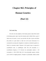
Weeks
Days
At beginning of fourth week,
embryonic disc is flat and trilaminar
21
22
Respiratory diverticulum
forms
4
Body folding commences
24
Body folding is complete, yielding threedimensional embryo with tube-within-atube body plan enclosed in amniotic sac
26
Respiratory diverticulum
branches into left and right
bronchial buds; stem of
diverticulum will differentiate
into trachea and larynx
28
5
Pericardioperitoneal canals
Branching yields secondary
bronchial buds, which
represent future lung lobes
35
36
6
Branching yields tertiary
bronchial buds, which represent
future bronchopulmonary
segments
Pleuropericardial folds begin to separate
primitive pericardial cavity into pericardial
cavity and two pleural cavities; latter are
initially continuous with
peritoneal cavity through
pericardioperitoneal
canals, but pair of
pleuroperitoneal
membranes form to
close off these canals
42
Expansion of amnion
encloses yolk sac and
connecting stalk in
common sheath, forming
umbilical cord
7
Formation of pericardial sac
is complete; lungs are
growing
Terminal bronchioles form
Respiratory bronchioles form;
surrounding mesenchyme
becomes highly vascular;
first terminal sacs
(primitive alveoli) form
Terminal sacs begin to
differentiate into mature
alveoli; alveoli continue to
form through eighth year
16
Pleuroperitoneal membranes have closed off
pericardioperitoneal canals; diaphragm
begins to differentiate
28
36
Birth
8 years
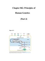
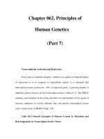
Time line. Development of the lungs, respiratory tree, and body cavities.
situs inversus is consistent with the diagnosis of Kartagener
syndrome (pronounced “KART-agayner”; see Chapters 3
and 12 for additional discussion of Kartagener syndrome), a
variant of PCD. Kartagener syndrome is caused by autosomal
recessive mutations in the DYNEIN AXONEMAL HEAVY CHAIN
5 (DNAH5) gene. Mutations in this gene result in immotile
cilia in the respiratory tract, leading to poor mucus transport
and frequent infections. Because cilia are also involved in
sperm transport, affected males are sterile. During embryonic
development, cilia in the node are involved in determination
of the left-right axis (covered in Chapter 3). Loss of node ciliary function in PCD leads to randomization of laterality, with
50% of affected individuals having situs inversus.
DEVELOPMENT OF LUNGS AND
RESPIRATORY TREE
Animation 11-1: Development of Lungs.
Animations are available online at StudentConsult.
Development of the esophagus, stomach, trachea, and
lungs from the foregut region is tightly linked (Fig. 11-1A).
Hence, defects in the development of the foregut region
often involve both the cranial level of the gastrointestinal system and the respiratory system (see Chapters 14
and 17 for further coverage of the development of the
foregut region). Development of the lungs begins on day
Chapter 11 — Development of the Respiratory System and Body Cavities
253
Caudal pharynx
Larynx
Esophagus
Trachea
Esophagus
Trachea
Trachea
Esophagus
Trachea
Lung
Lungs
Primary
lung bud
Primary bronchi
Lung
Primary
lung bud
Lung
Esophagus
Cystic
diverticulum Esophagus
Stomach
Esophagus
Pancreatic
rudiments
Lung
Stomach
Stomach
E10.5
E11.5
E12.5
Stomach
E13.5
A
Future trachea
and larynx
Esophagus
Left and right primary
bronchial buds
Mesencephalon
Pleural mesenchyme
Rhombencephalon
Diencephalon
Pharynx
Respiratory
diverticulum
28 days
Liver cords
Septum
transversum
Secondary
bronchial buds
Midgut
Allantois
30 days
Yolk sac
Tertiary bronchial
buds
25 days
B
38 days
Stomach
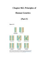
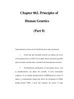
Figure 11-1. Development of the respiratory diverticulum. A, Four stages in development of the mouse foregut, showing origins of the esophagus, trachea, lungs, and stomach. The foregut epithelium has been stained with an antibody to E-cadherin. The branching pattern of the mouse
respiratory tree differs from that of the human, which is described in the text. B, The respiratory diverticulum first forms as an evagination of the
foregut on day twenty-two and immediately bifurcates into two primary bronchial buds between day twenty-six and day twenty-eight. Early in the
fifth week, the right bronchial bud branches into three secondary bronchial buds, whereas the left bronchial bud branches into two. By the sixth
week, secondary bronchial buds branch into tertiary bronchial buds (usually about ten on each side) to form the bronchopulmonary segments.
twenty-two with formation of a ventral outpouching of
the endodermal foregut called the respiratory diverticulum (Fig. 11-1B). This bud grows ventrocaudally
through the mesenchyme surrounding the foregut, and on
days twenty-six to twenty-eight, it undergoes a first bifurcation, splitting into right and left primary bronchial
(or lung) buds. These buds are the rudiments of the two
lungs and the right and left primary bronchi, and the
proximal end (stem) of the diverticulum forms the trachea and larynx. The latter opens into the pharynx via
the glottis, a passageway formed at the original point of
evagination of the diverticulum. As the primary bronchial
254
Larsen's Human Embryology
TABLE 11-1 STAGES OF HUMAN LUNG DEVELOPMENT
Stage of Development
Period
Events
Embryonic
Twenty-six days to six
weeks
Respiratory diverticulum arises as a ventral outpouching of foregut endoderm and
undergoes three initial rounds of branching, producing the primordia successively
of the two lungs, the lung lobes, and the bronchopulmonary segments; the stem
of the diverticulum forms the trachea and larynx
Pseudoglandular
Six to sixteen weeks
Respiratory tree undergoes fourteen more generations of branching, resulting in the
formation of terminal bronchioles
Canalicular
Sixteen to twenty-eight
weeks
Each terminal bronchiole divides into two or more respiratory bronchioles.
Respiratory vasculature begins to develop. During this process, blood vessels come
into close apposition with the lung epithelium. The lung epithelium also begins to
differentiate into specialized cell types (ciliated, secretory, and neuroendocrine cells
proximally and precursors of the alveolar type II and I cells distally)
Saccular
Twenty-eight to
thirty-six weeks
Respiratory bronchioles subdivide to produce terminal sacs (primitive alveoli).
Terminal sacs continue to be produced until well into childhood
Alveolar
Thirty-six weeks to term
Alveoli mature
Splanchnopleuric
mesoderm
Respiratory
bronchiole
Terminal sac
Terminal
bronchiole
28-36 weeks
Mature alveolus
36 weeks–
early childhood
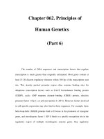
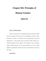
Figure 11-2. Maturation of lung tissue. Terminal sacs (primitive alveoli) begin to form between weeks twenty-eight and thirty-six and begin to
mature between thirty-six weeks and birth. However, only 5% to 20% of all terminal sacs eventually produced are formed before birth. Subsequent
septation of the alveoli is not shown.
buds form, the stem of the diverticulum begins to separate from the overlying portion of the pharynx, which
becomes the esophagus. During weeks five and twentyeight, the primary bronchial buds undergo about sixteen
rounds of branching to generate the respiratory tree of the
lungs. The pattern of branching of the lung endoderm is
regulated by the surrounding mesenchyme, which invests
the buds from the time that they first form. The stages of
development of the lungs are summarized in Table 11-1.
The first round of branching of the primary bronchial
buds occurs early in the fifth week (see Fig. 11-1B). This
round of branching is highly stereotypical and yields
three secondary bronchial buds on the right side and
two on the left. The secondary bronchial buds give rise to
the lung lobes: three in the right lung and two in the
left lung. During the sixth week, a more variable round of
branching typically yields ten tertiary bronchial buds
on both sides; these become the bronchopulmonary
segments of the mature lung.
By week sixteen, after about fourteen more branchings, the respiratory tree produces small branches called
terminal bronchioles (Fig. 11-2). Between sixteen and
255
Chapter 11 — Development of the Respiratory System and Body Cavities
twenty-eight weeks, each terminal bronchiole divides
into two or more respiratory bronchioles, and the
mesodermal tissue surrounding these structures becomes
highly vascularized. By week twenty-eight, the respiratory bronchioles begin to sprout a final generation of
stubby branches. These branches develop in craniocaudal
progression, forming first at more cranial terminal bronchioles. By week thirty-six, the first-formed wave of terminal branches are invested in a dense network of capillaries
and are called terminal sacs (primitive alveoli). Limited gas exchange is possible at this point, but the alveoli
are still so few and immature that infants born at this
age may die of respiratory insufficiency without adequate
therapy (covered in a following “In the Clinic” entitled
“Lung Maturation and Survival of Premature Infants”).
Additional terminal sacs continue to form and differentiate in craniocaudal progression both before and
after birth. The process is largely completed by two years.
About twenty-million to seventy-million terminal sacs
are formed in each lung before birth; the total number
of alveoli in the mature lung is three-hundred million
to four-hundred million. Continued thinning of the
squamous epithelial lining of the terminal sacs begins
just before birth, resulting in the differentiation of these
primitive alveoli into mature alveoli.
The development of the lung during fetal and postnatal life is often subdivided into four phases. The pseudoglandular phase begins around the beginning of the
fifth month of gestation. It is characterized by the presence of terminal bronchi consisting of thick-walled tubes
surrounded by dense mesenchyme. The canalicular
phase begins around the beginning of the sixth month
of gestation (Fig. 11-3A). It is characterized by thinning
of the walls of the tubes as the lumens of the bronchi
enlarge. During the canalicular phase, the lung becomes
highly vascularized. The saccular phase begins around
the beginning of the seventh month of gestation (Fig.
11-3B). It is characterized by further thinning of the tubes
to form numerous sacculi lined with type I and II alveolar cells (the former form the surface for gas exchange,
and the latter respond to damage to type I cells by dividing and replacing them; as covered in the “In the Clinic”
entitled “Lung Maturation and Survival of Premature
Infants,” type II cells are the source of pulmonary surfactant). The alveolar phase begins shortly before birth,
typically around the beginning of the ninth month of
gestation, and continues into postnatal life (Fig. 11-3C). It
is characterized by the formation of mature alveoli.
An important process of septation, which further subdivides the alveoli, occurs after birth. Each septum formed
during this process contains smooth muscle and capillaries.
The lung is a composite of endodermal and mesodermal tissues. The endoderm of the respiratory diverticulum
gives rise to the mucosal lining of the bronchi and to the
epithelial cells of the alveoli. The remaining components
of the lung, including muscle and cartilage supporting
the bronchi and the visceral pleura covering the lung, are
derived from the splanchnopleuric mesoderm, which covers the bronchi as they grow out from the mediastinum
into the pleural space. The lung vasculature is thought to
develop via angiogenesis (i.e., sprouting from neighboring
vessels; angiogenesis is covered in Chapter 13).
M
C
C
C
C
M
C
C
M
A
AW
M
S
M
M
S
S
AW
M
S
B
A
M
M
A
A
A
C
M
A
A
M
A
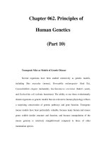
Figure 11-3. Histologic stages of normal human lung development.
A, Canalicular stage. B, Saccular stage. C, Alveolar stage. A, alveolus; AW,
airway; C, canaliculus; M, mesenchyme; S, saccule; Arrows, capillaries.
In the Research Lab
Induction of Lungs and Respiratory Tree
Experiments in mouse embryos have revealed that induction
of the respiratory tree requires Wnt signaling. After inactivation of β-catenin in the foregut endoderm, or in mice null for
Wnt2/2b, the foregut fails to express the transcription factor
Nkx2.1 (formerly called thyroid transcription factor-1; Titf1)
—the earliest marker of the respiratory tree—and the lungs
fail to form. Conversely, increasing Wnt/β-catenin signaling
leads to the conversion of esophagus and stomach endoderm
into lung endoderm that expresses Nkx2.1. Collectively, these
experiments demonstration that Wnt signaling is both sufficient
and necessary for formation of the respiratory tree, and that a
choice is made during development through inductive interactions to convert the foregut endoderm into either trachea and
lungs or esophagus and stomach.
256
Larsen's Human Embryology
In the Clinic
Esophageal Atresia and Tracheoesophageal Fistula
Esophageal atresia (EA; a blind esophagus) and tracheoesophageal fistula (TEF; an abnormal connection between
tracheal and esophageal lumens resulting from failure of the
foregut to separate completely into trachea and esophagus; also
called esophagotracheal fistula) are usually found together
and occur in 1 of 3000 to 5000 births (Fig. 11-4). However, many
variations in these defects are known, including an EA that connects to the trachea, forming a proximal TEF (with or without a
distal TEF; the latter is illustrated in Fig. 11-4), an isolated TEF (i.e.,
without an EA), and an isolated EA (i.e., without a TEF). In addition, both of these defects can be associated with other defects
(e.g., esophageal atresia with cardiovascular defects such as tetralogy of Fallot—covered in Chapter 12; tracheoesophageal fistula
with VATER or VACTERL association—covered in Chapter 3).
Both esophageal atresia and tracheoesophageal fistula are dangerous to the newborn because they allow milk or other fluids to be
aspirated into the lungs. Hence, they are surgically corrected in the
newborn. In addition to threatening survival after birth, esophageal atresia has an adverse effect on the intrauterine environment
before birth: the blind-ending esophagus prevents the fetus from
swallowing amniotic fluid and returning it to the mother via the
placental circulation. This leads to an excess of amniotic fluid
(polyhydramnios) and consequent distention of the uterus.
In the Research Lab
Esophageal Atresia and Tracheoesophageal Fistula
The cause of esophageal atresia is thought to be failure
of the esophageal endoderm to proliferate rapidly enough
during the fifth week to keep up with the elongation of the
embryo. However, the cause of tracheoesophageal fistula
and the reason why the two defects are usually found together
remain a puzzle. During development of the mouse embryo,
the anterior foregut expresses the transcription factor Sox2,
with highest levels of expression occurring in the future esophagus and stomach. In contrast, the future tracheal region of the
foregut expresses the transcription factor Nkx2.1. Moreover,
sonic hedgehog (Shh) is expressed in the ventral endoderm of
the foregut, where it controls cell proliferation, and fibroblast
growth factors (Fgfs) are expressed in the adjacent ventral mesenchyme. Disruption of the Shh pathway or the transcription
factor Nkx2.1 causes tracheoesophageal fistula. It is believed
that Sox2 expression in the foregut sets up a boundary separating the trachea and the esophagus in normal development,
and organ culture experiments suggest that Fgfs expressed by
the ventral mesenchyme regulate Sox2 expression. Moreover,
bone morphogenetic protein (Bmp) signaling is also required to
repress Sox2 expression in the future trachea. Finally, Sox2 and
Nkx2.1 reciprocally inhibit each other's expression, supporting
an important role for the establishment of a tissue boundary in
normal tracheal and esophagus development.
In the Clinic
Developmental Abnormalities of Lungs
and Respiratory Tree
Many lung anomalies result from failure of the respiratory diverticulum or its branches to branch or differentiate correctly. The most
severe of these anomalies, pulmonary agenesis, results when
the respiratory diverticulum fails to split into right and left bronchial
buds and to continue growing. Errors in the pattern of pulmonary
branching (branching morphogenesis) during the embryonic
and early fetal periods result in defects ranging from an abnormal
number of pulmonary lobes or bronchial segments to the complete absence of a lung. The complexity of branching morphogenesis can be appreciated by examining developing lungs in mouse
Trachea
Right bronchus
Left bronchus
Figure 11-4. Diagram of an infant with esophageal
atresia and tracheoesophageal fistula shows how the first
drink of fluid after birth could be diverted into the newly
expanded lungs (arrows).
Proximal, blind-ending
part of esophagus
Tracheoesophageal
fistula
Distal
esophagus
Chapter 11 — Development of the Respiratory System and Body Cavities
embryos in which the respiratory tree has been specifically stained
(Fig. 11-5); such images make clear how defects in branching morphogenesis can lead to lobe or bronchial segment anomalies.
Defects in the subdivision of the terminal respiratory bronchi
or in the formation of septae after birth can result in an abnormal
paucity of alveoli, even if the respiratory tree is otherwise normal.
Some of these types of pulmonary anomalies are caused by
intrinsic molecular and cellular defects of branching morphogenesis (see the following “In the Research Lab” entitled “Molecular
and Cellular Basis of Branching Morphogenesis”). However, the
primary cause of pulmonary hypoplasia—a reduced number
of pulmonary segments or terminal air sacs—often represents a
response to some condition that reduces the volume of the pleural
cavity, thus restricting growth of the lungs (e.g., protrusion of the
abdominal viscera into the thoracic cavity, a condition known as
congenital diaphragmatic hernia; covered in a later “In the Clinic”
entitled “Diaphragmatic Defects and Pulmonary Hypoplasia”).
Lung Maturation and Survival of Premature Infants
As the end of gestation approaches, the lungs undergo a rapid
and dramatic series of transformations that prepare them for air
breathing. The fluid that fills the alveoli prenatally is absorbed at
birth, the defenses that will protect the lungs against invading
pathogens and against the oxidative effects of the atmosphere
are activated, and the surface area for alveolar gas exchange
increases greatly. Changes in the structure of the lung take
place during the last three months, accelerating in the days just
preceding a normal term delivery. If a child is born prematurely,
the state of development of the lungs is usually the prime factor
determining whether he or she will live. Infants born between
twenty-four weeks and term—during the phase of accelerated
terminal lung maturation—have a good chance of survival
with appropriate (including intensive medical assistance at
the younger ages) neonatal support. Infants born earlier than
twenty-four weeks (during the canalicular phase of lung development) currently have a poor chance of survival (in neonatal
intensive care units, or NICUs, 10% to 15% of infants born at
22 to 23 weeks survive, but about 50% of these have profound
impairment; recently, an infant born at twenty-one weeks was
reported to survive). Unfortunately, surviving infants receiving
Figure 11-5. Whole mount of developing lungs from a mouse
embryo at E14.5. Lung and tracheal epithelium has been labeled with
an antibody to E-cadherin to show the pattern of branching (the pattern differs from the human pattern, which is described in the text).
257
intensive respiratory assistance may develop lung fibrosis that
results in long-term respiratory problems.
Although the total surface area for gas exchange in the lung
depends on the number of alveoli and on the density of alveolar
capillaries, efficient gas exchange will occur only if the barrier
separating air from blood is thin—that is, if the alveoli are thinwalled, properly inflated, and not filled with fluid. The walls of
the maturing alveolar sacs thin out during the weeks before birth.
In addition, specific alveolar cells (alveolar type II cells) begin to
secrete pulmonary surfactant, a mixture of phospholipids and
surfactant proteins that reduces the surface tension of the liquid
film lining the alveoli and thus facilitates inflation. In the absence
of surfactant, the surface tension at the air-liquid interface of the
alveolar sacs tends to collapse the alveoli during exhalation. These
collapsed alveoli can be inflated only with great effort.
The primary cause of the respiratory distress syndrome
of premature infants (pulmonary insufficiency accompanied by
gasping and cyanosis) is inadequate production of surfactant.
Respiratory distress syndrome not only threatens the infant with
immediate asphyxiation, but the increased rate of breathing and
mechanical ventilation required to support the infant's respiration
can damage the delicate alveolar lining, allowing fluid and cellular
and serum proteins to exude into the alveolus. Continued injury
may lead to detachment of the layer of cells lining the alveoli—a
condition called hyaline membrane disease. Chronic lung
injury associated with preterm infants causes a condition termed
bronchopulmonary dysplasia, in which the lungs become
inflamed and ultimately scarred, compromising their ability to
oxygenate the blood. In mothers at high risk for premature delivery, the fetus can be treated antenatally with steroids to accelerate lung maturation and the synthesis of surfactant.
Critically ill newborns were first successfully treated with
surfactant replacement therapy—administration of
exogenous surfactant—in the late 1970s. Although originally
extracted from animal lungs or human amniotic fluid, synthetic
surfactant preparations are now used. In addition to containing phospholipids, current preparations include some of the
supplementary proteins found in natural surfactant. Four native
surfactant proteins are known: hydrophobic surfactant proteins
B and C (Sp-B and Sp-C, respectively) and hydrophilic surfactant
proteins A and D (Sp-A and Sp-D, respectively). Sp-B seems
to act by organizing the surfactant phospholipids into tubular
structures, termed tubular myelin, which is particularly effective at reducing surface tension. Although Sp-C is not required
for tubular myelin formation, it does enhance the function of
surfactant phospholipids. Sp-A and -D apparently play important
roles in innate host defense of the lung against viral, bacterial,
and fungal pathogens.
A fatal disease called hereditary surfactant protein B
deficiency (hereditary SP-B deficiency) is a rare cause of
respiratory failure in both premature and full-term newborn
infants. Alveolar air spaces are filled with granular eosinophilic
proteinaceous material, and tubular myelin is absent. Even
though aggressive medical interventions have been applied in
these cases, including surfactant replacement therapy, infants
afflicted with this disease will die, typically within the first year,
if they do not receive a lung transplant.
Hereditary SP-B deficiency is an autosomal recessive condition. The genetic basis for this condition has been examined. In
most cases, a frameshift mutation in exon 4 of the human
SP-B gene has been identified. This mutation results in premature
termination of translation of the SP-B protein. Other mutations of
the SP-B gene have also been identified that result in synthesis of
defective forms of the SP-B protein. It has been demonstrated that
effects of SP-B deficiency extend beyond the disruption of translation of the SP-B gene. Results of studies of null mutations of the
sp-b gene in transgenic mice, for example, show that although
the amount of sp-c or sp-a mRNA is not affected, precursors of
258
Larsen's Human Embryology
the mature sp-c protein are not completely processed. In addition, the processing of pulmonary phospholipids is disrupted.
Similar disruptions of SP-C peptide and phospholipid processing
have been described in a human infant with SP-B deficiency.
More than fifteen different mutations in the SP-B gene have been
associated with hereditary SP-B deficiency. Mild mutations can
cause chronic pulmonary disease in infants. Although these studies have been useful in diagnosis, it is hoped that they will lead to
effective therapies for this usually fatal disease.
In the Research Lab
Approaches for Studying Lung Development
and Branching Morphogenesis
Organ Culture
Just after formation of the primary bronchial buds, the lung
primordia can be removed from embryonic birds or mice and
cultured in media free of serum and other exogenous growth
factors. Under these conditions, the lung primordia will grow
and branch for a few days. However, in the absence of an intact
vascular system, complete development is not possible. With this
limitation, it is possible to use these cultured lungs to analyze the
roles of growth factors and other agents in the branching process.
In one such study, a small peptide that served as a competitive
inhibitor of ligand binding to integrins resulted in abnormal morphology of the developing lung primordium. In another study,
incubation with monoclonal antibodies to specific sequences
of the extracellular matrix protein laminin resulted in reduction
of terminal buds and segmental dilation of the explanted lung
primordia. In another strategy, lung explants were treated with
antisense oligonucleotides, which bind with and inactivate the
mRNA of the specific factor of interest. Experiments with antisense
oligonucleotides against transcription factors such as Nkx2.1
resulted in a reduction in the number of terminal branches of the
lung primordium. It is possible to cleanly separate the endoderm
of the lung buds from the mesoderm and to culture each alone
or together and in the presence of purified factors. This can reveal
the mechanisms by which these layers and factors interact in vivo.
Transgenic and Gene-Targeting Technologies
Genetic strategies, including the generation of engineered lossof-function mutations (gene knockouts) and gain-of-function
transgenes, have provided important insights into lung development. Recent advances have enabled genes to be deleted only
in lung epithelial cells, either in the embryo or in the adult, thus
bypassing the early lethality of some null mutations. In addition,
transgenes can be selected that drive expression of proteins in
specific respiratory cell types. Among examples, a surfactant B
gene null mutation was described in the preceding “In the Clinic”
entitled “Lung Maturation and Survival of Premature Infants.”
Similar approaches have implicated many transcription factors
in the control of lung growth, differentiation, and branching.
These include the proto-oncogene N-myc, the homeodomain
protein Gata6, and the Lim homeodomain factor Lhx4 (previously
known as Gsh4). Similarly, the homeodomain-containing transcription factor Nkx2.1 and the winged helix transcription
factors Foxa1 and Foxa2 (previously known, respectively, as hepatic nuclear factor 3α and β) have been shown to be required for
the regulation of lung cell genes, including surfactant synthesis.
A dramatic result was obtained by targeted disruption of the
function of an Fgf receptor protein in the lung. A transgene consisting of the surfactant C promoter element and a mutant form
of the Fgf receptor that lacked a kinase sequence was constructed
and injected into fertilized eggs to generate transgenic mice.
Inclusion of the surfactant C promoter element in the transgene
resulted in its expression only in the airway epithelium. The
rationale behind the experiment is that formation of a functional
Fgf receptor requires dimerization of two normal Fgf protein
monomers. Therefore, dimerization of the mutant protein produced by the transgene with the endogenous wild-type (normal)
Fgf protein resulted in formation of inactive receptors only in the
lungs. As a consequence, other tissue of the embryos developed
normally, but branching of the respiratory tree in the transgenic
pups was completely inhibited. This resulted in formation of
elongated epithelial tubes that were incapable of supporting
normal respiratory function at birth (Fig. 11-6). Subsequent
gene-targeting experiments in mice demonstrated that fibroblast
growth factor 10 (Fgf10) and an isoform of its receptor in the
respiratory epithelium, the Fgf-receptor2, were critical for formation of both lungs and limbs. Similarly, ablation of Nkx2.1 blocked
formation of both thyroid and lung.
Genetic strategies have also been used to create models of human pulmonary disease such as cystic fibrosis. Mouse mutants in
which the c-AMP–stimulated chloride secretory activity of the cystic
fibrosis gene is absent or reduced have been created by homologous recombination. These mice express some, but not all, of the
abnormal phenotypes characteristic of the human disease. In other
experiments, transgenic mice have been created that carry the normal human cystic fibrosis gene to demonstrate that it is non-toxic
and, therefore, probably safe to use in human therapy. Currently,
various approaches to human gene therapy for cystic fibrosis are
being developed with viral- and DNA-based delivery systems. The
long-term goal is to insert the cystic fibrosis gene directly into the
somatic airway epithelial cells of afflicted infants and children.
Molecular and Cellular Basis of Branching Morphogenesis
As covered earlier in the chapter, endodermal bronchial buds
and subsequent airway branches grow into the mesenchyme
surrounding the thoracic gut tube. Deficiencies or abnormalities
in branching of the respiratory tree serve as the basis of many
Figure 11-6. Mutation of a fibroblast growth factor receptor specifically expressed in the lungs results in inhibition of branching of the
respiratory tree and formation of elongated epithelial tubes that end
bluntly. Stippling indicates the outline of where the lungs would form
and their branching pattern in a wild-type embryo.
Chapter 11 — Development of the Respiratory System and Body Cavities
forms of pulmonary hypoplasia (covered in the preceding “In
the Clinic” entitled “Developmental Abnormalities of Lungs and
Respiratory Tree”). Studies over the past several decades have
demonstrated that branching morphogenesis of the respiratory tree is regulated by reciprocal interaction between endoderm and surrounding mesoderm. For example, when mesenchyme in the region of the bifurcating bronchial buds is replaced
with mesenchyme from around the developing trachea, further
branching is inhibited. Conversely, replacement of tracheal mesenchyme with that from the region of the bifurcating bronchial
buds stimulates ectopic tracheal budding and branching. Based
on experiments such as these, components of the extracellular
matrix and growth factors have been implicated in the stimulation and inhibition of branching. For example, collagen types IV
and V, laminin, fibronectin, and tenascin—all components of the
extracellular matrix—are thought to play a permissive or a stimulatory role in branching of the bronchial buds. Likewise, regulation of expression of receptors for these matrix components has
been implicated in control of branching morphogenesis.
Many growth factors have been implicated in the growth,
differentiation, and branching morphogenesis of the lung. Among
them are retinoic acid (RA), transforming growth factorβ (Tgfβ),
Bmps, Shh, Wnts, Fgfs, epithelial growth factor (Egf), plateletderived growth factor (Pdgf), insulin-like growth factor (Igf), and
transforming growth factorα (Tgfα). These growth factors and
their receptors are expressed in specific cell populations during different phases of lung growth and branching, consistent with their
postulated roles in this complex process. For example, branching
during the pseudoglandular stage is apparently influenced
in part by the dynamic activity of RA, Shh, Fgf (especially Fgf10),
Bmp, and Tgfβ signaling pathways. Thus, experiments have shown
that Fgf10, produced by the mesenchyme overlying the tips of the
outgrowing bronchial buds, promotes both proliferation of the
endoderm and its outward chemotaxis (i.e., directed movement
according to the presence of so-called chemotactic factors in
the cellular environment). On the other hand, Shh, produced by
the endoderm, promotes proliferation and differentiation of the
overlying mesoderm. In addition, Shh negative regulates Fgf10
expression, thereby suppressing inappropriate branching.
The complex branching pattern of the mouse lung has been
examined three-dimensionally (see Fig. 11-5), and it was noted
that branching occurs in three geometric modes: domain branching—formation of branches arranged much like the bristles on a
brush; planar bifurcation—splitting of the tip of a branch into two
branchlets; and orthogonal bifurcation—involving two rounds of
planar bifurcation with 90-degree rotation between rounds to form
a rosette-like structure of four branches. Through iteration of these
three simple branching patterns, the more than a million branches
present in the mouse lung are generated. In addition, experiments support a model in which airway branching, following the
establishment of left-right asymmetry in the lung (e.g., three lobes
in the right lung and two in the left of humans), is controlled by a
master branch generator served by three slaves (i.e., subroutines that control discrete patterning events). The three subroutines consist of a periodicity clock, which times the formation of
branches, and two other routines—one that controls bifurcation,
259
and another that controls branch-point rotation. Sprouty2, an Fgf
signaling inhibitor, is a candidate gene for the periodicity clock.
Interactions between sprouty2, Fgf10, and Fgf receptor2 control
the master branch generator. The other two subroutines involve
interactions among the myriad signaling systems covered earlier
in this section. Finally, it is important to point out that the lungs in
mammals and the tracheal system in flies (see next section entitled
“Drosophila Tracheal System Development”) undergo extensive
branching to increase the surface area for gas exchange, and that
Fgf signaling (and presumably branching) is regulated by oxygen
levels in flies. In mammals, at least two families of factors likely act
as oxygen sensors in lung branching morphogenesis: hypoxiainducible factor (Hif) and vascular endothelial growth factor (Vegf).
Drosophila Tracheal System Development
The respiratory organ in Drosophila, the tracheal system, consists
of a branched network of tubes (Fig. 11-7). It is interesting to
note that given the central role for Fgf signaling in vertebrate
lung development just covered, formation of the tracheal system
also involves Drosophila orthologs of the Fgf signaling system.
Three components of this system have been identified during
development of the tracheal system: branchless, an Fgf-like
ligand; breathless, an Fgf receptor; and sprouty, an endogenous
Fgf inhibitor. Although at least thirty other genes are involved
in tracheal development, branchless and breathless are used
repeatedly to control branch budding and outgrowth. Sprouty
provides negative feedback regulation by antagonizing Fgf signaling, thereby limiting the amount of branching that occurs.
Molecular and Cellular Basis of Alveolar Differentiation
Growth factors such as Fgfs and Egf regulate not only early
growth and branching of the lung, but also later formation and
maturation of terminal sacs during the saccular stage. Later still,
PdgfA is required for the postnatal formation of alveolar septae–
containing myofibroblasts. Like Nkx2.1 and Foxa1/a2 (covered
in a preceding section of this “In the Research Lab” entitled
“Transgenic and Gene-Targeting Technologies”), cytokines,
glucocorticoids, and thyroxine stimulate surfactant synthesis before birth. It is hoped that these findings will lead to therapeutic
stimulation of adequate alveolar formation and differentiation
and surfactant synthesis within the lungs of premature infants.
Considerable effort has been spent in identifying genes that
regulate the differentiation of lung progenitor cells into specialized types such as ciliated, secretory (Clara), and neuroendocrine
cells. For example, analysis of lungs from mice lacking the gene
Mash1 (a member of the notch pathway; covered in Chapter 5)
has shown that they lack neuroendocrine cells, whereas in Hes1
(another member of the notch pathway) null mutants, neuroendocrine cells form prematurely and in larger numbers than normal. The gene Foxj1 (one of the many Fox transcription factors)
is required for the development of differentiated ciliated cells.
The formation of submucosal glands, which are the major source
of mucus production in the normal lung, is also regulated genetically. Mice lacking genes controlling the ectodysplasin (Eda/Edar)
signaling pathway (a gene involved in epithelial morphogenesis;
covered in Chapter 7) do not develop submuscosal glands.
These glands are also absent in humans lacking the EDA gene.
Figure 11-7. The Drosophila tracheal (respiratory) system consists of a network of interconnected epithelial tubes, visualized in a third-instar
larva by expression of green fluorescent protein driven by the breathless promoter. Breathless is an Fgf receptor ortholog that is required for
tracheal tube branching and outgrowth. Image shows a ventral view of the larva, with the head (anterior) to the left.
260
Larsen's Human Embryology
PARTITIONING OF COELOM AND
FORMATION OF DIAPHRAGM
Animation 11-2: Development of Body Cavities
and Diaphragm.
Animations are available online at StudentConsult.
At the beginning of the fourth week of development,
before body folding, the intraembryonic coelom
forms a horseshoe-shaped space that partially encircles the future head end of the embryo (Fig. 11-8).
A
Cranially, the intraembryonic coelom lies just caudal to
the septum transversum and represents the future
pericardial cavity. The two caudally directed limbs
of the horseshoe-shaped intraembryonic coelom represent the continuous future pleural and peritoneal
cavities. At about the mid-trunk and more caudal levels, the intraembryonic coelom on each side is continuous with the extraembryonic coelom or chorionic
cavity.
With body folding, changes occur in the position of
the intraembryonic coelom. The head fold moves the
Septum
transversum
Cut edge of
amnion
Extraembryonic
coelom (chorionic
cavity)
Oropharyngeal
membrane
Arrow in cranial
intraembryonic
coelom
Neural plate
Continuity
between
intraembryonic
and
extraembryonic
Arrow in
coeloms
caudal
intraembryonic
coelom
Septum
transversum
Heart rudiment
Yolk sac
Splanchnopleure
Cloacal
membrane
Amniotic
cavity
Somatopleure
Intraembryonic coelom
(future pericardial cavity) Heart rudiment
Cranial
body
fold
Oropharyngeal
membrane
Brain
B
Yolk sac
Yolk sac
C
Caudal
body fold
Amnion
Figure 11-8. The intraembryonic coelom prior to body folding. A, At the beginning of the fourth week, the intraembryonic coelom forms a
horseshoe-shaped space partially encircling the head end of the embryo. Diagram of the epiblast after removal of the amnion shows the position
of the neural plate, oropharyngeal and cloacal membranes, and intraembryonic coelom; the latter is continuous with the extraembryonic coelom
at about the mid-trunk and at more caudal levels. B, Cranial (top) and caudal (bottom) halves of embryos transected at the level indicated in A.
Arrows show continuity between the intraembryonic and extraembryonic coeloms. C, Midsagittal view through the right side of an embryo at the
level indicated in A. Arrows show the directions of the head and tail body folds.
Chapter 11 — Development of the Respiratory System and Body Cavities
future pericardial cavity caudally and repositions it on
the anterior (ventral) side of the developing head (Fig.
11-9A). The septum transversum, which initially constitutes a partition that lies cranial to the future pericardial
cavity, is repositioned by the head fold to lie caudal to
the future pericardial cavity. The developing heart (covered further in Chapter 12), which initially lies ventral to
the future pericardial cavity, is repositioned dorsally and
quickly begins to bulge into the pericardial cavity. Thus,
after formation of the head fold, the intraembryonic coelom is reshaped into a ventral cranial expansion (primitive pericardial cavity); two narrow canals called
pericardioperitoneal canals (future pleural cavities) that lie dorsal to the septum transversum; and two
more caudal areas (which merge to form the future peritoneal cavity), where the intraembryonic and extraembryonic coeloms are broadly continuous (Fig. 11-9B).
During the fourth and fifth weeks, continued folding and differential growth of the embryonic axis cause a
gradual caudal displacement of the septum transversum.
The ventral edge of the septum finally becomes fixed to the
anterior body wall at the seventh thoracic level, and the
dorsal connection to the esophageal mesenchyme becomes
Developing brain
261
fixed at the twelfth thoracic level. Meanwhile, myoblasts
(muscle cell precursors) differentiate within the septum
transversum. These cells, which will form part of the future
diaphragm muscle, are innervated by spinal nerves at a
transient, cervical level of the septum transversum—that
is, by fibers from the spinal nerves of cervical levels three,
four, and five (C3, C4, C5). These fibers join to form the
paired phrenic nerves, which elongate as they follow the
migrating septum caudally.
PERICARDIAL SAC IS FORMED BY
PLEUROPERICARDIAL FOLDS THAT GROW
FROM LATERAL BODY WALL
IN A CORONAL PLANE
During the fifth week, the pleural and pericardial cavities are divided from each other by pleuropericardial
folds that originate along the lateral body walls in a coronal plane (Fig. 11-10; see Fig. 11-9B for orientation). These
septae appear as broad folds of mesenchyme and pleura
that grow medially toward each other between the heart
and the developing lungs. At the end of the fifth week,
the folds meet and fuse with the foregut mesenchyme,
Forebrain
Amnion
Foregut
Oropharyngeal
membrane
Pericardial
coelom
Developing heart tube
Pericardial coelom
Oropharyngeal
membrane
Developing heart (solid cord)
Septum
transversum
Septum tranversum
Stomodeum
A
Neural tube
Dorsal aortae
Left pericardioperitoneal canal
Lung bud
Communication
between
intraembryonic
coelom (primitive
peritoneal cavity) and
extraembryonic coelom
(chorionic cavity)
Foregut
Stomodeum
Primitive
pericardial
cavity
Level of Figure 11-10
B
Level of Figures
11-11, 11-12, 11-13
Septum
tranversum
Figure 11-9. Body folding changes the shape of the intraembryonic coelom. A, The head end of the embryo before (left) and after (right) formation of the head fold. B, Initial subdivision of the intraembryonic coelom into a primitive pericardial cavity, paired pericardioperitoneal canals,
and paired primitive peritoneal cavities. The latter are continuous on each side with the extraembryonic coelom. Subsequent lateral body folding
progressively separates the intraembryonic and extraembryonic coeloms as the yolk stalk narrows.
262
Larsen's Human Embryology
thus subdividing the primitive pericardial cavity into
three compartments: a fully enclosed, ventral definitive
pericardial cavity and two dorsolateral pleural cavities. The latter are still continuous with the more caudal peritoneal cavities through the pericardioperitoneal
canals. The name pericardioperitoneal is retained for these
canals, even though they now provide communication
between pleural and peritoneal cavities.
As the tips of the pleuropericardial folds grow medially
toward each other, their roots migrate toward the ventral midline (Fig. 11-10B, C). By the time the tips of the
folds meet to seal off the pericardial cavity, their roots
take origin from the ventral midline. Thus, the space that
originally constituted the lateral portion of the primitive
pericardial cavity is converted into the ventrolateral part
of the right and left pleural cavities.
The pleuropericardial folds are three-layered, consisting of mesenchyme sandwiched between two epithelial
layers; all three layers are derived from the body wall.
The thin definitive pericardial sac retains this threefold
composition, consisting of inner and outer serous membranes (the inner serous pericardium and the outer
mediastinal pleura) separated by a delicate filling of
mesenchyme-derived connective tissue, the fibrous
pericardium. The phrenic nerves, which originally run
through the portion of the body wall mesenchyme incorporated into the pleuropericardial folds, course through
the fibrous pericardium of the adult.
PLEUROPERITONEAL MEMBRANES
GROWING FROM POSTERIOR AND
LATERAL BODY WALL SEAL OFF
PERICARDIOPERITONEAL CANALS
Recall that the septum transversum is repositioned by
the head fold to lie ventral to the paired pericardioperitoneal canals (Fig. 11-11; see Fig. 11-9B for orientation).
At the beginning of the fifth week, a pair of membranes,
the pleuroperitoneal membranes, arise along an
oblique line connecting the root of the twelfth rib with
the tips of ribs twelve through seven (Fig. 11-12; see Fig.
11-9B for orientation). These membranes grow ventrally
to fuse with the septum transversum, thus sealing off the
pericardioperitoneal canals. The left pericardioperitoneal
canal is larger than the right and closes later. Closure of
both canals is complete by the seventh week. The membranes that close these canals are called pleuroperitoneal
membranes because they do not contact the septum transversum until after the pericardial sac is formed; thus, after
they fuse with the septum transversum, they separate the
definitive pleural cavities from the peritoneal cavity.
DIAPHRAGM IS A COMPOSITE DERIVED
FROM FOUR EMBRYONIC STRUCTURES
The definitive musculotendinous diaphragm incorporates derivatives of four embryonic structures: (1) septum
Foregut
Bronchial bud
Bronchial bud
Pleuropericardial
fold
Foregut
Heart
Phrenic
nerve
Phrenic nerve
Pleuropericardial
fold
Heart
B
Pleural cavity
Lung
Parietal pleura
A
Fibrous
pericardium
Phrenic nerve
Serous
pericardium
Heart
C
Definitive pericardial
cavity
Figure 11-10. Subdivision of the primitive pericardial cavity. A, During the fifth week, pleuropericardial folds grow out from the lateral body
wall toward the midline, where they fuse with each other and with mesoderm associated with the esophagus. Simultaneously, the roots of these
folds migrate ventrally so that they ultimately connect to the ventral (anterior) body wall. B, The phrenic nerves initially embedded in the body
wall are swept into these developing partitions. C, The pleuropericardial folds with their associated serous membrane form the pericardial sac and
transform the primitive pericardial cavity into a definitive pericardial cavity and right and left pleural cavities.
Chapter 11 — Development of the Respiratory System and Body Cavities
transversum, (2) pleuroperitoneal membranes, (3) mesoderm of the body wall, and (4) esophageal mesoderm (Fig.
11-13A; see Fig. 11-9B for orientation). Some of the myoblasts that arise in the septum transversum emigrate into
the pleuroperitoneal membranes, pulling their phrenic
nerve branches along with them. Most of the septum
transversum then gives rise to the non-muscular central
tendon of the diaphragm (Fig. 11-13B).
Pericardioperitoneal
canals
Foregut
Lung bud
Septum
transversum
263
The bulk of the diaphragm muscle within the pleuroperitoneal membranes is innervated by the phrenic
nerve. However, the outer rim of diaphragmatic muscle arises from a ring of body wall mesoderm (see Figs.
11-12B, 11-13A); this mesoderm is derived from somatic
mesoderm and is invaded by myoblasts arising from
the myotomes of neighboring somites. Therefore, the
peripheral musculature of the diaphragm is innervated
by spinal nerves from thoracic spinal levels T7 through
T12. Finally, mesoderm arising from vertebral levels L1
through L3 condenses to form two muscular bands—the
right and left crura of the diaphragm, which originate
on the vertebral column and insert into the dorsomedial
diaphragm (see Fig. 11-13B). The right crus originates on
vertebral bodies L1 through L3, and the left crus originates on vertebral bodies L1 and L2.
Primitive
pericardial
cavity
In the Clinic
Arrow in
peritoneal
cavity
Figure 11-11. In the future thoracic region, the septum transversum forms a ventral partition beneath the paired pericardioperitoneal
canals (arrows), which interconnect the primitive pericardial cavity cranially and peritoneal cavities caudally.
Diaphragmatic Defects and Pulmonary Hypoplasia
As covered earlier in the chapter, pulmonary hypoplasia
often occurs in response to some conditions that reduce the
volume of the pleural cavity, thereby restricting growth of the
lungs. In congenital diaphragmatic hernia, the developing abdominal viscera may bulge into the pleural cavity (Fig.
11-14). If the mass of displaced viscera is large enough, it will
stunt growth of the lungs, typically on both sides. Congenital
diaphragmatic hernia occurs in about 1 of 2500 live births.
The left side of the diaphragm is involved four to eight times
more often than is the right (i.e., about 80% of diaphragmatic
hernias occur on the left side), probably because the left
pericardioperitoneal canal is larger and closes later than the
right. Most diaphragmatic hernias (i.e., 95%) occur posterolaterally within the diaphragm and are referred to clinically
as Bochdalek hernias. However, diaphragmatic hernias can
rarely occur through the esophageal hiatus or more anteriorly
Pericardioperitoneal
canal
Aorta
Pleuroperitoneal
membrane
Foregut
Inferior
vena cava
Septum transversum
Body wall
A
B
Figure 11-12. Closure of the pericardioperitoneal canals (A, B). Between weeks five and seven, a pair of horizontal pleuroperitoneal membranes
grow from the posterior body wall to meet the septum transversum (arrows, A), thus closing the pericardioperitoneal canals. These membranes
form the posterior portions of the diaphragm and completely seal off the pleural cavities from the peritoneal cavity. Arrows in B indicate invasion
of the developing diaphragm by muscle fibers from the adjacent body wall.
264
Larsen's Human Embryology
Aorta
Central tendon
Pleuroperitoneal
membrane
Inferior vena cava
Esophageal
mesoderm
Esophagus
Inferior vena
cava
Foregut
Costal margin
Septum
transversum
Body wall
A
Aorta
Superior view
of
developing diaphragm
L2
L3
B
Left and right
diaphragmatic crura
Rib
Vertebrae
Inferior view
of diaphragm
Figure 11-13. Formation of the diaphragm. The definitive diaphragm is a composite structure, including elements of the septum transversum,
pleuroperitoneal membranes, and esophageal mesenchyme, as well as a rim of body wall mesoderm. A, Superior view. B, Inferior view.
Parasternal defect
Inferior
vena cava
Central tendon
of diaphragm
Esophagus
Left lung
Small intestine
Congenital
absence of large
area of diaphragm
A
Aorta
Colon
Heart
Spleen
Diaphragm
Stomach
B
Figure 11-14. Diaphragmatic hernia. This defect most often occurs through failure of the left pleuroperitoneal membrane to seal off the left
pleural cavity completely from the peritoneal cavity. A, Inferior view. B, Abdominal contents may herniate through the patent pericardioperitoneal
canal, preventing normal development of the lungs on both sides, which become compressed.
Chapter 11 — Development of the Respiratory System and Body Cavities
265
contribution to the amniotic fluid. Therefore, bilateral renal
agenesis—failure of both kidneys to form (covered in Chapter
15)—results in oligohydramnios. Also, in a condition called premature rupture of the membranes (PROM), the amnion
ruptures early and amniotic fluid is lost, resulting in oligohydramnios. Presumably, oligohydramnios, regardless of its cause,
results in pulmonary hypoplasia due to excessive loss of fluid
from the fetal lungs and resulting decreased fluid pressure within
the maturing respiratory tree. Compression of the fetal chest by
the uterine wall has been postulated to play a role.
In the Research Lab
Figure 11-15. Eventration of the diaphragm. Failure of the pleuroperitoneal membranes to differentiate normally during fetal life may
allow abdominal organs to dilate the abnormally thin regions of the
diaphragm and eventrate into the pleural cavity.
(i.e., retrosternally or parasternally), where they are referred
to clinically as Morgagni hernias. The mortality rate from
diaphragmatic hernias is high, averaging about 50%, but the
prognosis depends on the type of hernia. For example, rightsided Bochdalek hernias have a worse prognosis than do left-sided
hernias, and Morgagni hernias usually have only minor clinical
consequences. Diaphragmatic hernias can be surgically corrected at birth and have also rarely been corrected by surgery
during fetal life (covered in Chapter 6). However, if the hernia
has resulted in severe pulmonary hypoplasia, the newborn
may die of pulmonary insufficiency or pulmonary hypertension
even if the hernia is repaired.
If the development of muscle tissue in the diaphragm is
deficient, the excessively compliant diaphragm may allow the
underlying abdominal contents to balloon or eventrate into
the pulmonary cavity (Fig. 11-15). This condition can also result
in pulmonary hypoplasia and hypertension, which may be fatal.
Oligohydramnios and Pulmonary Hypoplasia
As covered earlier in the chapter, pulmonary hypoplasia can
result from failure of proper branching morphogenesis during
development of the lungs and respiratory tree, as well as from
diaphragmatic defects as just described. Another classic cause of
pulmonary hypoplasia is oligohydramnios, the condition in
which there is an insufficient amount of amniotic fluid. The causes
of oligohydramnios and how they result in pulmonary hypoplasia
are complex. During in utero life, the lung acts like an exocrine
gland, producing fluid that provides a substantial contribution to
the amniotic fluid. In addition, once the kidneys begin to function, after about sixteen weeks, fetal urine provides a substantial
Congenital Diaphragmatic Hernia
Little is known about the molecular mechanisms of diaphragm
formation and how this process fails to occur, resulting in congenital diaphragmatic hernia (CDH). However, it was shown in
a screen of fetal mice harboring ENU-induced genetic mutations
that CDH resulted from a mutation in the Fog2 (friend of Gata2;
Gata2 is a transcription factor) gene. In addition, pulmonary
hypoplasia occurred early in gestation, and Fog2 was expressed
throughout the pulmonary mesenchyme during stages of
branching morphogenesis, suggesting a direct role of Fog2
in pulmonary development. Screening of DNA from patients
with congenital diaphragramatic defects revealed mutations in
FOG2, demonstrating a role for this gene in development of the
diaphragm in both mouse and human.
Additional evidence that FOG2 is critical for normal diaphragm formation comes from studies that have shown that
Fog2 is an important regulator of Gata4 in the developing
heart, and that both genes are co-expressed during cardiac
embryogenesis. Homozygous mice null for Gata4 also have
CDH, suggesting that abnormal regulation of Gata4 by Fog2
might be important for diaphragm development. Furthermore,
Fog2 binds to the ligand-binding domain of chicken ovalbumin
upstream promoter transcription factor II (Coup-tfII). CouptfII has been shown to be necessary for Fog2 to repress the
transcription of a Gata4. It is important to note that mice with
tissue-specific mutations of Coup-TFII have CDH, and Coup-TFII
is located on human chromosome 15q26.2—a genomic region
that is deleted in some CDH patients.
Embryology in Practice
Lung Mass
A mid-gestation ultrasound reveals a mass that fills much of the left
hemi-thorax of a fetus at twenty weeks of gestation. The remainder of the examination and the pregnancy history are unremarkable. Chromosome studies from an earlier amniocentesis, which
was done because of advanced maternal age, were normal.
Careful review of the ultrasound shows normal diaphragm
and abdominal contents, which argues against the presence
of a congenital diaphragmatic hernia. The two remaining
considerations for an interthoracic mass include bronchopulmonary sequestration (BPS) and congenital cystic adenomatoid
malformation (CCAM).
Examination of the mass by color Doppler ultrasound
demonstrates lack of a systemic arterial blood supply, which
is always seen in cases of BPS. The presumptive diagnosis of a
CCAM is made.
The parents are counseled that the effects on the fetus
depend on the size of the lesion. Smaller lesions may have no
266
Larsen's Human Embryology
Fetal heart compressed
against chest wall
Large CCAM
(solid and cystic)
A
Right lung
effect on the pregnancy, but larger masses can impact the fetus
by compression of the thoracic contents. Close follow-up with
serial ultrasounds is planned.
Unfortunately, these ultrasounds show progressive growth of
the mass, causing a mediastinal shift and resultant lung hypoplasia (Fig. 11-16A). The mass also impinges on the heart, leading to
cardiovascular compromise and fetal hydrops. Compression of
the esophagus and reduced swallowing of amniotic fluid lead to
polyhydramnios. At thirty weeks of gestation, the pregnancy is
spontaneously lost as the result of fetal cardiac failure. An autopsy
reveals that the chest is almost completely filled with a solid
mass, with compression of the lungs and heart (Fig. 11-16B).
As the name suggests, congenital cystic adenomatoid
malformation (CCAM) consists of proliferative overgrowths
of abnormal lung tissue that occur without a known cause
(adenomatoid malformation means “a benign tumor”). CCAM is
differentiated from BPS by the lack of a systemic arterial supply.
BPS is typically a wedge-shaped, left-sided anomaly surrounded
by its own visceral pleura. Several subtypes of CCAM (also called
congenital pulmonary airway malformation, or CPAM) have
been designated on the basis of cyst size. The risk of malignant
transformation prompts surgical resection in surviving infants at
many centers, irrespective of type.
Suggested Readings
CCAM from
left lung
B
Liver
Figure 11-16. Congenital cystic adenomatoid malformation (CCAM).
A, Sonogram at twenty-nine weeks. B, Autopsy photo of the stillborn
fetus showing the enlarged left lung mass after removal of the chest wall.
Ahlfeld SK, Conway SJ. 2012. Aberrant signaling pathways of the lung
mesenchyme and their contributions to the pathogenesis of bronchopulmonary dysplasia. Birth Defects Res A Clin Mol Teratol 94:3–15.
De Langhe SP, Reynolds SD. 2008. Wnt signaling in lung organogenesis. Organogenesis 4:100–108.
Domyan ET, Sun X. 2011. Patterning and plasticity in development of
the respiratory lineage. Dev Dyn 240:477–485.
Holder AM, Klaassens M, Tibboel D, et al. 2007 Genetic factors in congenital diaphragmatic hernia. Am J Hum Genet 80:825–845.
Maeda Y, Dave V, Whitsett JA. 2007. Transcriptional control of lung
morphogenesis. Physiol Rev 87:219–244.
Metzger RJ, Klein OD, Martin GR, Krasnow MA. 2008. The branching
programme of mouse lung development. Nature 453:745–750.
Morrisey EE, Hogan BL. 2010. Preparing for the first breath: genetic and
cellular mechanisms in lung development. Dev Cell 18:8–23.
Ornitz DM, Yin Y. 2012. Signaling networks regulating development
of the lower respiratory tract. Cold Spring Harb Perspect Biol 4:1–19.
Warburton D. 2008. Developmental biology: order in the lung. Nature
453:733–735.
Chapter 12
Development of the Heart
SUMMARY
In response to inductive and permissive signals emanating from the endoderm, ectoderm, and midline mesoderm, cardiogenic precursors form a cardiac primordium
within the splanchnic mesoderm at the cranial end of
the embryonic disc called the cardiac crescent, or
first heart field. In response to signals from the underlying endoderm, a subpopulation of cells within the first
heart field form a pair of lateral endocardial tubes
through the process of vasculogenesis. The cranial and
lateral folding of the embryo during the fourth week
results in the fusion of these tubes along the midline in
the future thoracic region, where they form a single primary heart tube. This tube consists of a single endocardial tube with adjacent mesoderm differentiating into
cardiomyocytes.
The heartbeat is initiated around the twenty-first
day, and its continual beating is required for normal
heart development. Between weeks four and eight, the
primary heart tube undergoes a series of events, including
looping, remodeling, realignment, and septation,
eventually leading to the transformation of a single heart
tube into a four-chambered heart, thus laying down the
basis for the separation of pulmonary and systemic circulations at birth.
Starting at the inflow end, the primary heart tube
initially consists of the left and right horns of the
sinus venosus, the primitive atrium, the atrioventricular canal, the primitive left ventricle,
and a short outflow region. Lengthening of the primary
heart tube and proper cardiac bending and looping are
driven through the addition of cardiac precursor cells by
the second heart field. At the outflow end, the main
additions are the primitive right ventricle and the
outflow tract that connects with the aortic sac at the
arterial orifice. As the outflow tract lengthens, proximal
(conus) and distal (truncus) components can be distinguished. Septation of the outflow tract leads to separate
left and right ventricular outlets and to formation of the
ascending aorta and pulmonary trunk. At the inflow end,
the second heart field also contributes myocardium to
the sinus venosus wall, the body of the right and left
atrium, and the atrial septa.
Venous blood initially enters the sinus horns through
paired, symmetrical common cardinal veins. However, as covered in Chapter 13, changes in the venous
system rapidly shift the entire systemic venous return to
the right, so that all blood from the body and umbilicus
enters the future right atrium through the developing
superior and inferior caval veins. The left sinus
horn becomes the coronary sinus, which collects
blood from the coronary circulation. A process of intussusception incorporates the right sinus horn and the
ostia of the caval veins into the posterior wall of the
future right atrium. In this process, the pulmonary
vein developing within the dorsal mesocardium shifts
to the future left atrium as a result of the development
of a dorsal mesenchymal protrusion. Subsequently,
the walls of the pulmonary vein are partially incorporated into the atrial wall, forming the larger part of the
dorsal left atrial wall. In the fifth and sixth weeks, the
atrial septum starts to develop. This is a two-step process.
It begins with the formation of the septum primum
(primary atrial septum), which is followed by formation of the septum secundum (secondary atrial
septum). The formation of this atrial septal complex
results in separation of the right and left atria. However,
the two septa do not fuse until after birth, allowing for
right-to-left shunting of blood throughout gestation.
The mitral (bicuspid) and tricuspid atrioventricular valves develop from atrioventricular cushion
tissue during the fifth and sixth weeks. Meanwhile, the
heart undergoes remodeling, bringing the future atria
and ventricles into correct alignment with each other
and aligning both ventricles with their respective future
outflow vessels. During expansion of the primitive right
and left ventricles, a muscular ventricular septum
forms that partially separates the ventricles. During the
seventh and eighth weeks, the outflow tract of the heart
completes the process of septation and division. During
this process, remodeling of the distal outflow tract cushion tissue (truncal cushions) results in the formation
of the semilunar valves of the aorta and pulmonary
artery. Fusion of the proximal outflow tract cushions
(conal cushions) creates the outlet septum, resulting
in the separation of left and right ventricular outlets.
Complete ventricular septation depends on fusion of
the outflow tract (conotruncal) septum, the muscular
ventricular septum, and the atrioventricular cushion
tissues.
The myocardium of the heart differentiates into working myocardium and myocardium of the conduction
system. The epicardium grows out from the pro-
epicardial organ covering the myocardium. It contributes to the formation of the coronary vasculature, which
is necessary for oxygenation of the thickening myocardial wall and myocardial cell population.
267
268
Larsen's Human Embryology
Weeks
Days
Cranial lateral plate mesoderm initiates vasculogenesis to form
lateral endocardial tubes; myocardiogenesis begins
19
Lateral body folding brings endocardial tubes and surrounding
cardiogenic mesoderm together in thoracic region
3
20
Endocardial tubes surrounded by myocardium fuse to
form primary heart tube, which is divided into incipient
chambers by sulci
21
Atrioventricular sulcus
Myocardium invests endocardial heart tube and forms
cardiac jelly
22
Heart begins to beat
4
23 Heart begins to loop
Septum primum begins to form
28 Heart looping is complete
Epicardium
forms
Muscular ventricular
septum begins to form
5
Endocardial cushion tissues form
33
Outflow tract endocardial
cushions begin to form
35
6
42
7
46
Atrioventricular
endocardial cushions
fuse to form
atrioventricularseptum
Definitive atria and auricles are present
Foramen secundum and foramen ovale
form as septum primum meets
atrioventricular septum
Muscular ventricular
septum ceases to grow
8
56
Aortic and pulmonary outflow
tracts are fully separated by
fusion of outflow tract cushions;
ventricular septation is
completed by joining of
outflow tract, atrioventricular, and
muscular interventricular septa
Coronary sinus is formed
9
63
10
Semilunar and atrioventricular
valves are complete
Time line. Formation of the heart.
Clinical Taster
A full-term boy is born to a primigravid (first gestation)
mother after an uncomplicated pregnancy. The delivery goes
smoothly, with healthy Apgar scores of 8/10 at one minute
and 9/10 at five minutes. All growth parameters (length,
weight, and head circumference) are normal, ranging between the 10th and 25th centiles. The newborn examination
is also normal, and the infant is returned to his mother to
begin breast feeding.
The boy initially feeds well, but he becomes sleepy and
disinterested in feeding as the day progresses. At twenty hours
after birth, he exhibits decreased peripheral perfusion, cyanosis,
and lethargy. A pulse oximeter shows oxygen saturation in
the low 80% range (normal equals >90%) with increasing
respiratory distress. Paradoxically, blood oxygen saturation
worsens after administration of oxygen. The boy is emergently
transferred to the neonatal intensive care unit in worsening
shock. There, he is intubated, central intravascular catheters are
placed, and he is started on prostaglandins.
Chapter 12 — Development of the Heart
269
First heart field
Head
fold
Primitive
node
Right
heart
field
Cardiac
progenitors
Second heart
field
Left
heart
field
Notochord
Primitive
streak
A
B
C
Figure 12-1. Formation of the first heart field seen in ventral views. A, Location of cardiogenic progenitors in the early primitive streak. B, Location
of cardiogenic precursors (red gradated regions) within the mesoderm shortly after gastrulation and during initial specification. C, Location of the first
heart field (red) containing specified cardiogenic cells. The crescent-like arrangement of the progenitors is due to their migration pattern, local cardiogenic induction signals, and development of the body folds. Medial and slightly caudal to the first heart field lies the second heart field (orange).
A chest x-ray shows cardiomegaly (enlarged heart) and
increased pulmonary vascularity (indicative of increased blood
flow). An echocardiogram shows a very small left ventricle
with a small aortic outflow tract, leading to the diagnosis of
hypoplastic left heart syndrome (HLHS).
HLHS is a shunt-dependent lesion: survival of these patients
depends on maintaining a patent ductus arteriosus (PDA)
to carry blood from the pulmonary artery to the aorta and out
to perfuse the systemic circulation. Supplemental oxygen lowers
resistance to pulmonary blood flow, causing blood to circulate
to the lungs instead of crossing the PDA. Thus, administering
supplemental oxygen actually decreases blood oxygen saturation. Administration of prostaglandins prevents the physiological
closure of the ductus arteriosus, maintaining systemic perfusion
until surgery can be performed. The first-stage surgery, called
the Norwood procedure, connects the right ventricular outflow
tract to the aorta, and a separate shunt is used to provide blood
flow to the lungs. More surgeries follow at about six months and
two to three years of age. Occasionally, heart transplantation is
performed. The five-year survival rate for HLHS is around 70%.
established during gastrulation (left-right patterning is
covered in Chapter 3 and later in this chapter).
Cardiac progenitor cells are derived from intraembryonic mesoderm emerging from the cranial third of the
primitive streak during early gastrulation. These progenitors
leave the primitive streak and migrate in a cranial-lateral
direction to become localized on either side of the primitive
streak (Fig. 12-1A, B). The cardiac progenitor cells eventually
become localized within the cranial lateral plate mesoderm
on both sides of the embryo, extending and arcing cranial
to the developing head fold, forming a cardiac crescent
(Fig. 12-1C). Cells in the cardiac crescent constitute the socalled first heart field. It is thought that the cardiac cell
lineage is specified from mesodermal cells within the first
heart field. As discussed later, the first heart field is not the
sole source of cardiogenic cells for the developing heart, as
medial to the first heart field, there is already a population
of second heart field cells (Fig 12-1C).
In the Research Lab
ESTABLISHING CARDIAC LINEAGE
The heart is the first organ to function in human embryos.
It begins beating as early as the twenty-first day, and starts
pumping blood by the twenty-fourth to twenty-fifth day.
Much of cardiac development, including remodeling and
septation, occurs while the heart is pumping blood. This
is necessary to provide nutrients and oxygen and to dispose of wastes during embryonic and fetal development,
but this mechanical and electrical activity also plays an
important role in the morphogenesis of the heart. The
embryonic heart is first morphologically identifiable as
a single tube composed of contractile myocardium surrounding an inner endocardial (endothelial) tube, with
an intervening extracellular matrix. The heart is also
an asymmetrical organ whose left-right patterning is
Specification of Cardiac Progenitor Cells
To what degree cardiac progenitor cells within the epiblast and
the primitive streak are specified remains unknown. Activin and
Tgfβ produced by the hypoblast of the chick induce cardiogenic
properties in some of the overlying epiblast cells (Fig. 12-2A, B).
Other members of the Tgfβ superfamily, including nodal and
Vg1, also play a role in inducing cardiogenic properties in the
epiblast. During gastrulation, cardiac precursors residing in the
primitive streak are uncommitted, but these progenitors become
specified to become cardiogenic mesoderm soon after migrating
into the lateral plate. Mesp1 (mesoderm posterior 1) and Mesp2
(mesoderm posterior 2), members of the basic HLH family of
transcription factors, are expressed transiently during the primitive streak stage. Both are required for migration of the cardiac
progenitor cells into the cranial region of the embryo, and both
have been implicated in the specification of the early cardiovascular
270
Larsen's Human Embryology
lineage. Interaction of cranial lateral mesoderm with the endoderm is required for this cardiac specification. The endoderm
secretes several signaling molecules—including Bmp, Fgf,
activin, insulin-like growth factor 2, and Shh—that promote cell
survival and proliferation of cardiogenic cells. One particularly
important growth factor is Bmp2, which is essential for stimulating the expression of early cardiogenic transcription factors, such
as Nkx2.5 (Nkx2 transcription factor related, locus 5) and Gata
(proteins that bind to a DNA GATA sequence) within the lateral
mesoderm. In the chick embryo, Bmp2 can induce expression
of myocardial cell markers in ectopic regions (i.e., outside their
proper position), whereas mouse embryos lacking Bmp2 fail to
develop hearts. However, cardiac specification of the mesoderm
still occurs in these embryos, likely as the result of overlapping
functions of other Bmp family members with Bmp2.
Bmp signaling specifies the cardiogenic lineage, but its effect
on the mesoderm is limited to the lateral mesoderm. Why? The
reason is that Bmp antagonists and inhibitors are released from
midline tissues. The notochord synthesizes and releases chordin
Epiblast
Bmp4
Hypoblast
Activin
Tgf
A
PN
Mesoderm
Endoderm
B
NF
NF
Wnt1/3a
Bmp2
Chordin
Noggin
C
Bmp2
Figure 12-2. Induction of the first heart field. A, B, Before and during
gastrulation, Tgfβ and activin released by the hypoblast induce cardiogenic potential in a subset of epiblast cells and newly forming mesodermal cells. C, Bmps, released from the newly formed endoderm, signal
the formation of a cardiogenic lineage from the mesoderm (red cells),
but their influence is limited to the lateral mesoderm because of the
release of chordin and noggin from the notochord and Wnt1/3a from
the forming neuroectoderm. NF, Neural fold; PN, primitive node.
and noggin, two proteins that sequester Bmps and prevent binding to their receptors (Fig. 12-2C). If chordin activity is inhibited in
cranial paraxial mesoderm, the medial mesoderm has the capacity
to form cardiac cells. In addition, the developing neural plate
ectoderm releases Wnt1 and Wnt3a, which also antagonize Bmp
signaling. If Wnt signaling is abrogated in mouse embryos, multiple hearts are generated. Therefore, because of the antagonizing
effects of chordin/noggin and Wnt signaling on Bmp signaling,
the influence of Bmp on mesoderm is limited to lateral regions.
But why is the cardiogenic region limited to the cranial portion of the lateral mesoderm? We know that the caudal lateral
plate mesoderm is capable of responding to cardiac specification
signals: if it is grafted into the cranial region, it transforms into
cardiogenic cells. As covered above, Wnt1/Wnt3a and chordin/
noggin inhibit the effects of Bmps on mesoderm. However, other
Wnts (e.g., Wnt8) expressed in the cranial and caudal mesoderm
also inhibit Bmp effects on the mesoderm. Knowing that Bmp
signaling is required for cardiac mesoderm formation, how can
Bmp still exert its influence on the cranial lateral mesoderm in the
presence of these Wnts but not on the caudal lateral plate? The
answer is that other molecules secreted by the cranial endoderm
antagonize the negative effects of Wnts on Bmp-driven heart
formation. These include secreted frizzled-like proteins (sFrps) that
sequester Wnts and Dickkopfs that bind to and inhibit the Wnt
co-receptors of the Lrp (low-density lipoprotein receptor–related
protein) class (Fig. 12-3). Hence, in the absence of Wnt signaling,
the effect of Bmp is to promote the cardiac lineage in the cranial
portion of the lateral mesoderm, whereas in the presence of Wnt
signaling, Bmp initiates a blood vessel–forming capacity in the
caudal portion of the lateral plate mesoderm. However, recent
studies suggest that canonical Wnt signaling has biphasic effects
on cardiogenesis depending on the time of action, promoting
cardiac specification during gastrulation but later obstructing it.
Non-canonical Wnt signaling (Wnt5a and Wnt11) also promotes
cardiogenesis.
Several cardiac transcription factors are activated within the
first heart field. The earliest transcription factors with limited
expression within the cardiac lineage include Nkx2.5, Tbx5,
and members of the Gata family. Nkx2.5 is expressed in cardiac
progenitor cells soon after the onset of gastrulation under the
influence of endodermally derived Bmp. Downstream targets
of Nkx2.5 include several other cardiac genes, such as Mef2c,
ventricular myosin, and Hand1. A human ortholog of NKX2.5
has been mapped to chromosome 5q35.2, and mutations in
this gene are associated with human congenital heart disease,
including atrial septal defects, ventricular septal defects, and
defects in the conduction system. Nkx2.5 knockout mice die in
utero but still form a heart, albeit one without left ventricular
markers, with incorrect looping, and with a deranged cranialcaudal identity. So Nkx2.5 expression is not solely responsible
for dictating the cardiac cell lineage. Mice null for Gata4 have
fewer cardiomyocytes. Mice lacking Gata5 are normal but
exhibit elevated Gata4 levels, suggesting a compensatory effect
for the loss of Gata5. Gata5 null mice also lacking one of the
Gata4 genes exhibit profound cardiac defects, whereas mice
with normal Gata5 genes lacking one of the Gata4 genes are
normal. This suggests that Gata4 and Gata5 act cooperatively in
directing early cardiac lineage. Nkx2.5 and Gatas may mutually
reinforce cardiac expression of each other's cardiac expression,
as each contains promoter regions for the other.
In summary, the program of early cardiac specification
is quite flexible, but it requires the presence of particular
morphogens providing a permissive environment for lineage
specification. Moreover, no single transcription factor or signaling molecule has been identified that is solely responsible for
encoding myocardial specification and differentiation. Rather,
it seems that a combination of factors working together is
needed to stably specify the cardiac cell lineage.
Chapter 12 — Development of the Heart
271
sFrps/dickkopfs
Bmp signal minus
Wnt8 signal
Cardiogenic
field (Nkx2.5)
Blood
forming field
Remaining Bmp
signal
Wnt8
expression
Bmp signal plus
Wnt8 signal
A
B
D
Uninhibited
Wnt8
C
Figure 12-3. Regional specification of cardiogenic mesoderm. A, Pattern of Bmp signaling on the mesoderm remaining after accounting for
chordin/noggin and Wnt1/3a inhibition. B, Pattern of Wnt8 expression in the mesoderm. C, Spatial distribution of secreted frizzled-related proteins (sFrps) and dickkopf expression (both Wnt antagonists) in the underlying endoderm, and remaining pattern of uninhibited Wnt8 activity in
the mesoderm. D, Pattern of expression of the cardiogenic marker Nkx2.5 as a result of Bmp signaling in the absence of Wnt inhibition. In the
presence of Bmp and Wnt8 signaling, blood-forming fields are primed.
FORMATION OF PRIMARY HEART TUBE
Animation 12-1: Formation of Primitive Heart Tube.
Animations are available online at StudentConsult.
With formation of the intraembryonic coelom, the lateral
plate mesoderm is subdivided into somatic and splanchnic layers; the first heart field forms within the splanchnic mesodermal subdivision. During the process of body
folding (covered in Chapter 4), the cranialmost portion
of the first heart field is pulled ventrally and caudally to
lie ventral to the newly forming foregut endoderm (Fig.
12-4). As the lateral body folds move medially, they bring
the right and left sides of the first heart field together,
and the two limbs of the first heart field fuse at the midline, caudal to the head fold and ventral to the foregut
(Fig. 12-5A-D). This fusion occurs at the site of the anterior intestinal portal and progresses in a cranial-to-caudal
direction as the foregut tube lengthens. As the two limbs
of the first heart field fuse, a recognizable pair of vascular elements called the endocardial tubes develops
within each limb of the first heart field (Fig. 12-5B, C).
These vessels form within the first heart field from a
seemingly distinct progenitor population from other
endothelial subtypes through mechanisms that still are
not well understood. The cells of the endocardial tubes
coalesce into a single tube as the limbs of the first heart
field join to make the primary heart tube (Fig. 12-5C, D).
If fusion of the first heart field limbs fails, two tube-like
structures form rather than a single primary heart tube,
Foregut
First aortic arch artery
Fusing endocardial
heart tubes
Pericardial cavity
Dorsal
mesocardium
Septum
transversum
Dorsal aorta
Vitelline veins
Amniotic
cavity
Hindgut
Allantois
Umbilical arteries
Figure 12-4. Formation of the first aortic arch artery and dorsal aorta
during the third week. The paired dorsal aortae develop in the dorsal mesoderm on either side of the notochord and connect to the
fusing endocardial heart tubes while body folding ensues. As flexion
and growth of the head fold (large curved arrow) carry the forming
primary heart tube into the cervical and then into the thoracic region,
the cranial ends of the dorsal aortae are pulled ventrally until they form
a dorsoventral loop—the first aortic arch artery. A series of four more
aortic arch arteries will develop during the fourth and fifth weeks.
272
Larsen's Human Embryology
Left
dorsal
aorta
Neural groove
Amniotic
cavity
Intraembryonic
coelom
Differentiating
cardiomyocytes
Endocardial
tubes
B
20 days
Neural groove
Left
dorsal
aorta
Amniotic
cavity
Foregut
A
20 days
Myocardium
Dorsal
Mesocardium
Cardiac jelly
Pericardial cavity
C
21 days
Neural tube
Endocardial tubes
Dorsal aorta
Foregut
Pericardial cavity
Dorsal
Mesocardium
Cardiac
jelly
Myocardium
D
Fusing
endocardial
heart tubes
Figure 12-5. Formation of the primary heart tube. During the process of body folding in the third week, the cranialmost portion of the first heart
field is pulled ventrally and caudally to lie beneath the newly forming foregut. A, Ventral view; dashed horizontal line indicates the level of sections
illustrated in B and C; curved solid line indicates the anterior (cranial) intestinal portal of the developing foregut; vertical solid line indicates the
notochord; red, first heart field; orange, second heart field. As the lateral body folds (arrows) fuse in the midline in a cranial-to-caudal progression, they also bring the right and left sides of the first heart field together (red). B, C, Drawings of cross sections at the level indicated by the
dashed line in A, with C at a later stage than B. D, Scanning electron micrograph of a cross section. As the two limbs of the first heart field fuse,
a recognizable pair of vascular elements called the endocardial tubes develops within each limb of the first heart field. These endocardial tubes
then fuse to form the primary heart tube.
Chapter 12 — Development of the Heart
A
B
273
C
Figure 12-6. Scanning electron micrographs of developing mouse embryos. A-C, Head folding progressively translocates the developing endocardial tubes from a region initially just cranial to the neural plate to the thoracic region (arrow in A, cardiogenic region).
Neural groove
Foregut
First aortic arch artery
Embryonic
ectoderm
Amniotic
ectoderm
Myocardium
Pericardial
cavity
Cardiac jelly
Endocardium
Septum
transversum
Sinus horn
Yolk sac
22 days
Figure 12-7. Composition of the primary heart tube walls. By twenty-two days, the endocardium of the primary heart tube is invested by an
acellular layer of cardiac jelly and a layer of myocardial cells. The myocardium is derived from a mass of splanchnic mesoderm that encloses the
endocardial heart tube. The myocardium then secretes the extracellular cardiac jelly between itself and the endocardium.
leading to cardia bifida (however, both tubes persist,
contract, and continue to undergo cardiogenesis, including looping; looping is covered below). The primary heart
tube harbors progenitors for the atria and left ventricle, as
well as endocardium. As the fusion process continues, cell
proliferation in the first heart field continues to add the
more caudal segments of the heart, including the atrioventricular canal, the primitive atria, and a portion of the
sinus venosus (covered later in the chapter). Late in the
third week, cranial body folding brings the developing
heart tube into the thoracic region (Figs. 12-4, 12-6; also
covered in Chapters 4 and 11).
By the twenty-first to twenty-second day, the primitive endocardial tube is surrounded by a mass of splanchnic mesoderm containing myocardial progenitors that
aggregate around fused endocardial tubes to form the
myocardium. A thick layer of acellular extracellular
matrix, the cardiac jelly, is deposited mainly by the
developing myocardium, separating it from the endocardial tube (Fig. 12-7). The epicardium (visceral lining of
the pericardial cavity covering the heart) is formed later
by a population of mesodermal cells that are independently derived from splanchnic mesoderm that migrates
onto the outer surface of the myocardium (covered later
in the chapter).
A series of constrictions and expansions develop in the
primary heart tube (Fig. 12-8). Over the next five weeks,
as the tubular heart lengthens, these expansions contribute to the various heart chambers. Starting at the caudal
(inflow) end, the sinus venosus consists of the partially
confluent left and right sinus horns, into which the
common cardinal veins (covered later in the chapter)
274
Larsen's Human Embryology
Neural fold
Neural groove
Neural fold
First aortic arch arteries
Foregut
Primitive left
ventricle
Outflow
tract
Atrioventricular
sulcus
Outflow Primitive
left ventricle
22 days tract
Primitive left
ventricle
Outflow tract
Primitive
atrium
Pericardial cavity
Sinus venosus
Primitive
right ventricle
Primitive
atrium
Yolk sac
Sinus horns
24 days
Primary
muscular fold
Aortic sac
Distal outflow
tract
Proximal
outflow tract
Primitive
atrium
Auricles
Sinus
venosus
25 days
Aortic sac
Distal
outflow
tract
Sinus
venosus
Proximal
outflow
tract
Primitive
left
ventricle
Primitive
right ventricle
Primitive
right
ventricle
29 days
Primary
muscular fold
Primitive
left ventricle
26 days
Atrioventricular
sulcus
Figure 12-8. Regionalization of the heart tube during its lengthening. As the heart tube lengthens and adds to the outflow segment, looping of
the heart tube repositions the outflow tract ventrally and to the right, shifts the primitive left ventricle to the left, and shifts the primitive atrium
dorsally and cranially. Addition of myocardium at the arterial end forms the right ventricle and the future proximal and distal segments of the
outflow tract. The primitive left ventricle will form the definitive left ventricle, and the primitive atrium will give rise to a portion of the atrial wall
and auricles of the heart. During this process, deepening external folds and grooves increasingly distinguish each segment of the heart tube.
Chapter 12 — Development of the Heart
drain. Cranial to the sinus venosus, the next chamber is
the primitive (or common) atrium, which, as a result
of the subsequent formation of the atrial septal complex,
eventually becomes divided into the right and left atria.
Connected in series with the atrium are the atrioventricular canal, the primitive left ventricle, and the
developing primitive right ventricle and outflow
tract. The primitive left ventricle is separated from the
primitive right ventricle by a primary muscular fold
(formerly referred to as the bulboventricular fold),
the latter contributing to the muscular ventricular
septum. Whereas the atria, atrioventricular canal, and
left ventricle are largely derived from the first heart field,
the right ventricle and outflow tract are not. Rather, they
are derived from an additional source of cardiac precursor cells, referred to as the second heart field. The outflow tract forms the outflow region for both the left and
right ventricles. The outflow tract can be subdivided into
a proximal outflow tract (conus arteriosus), which
eventually becomes incorporated into the left and right
ventricles, and the distal outflow tract (truncus arteriosus), which eventually splits to form the ascending
aorta and pulmonary trunk. The distal outflow tract is
connected at its cranial end to a dilated expansion called
the aortic sac. The aortic sac is continuous with the first
aortic arch artery and, eventually, is continuous with the
other four aortic arch arteries as they develop. The aortic
arch arteries form major arteries transporting blood to the
head and trunk (covered in Chapter 13).
The primary heart tube is initially suspended in the
developing pericardial cavity by a dorsal mesocardium
(dorsal mesentery of the heart) formed by splanchnic
mesoderm located beneath the foregut. Subsequently, this
dorsal mesocardium ruptures over almost the entire length
of the heart tube, with the exception of the caudalmost
aspect, where a small but very important component of
the dorsal mesocardium persists. As a result, the heart is
left suspended in the pericardial cavity by its developing
arterial and venous poles, with the region of the ruptured
dorsal mesocardium becoming the transverse pericardial sinus within the pericardial sac of the definitive
heart (Fig. 12-9). Ligatures sometimes are passed through
this space and around the vessels at either pole to control
blood flow in children or adults undergoing surgery.
As noted earlier, not all of the cardiac cells found in
the mature heart are derived from the first heart field.
Rather, additional sources of cardiogenic precursors are
recruited from the mesoderm immediately adjacent and
medial to the initial cardiac crescent (Fig. 12-10). While
the developing primary heart tube continues to expand,
there is continued recruitment of cardiac progenitor cells
from outside the original first heart field at both the arterial (cranial) and venous (caudal) poles. The source of
Amnion
Foregut
First aortic
arch artery
Endocardial
heart tube
Pericardial
cavity
Yolk sac
Vitelline
veins
Perforated dorsal
mesocardium
Dorsal aorta
Figure 12-9. Formation of the transverse pericardial sinus of the definitive pericardial cavity by rupture of the dorsal mesocardium early in the
fourth week. Arrow passes through the transverse pericardial sinus.
Cardiac crescent
(first heart field)
Second
heart field
(arterial pole)
PhA
Second
heart field
(venous pole)
PhA
RV
Primary
heart tube
RA
OFT
LA
OFT
LV
PA
RV
LV
Second
heart field
A
275
B
C
D
Figure 12-10. The second source of cardiogenic progenitors for the heart, the second heart field (shown in orange in A-D). A, Location of
the second heart field relative to the first heart field before body folding. The second heart field is located within the splanchnic mesoderm just
medial and slightly caudal to the first heart field (first heart field shown in red). B, After formation of the primary heart tube, the second heart
field becomes located dorsal to the dorsal mesocardium and runs along the craniocaudal axis. C, With rupture of the dorsal mesocardium, the
second heart field is divided into a caudal segment, responsible for adding to the venous pole of the heart, and a cranial segment, responsible
for lengthening the heart tube at the arterial pole. D, Ventral view of the looped heart shows the contributions of the first and second heart fields
(contributions of the second heart field to the atria are not visible in this view). LA, Left atrium; LV, left ventricle; OFT, outflow tract; PA, primitive
atrium; PhA, pharyngeal arch; RA, right atrium; RV, right ventricle.