Quy đổi đơn vị gray, rem, msv
Bạn đang xem bản rút gọn của tài liệu. Xem và tải ngay bản đầy đủ của tài liệu tại đây (1.82 MB, 38 trang )
PATIENT RADIATION
DOSES IN DIAGNOSTIC
RADIOLOGY
EDWARD L. NICKOLOFF, D.Sc.
ZHENG FENG LU, Ph.D.
DEPARTMENT OF RADIOLOGY
COLUMBIA UNIVERSITY &
NEW YORK-PRESBYTERIAN HOSPTIAL
NEW YORK, NY
ORGANIZATION OF THE
PRESENTATIONS
• PART 1: INTRODUCTION & REVIEW
– REVIEW OF DIFFERENT UNITS OF
RADIATION MEASUREMENTS
– FACTORS THAT INFLUENCE PATIENT
RADIATION DOSE
– PRACTICAL METHODS FOR ESTIMATING
PATIENT RADIATION DOSES
– WITH REFERENCES
1
ORGANIZATION OF THE
PRESENTATIONS
• PART 2: TYPICAL RADIATION DOSE
VALUES, RISKS & DEALING WITH
PUBLIC
– N.E.X.T. SURVEYS
– REFERENCE VALUES
– FETAL DOSE CALCULATION GUIDES
– REVIEW OF SOME BIOLOGICAL RISKS
– DEALING WITH THE PUBLIC
- WITH REFERENCES
PATIENT RADIATION
DOSES IN DIAGNOSTIC
RADIOLOGY… part 1
ZHENG FENG LU, Ph.D.
DEPARTMENT OF RADIOLOGY
COLUMBIA UNIVERSITY &
NEW YORK-PRESBYTERIAN HOSPTIAL
NEW YORK, NY
2
Radiation Quantity and Unit
• EXPOSURE (X):
Amount of ion pairs
created in air by x-ray
or gamma radiation.
Unit is Roentgen.
• 1 R = 2.58x10⁻⁴(C/kg)
3
Radiation Quantity and Unit
• ABSORBED DOSE (D): Energy
absorbed from ionizing radiation per
unit mass.
• SI Unit is J/kg or Gray (Gy).
• Conventional unit is rad.
1 Gy = 100 rad or 1 rad = 10 mGy
• Soft tissue f-factor: 0.93 for diagnostic.
Radiation Quantity and Unit
• Equivalent Dose (H): Converts absorbed
dose to equivalent tissue damage for
different types of radiation.
• ICRP 92: radiation-weighted dose
• For X-ray, the weighting factor WR is 1.
• SI unit is Sievert (Sv).
• Conventional unit is rem.
1 Sv = 100 rem or 1 rem = 10 mSv
4
Radiation Quantity and Unit
• Effective Dose (E):
• Concerns different tissue radiosensitivity
• Tissue weighting factors were established
• Assigned the proportion of the risk of
stochastic effects (Includes fatal + non-fatal
cancer risks + serious hereditary effects to all
generations) resulting from irradiation of that
tissue compared to a uniform whole body
irradiation.
• Weighting individual tissue dose to derive the
whole body equivalent.
E = WT WR DT
T
Evolving Tissue-Weighting Factors
Tissue Type
ICRP 26 (1977) ICRP 60 (1991)
Gonads
0.25
0.20
Red Bone Marrow
0.12
0.12
Colon
Lungs
0.12
0.12
0.12
Stomach
0.12
Bladder
0.05
Breast
0.15
0.05
Liver
0.05
Esophagus
0.05
Thyroid
0.03
Skin
0.05
0.01
Bone Surface
0.03
0.01
Remainder
0.30
0.05
Total
1.00
1.00
5
Tissue Type
ICRP 60
(1991)
ICRP Draft
(proposed in 2005)
Gonads
0.20
0.08
Red Bone Marrow
0.12
0.12
Colon
0.12
0.12
Lungs
0.12
0.12
Stomach
0.12
0.12
Bladder
0.05
0.04
Breast
0.05
0.12
Liver
0.05
0.04
Esophagus
0.05
0.04
Thyroid
0.05
0.04
Skin
0.01
0.01
Bone Surface
0.01
0.01
Brain
0.01
Salivary Glands
0.01
Remainder
0.05
0.12
W
NE
www.icrp.org
OUTLINE
Part I
2. FACTORS THAT INFLUENCE
PATIENT RADIATION DOSE
Radiography
Fluoroscopy
Mammography
Computed Tomography
6
Dose Affecting Factors
• X-RAY BEAM ENERGY (KVP):
higher kVp results in lower dose.
• ADDED FILTRATION: Higher added
filtration results in lower dose.
• COLLIMATION: Aggressive
collimation reduces the irradiated
area as well as scatter radiation.
• GRIDS: Grids reduce scatter
radiation but increase patient dose.
More Dose Affecting Factors
•
IMAGE RECEPTOR: Faster speed
image receptor reduces patient dose.
•
TUBE CURRENT AND EXPOSURE
TIME (mAs): The patient dose is
proportional to mAs.
•
PATIENT SIZE: It is beneficial to
optimize the technique chart for
various patient size and anatomic
areas.
7
CR:
•
•
•
CR plates have lower speed, typically
speed 200;
Data manipulation tools available for
digital image processing;
More added filtration and higher kVp
may be used to reduce patient dose.
DR:
•
•
Usually, DR speed is faster.
DR speed can be programmed according
to the acceptable image noise level.
Dose Affecting Factors in
Mammography
•
•
•
•
•
•
•
Target materials: Molybdenum/Rhodium.
Filter materials: filter target combination.
Grids: The Bucky factor for mammography
grids is usually in the range of 2-3.
Mag mode: magnification increases dose.
Compression
Breast size and tissue composition
kVp is 24-30 kVp. RBE for such low energy
x-ray photons is higher (BJR 79(2006):195200).
8
Dose Affecting Factors in
Fluoroscopy
•
•
•
Pulsed fluoroscopy vs. continuous
fluoroscopy;
Modern fluoroscopy systems are
entirely automated.
Various programmable features are
available.
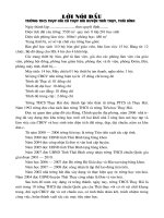
Continuous Fluoro Vs. Pulsed Fluoro
22 cm FOV, 0.2 mm Cu filter
10
ESER (R/min)
1
continuous
15 p/s
7.5 p/s
3 p/s
0.1
0.01
0.001
5
10
15
20
Acrylic Thickness (cm)
9
ESER Reduction With Added Filtration
22 cm FOV, continuous fluoroscopy
ESER (R/min)
10
0.0mm Cu
0.1mm Cu
0.2mm Cu
0.3mm Cu
1
0.1
0.01
5
10
15
20
Acrylic Phantom Thickness (cm)
10
ADULT DIAGNOSTIC CORONARY ANGIOGRAPHY
(BASELINE: 16cm FoV, C PLUS, 30pps, GRID, 25cm PMMA)
PERCENT RADIATION DOSE (%)
100
C PLUS, 16cm Fov,
30pps
90
C+ --> C NORMAL
80
30pps --> 15pps
70
60
16cm FoV --> 25cm
FoV
50
C- FLUORO & C
NORMAL RECORD
40
30
SID & SSD
OPTIMIZED
20
AUTO
COLLIMATION
10
AUTO POSITION
0
SELECTABLE VARIABLES
!"
#
#
$%
11
Factors Affecting CTDI
•
•
•
•
•
X-RAY BEAM ENERGY (KVP): higher kVp
results in higher CTDI values.
X-RAY TUBE CURRENT (mA): dose is
proportional to mAs.
TUBE ROTATION TIME: dose is
proportional to mAs.
PITCH: inversely proportional to dose.
X-RAY BEAM COLLIMATION: thinner
collimation results in higher CTDI values.
Factors Affecting CTDI
(…continued)
•
•
•
•
•
•
PATIENT SIZE: smaller patient size results
in higher CTDI values.
DOSE REDUCTION TECHNIQUE, i.e., mA
modulation technique
DETECTOR CONFIGURATION
SLICE THICKNESS
ADDED FILTRATION
GEOMETRIC EFFICIENCY
12
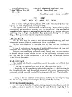
Take a guess
Measured CTDIvol (mGy/100mAs)
If the body size is reduced from 32 cm in
diameter to 16 cm in diameter, the CTDI
will be
.
• A. the same
• B. increased by 50%
• C. doubled
• D. more than doubled
40
35
30
25
20
15
10
5
0
10
15
20
25
30
Body Phantom Diameter (cm)
measured CTDIvol at 80 kVp
measured CTDIvol at 100 kVp
measured CTDIvol at 120 kVp
measured CTDIvol at 140 kVp
13
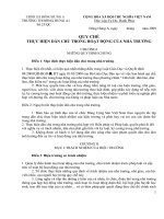
Solid State Integrating Dosimeter
COMPARISON OF BODY CT RADIATION DOSE PER
100 mAs vs. WEIGHT
18
16
RADIATION DOSE
(mGy / 100 mAs)
14
12
10
8
6
4
2
0
0
50
100
150
200
250
PATIENT WEIGHT (lbs.)
CTvol / 100 mAs
Linear (MEAS. / 100 mAs)
MEAS. / 100 mAs
Linear (CTvol / 100 mAs)
14
OUTLINE
Part I
3. PRACTICAL METHODS FOR
ESTIMATING PATIENT RADIATION
DOSES WITH REFERENCES
Phantoms
• Acrylic phantoms
• Anthropomorphic
phantoms:
• Mathematical phantoms:
Reference Man
15
!
"
#
&'
-.
1
2#
&
'
$#
%
( )! *
/!
0
) 3
$
)+ ) ,
! %
# ( $
E = WT WR DT
T
('
# ( $
Limitations of Tabular Conversion Factors
• The reference person (male 154lb, female
128lb) has a fixed size.
• The number of exam types is limited.
• The number of exam settings is limited:
field size, SID, etc.
• The number of organ types is limited.
• The data were based upon cancer
detriment index published earlier (need
updated).
16
&
'
%
")
- &4!
)4
,' "
!),
# 0 4!4 ') ' ,, )
)
,!
)) )
'
)!
0
,
)!
0
!
- -)!
#
0 5#0
- /. !
#
!
60 ,
7!
.0
8# )
,
00 . 9 ! ) :
*
) "+
,
,
(
, ),)",
! "+
./00
)
1)
Steps for Tissue Dose Estimation
!
"
%,
234
)
) 5
-*
)
) 6 #3"
$
) "
5
-1
"
!
"
# $
-4
" )) 7
"
(
-
17
! "+
1)
3.5
Lung
549
18
&
'
%
*
"
.//
*
) "+
,
,
(
, ),)",
)
ACR CT ACCREDITATION FORM
Section 11 - Radiation Dosimetry ( Adult Body)
Use the TAB key to move between data entry cells in the column named Measured .
CTDI Body Phantom (32-cm diameter PMMA Phantom)
Measured
kVp
120
mA
480
Exposure time per rotation (s)
Z axis collimation T (mm)
0.5
1
# data channels used (N)
3
1
6
1
Axial (A): Table Increment (mm) = (I)
OR
1
Helical (H):Table Speed (mm/rot) = (I)
Active Chamber length (mm)
100
Chamber correction factor
1.98
24
19
CTDIvol and DLP
CTDI vol =
) " )
"
7
7
)
1
CTDI w
pitch
)
7
(
7
!
8
DLP = CTDI vol× scan length
Effective Dose in CT
• European Guidelines on Quality Criteria for CT
( />
Region of body
Head
Normalized Effective Dose
(mSv/mGy-cm)
0.0023
Neck
0.0054
Chest
0.017
Abdomen
0.015
Pelvis
0.019
20
Software Resourses
Software programs to calculate organ
dose using Monte Carlo Techniques:
• www.hpa.org.uk (NRPB):
XDOSE, CHILDOSE, CTDOSE
• www.vamp-gmbh.de (company for CT ):
ImpactDose
PATIENT RADIATION
DOSES IN DIAGNOSTIC
RADIOLOGY… part 2
EDWARD L. NICKOLOFF, D.Sc.
DEPARTMENT OF RADIOLOGY
COLUMBIA UNIVERSITY &
NEW YORK-PRESBYTERIAN HOSPTIAL
NEW YORK, NY
21
TYPICAL PATIENT
RADIATION DOSES
NATIONAL EVALUATION OF X-RAY
TRENDS (N.E.X.T.) SURVEY 1990-2002
EXAMINATION
& PROJECTION
1st
QUARTILE
MEDIAN
(mGy)
3RD
QUARTILE
(mGy)
0.11
2.4
2.8
48.7
3.30
58
15
1.4
0.46
3.4
4.2
69.8
4.83
75
19
1.9
(mGy)
CHEST PA
ABDOMEN AP
LS SPINE AP
GI FLUORO / min
GI SPOT (1)
CTDIvol HEAD
CTDIvol BODY
MAMMO
.08
1.7
2.0
33.9
2.21
43
11
1.0
* FROM: WWW.CRCPD.ORG WEBSITE & ACR MAMMO
22
DIAGNOSTIC RADIOLOGY DOSE
REFERENCE LEVELS (DRL)
DIAGNOSTIC REFERENCE LEVELS
• VOLUNTARY FOR COMPARISON
– BASED UPON NATIONWIDE SURVEYS
– NOT FOR REGULATORY PURPOSES
• GUIDANCE LEVEL FOR INVESTIGATION…. IF ABOVE
– MAY BE APPROPRIATE BECAUSE OF PATIENT
SIZE OR CLINICAL COMPLEXITY
– MAY BE SUBOPTIMAL USAGE OF EQUIPMENT
– MAY BE EQUIPMENT PROBLEMS
• TYPICALLY REFERENCE LEVEL IS THIRD QUARTILE
OR ABOUT 80% OF SURVEY
– MEAN + 0. 70
→ 75 %
– MEAN + 1.00
→ 84 %
• DIRECTED TOWARDS RADIATION DOSE REDUCTION
23
ACR / AAPM REFERENCE VALUES
FOR ADULTS
EXAMINATION
& PROJECTION
REFERENCE VALUE
( mGy / IMAGE )
CHEST PA
ABDOMEN AP
0.25
4.50
LS SPINE AP
CERVICAL SPINE AP
GI FLUORO / min
CTDIc HEAD
CTDIp BODY
MAMMO
5.00
1.25
65.0
60.0
40.0
3.00 (MQSA)
ACRIN MAMMOGRAPHY DATA
PARAMETER
SCREEN-FILM
FFDM
MEAN THICK.
5.29 cm
5.28 cm
1.37 cm
1.45 cm
2.37 mGy
1.88 mGy
0.99 mGy
0.68 mGy
3.36 mGy
2.56 mGy
1
THICK.
MEAN DOSE
1
DOSE
MEAN + 1
DOSE
From Drs. Eric Berns & Ed Hendrick at Northwesterm Univ.
24
UK DIAGNOSTIC REFEENCE LEVELS 2000
EXAMINATION
& PROJECTION
SKULL AP/PA
SKULL LAT
DRL
( mGy / IMAGE )
3.0
1.5
CHEST PA
CHEST LAT
THOR. SPINE AP
THOR. SPINE LAT
LS SPINE AP
LS SPINE LAT
0.2
1.0
3.5
16
6.0
14.0
WEBSITE: www.hpa.org.uk/radiation
UK DIAGNOSTIC REFEENCE LEVELS 2000
EXAMINATION
& PROJECTION
ABDOMEN AP
PELVIS AP
DRL as DAP
(Gy-cm2 )
3.0 (6 mGy/image)
3.0 (4 mGy/image)
BARIUM SWALLOW*
BARIUM MEAL*
BARIUM ENEMA*
RETRO. PYLEO.*
Dx CORONARY
ANGIOGRAPHY*
11
13
31
13
36
* FOR ENTIRE PROCEDURE
25