- Trang chủ >>
- Sư phạm >>
- Sư phạm hóa
Effect of chitosan
Bạn đang xem bản rút gọn của tài liệu. Xem và tải ngay bản đầy đủ của tài liệu tại đây (693.44 KB, 6 trang )
Article
pubs.acs.org/JAFC
Effect of Chitosan, O-Carboxymethyl Chitosan, and N-[(2-Hydroxy-3N,N-dimethylhexadecyl ammonium)propyl] Chitosan Chloride on
Overweight and Insulin Resistance in a Murine Diet-Induced Obesity
Xiaofei Liu,*,†,‡ Xiaona Zhi,†,‡ Yunfei Liu,†,‡ Bo Wu,†,‡ Zhong Sun,§ and Jun Shen∥
†
Department of Polymer Materials Science and Engineering, College of Materials Science and Engineering, and ‡Tianjin Key
Laboratory of Composite and Functional Materials, Tianjin University, Tianjin, 300072, People's Republic of China
§
Department of Health Statistics and ∥Department of Sanitary Chemistry, College of Public Health, Tianjin Medical University,
Tianjin, 300070, People's Republic of China
ABSTRACT: Two water-soluble chitosan derivatives, O-carboxymethyl chitosan (O-CM-chitosan) and N-[(2-hydroxy-3-N,Ndimethylhexadecyl ammonium)propyl] chitosan chloride (N-CQ-chitosan), were prepared, and the therapeutic effects of
chitosan, O-CM-chitosan, and N-CQ-chitosan on insulin resistance were simultaneously evaluated by rats fed on a high-fat diet.
The parameters of high-fat diet-induced rats indicated that chitosan and its two derivatives not only have low cytotoxicity but can
control overnutrition by fat and achieve insulin resistance therapy. However, the results in experiment in vivo showed that the
therapeutic degree varied by the molecular weight and surface charge of chitosan, O-CM-chitosan, and N-CQ-chitosan. N-CQchitosan with a MW of 5 × 10⁴ decreased body weight, the ratio of fat to body weight, triglyceride, fasting plasma glucose, fasting
plasma insulin, free fatty acid, and leptin by 11, 17, 44, 46, 44, 87, and 64% and increased fecal lipid by 95%, respectively.
KEYWORDS: chitosan, derivatives, high-fat rats, body weight, insulin resistance
■
INTRODUCTION
Overweight and obesity have attracted strong attention,1 which
are important contributors to cardiovascular disease,2 type II
diabetes mellitus,3 and several common cancers.4 Type II
diabetes mellitus is a result of the development of insulin
resistance (IR), which is linked to both genetic and
environment factors. Obesity, the most important factor, is
usually a combination of polygenetic and environmental
origins.5 Therefore, restricted calorie intake, weight reduction,
and physical activity can improve insulin sensitivity.6
There are many reports concerned with a role for the plasma
free fatty acid (FFA) elevation in the development of insulin
resistance, which mainly occurred in skeletal muscles and
liver.7−10 Jiao et al.11 have reported that high-fat diet-induced
insulin resistance in Sprague−Dawley rat is closely associated
with the plasma FFA elevation as well as heterotopic deposition
of triglyceride (TG) in liver and skeletal muscle.
Leptin (16 kDa) is the ob gene product and is produced by
adipose tissue. Leptin controls body composition mostly via
hypothalamic receptors that regulate food intake and body
weight.12 However, research shows that the leptin level in
organisms is positively related with body fat content13 and
insulin levels;14 that is, leptin synthesis and secretion are
markedly increased in obesity in both humans and rodents,15
and there has been a high incidence of insulin resistance in
obesity.
Chitosan is the deacetylated form of the polysaccharide
chitin (a byproduct of crustaceans), and it appears to be blind
to negatively charged lipids in animal trials, thus reducing the
animals' gastrointestinal uptake16 and lowering their serum
cholesterol.17 However, because of its poor water solubility, the
applications of chitosan are limited in medicine and the food
© 2012 American Chemical Society
industry. Chemical modifications such as carboxylation and
quanternization have been adopted to overcome the limited
solubility, because of the existence of living amidos and
hydroxys. 18 Therefore, we can get negatively charged
carboxymethylated chitosan and positively charged quanternized chitosan via carboxylation and quaternization.
In recent years, some reported studies have almost
investigated the effect of chitosan on body weight and plasma
lipid level,16,19,20 but its effect on insulin resistance is hardly
known.21 In accordance with obesity is associated with insulin
resistance, we hypothesized that chitosan improves insulin
resistance. In our study, two water-soluble chitosan derivatives,
O-carboxymethyl chitosan (O-CM-chitosan), with a negative
surface charge, and N-[(2-hydroxy-3-N,N-dimethylhexadecyl
ammonium)propyl] chitosan chloride (N-CQ-chitosan), with a
positive surface charge, were prepared, and the therapeutic
effects of chitosan, O-CM-chitosan, and N-CQ-chitosan in
insulin resistance were simultaneously evaluated.
■
CHEMICALS
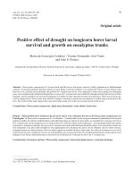
Preparation of O-CM-Chitosan and N-CQ-Chitosan. OCM-chitosan and N-CQ-chitosan were prepared following our
previous report.27 The synthetic process of O-CM-chitosan and
N-CQ-chitosan is shown in Figure 1.
Characterization of Chitosan, O-CM-Chitosan, and NCQ-Chitosan. After it was dried completely at 50 °C, the
samples could be used for Fourier transform infrared (FT-IR)
Received:
Revised:
Accepted:
Published:
3471
September 22, 2011
February 28, 2012
March 8, 2012
March 8, 2012
dx.doi.org/10.1021/jf205226r | J. Agric. Food Chem. 2012, 60, 3471−3476
Journal of Agricultural and Food Chemistry
Article
experiment. The N−H bending (1590 cm−1) of the primary
amine disappeared due to the change of the primary amine in
N-CQ-chitosan. The peak at 1500 cm−1 was assigned to NH3+.
Therefore, it can be concluded that the substitution mainly
occurred at the amino groups of chitosan. All of these results
proved that the synthesis of carboxymethylated and quaternized
chitosan was successful.27
■
MATERIALS AND METHODS
Materials. Chitosan [weight-average molecular weight (MW) of 5
× 104 and 15 × 104, with a degree of deacetylation of 0.85], a
commercial material, was supplied by Qingdao Medicine Institute,
Shandong, China. O-CM-chitosan was prepared in our previous
report,22 and N-CQ-chitosan was prepared from the above raw
chitosan with epoxy chloropropane and N,N-dimethylhexadecyl
amine.23 The degrees of substitution of O-CM-chitosan and N-CQchitosan were 0.72 and 0.41.24,25 All other reagents were analytical
grade provided by No. 3 Chemical Reagent Factory of Tianjin, China.
Solubility of Chitosan, O-CM-Chitosan, and N-CQ-Chitosan.
The solubility of chitosan, O-CM-chitosan, and N-CQ-chitosan was
determined in H2O, acetic acid (HAc), methanol, ethanol, CHCl3,
ether, DMSO, formamide, and DMF.
Cytotoxicity to Hepatocytes with Chitosan and Its Derivatives Analysis. Chitosan and its derivatives were investigated for their
possible cytotoxic effects, and the cytotoxicity for hepatocytes, which
were obtained from male Wistar Rats (Experimental Animal Center of
Tianjin Medical University, Tianjin, China) by collagenase perfusion,
was measured by the MTT assay. Cells were seeded in 96-well plates
at an initial density of 1 × 10⁴ cells/well in 100 μL of growth medium
and incubated overnight. The media were replaced by fresh, serumfree media containing chitosan, O-CM-chitosan, and N-CQ-chitosan
at various MW. Chitosan, O-CM-chitosan, and N-CQ-chitosan were
dissolved in HBBS/HEPES at a concentration of 100 μg/mL. After an
additional incubation for another 24 h, 15 μL of MTT (5 mg/mL)
solution was added into each well and incubated for 4 h and further
dissolved in 150 μL of dimethylsulfoxide (DMSO). All chitosan, OCM-chitosan, and N-CQ-chitosan were UV sterilized. The absorbance
at 490 nm was measured by an enzyme-linked immunosorbent assay
(ELISA) plate reader (Bio-Rad, Microplate Reader). The percentage
cell viability was calculated according to the following equation:
percentage cell viability = OD490(sample)/OD490(control) × 100, where
OD490(sample) represents a measurement from a well treated with
chitosan, O-CM-chitosan, and N-CQ-chitosan and OD490(control)
represents a well treated without any sample.
Animal Experimental Design. Clean male Wistar Rats (n = 120),
with a mean mass of 65 ± 15 g, were provided by the Experimental
Animal Center of Tianjin Medical University, Tianjin, China. All rats
were kept in cages with stainless steel bottoms in a room controlled at
23 ± 1 °C and 55 ± 5% humidity under a 12 h light−dark cycle with
lighting from 8:00 a.m. to 8:00 p.m. Rats were allowed to have free
access to food and water. All animal protocols were approved by the
Institutional Animal Care and Use Committee of Tianjin Medical
University.
After acclimation for 7 days, rats were randomly divided into eight
groups with 15 rats in each group: group C (normal fat control group),
group F (high-fat control group), group TA (chitosan, MW of 5 × 10⁴
and 15 × 104), group TB (O-CM-chitosan, synthesized from chitosan
with MW of 5 × 10⁴ and 15 × 104), and group TC (N-CQ-chitosan,
synthesized from chitosan with MW of 5 × 10⁴ and 15 × 104). The
final concentration of each sample group was 100 mg/L in the mass of
diet. Rats of group C were fed on a commercial diet (provided by
Tianjin Laboratory Animal Co. Ltd., Tianjin, China). Rats of group F
received a high-fat diet containing 10% (w/w) lard, 12% (w/w)
reconstituted skim milk, 10% (w/w) yolk powder, and 7% (w/w)
casein in commercial diets. The sample groups, including groups TA,
TB, and TC, all were fed the same diet as group F but with chitosan,
O-CM-chitosan, and N-CQ-chitosan added, respectively.
During the first 6 weeks of the experimental period, all of the rats
were fed a high-fat diet to establish the high-fat diet-induced model
Figure 1. Chemical structures of chitosan, O-CMCs, and N-CQCs.
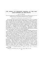
analysis with the standard KBr pellet method. Figure 2 is the
FT-IR spectra of chitosan, O-CM-chitosan, and N-CQ-
Figure 2. FT-IR spectra of (a) chitosan, (b) O-CMCs, and (c) NCQCs.
chitosan. Figure 2a shows the basic characteristics of chitosan
at 3455 (O−H stretch), 2867 (C−H stretch), 1654 (N−H
bend), 1154 (bridge-O stretch), and 1094 cm−1 (C-Ostretch).
The FT-IR spectrum of O-CMCs (Figure 2b) showed
characteristic absorptions due to the −COOH group at 1737
cm−1, confirming a successful carboxymethylation. The
absorption at 1519 cm−1 was assigned to −NH3+. In Figure
2c, the peaks at 2920 and 2870 cm−1 were broadened in N-CQchitosan, as compared with that of chitosan, which were
attributed to the methyl groups and long carbon chain of the
quaternary ammonium salt. Characteristic peaks of the hydroxyl
and second hydroxyl groups of chitosan between 1160 and
1080 cm−1 were unchanged in N-CQ-chitosan, which indicated
that no groups were introduced to the C-3 and C-6 during
3472
dx.doi.org/10.1021/jf205226r | J. Agric. Food Chem. 2012, 60, 3471−3476
Journal of Agricultural and Food Chemistry
Article
Table 1. Solubility of Chitosan, O-CM-Chitosan, and N-CQ-Chitosan in Water and Organic Solventsa
solvent
a
sample
H2O
HAc
methanol
ethanol
CHCl3
ether
DMSO
formamide
DMF
chitosan
O-CM-chitosan
N-CQ-chitosan
−
±
±
++
++
++
±
±
±
±
±
±
−
−
−
−
−
−
−
±
±
−
−
−
−
±
±
++, highly soluble; ±, partially soluble or swollen; and −, insoluble.
except the rats of group C with a commercial diet. Then, in the
following 6 weeks, all sample groups were given corresponding diets.
During the 12 week experimental period, body weight and food intake
were recorded weekly. At the end of the experimental period of
continuous feeding in 12 weeks, rats were deprived of food overnight,
and their blood was collected from the femoral artery puncture under
ether anesthesia. The serum samples were stored in a −20 °C freezer
for further analysis.
Ratio of Fat to Body Weight Analysis. The ratio of fat to body
weight was calculated according to the formula: the ratio of fat to body
weight = (epididymal fat pad weight + perinephrit fat weight)/body
weight (g) × 100.
Fecal Lipid Analysis. During the experimental period, the feces
excreted were collected every day. Fecal lipids were determined
gravimetrically by a modification of the Saxon method.20
Assay of Blood Samples. The TG concentration was determined
using a enzyme colorimetric assay kit (Zhongsheng Beikong Biotech
Co., Ltd., Beijing, China). A glucose oxidase method was employed to
measure fasting plasma glucose (FPG), and a rat insulin ELISA kit
(Jiancheng Biological Engineering Research Institute, Nanjing, China)
was used to measure fasting plasma insulin (FPI). Homeostasis model
assessment for insulin resistance (HOMA-IR) was calculated as an
indicator of insulin resistance according to the formula: HOMA-IR =
FPG (mM) × FPI (mIu/L)/22.5.26 The FFA was detected by using
FFA kit (Jiancheng Biological Engineering Research Institute). Leptins
were determined using a rat leptin ELISA kit (Aditteram Diagnostic
Laboratories, Inc., TX).
Statistical Analysis. The data were expressed as means ± standard
deviations (SDs). One-way analysis of variance was carried out, and
the statistical comparisons among the groups were performed by
Fisher's protected LSD test using a statistical package program to
evaluate whether or not there was a significant difference at p < 0.05.
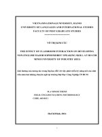
Figure 3. Cell viability of hepatocytes after treatment with chitosan, OCM-chitosan, and N-CQ-chitosan. Data are presented as means ±
SDs, n = 3.
polymer aggregation on cell surfaces impairing the important
membrane functions.32
Effect of Chitosan and Its Derivatives on Overweight.
The effects of chitosan and its derivatives on body weight
content are shown in Table 2. As compared to group F,
chitosan and its derivatives all had a function on decreasing
body weight content. By the administration of chitosan and its
derivatives for 12 weeks, the content in group TC with MW of
5 × 10⁴ was even lower than that in group C. Chitosan and its
derivatives all significantly decreased the body weight. Moreover, chitosan and its derivatives with lower MW had a better
lowering body weight effect.
The effect of chitosan and its derivatives on the ratio of fat to
body weight was also examined, which is a parameter of
evaluating obesity level of the rats. As shown in Table 2, the rat
of fat to body weight in each experimental group was all
decreased to some degree in comparison with group F. As
compared with group F, there are all significant differences in
the ratio of fat to body weight among these groups, and the
most reduction of the ratio of fat to body weight was found in
group TC with MW of 5 × 10⁴.
Then, the content of fecal lipid excretion was examined. The
result indicated that chitosan and its derivatives all increased
the content of fecal lipid excretion as compared with group F
(Table 2). An increase of fecal lipid excretion is a well-known
mechanism for the lipid-lowering effect, and there are all
significant differences in the content of fecal lipid excretion
among these groups. In particular, N-CQ-chitosan with MW of
5 × 10⁴ markedly increased the content of fecal lipid excretion.
Blood Samples Analysis. To investigate the effect of
chitosan and its derivatives on insulin resistance, TG, FPG, FPI,
FFA, and plasma leptin were measured after the end of the
experiment for 12 weeks. With respect to TG (Figure 4), a
remarkable reduction of TG content was found in MW of 5 ×
■
RESULTS
Solubility of Chitosan, O-CM-Chitosan, and N-CQChitosan. The solubility of chitosan and its derivatives was
studied in our previous report.28 The results demonstrated that
O-CM-chitosan and N-CQ-chitosan were well soluble in both
water and organic solvents (Table 1). The lipotropy of the long
carbon chain and the hydrophilicity of the carboxyl group and
quaternary ammonium salt group within the molecule changed
O-CM-chitosan and N-CQ-chitosan into an amphiphilic
polymer.
Cytotoxicity to Hepatocytes with Chitosan and Its
Derivatives Analysis. Viability data of hepatocytes are shown
in Figure 3. Chitosan and its derivatives (O-CM-chitosan and
N-CQ-chitosan) were confirmed to have a lower cytotoxicity to
hepatocytes, which were in agreement with previous
reports.29−31 The cell viabilities in the chitosan and its
derivatives were 87.4 (chitosan with MW of 5 × 10⁴), 80.9
(O-CM-chitosan with MW of 5 × 10⁴), and 83.5% (N-CQchitosan with MW of 5 × 10⁴), at concentration of 100 μg/mL.
The cell viability of the two chitosan derivatives was slightly
lower than that of chitosan, indicating that the introduction of
carboxymethyl and quaternary ammonium salt groups endowed
a slightly high cytotoxicity to hepatocytes. Some reports
showed that cationic polymers had cytotoxicity caused by
3473
dx.doi.org/10.1021/jf205226r | J. Agric. Food Chem. 2012, 60, 3471−3476
Journal of Agricultural and Food Chemistry
Article
Table 2. Lipid-Lowering Effect of Chitosan and Its Derivativesa
body weight (g)
group
group
group
group
group
group
group
group
group
a
2th week
C
F
TA (MW =5 × 10⁴)
TA (MW =15 × 104)
TB (MW =5 × 10⁴)
TB (MW =15 × 104)
TC (MW =5 × 10⁴)
TC (MW =15 × 104)
169.4
181.3
149.4
144.3
167.8
156.3
167.3
166.2
±
±
±
±
±
±
±
±
10.9*
9.4
27.1*
14.4*
10.2*
14.9*
11.9*
22.4*
sixth week
319.1
347.6
338.4
348.2
332.3
345.6
333.6
345.5
±
±
±
±
±
±
±
±
12th week
24.3*
15.7
11.7*
13.8*
25.9*
11.7*
23.6*
16.0*
419.0
460.0
430.7
442.4
419.4
432.6
409.7
427.8
±
±
±
±
±
±
±
±
34.6*
24.7
27.3*
26.9*
29.2*
30.5*
29.3*
25.3*
ratio of fat to body weight (%)
2.685
4.344
3.906
4.038
3.868
3.897
3.621
3.858
±
±
±
±
±
±
±
±
0.18*
0.33
0.11*
0.23*
0.50*
0.34*
0.28*
0.21*
fecal lipid (%)
2.78
4.30
4.91
4.72
5.74
5.04
8.37
7.02
±
±
±
±
±
±
±
±
0.56*
0.42
0.51*
0.64*
0.73*
0.25*
1.58*
0.99*
Data are presented as the mean ± SD, n = 15 in each group, *p < 0.05 vs group F.
It can be concluded that chitosan and its two derivatives had
down-regulated effects on FPG and FPI, and the order of the
improved effects was as follows: N-CQ-chitosan > O-CMchitosan > chitosan. It is probably due to amphiphilicity and
solubility of N-CQ-chitosan and O-CM-chitosan. Meanwhile,
HOMA-IR was calculated (Table 3) according to the formula
as described previously.26 It can be seen that HOMA-IR was
significantly decreased after treatment with chitosan and its two
derivatives for 12 weeks, which achieved the therapeutic result.
Then, the effects on the content of FFA and leptin in the
rats' blood serum were investigated, which were effective
among all sample groups (TA, TB, and TC) from Figures 5 and
Figure 4. Effect of chitosan and its derivatives on TG after treatment
with chitosan, O-CM-chitosan, and N-CQ-chitosan. Data are
presented as the mean ± SD, n = 15 in each group, (a) p < 0.05 vs
group F.
104 (lower MW) groups. Besides, the TG concentration in
serum was significantly decreased when compared with group
F, and the content of TG in each sample group was almost
lower than that in group C except group TA with a MW of 15
× 104.
As shown in Table 3, the levels of FPG and FPI in group F
were significantly higher than those in groups TA, TB, and TC.
Figure 5. Effect of chitosan and its derivatives on the content of FFA
in the rats' blood serum after treatment with chitosan, O-CM-chitosan,
and N-CQ-chitosan. Data are presented as the mean ± SD, n = 15 in
each group, (a) p < 0.05 vs group F.
Table 3. Effect of Chitosan and Its Derivatives on IRa
group
group C
group F
group TA (MW =
5 × 10⁴)
group TA (MW =
15 × 104)
group TB (MW =
5 × 10⁴)
group TB (MW =
15 × 104)
group TC (MW =
5 × 10⁴)
group TC (MW =
15 × 104)
FPG (mM)
FPI (mIu/L)
HOMA-IR
1.74 ± 0.44*
4.70 ± 0.22
3.57 ± 0.54*
6.36 ± 0.43*
9.82 ± 0.69
6.31 ± 0.94*
0.50 ± 0.16*
2.06 ± 0.24
1.02 ± 0.30*
3.62 ± 0.26**
6.47 ± 1.02*
1.05 ± 0.24*
3.35 ± 0.95**
5.54 ± 0.37*
0.84 ± 0.29*
3.48 ± 0.56*
5.78 ± 0.33*
0.91 ± 0.20*
2.55 ± 0.37**
5.46 ± 0.27*
0.62 ± 0.12*
3.14 ± 0.36**
5.51 ± 0.33*
0.78 ± 0.14*
6. When groups TA, TB, and TC are compared with group F,
there are all significant reductions in the content of FFA in the
serum, and the plasma FFA content in each sample group was
all lower than that in group C. It is thus clear that chitosan and
its derivatives have a strong effect on the content of FFA.
Furthermore, each sample group with MW of 5 × 10⁴ markedly
decreased the content of FFA in the plasma.
On the other hand, chitosan and its derivatives were proved
to cause a significant decrease in the content of leptin in the
serum except group TA with MW of 15 × 104 (Figure 6). Also,
N-CQ-chitosan had a better improved effect than O-CMchitosan and chitosan, and it may be due to the quaternary
ammonium cation of N-CQ-chitosan.
Data are presented as the mean ± SD, n = 15 in each group, *p <
0.05 vs group F, and **p < 0.01 vs group F.
a
3474
dx.doi.org/10.1021/jf205226r | J. Agric. Food Chem. 2012, 60, 3471−3476
Journal of Agricultural and Food Chemistry
Article
> chitosan. This is probably because N-CQ-chitosan and OCM-chitosan are amphiphilic polymer and have better solubility
than chitosan, and the positive surface charge of N-CQchitosan made it easy to bind with negtively charge lipid.
In addition to those observations, the levels of FPG, FPI, and
leptin in the rats' blood serum were also significantly decreased.
Treatment with chitosan (MW = 5 × 10⁴), O-CM-chitosan
(MW = 5 × 10⁴), and N-CQ-chitosan (MW = 5 × 10⁴) for 12
weeks resulted in a 24, 29, and 46% decrease in FPG level, a 36,
43, and 44% decrease in FPI, and a 31, 42, and 64% decrease in
plasma leptin, respectively. The order of therapeutic effect is as
follows: N-CQ-chitosan > O-CM-chitosan > chitosan. It may
be due to the amphiphilicity, solubility, and surface charge of
chitosan and its two derivatives. Indeed, leptin is generally
believed to have an insulin-sensitizing effect.14 Thus, the
reduced FPG and leptin were the result of the alleviation of
insulin resistance. Moreover, leptin also contributes to
preventing excess lipid accumulation.15 So, this phenomenon
indicated that chitosan and its derivatives could decrease body
fat through controlling the level of leptin in the serum and
reduce fat toxicity, improve insulin sensitivity,7−10 and then
reduce the incidence of type II diabetes mellitus and some
complications related to obesity.
It has been confirmed in this study that such a low weight
gain was not caused by growth retardation due to any toxicity
of chitosan and its derivatives. With regard to the mechanism, it
is considered that the chitosan and its derivatives dissolved in
the stomach to form an emulsion with intragastric oil droplets
and would begin to precipitate in the small intestine.20
Moreover, chitosan and its derivatives also reduced the levels
of TG, FPG, FPI, FFA, and leptin in the rats' blood serum,
which actually improved insulin resistance, and the different
surface charge and MW of chitosan and its derivatives had
different effects on these parameters. These results imply that a
suitable chitosan and its derivatives intake would be useful to
control overnutrition by fat and to improve insulin resistance.
Figure 6. Effect of chitosan and its derivatives on the content of leptin
in the rats' blood serum after treatment with chitosan, O-CM-chitosan,
and N-CQ-chitosan. Data are presented as the mean ± SD, n = 15 in
each group, (a) p < 0.05 vs group F, and (b) p < 0.01 vs group F.
■
DISCUSSION
The result of the present investigation made clear that chitosan
had the potential ability to decrease body weight. Some studies
have demonstrated that chitosan could bind to negatively
charged lipids, thus reducing their gastrointestinal uptake.16,20
Nauss et al.33 have suggested that a soluble form of chitosan
would be able to interfere with intraluminal lipid absorption
through the interaction with micelle formation or emulsification
of lipids in the enteric phase. On the other hand, it has also
been suggested that the effect of chitosan on body weight is
substantially less and unlikely to be of clinical significance.19 It
is therefore noteworthy to find that chitosan is an effective
treatment for overweight and obesity.
According to our experiment in vivo, groups with chitosan
and its derivatives had different inhibiting effects on body
weight of the diet-induced rats. Furthermore, the lipid-lowering
effect of N-CQ-chitosan with MW of 5 × 10⁴ was better. As
compared with the high-fat control group, there was a
reduction in body weight by 11%, in the rate of fat to body
weight by 17%, and it elevated fecal lipid excretion by 95%.
Therefore, we may consider that feeding chitosan and its
derivatives resulted in down-regulated effects on lipid and
improved obesity, which was in accordance with the above
studies.16,20 The phenomenon that chitosans form highly
viscous solutions in dilute acids may cause distension of the
duodenum in animals and thereby increase satiety.34 The effect
could account for the lower body weights observed for rats fed
on chitosan and its derivatives in the present experiment in
vivo.
Moreover, it was found that a lower MW of chitosan and its
derivatives had a better therapeutic effect from all our data,
which is probably because lower MW of them were absorbed
by rats easily.35 Chitosan (MW of 5 × 10⁴) reduced TG and
FFA by 28 and 19%, O-CM-chitosan (MW of 5 × 10⁴)
decreased TG and FFA by 36 and 61%, and N-CQ-chitosan
(MW of 5 × 10⁴) reduced TG and FFA by 44 and 87%,
respectively. It was probably that lipid was probably bound to
chitosan and its derivatives and excreted with feces, which was
in agreement with our above analysis. The order of lipidlowering in vivo is as follows: N-CQ-chitosan > O-CM-chitosan
■
AUTHOR INFORMATION
Corresponding Author
*Tel: +86 22 2740 8099. Fax: +86 22 2740 4724. E-mail:
Funding
The work was financially supported by National Natural
Science Foundation of China.
Notes
The authors declare no competing financial interest.
■
REFERENCES
(1) Flegal, K. M.; Carroll, M. D.; Ogden, L. G.; Johnson, C. L.
Prevalence and trends in obesity among US adults, 1999−2008. J. Am.
Med. Assoc. 2010, 303, 235−241.
(2) Whitlock, G.; Lewington, S.; Ni, M. C. Coronary heart disease
and body mass index: A systematic review of the evidence from large
prospective cohort studies. Semin. Vasc. Med. 2002, 2, 369−381.
(3) Carey, V. J.; Walters, E. E.; Colditz, G. A.; Solomon, C. G.;
Willett, W. C.; Rosner, B. A.; Speizer, F. E.; Manson, J. E. Body fat
distribution and risk of non-insulin-dependent diabetes mellitus in
women. Am. J. Epidemiol. 1997, 145, 614−619.
(4) Calle, E. E.; Rodriguez, C.; Walker-Thurmond, K.; Thun, M. J.
Overweight, obesity, and mortality from cancer in a prospectively
studied cohort of U.S. adults. N. Engl. J. Med. 2003, 348, 1625−1638.
(5) Cummings, D. E.; Schwartz, M. W. Genetics and pathophysiology of human obesity. Annu. Rev. Med. 2003, 54, 453−471.
3475
dx.doi.org/10.1021/jf205226r | J. Agric. Food Chem. 2012, 60, 3471−3476
Journal of Agricultural and Food Chemistry
Article
(26) Iwai, H.; Ohno, Y.; Aoki, N. The effect of leptin, tumor necrosis
factor-α (TNF-α), and nitric oxide (NO) production on insulin
resistance in otsuka long-evans fatty rats. Endocr. J. 2003, 50, 673−680.
(27) Liu, X. F.; Yang, F.; Song, T.; Zeng, A. R.; Wang, Q.; Sun, Z.;
Shen, J. Synthesis of carboxymethylated and quaternized chitosans and
their therapeutic effect on nonalcoholic fatty liver disease. J. Agric. Food
Chem. 2011, 59, 10683−10692.
(28) Liu, X. F.; Yang, F.; Song, T.; Zeng, A. R.; Wang, Q.; Sun, Z.;
Shen, J. Therapeutic effect of carboxymethylated and quanternized
chitosan on insulin resistance in high-fat-diet-induced rats and 3T3-L1
adipocytes. J. Biomater. Sci. 2011, 1−14, DOI: 10.1163/
092050611X579771.
(29) Thanou, M.; Florea, B. I.; Geldof, M.; Junginger, H. E.;
Borchard, G. Quaternized chitosan oligomers as novel gene delivery
vectors in epithelial cell lines. Biomaterials 2007, 23, 153−159.
(30) Anitha, A.; Rani, V. V. D.; Krishna, R.; Sreeja, V.; Selvamurugan,
N.; Nair, S. V.; Tamura, H.; Jayakumar, R. Synthesis, characterization,
cytotoxicity and antibacterial studies of chitosan,O-carboxymethyl and
N,O-carboxymethyl chitosan nanoparticles. Carbohydr. Polym. 2009,
78, 672−677.
(31) Zhang, Y. F.; Yin, P.; Zhao, X. Q.; Wang, J.; Wang, J.; Wang, C.
D.; Ren, L.; Zhang, Q. Q. O-Carboxymethyl-chitosan/organosilica
hybrid nanoparticles as non-viral vectors for gene delivery. Mater. Sci.
Eng. 2009, 29, 2045−2049.
(32) Wong, K.; Sun, G.; Zhang, X.; Dai, H.; Liu, Y.; He, C.; Leong, K.
W. PEI-g-chitosan, a novel gene delivery system with transfection
efficiency comparable to polyethylenimine in vitro and after Liver
Administration in vivo. Bioconjugate Chem. 2009, 17, 152−158.
(33) Mhurchi, C. N.; Dunshea-Mooji, C.; Bennrtt, D.; Rodegers, A.
Effect of chitosan on weight loss in overweight and obese individuals: a
systematic review of randomized of controlled trials. Obes. Rev. 2005,
6, 35−42.
(34) Sugano, M.; Watanabe, S.; Kishi, A.; Izume, M.; Ohtakara, A.
Hypocholesterolemic action of chitosans with different viscosity in
rats. Lipids 1988, 23, 187−191.
(35) Liu, X. F.; Zeng, A. R.; Li, L.; Yang, F.; Wang, Q.; Sun, Z.; Shen,
J. Synthesis of a novel amphiphilic quaternized chitosan and its
distribution in rats. J. Biomater. Sci. 2011, 22, 1115−1130.
(6) Greenfield, J. R.; Campbell, L. V. Insulin resistance and obesity.
Clin. Dermatol. 2004, 22, 289−295.
(7) Itani, S. I.; Ruderman, N. B.; Schmieder, F.; Boden, G. Lipidinduced insulin resistance in human muscle is associated with changes
in diacylglycerol, protein kinase C, and I kappa B-alpha. Diabetes 2002,
51, 2005−2011.
(8) Lam, T. K.; Yoshii, H.; Haber, C. A.; Bogdanovic, E.; Lam, L.;
Fantus, I. G. Free fatty acid-induced hepatic insulin resistance: a
potential role for protein kinase C-delta. Am. J. Physiol. Endocrinol.
Metab. 2002, 283, 682−691.
(9) Santomauro, A. T.; Boden, G.; Silva, M. E.; Rocha, D. M.; Santos,
R. F.; Ursich, M. J. Overnight lowering of free fatty acids with
Acipimox improves insulin resistance and glucose tolerance in obese
diabetic and nondiabetic subjects. Diabetes 1999, 48, 1836−1841.
(10) Boden, G.; She, P.; Mozzoli, M.; Cheung, P.; Gumireddy, K.;
Reddy, P.; Xiang, X. Q.; Luo, Z. J.; Ruderman, N. Free fatty acids
produce insulin resistance and activate the proinflammatory nuclear
factor-kappa B pathway in rat liver. Diabetes 2005, 54, 3458−3465.
(11) Jiao, K.; Liu, H.; Chen, J. K.; Tian, D. M.; Hou, J. F.; Kaye, A. D.
Roles of plasma interleukin-6 and tumor necrosis factor-α and FFA
and TG in the development of insulin resistance induced by high-fat
diet. Cytokine 2008, 42, 161−169.
(12) Temelkova-Kurktschiev, T. S.; Koehler, C.; Henkel, E.;
Leonhardt, W.; Fuecker, K.; Hanefeld, M. Postchallenge plasma
glucose and glycemic spikes are more strongly associated with
atherosclerosis than fasting glucose or HbA1c level. Diaberes Care
2000, 23, 1830−1834.
(13) Nagi, D. K.; Bennett, P. H.; Knowler, W. C. Intact
proinsulin,des 31,32 proinsulin, and specific insulin concentrations
among nondiabetic and diabetic subjects in populations at varying risk
of type 2 diabetes. Diabetes Care 1998, 21, 127−133.
(14) Takekoshi, K. Leptin directly stimulates catecholamine secretion
and synthesis in cultured porcine adrenal medullary chromaffin cells.
Biochem. Biophys. Res. Commun. 1999, 261, 426−431.
(15) Campbell, F. M.; Gordon, M. J.; Hoggard, N.; Dutta-Roy, A. K.
Interaction of Free Fatty Acids with Human Leptin. Diabetes 1998,
247, 654−658.
(16) Deuchi, K.; Kanauchi, O.; Imasato, Y.; Kobayashi, E. Effect of
the viscosity or deacetylation degree of chitosan on fecal fat excreted
from rats fed on a high fat diet. Biosci., Biotechnol., Biochem. 1995, 59,
781−785.
(17) Ormrod, D. J.; Holmes, C. C.; Miller, T. E. Dietary chitosan
inhibits hypercholesterolaemia and atherogenesis in the apolipoprotein
deficient mouse model of atherosclerosis. Atherosclerosis 1998, 138,
329−334.
(18) Mohamed, E. B. Chemical modification of chitosan: synthesis
and biological activity of new heterocyclic chitosan derivatives. Polym.
Int. 2008, 57, 254−261.
(19) Nauss, J. L.; Thompson, J. L.; Nagyvary, J. The binding of
micellar lipids to chitosan. Lipids 1983, 18, 714−719.
(20) Flourie, B.; Vidon, N.; Florent, C. H.; Bernier, J. J. Effect of
pectin on jejunal glucose absorption and unstirred layer thickness in
normal man. Gut 1984, 25, 936−941.
(21) Hernandez-Gonzales, S. O.; Gonzalez-OOrtiz, M.; MartinezAbundis, E.; Robles-Cervantes, J. A. Chitosan improves insulin
sensitivity as determined by the euglycemic-hyperinsulinemic clamp
technique in obese subjects. Nutr. Res. (N.Y.) 2010, 30, 392−395.
(22) Liu, X. F.; Song, L.; Li, L. Antibacterial effects of chitosan and its
water-soluble derivatives on E. coli, plasmids DNA and mRNA. J. Appl.
Polym. Sci. 2007, 103, 3521−3528.
(23) Guo, Z. Y.; Xing, R.; Liu, S.; Zhong, Z.; Ji, X.; Wang, L.; Li, P. C.
The influence of molecular weight of quaternized chitosan on
antifungal activity. Carbohydr. Polym. 2007, 71, 694−697.
(24) Sang-Hoon, L.; Samuel, M. H. Synthesis and antimicrobial
activity of a water-soluble chitosan derivative with a fiber-reactive
group. Carbohydr. Res. 2004, 339, 313−319.
(25) Sun, S. L.; Wang, A. Q. Adsorption kinetic chitosan of Cu(II)
ions using N,O-carboxymethyl-chitosan. J. Hazard. Mater. 2006, 131,
103−111.
3476
dx.doi.org/10.1021/jf205226r | J. Agric. Food Chem. 2012, 60, 3471−3476