PDF Immunology of Pregnancy PDF Download (Medical Intelligence Unit) by Gil Mor (Editor)
Bạn đang xem bản rút gọn của tài liệu. Xem và tải ngay bản đầy đủ của tài liệu tại đây (22.54 MB, 338 trang )
MEDICAL
INTELLIGENCE
UNIT
Immunology
of Pregnancy
Gil Mor, M.D., Ph.D.
Department of Obstetrics and Gynecology
Reproductive Immunology Unit
Yale University School of Medicine
New Haven, Connecticut, U.S.A.
L A N D E S B I O S C I E N C E / EUREKAH.COM
GEORGETOWN, TEXAS
U.S.A.
S P R I N G E R SCIENCE+BUSINESS M E D I A
NEW YORK, NEW YORK
U.S.A.
IMMUNOLOGY OF PREGNANCY
Medical Intelligence Unit
Landes Bioscience / Eurekah.com
Springer Science+Business Media, Inc.
ISBN: 0-387-30612-9
Printed on acid-free paper.
Copyright ©2006 Eurekah.com and Springer Science+Business Media, Inc.
All rights reserved. This work may not be translated or copied in whole or in part without the written
permission of the publisher, except for brief excerpts in connection with reviews or scholarly analysis. Use in
connection with any form of information storage and retrieval, electronic adaptation, computer software, or
by similar or dissimilar methodology now known or hereafter developed is forbidden.
The use in the publication of trade names, trademarks, service marks and similar terms even if they are not
identified as such, is not to be taken as an expression of opinion as to whether or not they are subject to
proprietary rights.
While the authors, editors and publisher believe that drug selection and dosage and the specifications and
usage of equipment and devices, as set forth in this book, are in accord with current reccrmmendations and
practice at the time of publication, they make no warranty, expressed or implied, with respect to material
described in this book. In view of the ongoing research, equipment development, changes in governmental
regulations and the rapid accumulation of information relating to the biomedical sciences, the reader is urged to
carefully review and evaluate the information provided herein.
Springer Science+Business Media, Inc., 233 Spring Street, New York, New York 10013, U.S.A.
Please address all inquiries to the Publishers:
Landes Bioscience / Eurekah.com, 810 South Church Street, Georgetown, Texas 78626, U.S.A.
Phone: 512/ 863 7762; FAX: 512/ 863 0081
Printed in the United States of America.
9 8 7 6 5 4 3 2 1
Library of Congress Cataloging-in-Publication Data
Immunology of pregnancy / [edited by] Gil Mor.
p. ; cm. ~ (Medical intelligence unit)
Includes bibliographical references and index.
ISBN 0-387-30612-9 (alk. paper)
1. Pregnancy—Immunological aspects.
[DNLM: 1. Pregnancy-immunology. 2. Immunity-Pregnancy. W Q 200 13272 2006] I. Mor, Gil. II.
Title. Ill, Series: Medical intelligence unit (Unnumbered : 2003)
RG557.I48 2006
6l8.2'079-dc22
2005030784
To my wife Anette for her unconditional love and support
CONTENTS
Preface
Immunology of Implantation: An Introduction
GilMor
Pregnancy Represents an Allograft
General Concepts of Immunology
Maternal Immune Response to the Trophoblast
The Role of the Innate Immune System in Pregnancy
Apoptosis and Implantation
1. Evolution of the Mammalian Reproductive Tract and Placentation
Susan Richman and Frederick Naftolin
Mammalian Reproduction
Secondary Use of Immune Mechanisms for Reproduction
The Role of the Endometrial Cycle
Placentas and Placentation
Maternal-Fetal Immune Function
Placental Contribution and Graft Tolerance
2. Toil-Like Receptors and Pregnancy
Vikki M. Abrahams and GilMor
Infections and the Innate Immune
Toll-Like Receptors
Toll-Like Receptor Expression
Toll-Like Receptors and Pregnancy
Toll-Like Receptor Signaling
Toll-Like Receptor Signaling in Trophoblast Cells
Toll-Like Receptors and Apoptosis
Infection, Toll-Like Receptors and Pregnancy Complications
1
1
2
2
5
5
7
7
8
9
10
11
12
15
15
16
16
17
18
18
19
20
3. IL-10 and Pregnancy
Shaun P. Murphy and Surendra Sharma
IL-10 Gene, Protein, and Expression
IL-10 Receptor and Signaling
Pregnancy Pathologies Associated with Abnormal IL-10 Expression....
26
4. Thl/Th2 Balance of the Implantation Site in Humans
Shigeru Saito, Satomi Miyazaki and Yasushi Sasaki
T Cells Change the Implantation Window and Promote Embryo
Implantation in Mice
Immunocompetent Cells in Human Endometrium
and Early Pregnant Decidua
T h l / T h 2 Balance in Normal Human Pregnancy
T h l / T h 2 Balance in Sporadic Abortion or Unexplained
Recurrent Spontaneous Abortion
Regulatory T Cells in Pregnancy
T h l / T h 2 Balance at Implantation Stage
37
26
28
30
37
39
41
43
45
46
5. The Regulation of Human Trophoblast Apoptosis and Survival
during Pregnancy
Shawn L. Straszewski-Chavez and GilMor
Death Receptor-Mediated Apoptosis
The Extrinsic Pathway
The Intrinsic Pathway
The Apoptotic Cascade in Trophoblast Cells
Endogenous Regulators of Trophoblast Apoptosis
Exogenous Regulation of Trophoblast Apoptosis
Trophoblast Apoptosis and Complicated Pregnancies
The Future of Trophoblast Apoptosis
6. Macrophages and Pregnancy
GilMor, Roberto Romero and Vikki M. Abrahams
Apoptosis and Implantation
Role of Apoptotic Cell Phagocytosis
in Pregnancy-Associated Diseases
7. Potential Role of Glucocorticoids in the Pathophysiology
of Intrauterine Growth Restriction (lUGR)
Seth Culler, YuehongMa and Men-Jean Lee
Excess Placental Fibrin and ECM Proteins Are Noted
in Pregnancies with lUGR/PE
Plasminogen Activator Inhibitor (PAI-1):
Role in Fibrin Deposition in Pregnancy
Role of TGF-(3 and Hypoxia on the Expression of PAJ-1
and ECM Proteins
Evidence That Glucocorticoids Stimulate PAI-1 and ECM Protein
Expression in Placenta by Enhancing the Action of TGF-P
8. NK Cells and Pregnancy
Mikael Eriksson, Satarupa Basu and Charles L Sentman
Uterine NK Cells
Recruitment of NK Cells into the Endometrium and Decidua
Function and Regulation of uNK Cells
NK Cells in Reproductive Disorders
49
49
50
51
52
52
56
56
57
63
64
68
73
73
7A
75
75
84
85
86
87
90
9. The Role of Corticotropin-Releasing Hormone (CRH)
on Implantation and Immunotolerance of the Fetus
96
Sophia N. Kalantaridou, Antonis Makrigiannakis, Emmanouil Zoumakis
and Ceorge P. Chrousos
Intrauterine CRH
96
CRH Promotes Blastocyst Implantation
and Early Maternal Tolerance
97
10. Indoleamine 2,3 Dioxygenase-Dependent T Cell Suppression
and Pregnancy
Babak Baban, Phillip R. Chandler and Andrew L. Mellor
Indoleamine 2,3 Dioxygenase (IDO)
IDO-Dependent T-Cell Suppression by Specific Subsets
of Dendritic Cells
IDO Expression at the Maternal-Fetal Interface
Extinction of Paternal IDO Gene Expression
in Trophoblast Giant Cells
IDO-Dependent and IDO-Independent Regulation
of Anti-Fetal T Cell Immunity
11. Leukemia Inhibitory Factor in Reproduction
Levent M. Senturk andAydin Arid
LIF in Endometrium
Potential Role of LIF in Implantation
LIF in the Human Fallopian Tube
LIF in Ovarian Follicle
Clinical Applications of LIF
12. Characterization of Human Dendritic Cells
at the Materno-Fetal Interface
Ulrike Kdmmerer, Lorenz Rieger, Arnd Honig and Eckhard Kdmpgen
Dendritic Cells within the Immune System
Characterization of Human Dendritic Cells
in Endometrium/Decidua
The Functional Role of Decidual Dendritic Cells
13. MHC Molecules of the Preimplantation Embryo
and Trophoblast
Martina Comiskey, Carol M. Warner and Danny J. Schust
Evolution of the M H C
M H C and Reproductive Behavior
M H C Class I in Preimplantation Embryos
Qa-2, The Preimplantation Embryo Development {Fed)
Gene Product
HLA-G Is the Proposed Human Functional Homolog
of Mouse Qa-2
Implantation and M H C Class I in the Trophoblast
14. Actions of Seminal Plasma Cytokines in Priming Female
Reproductive Tract Receptivity for Embryo Implantation
Sarah A. Robertson, John J. Bromfield, Danielle J. Glynn,
David J. Sharkey andMelindaJ. Jasper
Semen Exposure and Pregnancy Outcome
Active Factors in Semen
Consequences of the Post-Mating Inflammatory Response
101
102
102
103
104
105
109
Ill
112
113
114
115
122
122
123
126
130
132
133
134
136
136
138
148
149
149
150
sperm Selection and Clearance of Seminal Debris
Priming the Maternal Immune System to Paternal Antigens
Induction of Maternal Immune Tolerance for Implantation
Contribution to Tissue Remodelling
Activation of Embryotrophic Cytokines
151
152
152
153
154
15. B7 Family Molecules in the Placenta
Margaret G. Petrojf
B7-1 andB7-2
B7-H1 andB7-DC
B7-H2
B7-H3
B7-H4
159
16. The Role of Regulatory T Cells in Materno-Fetal Tolerance
Varuna R. Aluvihare and Alexander G. Betz
Mechanisms Mediating Fetal Immune Evasion
Markers and Characteristics of Regulatory T Cells
Regulatory T Cell Function
Other Cells with Regulatory Function
Regulatory T Cells Mediate Maternal Tolerance to the Fetus
Interaction of Regulatory T Cells with Fetal Immune
Evasion Mechanisms
Implications of Pregnancy-Induced Regulatory T Cell Expansion
171
17. The Eutherian Fetoembryonic Defense System Hypothesis:
An Update
Gary F. Clark, Anne Dell, Howard Morris andManish S. Patankar
In the Beginning: A Model for the Protection of the Gametes
The Extension of Protection to the Developing Eutherian:
Eu-FEDS
Eu-FEDS: The Strong Linkage to Pathogenesis
Mimicry or Acquisition?
AIDS: A Glycobiological Disease Linked to Eu-FEDS?
SIV Infection of Its Natural Hosts:
The "Perfect Eu-FEDS Pathogen"?
Cancer and the Protection of the Developing Eutherian
The Future
18. The Nature and Role of the Decidual T Cells
Lucia Mincheva-Nilsson and Vladimir Baranov
T Cells Are Constitutive Members of the Decidua-Associated
Lymphoid Tissue (DALT)
Characterization of the Decidual T Cells According
to TCR Usage and Phenotype
160
161
164
165
166
171
173
174
175
175
176
176
179
180
183
185
185
187
189
190
190
195
195
196
19. Trophoblast Cells as Immune Regulators
GilMor and Vikki M. Abrahams
Challenging the Medawar Hypothesis
The Trophoblast and Implantation
Cross Talk between the Trophoblast
and the Innate Immune System
TLRs and Pregnancy Complications
215
20. Inherited Thrombophilias and Early Pregnancy Loss
Jens Langhojf-Roos, Michael J. PaidaSy De-Hui Ku, Yale S. Arkel
and Charles J. Lockwood
Pregnancy Related Hemostatic Alterations
Inherited Thrombophilias: Factor V Leiden
Prothrombin Gene Mutation G2010A
Antithrombin Deficiency
Protein C Deficiency
Protein S Deficiency
Protein Z Deficiency
Hyperhomocysteinemia and Methylenetetrahydrofolate Reductase
Thermolabile Mutant Gene Mutation (MTHFR C677T)
Elevated Levels of Type-1 Plasminogen Activator Inhibitor (PAI-1)
and Homozygosity for the 4G/4G Mutation in the PAI-1 Gene ...
Screening for Inherited Thrombophilia Conditions in Patients
with a History of Fetal Loss
Early Pregnancy Loss
Screening Patients for Thrombophilia
Prevention of Adverse Pregnancy Outcome in Patients
with Inherited Thrombophilias
Antenatal Administration of Prophylactic Heparin to Prevent
Recurrent Adverse Pregnancy Outcomes in Women
with Thrombophilia
229
21.
Bi-Directional Cell Trafficking during Pregnancy:
Long-Term Consequences for Human Health
Kristina M. Adams and]. Lee Nelson
Fetal Mc in SSc
How Might Fetal Mc Contribute to Disease Pathogenesis in SSc?
Fetal Mc in Autoimmune Thyroid Disease
Fetal Mc in Other Autoimmune Diseases
Maternal Mc in Autoimmune Disease
How Might Maternal Mc Contribute to Disease Pathogenesis?
Technical and Study Design Considerations
217
218
222
224
229
229
230
230
230
230
231
232
233
233
234
236
237
237
244
245
247
247
248
249
249
250
22. Term and Preterm Parturition
253
Roberto Romero, Jimmy Espinoza, Joaquin Santolaya,
Tinnakom Chaiworapongsa and Moshe Mazor
Normal Duration of Pregnanq^
253
An Overview of Parturition and Labor
254
The Common Pathway of Parturition: Components
256
Increased Uterine Contractility
256
Cervical Ripening
258
Decidual/Fetal Membrane Activation
259
The Role of Prostaglandins
260
A Role for the Fetus in the Timing of the Onset of Labor
261
Possible Routes for the Fetus to Signal the Onset of Labor
261
Parturition as an Inflammatory Process
262
Role of the Placenta
263
Premature Parturition as a Syndrome
263
Intrauterine Infection and Inflammation
264
Frequency of Intrauterine Infection in Spontaneous Preterm Birth ... 265
Intrauterine Infection as a Chronic Process
265
Fetal Involvement
266
Preterm Labor and Preterm PROM as "Adaptive Responses"
266
Gene-Environment Interactions
267
Uteroplacental Ischemia
268
Uterine Overdistension
269
Abnormal Allograft Reaction
269
Allergy-Induced Preterm Labor
270
Cervical Insufficiency
270
Endocrine Disorders
271
Randomized Clinical Trials of Progesterone and Progestins
in Preventing Preterm Delivery
273
23.
Interleukin-1 and Implantation
Jan-S. Kriissel, Jens Hirchenhain, Andrea SchanZy Alexandra P. Hess,
Hong-Yuan Huang, Carlos Simon and Mary Lake Polan
Cytokines and Implantation
Expression of IL-1 in Human Embryos
The Role of IL-1 during Implantation
The IL-1 System as a Regulator of Implantation
24. Immunology and Pregnancy Losses:
HLA, Autoantibodies and Cellular Immunity
Joanne Kwak-Kim, Joon Woo Kim and Alice Gilman-Sachs
Histocompatibility Gene Products and Their Role
in Pregnancy Loss
Autoimmune Responses
Cellular Immune Responses in Pregnancy Loss
Index
294
294
296
298
299
303
303
304
307
317
EDITOR
Gil Mor
Department of Obstetrics and Gynecology
Reproductive Immunology Unit
Yale University School of Medicine
New Haven, Connecticut, U.S.A.
Preface, Chapters 2, 5, 6, 19
CONTRIBUTORS
Vikki M. Abrahams
Department of Obstetrics
and Gynecology
Yale University School of Medicine
New Haven, Connecticut, U.S.A.
Chapters 2, 6, 19
Kristina M. Adams
Division of Clinical Research
Fred Hutchinson Cancer Research
Center
and
Departments of Obstetrics
and Gynecology
University of Washington
Seattle, Washington, U.S.A.
Chapter 21
Yale S. Arkel
The Program for Thrombosis
and Hemostasis in Women's Health
Department of Obstetrics, Gynecology
and Reproductive Sciences
Yale University School of Medicine
New Haven, Connecticut, U.S.A.
Chapter 20
Babak Baban
Program in Molecular Immunology
Institute of Molecular Medicine
and Genetics
Medical College of Georgia
Augusta, Georgia, U.S.A.
Chapter 10
Varuna R. Aluvihare
MRC Laboratory of Molecular Biology
Cambridge, England, U.K.
Chapter 16
Vladimir Baranov
Department of Immunology
University of Umea
Umea, Sweden
Chapter 18
Aydin Arici
Department of Obstetrics
and Gynecology
Yale University School of Medicine
New Haven, Connecticut, U.S.A.
Chapter 11
Satarupa Basu
Department of Microbiology
and Immunology
Dartmouth Medical School
Lebanon, New Hampshire, U.S.A.
Chapter 8
Alexander G. Betz
MRC Laboratory of Molecular Biology
Cambridge, England, U.K.
Chapter 16
John J. Bromfield
Department of Obstetrics
and Gynaecology
University of Adelaide
Adelaide, South Australia, Australia
Chapter 14
Tinnakorn Chairowapongsa
Department of Obstetrics
and Gynecology
, Wayne State University School
of Medicine
Detroit, Michigan, U.S.A.
Chapter 22
Phillip R. Chandler
Program in Molecular Immunology
Institute of Molecular Medicine
and Genetics
Medical College of Georgia
Augusta, Georgia, U.S.A.
Chapter 10
George P. Chrousos
First Department of Pediatrics
School of Medicine
University of Athens
Athens, Greece
and
Pediatric and Reproductive
Endocrinology Branch
National Institute of Child Health
and Human Development
National Institutes of Health
Bethesda, Maryland, U.S.A.
Chapter 9
Gary F. Clark
Department of Physiological Sciences
Eastern Virginia Medical School
Norfolk, Virginia, U.S.A.
Chapter 17
Martina Comiskey
Biology Department
Northeastern University
Boston, Massachusetts, U.S.A.
Chapter 13
Anne Dell
Department of Biological Sciences
Imperial College London
London, U.K.
Chapter 17
Mikael Eriksson
Department of Microbiology
and Immunology
Dartmouth Medical School
Lebanon, New Hampshire, U.S.A.
Chapter 8
Jimmy Espinoza
Department of Obstetrics
and Gynecology
Wayne State University School
of Medicine
Detroit, Michigan, U.S.A.
Chapter 22
Alice Gilman-Sachs
Department of Microbiology
and Immunology
Rosalind Franklin University
of Medicine and Science
North Chicago, Illinois, U.S.A.
Chapter 24
Danielle J. Glynn
Department of Obstetrics
and Gynaecology
University of Adelaide
Adelaide, South Australia, Australia
Chapter 14
Seth GuUer
Department of Obstetrics
and Gynecology and Reproductive
Sciences
Yale University School of Medicine
New Haven, Connecticut, U.S.A.
Chapter 7
Alexandra P. Hess
Department of Obstetrics
and Gynecology
Stanford University Medical Center
Stanford, California, U.S.A.
Chapter 23
Jens Hirchenhain
Department of Obstetrics
and Gynecology, ART/REI-Unit
Heinrich-Heine-University Medical
Center
Dusseldorf, Germany
Chapter 23
Arnd Honig
Department of Obstetrics/Gynecology
University of Wuerzberg
Wuerzburg, Germany
Chapter 12
Hong-Yuan Huang
Department of Obstetrics
and Gynecology
Chang Gung Memorial Hospital
Taipei, Taiwan
Chapter 23
Melinda J. Jasper
Department of Obstetrics
and Gynaecology
University of Adelaide
Adelaide, South Australia, Australia
Chapter 14
Sophia N. Kalantaridou
Division of Reproductive Endocrinology
Department of Obstetrics
and Gynecology
School of Medicine
University of loannina
loannina, Greece
Chapter 9
Ulrike Kammerer
Department of Obstetrics/Gynecology
University of Wuerzberg
Wuerzburg, Germany
Chapter 12
Eckhard Kampgen
Department of Dermatology
University of Wuerzberg
Wuerzburg, Germany
Chapter 12
Joon Woo Kim
Rheumatology Division
Department of Medicine
Feinberg School of Medicine
Northwestern University
Chicago, Illinois, U.S.A.
Chapter 24
Jan-S. Kriissel
Department of Obstetrics
and Gynecology, ART/REI-Unit
Heinrich-Heine-University Medical
Center
Dusseldorf, Germany
Chapter 23
De-Hui Ku
The Program for Thrombosis
and Hemostasis in Women's Health
Department of Obstetrics, Gynecology
and Reproductive Sciences
Yale University School of Medicine
New Haven, Connecticut, U.S.A.
Chapter 20
Joanne Kwak-Kim
Department of Obstetrics
and Gynecology
Department of Microbiology
and Immunology
Rosalind Franklin University
of Medicine and Science
North Chicago, Illinois, U.S.A.
Chapter 24
Jens LanghofF-Roos
The Program for Thrombosis
and Hemostasis in Women's Health
Department of Obstetrics, Gynecology
and Reproductive Sciences
Yale University School of Medicine
New Haven, Connecticut, U.S.A.
Chapter 20
Men-Jean Lee
Department of Obstetrics
and Gynecology
New York University School of Medicine
New York, New York, U.S.A.
Chapter 7
Charles J. Lockwood
The Program for Thrombosis
and Hemostasis in Women's Health
Department of Obstetrics, Gynecology
and Reproductive Sciences
Yale University School of Medicine
New Haven, Connecticut, U.S.A.
Chapter 20
Yuehong Ma
Yale University School of Medicine
Department of Obstetrics
and Gynecology and Reproductive
Sciences
New Haven, Connecticut, U.S.A.
Chapter 7
Antonis Makrigiannakis
Department of Obstetrics
and Gynecology
School of Medicine
University of Crete
Heraklion, Greece
Chapter 9
Moshe Mazor
Department of Obstetrics
and Gynecology
Soroka Medical Center
Beer Sheva, Israel
Chapter 22
Andrew L. Mellor
Program in Molecular Immunology
Institute of Molecular Medicine
and Genetics
Medical College of Georgia
Augusta, Georgia, U.S.A.
Chapter 10
Lucia Mincheva-Nilsson
Department of Clinical Immunology
University of Umea
Umea, Sweden
Chapter 18
Satomi Miyazaki
Department of Obstetrics
and Gynecology
Toyama Medical
and Pharmaceutical University
Sugitani Toyama, Japan
Chapter 4
Howard Morris
Department of Biological Sciences
Imperial College London
London, U.K.
and
M-SCAN Mass Spectrometry Research
and Training Centre
Silwood Park, Ascot, U.K.
Chapter 17
Shaun P. Murphy
Department of Pediatrics
Women and Infants Hospital
Brown University
Providence, Rhode Island, U.S.A.
Chapter 3
Frederick Naftolin
Department of Obstetrics, Gynecology
and Reproductive Sciences
Yale University School of Medicine
New Haven, Connecticut, U.S.A.
Chapter 1
J. Lee Nelson
Division of Clinical Research
Fred Hutchinson Cancer Research
Center
Seattle, Washington, U.S.A.
and
Division of Rheumatology
University of Washington
Seattle, Washington, U.S.A.
Chapter 21
Michael J. Paidas
The Program for Thrombosis
and Hemostasis in Women's Health
Department of Obstetrics, Gynecology
and Reproductive Sciences
Yale University School of Medicine
New Haven, Connecticut, U.S.A.
Chapter 20
Manish S. Patankar
Department of Obstetrics
and Gynecology
Division of Gynecologic Oncology
University of Wisconsin-Madison
Madison, Wisconsin, U.S.A.
Chapter 17
Margaret G. Petroff
Department of Anatomy
and Cell Biology
University of Kansas Medical Center
Kansas City, Kansas, U.S.A.
Chapter 15
Mary Lake Polan
Department of Obstetrics
and Gynecology
Stanford University Medical Center
Stanford, California, U.S.A.
Chapter 23
Susan Richman
Department of Obstetrics, Gynecology
and Reproductive Sciences
Yale University School of Medicine
New Haven, Connecticut, U.S.A.
Chapter 1
Lorenz Rieger
Department of Obstetrics
and Gynecology
University of Wuerzberg
Wuerzburg, Germany
Chapter 12
Sarah A. Robertson
Department of Obstetrics
and Gynaecology
University of Adelaide
Adelaide, South Australia, Australia
Chapter 14
Roberto Romero
Perinatal Research Branch
National Institute of Child Health
and Human Development
National Institutes of Health
Detroit, Michigan, U.S.A.
Chapters 6, 22
Shigeru Saito
Department of Obstetrics
and Gynecology
Toyama Medical
and Pharmaceutical University
Sugitani Toyama, Japan
Chapter 4
Joaquin Santolaya
Department of Obstetrics
and Gynecology
Wayne State University School
of Medicine
Detroit, Michigan, U.S.A.
Chapter 22
Yasushi Sasaki
Department of Obstetrics
and Gynecology
Toyama Medical
and Pharmaceutical University
Sugitani Toyama, Japan
Chapter 4
1
Andrea Schanz
Department of Obstetrics
and Gynecology, ART/REI-Unit
Heinrich-Heine-University Medical
Center
Dusseldorf, Germany
Chapter 23
DannyJ. Schust
Department of Obstetrics
and Gynecology
Boston Medical Center
Boston University
Boston, Massachusetts, U.S.A.
Chapter 13
Charles L. Sentman
Department of Microbiology
and Immunology
Dartmouth Medical School
Lebanon, New Hampshire, U.S.A.
Chapter 8
Levent M. Senturk
Department of Obstetrics
and Gynecology
Division of Reproductive Endocrinology
Istanbul University Cerrahpasa School
of Medicine
Istanbul, Turkey
Chapter 11
David J. Sharkey
Department of Obstetrics
and Gynaecology
University of Adelaide
Adelaide, South Australia, Australia
Chapter 14
Surendra Sharma
Department of Pediatrics
Women and Infants Hospital
Brown University
Providence, Rhode Island, U.S.A.
Chapter 3
Carlos Simon
Department of Obstetrics
and Gynecology
Valencia University Medical Center
and Instituto Valenciano
de Infertilidad
Valencia, Spain
Chapter 23
Shawn L. Straszewski-Chavez
Department of Molecular, Cellular
and Developmental Biology
Yale University
New Haven, Connecticut, U.S.A.
Chapter 5
Carol M. Warner
Biology Department
Northeastern University
Boston, Massachusetts, U.S.A.
Chapter 13
Emmanouil Zoumakis
First Department of Pediatrics
School of Medicine
University of Athens
Athens, Greece
and
Pediatric and Reproductive
Endocrinology Branch
National Institute of Child Health
and Human Development
National Institutes of Health
Bethesda, Maryland, U.S.A.
Chapter 9
PREFACE
Immunology of Implantation:
An Introduction
GilMor
Pregnancy Represents an Allograft
C
ases of recurrent abortions, preeclampsia or babies born with hemolytic diseases of the
newborn still puzzle us with the of the question "Why did your mother reject you?"
Although, after looking at the complexity of the maternal-fetal immune interaction
and the cases of successftil pregnancies, with surprise and admiration the question now becomes: "Why didn't your mother reject you?"
Medawar, in the early 1950s, recognized for the first time the unique immunology of the
maternal-fetal interface and its potential relevance for transplantation. In his original work, he
described the "fetal allograft analogy" where the fetus is viewed as a semiallogeneic conceptus
that evaded rejection. The approaches over the next 50 years have followed the methodology
and development of transplantation immunity or more recently tumor immunity, unveiling
new hypotheses and redefining old concepts.
The objective of this book is to review some of the significant events involved in human
implantation related to the interaction between the maternal immune system and the fetus.
The volume focuses on the main aspects of reproductive immunology, both from basic sciences
and clinical points of view. Although there are still gaps in our knowledge, the advances accomplished in the last five years have proved the importance of understanding the role of the
immune system during pregnancy. This not only represents a fascinating field for research, but
it has the potential for new areas of treatment and diagnosis.
Defining Immunology ofPregnancy
Colbern and Main in 1991 redefined the conceptual framework of reproductive immunology as maternal-placental tolerance instead of maternal-fetal tolerance, focusing the interaction of the maternal immune system on the placenta and not on the fetus. ^ The embryo in
early development divides into two groups of cells, an internal, the inner cell mass, which give
rise to the embryo and an external layer, the embryonic trophoblast that becomes trophoblast
cells and later the placenta. The cells from the placenta are the only part of the fetus to interact
directly with the mother's uterine cells, and therefore the maternal immune system, and are
able to evade immune rejection. The fetus itself has no direct contact with maternal cells.
Moreover, the fetus per se is known to express paternal major histocompatibility complex (MHC)
antigens and is rejected as allograft if removed from its cocoon of trophoblast and transplanted
to the thigh muscle or kidney capsule of the mother.
This book we will focus on the interaction between trophoblast cells and the maternal
immune system.
Immunology ofPregnancy, edited by Gil Mor. ©2006 Eurekah.com
and Springer Science+Business Media.
Immunology ofPregnancy
General Concepts of Immunology
Types of Immune Response
The immune system eliminates foreign material in two ways: natural/innate immunity and
adaptive immunity. Natural immunity produces a relatively unsophisticated response that prevents access of pathogens to the body. This is a primitive evolutionary response that occurs
without the need of prior exposure to similar pathogens. For example, macrophages and granulocytes engulf invading microorganisms at the site of entry. Adaptive immunity is an additional, more sophisticated response found in higher forms such as humans. Cells of the innate
immune system process phagocytosed foreign material and present its antigens to cells of the
adaptive immunity for possible reactions. This immune response is highly specific and normally is potentiated by repeated antigenic encounters.
Adaptive immunity consists of two types of immune responses: humoral immunity, in which
antibodies are produced and, cellular immunity, which involves cell lysis by specialized lymphocytes (cytolytic T cells). Adaptive immunity is characterized by an anamnestic response
that enables the immune cells to 'remember' the foreign antigenic encounter and react to further exposures to the same antigen faster and more vigorously and by the use of cytokines for
communication and regulation of the innate immune response.
Cytokines: Th-l and Th-l Type
Immune cells mediate their effects by releasing cytokines and thus establishing particular
microenvironments. T helper lymphocytes (Th) that originate from the thymus play a major
role in creating a specific microenvironment for a particular organ or tissue. Following an
immune challenge, immune cells produce cytokine, the type of which determines their differentiation into T helper-1 (Th-1) or T-helper 2 (Th-2) lymphocytes. For example, Th-1 lymphocytes secrete interleukin-2 (IL-2) and interferon-y (INF-y) setting the basis for a pro-inflammatory environment. Conversely, the Th-2 lymphocytes secrete cytokines such as IL-4
and IL-10 which are predominately involved in antibody production following an antigenic
challenge. The actions of the two types of lymphocytes are closely intertwined, both acting in
concert and responding to counter regulatory effects of their cytokines. For example Thl
cytokines produce pro-inflammatory cytokine that while acting to reinforce the cytoytic immune response, also down-regulate the production of Th-2 type cytokines.
Each of the different components of the immune system interacts, at different stages and
circumstances, with the trophoblast. Our objective is to understand the type of interaction and
its role in the support of a normal pregnancy.
In the following pages I will summarize some of the main hypotheses proposed to explain
the trophoblast-maternal interaction.
Maternal Immune Response to the Trophoblast
The Pregnant Uterus as an Immune Privileged Site
Implantation is the process by which the blastocyst becomes intimately connected with the
maternal endometrium/decidua. During this period, the semi-allogenic fetus is in direct contact with the maternal uterine and blood-borne cells; however, as I pointed above, fetal rejection by the maternal immune system, in the majority of the cases, is prevented by mechanism(s)
yet undefined. A number of mechanisms have been proposed to account for the
immune-privileged state of the decidua. The different hypothesis can be summarized in five
main ideas: (i) a mechanical barrier effect of the trophoblast, (ii) suppression of the maternal
immune system during pregnancy, (iii) the absence of MHC class I molecules in the trophoblast, (iv) cytokine shift, and more recently (v) local immune suppression mediated by the Fas/
FasL system. I will discuss some of these hypotheses in brief and refer to the chapter where it is
discussed in detail.
Immunology of Implantation: An Introduction
Mechanical Barrier
The concept of mechanical barrier was proposed to explain the lack of immune response in
organs such as the brain, cornea, testicles and kidneys. We refer to these tissues as immune
privileged sites where an immune response represents a dangerous condition for the tissue.
Immune privilege sites are also organs or tissues of the body which, when grafted to conventional (nonprivileged) body sites, experience extended or indefinite survival. Whereas foreign
grafts placed at nonprivileged sites are rejected promptly. The pregnant uterus is an example of
an immune privilege site.
The first reasonable explanation of immune privilege was proposed by Peter Medawar in
the late 1940s.^ Medawar proposed that organs such as the anterior chamber of the eye and the
brain resided behind blood:tissue barriers. The existence of a mechanical barrier, (in the brain
the blood brain barrier [BBB]), prevents the movement of immune cells in and out of the
tissue. This barrier created a state of "immunologic ignorance" in which antigens within were
never detected by the immune system without. The pregnant uterus was proposed to have a
mechanical barrier formed by the trophoblast and the decidua, which prevented the movement
of activated T cells from the periphery to the implantation site. Similarly, this barrier would
isolate the fetus and prevent the escape of fetal cells to the maternal circulation.
Challenging the mechanical barrier effect theory are studies showing that the
trophoblast-decidual interface is less inert or impermeable than first envisioned. Evidence for
traffic in both directions across the maternal-fetus interface includes the migration of maternal
cells into the fetus and the presence of fetal cells in the maternal circulation.
This is the case of almost all the immune privilege tissues, including the brains BBB. Conclusive evidence has shown that immune cells circulate through all parts of the brain, indicating that immune cells are not deterred by mechanical barriers.
The studies described by Adams and Lee Nelson in this book further demonstrate the
bi-directional traffic across the maternal-fetal interface.
Systemic Immune Suppression
The second theory postulates the existence of nonspecific immune suppression during pregnancy. Numerous factors produced and isolated from the maternal placenta interface or from
the serum have been associated with immunosuppressive activity. Some studies have suggested
that human placental lactogen, human placental protein 14, and pregnancy associated plasma
protein-A may have immune-depressant activity on lymphocytes. Soluble suppressor activity
has also been identified in supernatants and cytosol fractions from placental explants and uterine secretions (for review see ref 6). Although all these studies have shown an immunologic
effect, it is important to keep in mind that many of these factors have only been partially
purified and their action has been tested using in vitro assays for lymphocytes or NK cell
activity. These assays are very sensitive to impurities, and upon further purification many of
these factors have lost their "immunosuppressive" effects.
Progesterone has been suggested to have immunosuppressive effects.^ Progesterone, in vitro,
was described to be highly suppressive of mitogen activation and cytotoxic T-cell generation.^
Similarly, progesterone was shown to blunt an inflammatory response in an in vivo rat model.
Other studies have shown that progesterone inhibits cytotoxic and natural killer cell activity as
well as prostaglandin F 2a synthesis. It has also been shown that progesterone activates regulatory T cells of a suppressor phenotype by induction of a 34 kDa protein from lymphocytes.^'^^
The concept of systemic immunosuppressive has been studied by numerous investigators
and for many years became an accepted explanation. Indeed, as described above, a wide array
of materials in human serum have been found to have profound in vitro immunosuppressive
activity. However, from an evolutionary point of view, it is difficult to conceive pregnancy as a
stage of immune suppression. In cultures where a pregnant woman is exposed to poor sanitary
conditions, a suppressed immune system would make fetus survival impossible. Furthermore,
there are recent studies clearly demonstrating that maternal antiviral immunity is not affected
Immunology ofPregnancy
by pregnancy. The obvious observation that HIV+ pregnant women do not suffer from AIDS-like
disease argues against the existence of such nonspecific immune suppression.
Lack ofExpression ofHLA Antigens
The third, more recently postulated theory is based on the fact that polymorphic class I and
II molecules have not been detected on the trophoblast.^^ Dr. Schust's chapter discusses the
subject in greater detail. Major histocompatibility complex (MHC) class I antigens are expressed on the surface of most nucleated cells and serve as important recognition molecules
concerned with vertebrate immune responses. In humans, these antigens are also known as
human leukocyte antigens (HLA). HLA class I genes are located on the same chromosomal
region (6p.21.3). They have been subdivided into two groups, namely the HLA class la and the
HLA class lb genes, according to their polymorphism, tissue distribution and functions. HLA-A,
-B and -C class la genes exhibit a very high level of polymorphism, are almost ubiquitously
expressed among somatic tissue and their immunological functions are well established: they
modulate antiviral and antitumoral immune responses through their interaction with T and
NK cell receptors. In contrast, HLA-E, F and G class lb genes are characterized by their limited
polymorphism and their restricted tissue distribution. Their roles are still poorly understood.
The human placenta does not express HLA-A and HLA-B class I antigens but expresses HLA-G
and HLA-C molecules. ^^ Where are those genes expressed? Dr. Schust's review discusses this
question.
Cytokine Shift
The proliferation, invasion and differentiation of trophoblast cells during implantation is a
tightly controlled process coordinated by a system of intercellular signals mediated by cytokines,
growth factors and hormones. ^^' An extensive array of cytokines is produced at the trophoblast-maternal interface that contributes to the well being of the feto-placental unit. Furthermore, these cytokines to a great extent regulate maternal immune responses, which play an
important role for a successful pregnancy outcome.
It is now recognized that cyokines have extremely diverse biological effects which may involve cell growth, differentiation and function. Their role in regulating human placenta development and implantation has been much discussed in recent years. The field of cytokines
and implantation could be divided in two aspects, one is their role as regulators of the immune
response and second as factors controlling trophoblast cell growth and implantation. This subject is extensively reviewed by Dr. Shigeru Saito, Dr. Surendra Sharma, Dr. Jan-S. Kriissel and
Dr. Aydin Arici.
Local Immune Suppression
The last main hypothesis that we will discuss in this review is the "specific antipaternal
suppressor/regulatory mechanism" observed during pregnancy. The first set of observations
pointing towards the importance of local immune regulation was from Rossant and colleagues.
Their observations were done using the Mus musculusiMus caroli system (for more details in the
model see ref 16). They have shown that the transfer of M musculus eggs into M. caroli is
always successful; in contrast, there is almost a constant time schedule for failure of Af. caroli
embryos in the M. musculus uterus. In such a case, cotransferred adjacent M. musculus embryos
do survive, whereas all the M. caroli embryos die from almost the same program. A strong
immune infiltrate consisting of CTL and NK cells is observed around day 9.5. By day 13, the
embryos are all completely reabsorbed. ^'^ It was later shown that M. caroli embryos can survive
until delivery, provided that M. musculus placenta was used. ^^'^^ These results suggested that an
important part of the placenta in M. caroli origin was responsible for provoking death and
resorbtion of Af. musculus trrhryos.
This model was the first to describe these immunologically-mediated abortions and revealed the "immunological" role of the placenta. Furthermore, we consider that one of the
Immunology of Implantation: An Introduction
great merits of this model was to bring to focus the importance of local immunoregulatory
events.
More recently, evidence exists for specific immune suppression directed towards the paternally encoded histocompatibilty antigens. Here, the maternal T cells that recognize paternal
antigens on the trophoblast are selectively abrogated. The role of decidual T cells during pregnancy is discussed by Dr. Lucia Mincheva-Nilsson.
The Role of the Innate Immune System in Pregnancy
During normal pregnancy, several of the cellular components of the innate immune system
are found at the site of implantation. Furthermore, from the first trimester onwards, circulating monocytes, granulocytes and NK cells increase in number and acquire an activated phenotype. This evidence suggests that the innate immune system is not indifferent to the fetus and
may have a role not only in host protection to infections, but also as important players in the
feto-maternal immune adjustment.
Vikki Abrahams, Ulrike Kaemmerer, Ali Ashkar and I discuss the possible roles of cells of
the innate immune system during pregnancy.
Furthermore, Dr. Abrahams' chapter presents evidence supporting the hypothesis that the
trophoblast can function as an immune cell, capable of recognizing and responding to bacterial
antigens.
Apoptosis and Implantation
During implantation, the uterine endometrium undergoes morphological and physiological changes to accommodate the embryo. This process of accommodation implies that the
embryo has to degrade the endometrial extracellular matrix (ECM) to invade the uterus in
species with hemochorial placentation. Apoptosis has been observed in endometrial epithelial
cells at the embryo implantation site, and it is believed to be due to loss of contact with ECM.
Those apoptotic cells are removed either by throphoblast or by maternal macrophages.
Apoptosis marks unwanted cells with "eat me" signals that direct recognition, engulfment
and degradation by phagocytes."^^ This clearance process, far from being the end, represents an
active and coordinated event, which will send specific signals to the remaining cells either for
survival or death."^^ If the wrong message is sent by macrophages to the wrong cell type, it may
have profound consequences for the normal physiology of the tissue.
Dr. Shawn Chavez discusses in detail the regulation of apoptosis in trophoblast cells.
Summary
Important reproductive events, including implantation, trophoblast invasion, placental development and immune protection are regulated by immune cells and their products (cytokines)
produced at the maternal-fetal interface.
The maternal-fetal immune interaction is very complex, and it is difficult to perceive the
whole process based on one mechanism of action. Clearly there are multiple mechanisms of
peripheral and local tolerance induction during pregnancy that prevent fetal rejection while
maintaining a strong and active immune surveillance against viral or bacterial infections, which
may endanger the successful outcome and the survival of the species.
Some of these mechanisms are discussed in this book. In addition the chapters of Drs.
Romero, Lockwood, Kriissel, Kwak-Kim and Richman present a clinical view of the role of the
immune system in normal pregnancy and how its alterations may lead to complications of
pregnancy.
Immunology
of Pregnancy
References
1. Colbern GT, Main EK. Immunology of the maternal-placental interface in normal pregnancy. Semin
Perinatol 1991; 15:196.
2. Weetman AP. The immunology of pregnancy. Thyroid 1999; 9:643.
3. Medawar PB. Immunity to homologous grafted skin. III. The fate of skin homografcs transplanted
to the brain, to subcutaneous tissue, and to the anterior chamber of the eye. Br J Exp Pathol
1948; 29:58.
4. Cserr HP, Knopf PM. Cervical lymphatics, the blood-brain barrier and the immunoreactivity of
the brain: a new view. Immunol Today 1992; 13:507.
5. Streilein J. New Insights into immunologic tolerance. Transplantation Proceedings 1996; 28:2066.
6. Formby B. Immunologic response in pregnancy. Its role in endocrine disorders of pregnancy and
influence on the course of maternal autoimmune diseases. Endocrinol Metab Clin North Am 1995;
24:187.
7. Szekeres-Bartho J, Varga P, Kinsky R et al. Progesterone-mediated immunosuppression and the
maintenance of pregnancy. Res Immunol 1990; 141:175.
8. Szekeres-Bartho J, Szabo J, Kovacs L. Alteration of lymphocyte reactivity in pregnant women treated
with the progesterone receptor inhibitor ZK 98734. Am J Reprod Immunol 1989; 21:46.
9. Szekeres-Bartho J, Reznikoff-Etievant MP, Varga P et al. Lymphocytic progesterone receptors in
normal and pathological human pregnancy. J Reprod Immunol 1989; 16:239.
10. Szekeres-Bartho J, Varga P, Pejtsik B. ELISA test for the detection of an immunological blocking
factor in human pregnancy serum. J Reprod Immunol 1989; 16:19.
11. Kovats S, Main E, Librach C. HLA-G expressed in human trophoblast. Science 1990; 248:220.
12. Schmidt C, Orr H. Maternal/Fetal interactions: The roles of the M H C class I molecule HLA-G.
Crit Rev Immunol 1994; 13:207.
13. Wegmann T G , Guilbert LJ. Immune signaling at the maternal-fetal interface and trophoblast differentiation. Dev Comp Immunol 1992; 16:425.
14. Mellor AL, M u n n D H . Immunology at the maternal-fetal interface: lessons for T cell tolerance
and suppression. Annu Rev Immunol 2000; 18:367.
15. Rice A, Chard T. Cytokines in implantation. Cytokine Growth Factor Rev 1998; 9:287.
16. Chaouat G. Placental infdtration of resorbing CBAxDBA/2 embryos. J Reprod Immunol 1986;
134:1.
17. Croy BA, Rossant J, Clark DA. Recruitment of cytotoxic cells by ectopic grafts of xenogeneic, but
not allogeneic, trophoblast. Transplantation 1984; 37:84.
18. Rossant J, Mauro V, Croy B. Importance of trophoblast genotype for survival of interspecific murine
chimeras. J Embryol Exp Morphol 1982; 69:141.
19. Rossant J, Croy B, Clark D et al. Interspecific hybrids and chimeras in mice. J Exp Zool 1983;
288:223.
20. Savill J, Fadok V. Corpse clearance defines the meaning of cell death. Nature 2000; 407:784.
2 1 . Duvall E, Wyllie AH, Morris RG. Macrophage recognition of cells undergoing programmed cell
death. Immunology 1985; 56:351.
CHAPTER 1
Evolution of the Mammalian Reproductive
Tract and Placentation
Susan Richman and Frederick Naftolin
Abstract
P
hylogenetic analysis suggests that the internalization of reproduction and the development
of hemochorial placentation have been accompanied by conservation of primitive
genitourinary genes. The products include the renin-angiotensin system and the innate
immune system. This explains what might otherwise be considered an ectopic presence of
these systems in the mammalian reproductive tract and the interaction of the allograft: embryo
and maternal host.
Introduction
Evolution is a conservative process; it more often proceeds through utilization of previously
neutral characters than depending upon de novo mutation and selection: novel applications
generally arise via utilization of preexisting adaptive mechanisms. Classical evolutionary methodology uses the fossil record, in conjunction with observations of both extant species and
ethnographic evidence from surviving societies. For example, the length of human gestation
and challenges of delivery such as cephalo-pelvic disproportion appear consequential to the
assumption of an upright posture combined with cranial expansion. At the molecular level,
this is accomplished by complex combinations of gene duplication, exon shuffling, and transposition. For example, the ancient glycoprotein hormone chorionic gonadotropin (CG) acts as
a signal to maternal physiology to begin a series of adaptations to pregnancy. The mammalian
gene for CG s beta subunit arose by duplication of the LH beta subunit gene approximately 94
million years ago from the common ancestor of both eutherian mammals and anthropoid
primates. During that time span, the gene duplication was apparently followed by a frameshift
mutation in the third exon.^ The major difference in CG gene function from its ancestral LH
is in gene expression variants, composition and length of coding region. The translated products differ in the number of sugar chains attached, slowing the clearance of CG molecules from
the maternal bloodstream to 12 hours, from 30 minutes in the case of LH.'^ Analogous changes
occurring in the structure and function of the excretory apparatus have led to the development
of the mammalian reproductive tract and placentation.^
Mammalian Reproduction
The development of sexual reproduction fostered genetic variability, which has hastened
the pace of evolution. The transition from external to internal fertilization shielded reproduction from a hazardous external environment (predators, toxic chemicals, adverse temperature
and pH), which has resulted in the requirement for fewer gametes per successful conception.
Immunology ofPregnancy, edited by Gil Mor. ©2006 Eurekah.com
and Springer Science+Business Media.
Immunology ofPregnancy
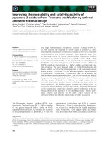
Invagination
^
External environment
..... Original excretory surface
that interfaces with •
Multi-layered animal
with internalization
of external
environment results
part of which
becomes the
reproductive tracts
Figure 1. Development of sexual reproduction: adaptation from external to internal reproduction.
Internal fertilization has been accomplished by the enfolding of excretory and reproductive
function. This adaptation accompanied the development of nonaquatic, terrestrial life forms,
including mammals (Fig. 1).
The higher proportion of live-born young resulting from this system requires a higher investment per oocyte, but furnishes greater overall reproductive success, gene transmission and
speciation. In humans, the allocation of resources that might have been devoted simply to
generation of innumerable eggs for external fertilization has been replaced by the cyclic modification of the reproductive organs, sexual activity, placentation, gestation, parturition and
lactation. All of this developed in the remnants of the ancient excretory tract, w^ith the preservation of many of its mechanisms for interacting v^ith an aquatic external environment.
Secondary Use of Immune Mechanisms for Reproduction
Molecular features of invertebrate immune systems such as the immune effector cells have
been retained in mammals. Three genes found in echinoderms encode highly conserved transcription factors; N F - K B , G A T A - 2 / 3 , and Runt-1, w^hich are rapidly upregulated in response
to bacterial challenges. SRCR family genes structurally resemble the mammalian macrophage
scavenger receptors. Vertebrates added to this successful strategy by:
1. Internalizing mucosal surfaces and increasing their complexity to form the reproductive
tracts—internalized but still aquatic environment.
2. Retaining control over the entirety of embryo development within the female reproductive
tract, allow^ing the young to be born at more advanced stages of development. This, in
combination w^ith maternal supervision and protection, facilitates evasion from predators.
Creating this microenvironment for gametogenesis, fertilization and implantation, was accomplished by the aforementioned "internalizing" of the extracorporeal space within the modern reproductive tract. In the process, ancient nonreproductive systems such as the
macrophage-cytokine system (innate or nonspecific immunity), which had evolved to interface
the genital precursor with the external environment and invading organisms, were modified to
accommodate the embryo. Mucosal immunity at body surfaces via TCR (T cell antigen receptor) Y^ lymphocytes emerged earlier in evolution than TCR a p , perhaps due to primitive
digestive tract exposure to injury and infection in early jawed vertebrates.^ The generation ofT
cells also occurs in gut associated lymphoid tissue, which was the early adaptive immune
Evolution of the Mammalian Reproductive Tract and Placentation
system, while the thymus evolved later, and its ontogeny is from pharyngeal pouch endoderm.
In humans, the third pouch develops into the thymus, while the second develops into the palatine
tonsil. The thymus also utilizes evolutionarily conserved immune-neuroendocrine effectors, as its
mesenchyme develops from neural crest cells. T and B cells, MHC and antibody production
constitute the adaptive or specific portion of the immune system.
Signals from the embryo-host interaction relay the presence of an allograft to the maternal
host, triggering the deployment of processes originally designed to protect against microbial or
environmental challenges.
A later chapter will describe how hormonal regulation of immunocytes prevents rejection of
the allograph embryo; however, the evolutionary relationship between the endometrium and
the embryo is a derivative function of the reproductive tract development.
The Role of the Endometrial Cycle
It is conventional to consider the ovarian and endometrial cycles as the fundamental processes involved in reproductive biology. However, the primary biologic goal is reproduction,
and menstruation is merely the avenue of reestablishing reproductive competence. In an evolutionary sense, each complete menstrual cycle signals a lost opportunity to perpetuate the germ
line.^
The superficial endometrium (flinctionalis) is the nexus of fetal signaling and the adhesion/
implantation mechanism. ^^ In higher primates, this portion of the endometrium will be shed
periodically. This occurs in the absence of signals (hCG, etc.) from the conceptus that drive the
corpus luteums cells to secrete the estrogen and progesterone that decidualize the endometrium
and maintain the embryo until its placenta is able to function independently. The complete
mechanism of menstruation (shedding of the flinctionalis) following ovulation remains unsettled; it appears that this process is triggered by the withdrawal of ovarian steroids from the
expiring corpus luteum that up regulate production of PGF2a.^^ VEGF secreted by the endometrial stromal and epithelial cells plays a role in the remodeling and regeneration from the
basalis layer that follows in the subsequent cycle, providing another opportunity to achieve
pregnancy.
The unique individual that is at the blastocyst stage will invade the receptive endometrium
and become essentially an allograft. This occurs in two steps: adhesion followed by implantation. The yolk sac-placenta provides nourishment until the definitive placenta develops. The
maternal host's reaction to invasion by the embryo includes ancestral innate immune reactions
to foreign proteins, modulated by estrogen, progesterone, and other signals from the maternal
gonad and/or embryo. At this point, immune function is primarily a TH1 response.^^
The human placenta is uniquely aggressive, and capable of invading through the endometrium to the myometrium and beyond, as in the case of placenta accreta/percreta. It is not
yet clear what role this characteristic plays in. Balancing the need for minimally encumbered
respiratory exchange, against the danger of overzealous invasion leading to maternal
exsanguinations or other complications. While the villous cytotrophoblasts are extraordinarily
efficient for this respiratory and nutrient exchange, the invasive extravillous cytotrophoblast
must be limited to invading only the decidua and superficial myometrium. Without this control, the placenta could implant on muscle that would not provide proper nourishment to the
conception and the mother would risk exsanguination from her large pelvic vessels. Potential
controlling autocrine/paracrine mechanisms include glycoproteins, cytokines, and growth factors.^ The proliferative, invasive and migratory activity of the villous cells declines with increasing gestational age, but it has not been established whether this is due to intrinsic cell
programming or extrinsic decidual factors. ^^
Immunoregulatory mechanisms are increasingly seen to be key regulators of this invasive
behavior. In vitro models of the maternal fetal interface involve co-culture of trophoblast and
decidual cell lines on collagen gel matices. Decidual TBF-B and dermatan sulfate proteoglycan