DSpace at VNU: Preparation of Well-aligned CuO Nanorods by Thermal Oxidation Method
Bạn đang xem bản rút gọn của tài liệu. Xem và tải ngay bản đầy đủ của tài liệu tại đây (659.22 KB, 5 trang )
VNU Journal of Science: Mathematics – Physics, Vol. 32, No. 4 (2016) 40-44
Preparation of Well-aligned CuO Nanorods by Thermal
Oxidation Method
Tran Thi Ha1, Bui Thi Huyen2, Nguyen Viet Tuyen2,
1
Hanoi University of Mining and Geology, Duc Thang,Bac Tu Liem, Ha Noi, Vietnam
2
Faculty of Physics, VNU University of Science, 334 Nguyen Trai, Hanoi, Vietnam
Received 15 November 2016
Revised 18 December 2016; Accepted 24 December 2016
Abstract: Well-aligned CuO nanorods were successfully prepared by thermal oxidation method.
The effect of annealing time and annealing temperature on the morphology of the nanorods were
studied by scanning electron microscopy. The results show that annealing temperature plays a
more critical role in affecting the diameter and density of nanorods. Besides SEM images, the
effect of annealing time and temperatures on the structure of the product were also studied by Xray diffraction and Raman spectroscopy. The diameter of CuO nanorods varies from 30 nm to
above 100 nm when annealing temperature changes from 400 °C to 600 °C, while the length of the
rods is up to several tens of micrometers. The most uniform nanorods with highest crystal quality
of CuO were obtained when annealing temperature is 500 °C and annealing time was 2 h as
suggested by SEM images together with Raman results.
Keywords: Cupric, Nanorods, Raman, Thermal oxidation.
1. Introduction
The distinctive characteristics of nanostructures of metal oxide semiconductor have drawn
considerable interest in recent years because of their special properties such as a large surface-tovolume ratio, enhanced activity, unique electronic and optical properties compared to those of bulk
materials [1-3]. The use of metal oxide nanostructures has become promising in solid state chemistry,
because of their controllable properties and structures. Among metal oxides, copper oxide is a narrow
band gap (~1.2 eV in bulk) p-type multifunctional semiconductor which has been recognized as an
industrially important material for various applications [4-7].
The reduction of CuO dimensions to the nanoscale results in significant deviation of some of its
physical properties from its bulk counterpart because of the “quantum-size effects”. Therefore, a
thorough understanding of the fundamental properties of CuO nanostructures is crucial to their
synthesis and applications and a key to the rational design of CuO nanostructure-based functional
devices. CuO nanorods can be applied in many different fields, such as gas sensor, magnetic storage
media, solar-energy transformation, electronics, high sensitivity glucose sensors, super hydrophobic
surfaces [8, 9] or low cost solar cells... The successful preparation of aligned CuO nanorods is
_______
Corresponding author. Tel.: 84-977128393
Email:
40
T.T. Ha et al. / VNU Journal of Science: Mathematics – Physics, Vol. 32, No. 4 (2016) 40-44
41
believed to enrich our understanding of its fundamental properties, which may lead to enhancement of
performance in its applications. In this paper, we report the preparation of uniform and well-aligned
CuO nanorods by thermal oxidation methods. The effect of oxidation temperature and time on
structure and morphology of the nanorods was investigated thoroughly by scanning electron
microscopy, Raman spectroscopy and Xray diffraction measurement.
2. Experiment
High purity copper wire (purity higher than 99% and diameter of about 1 mm) was used as raw
material. The native oxide layer on copper wire was removed by using chemical corrosive. First, Cu
wire was put into diluted acid (HCl 10%) for 1 hour, then rinsed with distilled water. CuO nanorod
was prepared from Cu wire by thermal oxidation process in an electrical furnace XD-1600MT.
Two sets of samples were prepared at different temperatures from 400 to 600 °C in 2 hours or 4
hours to study the influence of annealing temperature and time on the morphology and structure of the
products. The morphologies of the products were investigated by using Nova Nano SEM 450. Raman
spectra of samples were collected on Labram 800 from Horiba with excitation wavelength of 632.8
nm. Acquisition time was fixed at 120s for all samples with power at surface sample of about 0.2 mW.
Xray diffraction measurement was done on Bruker D5005 diffractometer, using the wavelength of
1.54056 Å of CuKα radiation.
3. Results and discussion
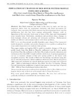
SEM images show that at all annealing temperature from 400 to 600°C, we obtained high density
and well-oriented nanorods (Fig.1). At 400°C, length and width of the obtained nanowires are 3-5 μm,
and 40-60 nm, respectively.
Figure 1. SEM images of CuO nanowires prepared by thermal oxidation in air in 2 h at different temperatures:
(a): 400°C, (b): 450°C, (c): 500°C, (d): 550°C and (e): 600 °C.
42
T.T. Ha et al. / VNU Journal of Science: Mathematics – Physics, Vol. 32, No. 4 (2016) 40-44
When annealing time was fixed at 2 h, higher annealing temperature resulted in thicker and shorter
rods. As annealing temperature increased from 400 to 600 °C, the diameter of nanorods increased
from 30 nm to 200 nm. The maximum length of the nanorods, which was above 20 micrometers, was
obtained at 500 °C and the length of nanorods decreased when annealing temperature was higher or
lower than 500 °C.
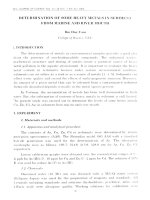
Figure 2. SEM images of CuO nanostructures annealed in 4 h at different temperatures (a): 400 °C, (b): 450 °C,
(c): 500 °C, (d): 550 °C and (e): 600 °C
The same trend was observed for the samples prepared at different temperatures in 4 h (Fig. 2). At
400 °C, we found only rods of low density. In temperature region from 450 to 500 °C, we obtained
well-aligned and uniform rods with diameter of about 100 nm and length of about 3 to 5 µm. At
temperature region from 550°C to 600°C, we obtained short and thick nanowires of 150 -300 nm in
diameter and length of these wires is shorter than 2 micrometers.
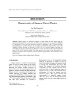
Figure 3. Raman spectra of CuO nanorods prepared in 2 h (a) and in 4 h
(b) in air at different temperatures from 400 to 600 °C.
T.T. Ha et al. / VNU Journal of Science: Mathematics – Physics, Vol. 32, No. 4 (2016) 40-44
43
Group theory shows that CuO, with a monoclinic structure and belonging to the space group C 62h,
has three Raman-active modes (Ag + 2Bg). Typical Raman spectra of the as-prepared nanorods
consist of three characteristic Raman peaks of CuO. The peak at around 288 cm-1 can be assigned to
the Ag mode, and the peaks at around 330 and 621 cm-1 corresponds to the B1g and B2g modes,
respectively [2].
Raman spectra of the CuO synthesized in 2 h and 4 h in air are shown in Fig. 3a and b,
respectively. The evolution of the Raman spectrum with increasing temperature in both cases is quite
similar. The high intensity and sharp Raman peaks confirm that CuO material with good crystalinity
was formed. The spectra contain characteristic peaks of CuO at 289 cm-1, 332 cm-1, and 630 cm-1. The
Ag and B1g Raman peaks of CuO nanorods prepared at high temperatures also shift to longer wave
number with decreasing temperature.
Quantum confinement due to the reduced size to nanoscale of the samples is not likely the reason
for the peak shift because the sample of smallest size, obtained at 500 °C, did not exhibit any shift.
The peak shift observed most clearly in samples prepared at 400 °C for both sets of sample prepared in
2 h or 4 h suggested that the reason might be the phonon confinement on defects formed in the
samples at low annealing temperature.
At low temperature region, we observed another peak at around 200 cm-1, which might be
attributed to vibration of copper sub-oxide (Cu1-xO) lattice. This peak grows as temperature decreases
from 450 to 400 °C. The decrease in intensity of this peak implies that higher annealing temperature is
preferred to get pure CuO nanowires. However, temperature higher than 550 °C results in fast
oxidation reaction, which may reduce the crystal quality of the obtained nanowires. This remark is
illustrated by the broadening of Raman peak of sample prepared at 600 °C. Then, together with SEM
images, Raman spectrum of the samples suggest that product of highest quality could be obtained at
around 500°C. The XRD patterns of the samples prepared at 500°C, in 2 and 4 h, shown in Fig. 4 also
confirm the pure phase of the products.
Figure 4. XRD patterns of CuO nanowires prepared in 2 h and 4 h in air.
All diffraction peaks could be well indexed as those of CuO of monoclinic structures both in
relative intensities and positions. The average size of the CuO nanocrystals is estimated to be 25 nm
and 20 nm for sample according to the Debye-Scherrer formula: D= (0.9 λ)/ (B cosθ), where D is the
particle size, B is the full width at half maximum intensity of the peak and λ is the monochromatic
44
T.T. Ha et al. / VNU Journal of Science: Mathematics – Physics, Vol. 32, No. 4 (2016) 40-44
wavelength of X-ray used in the equipment. The lattice constants are calculated for the two samples
and shown in Table 1. These values are similar to the reported values for CuO materials in literature
[4-6]
Table 1. Lattice parameters of CuO nanowires prepared at 500°C in 2 and 4 h.
Lattice parameters
2h
4h
a (Å)
4.7
4.7
b (Å)
3.4
3.3
c (Å)
5.1
5.1
Β
99°40’
99°20’
4. Conclusion
Well-aligned CuO nanowires were successfully prepared by thermal oxidation methods. By
controlling annealing time, annealing temperature, we could control the density and the aspect ratio of
the as-produced nanorods. When varying temperature from 400 to 600 °C, we obtained nanowires of
diameter ranging from 30 to over 100 nm, and length from 1 µm up to 20 µm with high density. The
nanorods prepared at 500°C have highest crystal quality, uniformity as well as well-orientation. The
successful preparation of CuO nanowire with low-cost thermal oxidation method may lead to the
enhancement of performance in its applications.
Acknowledgements
The research was financially supported by Asia Research Center via project CA.16.02A. The
authors would like to thank Faculty of Physics, VNU University of Science for letting use the
equipments.
References
[1] T.Yu, X.Zhao, Z.X.Shen, Y.H.Su, “Investigation of individual CuO nanorods by polarized micro-Raman
scattering”, Journal of crystal growth (2004) 268 590-595.
[2] Q. Zhang, K. Zhang, D. Xu, “CuO nanostructures: synthesis, characterization, growth mechanisms, fundamental
properties, and applications”, Progress in Materials Science, (2014) 60(1) 208–237.
[3] Xuchuan Jiang, Thurston Hericks,and Younan Xia, “CuO nanowires synthesized by heating copper substrates in
air”, Nano letters (2002) 2(12), 1333-1338.
[4] Aslani and V. Oroojpour, “CO gas sensing of CuO nanostructures, synthesized by an assisted solvothermal wet
chemical route”, Physica B: Condensed Matter (2011) 406(2) 144–149.
[5] M. Yang, J. He, X. Hu, C. Yan, and Z. Cheng, “CuO nanostructures as quartz crystal microbalance sensing layers
for detection of trace hydrogen cyanide gas,” Environmental Science and Technology (2011) 45(14) 6088–6094.
[6] Y. Li, J. Liang, Z. Tao, and J. Chen, “CuO particles and plates: synthesis and gas-sensor application”, Materials
Research Bulletin (2008) 43(8-9) 2380–2385.
[7] X. Wang and X. Xu, “Thermal conductivity of nanoparticle-fluid mixture”, Journal of Thermophysics and Heat
Transfer (1999)13(4) 474–480.
[8] X. Liu, Z. Jiang, J. Li, Z. Zhang, and L. Ren, “Super-hydrophobic property of nano-sized cupric oxide
films”, Surface and Coatings Technology, (2010) 204(20) 3200–3204.
[9] Wei Jiang, Jian He, Feng Xiao,Shaojun Yuan, Houfang Lu, Bin Liang, “ Preparation and antiscaling application
of superhydrophobic anodize CuO nanowire surfaces” (2015) 54 (27) 6874-6883.