DSpace at VNU: Constituents of the Rhizomes of Boesenbergia pandurata and Their Antiausterity Activities against the PANC-1 Human Pancreatic Cancer Line
Bạn đang xem bản rút gọn của tài liệu. Xem và tải ngay bản đầy đủ của tài liệu tại đây (1.59 MB, 8 trang )
Article
pubs.acs.org/jnp
Constituents of the Rhizomes of Boesenbergia pandurata and Their
Antiausterity Activities against the PANC‑1 Human Pancreatic Cancer
Line
Nhan Trung Nguyen,*,† Mai Thanh Thi Nguyen,† Hai Xuan Nguyen,† Phu Hoang Dang,†
Dya Fita Dibwe,§ Hiroyasu Esumi,‡ and Suresh Awale*,§
†
Faculty of Chemistry, University of Science, Vietnam National University, Ho Chi Minh City, 227 Nguyen Van Cu Street, District 5,
Ho Chi Minh City, Vietnam
‡
Research Institute for Biomedical Sciences, Tokyo University of Science, Chiba 278-8510, Japan
§
Division of Natural Drug Discovery, Institute of Natural Medicine, University of Toyama, 2630 Sugitani, Toyama 930-0194, Japan
S Supporting Information
*
ABSTRACT: Human pancreatic cancer cell lines have a
remarkable tolerance to nutrition starvation, which enables them
to survive under a tumor microenvironment. The search for
agents that preferentially inhibit the survival of cancer cells under
low nutrient conditions represents a novel antiausterity strategy in
anticancer drug discovery. In this investigation, a methanol extract
of the rhizomes of Boesenbergia pandurata showed potent
preferential cytotoxicity against PANC-1 human pancreatic cancer
cells under nutrient-deprived conditions, with a PC50 value of 6.6
μg/mL. Phytochemical investigation of this extract led to the
isolation of 15 compounds, including eight new cyclohexene
chalcones (1−8). The structures of the new compounds were
elucidated by NMR spectroscopic data analysis. Among the isolated compounds obtained, isopanduratin A1 (14) and
nicolaioidesin C (15) exhibited potent preferential cytotoxicity against PANC-1 human pancreatic cancer cells under nutritiondeprived conditions, with PC50 values of 1.0 and 0.84 μM, respectively.
P
reported preferential cytotoxicity against PANC-1 cells in
nutrient-deprived medium (NDM).4,7
In the present investigation, it was found that a methanol
extract of the rhizomes of B. pandurata displayed potent
preferential cytotoxicity against PANC-1 cells under nutrientdeprived conditions, with a PC50 value of 6.6 μg/mL. Purification
of this extract led to the isolation of eight new secondary
metabolites (1−8), together with seven known compounds (9−
15). Reported herein are the isolation, stucture determination,
and antiausterity activities of these compounds.
ancreatic cancer is one of the deadliest forms of malignancy
and is associated with the lowest five-year survival rates
known for cancer.1 It shows resistance to conventional anticancer
agents in clinical use.2 Pancreatic cancers are hypovascular in
nature, resulting in an inadequate supply of nutrition and oxygen
to aggressively proliferating cells. However, pancreatic cancer
cells show an extraordinary tolerance to starvation, enabling
them to survive in hypovascular (austerity) conditions.3 Thus,
the development of test compounds aimed at countering this
tolerance to nutrient deprivation is a novel antiausterity strategy
in anticancer drug discovery. Working under this hypothesis,
medicinal plants of different origin have been screened for the
discovery of antiausterity agents, using the PANC-1 human
pancreatic cancer cell line.4−12
Boesenbergia pandurata (Roxb.) Schltr. is a perennial medicinal
herb belonging to the Zingiberaceae family. It is cultivated in
some tropical countries in Southeast Asia including Vietnam,
Thailand, Myanmar, Indonesia, and Malaysia. In Vietnam, it is
known as “Ngai bun”, and the fresh rhizomes are mainly used as a
spice.13 The rhizomes are also used as traditional medicine to
cure flatulence, fatigue, and dysmenorrhea and to promote the
discharge of bile in Vietnam, Cambodia, Laos, and the People’s
Republic of China.14 This plant contains prenylated chalcones
and other flavonoids as the major bioactive constituents, with
© XXXX American Chemical Society and
American Society of Pharmacognosy
■
RESULTS AND DISCUSSION
A methanol-soluble extract of the rhizomes of B. pandurata was
partitioned between CHCl3 and water to give a CHCl3-soluble
fraction. The CHCl3 fraction was subjected to a series of column
chromatographic separation steps and preparative TLC to afford
eight new secondary metabolites (1−8), together with seven
known compounds. The known compounds nicolaioidesin A
(9),15 panduratin A (10),16 isopanduratin A (11),17 4hydroxypanduratin A (12),18 nicolaioidesin B (13),15 isopandurReceived: August 26, 2016
A
DOI: 10.1021/acs.jnatprod.6b00784
J. Nat. Prod. XXXX, XXX, XXX−XXX
Journal of Natural Products
Article
Chart 1
Figure 1. Connectivities (bold lines) deduced by the COSY and HSQC spectra and significant HMBC correlations (solid arrows) of compounds 1−8.
atin A1 (14),17 and nicolaioidesin C (15)15 were identified by
comparing their spectroscopic data with literature values.
Compound 1 was isolated as a yellowish, amorphous solid, and
its molecular formula was found to be C25H28O4 by HRESIMS.
The IR spectrum of 1 showed absorptions due to hydroxy (3500
cm−1), carbonyl (1640 cm−1), and phenyl (1450 cm−1) groups.
The 1H NMR spectrum displayed signals corresponding to a
phenyl group (δH 7.25, 7.07, 6.97), two magnetically equivalent
aromatic protons (δH 5.74), two olefinic methine protons (δH
5.58, 5.13), three aliphatic methines (δH 4.85, 3.06, 2.84), two
allylic methylenes (δH 2.31, 2.23, 2.15, 2.14), and three vinyl
methyls (δH 1.71, 1.51, 1.41). Its 13C NMR spectrum revealed 25
B
DOI: 10.1021/acs.jnatprod.6b00784
J. Nat. Prod. XXXX, XXX, XXX−XXX
Journal of Natural Products
Article
Figure 2. Key NOESY correlations observed for compounds 1−8.
shift for a vinyl methyl carbon C-5″ (δC 14.0) along with NOESY
correlations of H-2″ with H-4″ and of H-1″ with H-5″ (Figure 2).
Moreover, the relative configuration of the cyclohexenyl unit of 2
was established by coupling constant data and NOESY
spectroscopic analysis. The large coupling constant between H1′ and H-6′ (J = 11.8 Hz) indicated that they are in a trans-diaxial
orientation, and the small coupling constant between H-1′ and
H-2′ (J = 4.6 Hz) showed their cis relationship. This was
confirmed by the NOESY correlations between H-1′ and H-2′,
H-1′ and H-2‴/H-6‴, H-1′ and H-5′α, H-6′ and H-1″, and H-6′
and H-5′β (Figure 2). Therefore, the structure of compound 2
was concluded to be 3″-hydroxymethylpanduratin A.
Panduratin J (3) was obtained as a yellowish, amorphous solid
having the molecular formula C26H30O5, as determined by
HRESIMS. The IR spectrum of 3 showed absorptions due to
hydroxy, carbonyl, and phenyl groups. The 1H and 13C NMR
data resembled those of panduratin A (10),16 isolated from the
same plant extract, and indicated the presence of a substituted
cyclohexene ring, a phenyl ring, two magnetically equivalent
aromatic protons, and a methoxy group. However, 3 showed
signals corresponding to exomethylene (δH 4.81, 4.61; δC 150.1,
109.6) and oxymethine (δH 3.50; δC 75.4) groups instead of
signals corresponding to olefinic methine and vinyl methyl
groups as in the prenyl unit in compound 10. Thus, the presence
of a 3-methyl-2-hydroxybut-3-enyl moiety rather than a prenyl
moiety was proposed. The HMBC correlations of the H-4″
exomethylene protons (δH 4.81, 4.61) with the C-2″ oxymethine
carbon (δC 75.4) and the C-5″ methyl carbon (δC 18.2) indicated
that the exomethylene group occurs at C-4″. Similarly, the
location of the hydroxy group was determined to be C-2″ based
on HMBC correlations from the H-2″ oxymethine proton (δH
3.50) to the C-1″ methylene carbon (δC 37.4) and the C-4″
exomethylene carbon (δC 109.6) and from the H-4″ exomethylene protons (δH 4.81, 4.61) and the H-5″ methyl proton
(δH 1.62) to the C-2″ oxymethine carbon (δC 75.4) (Figure 1).
Moreover, the partial structure C-1″−C-2″ was deduced from
the COSY and HSQC spectra and the downfield shift of C-2″ (δH
3.50; δC 75.4). Finally, the coupling constants and NOESY
correlations suggested 3 as having the same relative configuration
as 2 in the cyclohexenyl chalcone unit. Therefore, the structure of
panduratin J (3) was elucidated as shown.
carbon signals including those for a ketone carbonyl carbon (δC
210.4), 12 aromatic carbons, four olefinic carbons (δC 137.2,
132.7, 123.5, 123.1), three methine carbons (δC 54.8, 47.3, 46.0),
two methylenes (δC 35.5, 29.8), and three vinyl methyls (δC 26.0,
21.7, 18.0). These data were similar to those of nicolaioidesin A
(9),15 an isolate obtained from the same extract, except for the
disappearance of signals due to a methoxy group at C-4 in 9 (δH
3.70; δC 55.7). Thus, compound 1 was assigned tentatively as 4hydroxynicolaioidesin A, which was confirmed by the HMBC
spectrum (Figure 1). The relative configuration of 1 was
determined from the coupling constant data and NOESY
analysis. The large coupling constant between H-1′ and H-6′ (J =
11.4 Hz) and between H-1′ and H-2′ (J = 10.2 Hz) indicated that
they are in a trans-diaxial orientation. This was supported by the
NOESY correlations between H-1′ and H-1″, H-1′ and H-2‴/H6‴, H-1′ and H-5′α, H-2′ and H-6′, and H-6′ and H-5′β (Figure
2). Therefore, the structure of compound 1 was assigned as 4hydroxynicolaioidesin A.
Compound 2 was obtained as a yellowish, amorphous solid,
and its molecular formula was determined as C26H30O5 by
HRESIMS. The IR spectrum of 2 exhibited absorption bands for
hydroxy (3600 cm−1), carbonyl (1640 cm−1), and phenyl (1460
cm−1) groups. The 1H NMR spectrum showed signals due to a
phenyl ring (δH 7.22, 7.18, 7.05), two magnetically equivalent
aromatic protons (δH 5.95), two olefinic methine protons (δH
5.43, 5.19), an oxymethylene (δH 3.74), three aliphatic methines
(δH 4.84, 3.45, 2.34), two allylic methylenes (δH 2.38, 2.34, 2.13,
2.00), two vinyl methyls (δH 1.78, 1.56), and a methoxy group
(δH 3.77). The 13C NMR spectrum displayed 26 carbon signals
including a ketone carbonyl carbon (δC 207.5), 12 aromatic
carbons, four olefinic carbons (δC 137.8, 136.4, 125.3, 121.9),
one oxymethylene (δC 68.7), three methine carbons (δC 54.7,
43.3, 37.8), two methylenes (δC 36.8, 29.2), two vinyl methyls
(δC 23.1, 14.0), and one methoxy (δC 55.8). These data closely
resembled those of panduratin A (10),16 a major compound of
B. pandurata, except for the appearance of signals for a
hydroxymethyl group in 2 instead of one of the vinyl methyls
in 10. The hydroxymethyl group was determined to be at C-4″
based on the HMBC correlations of H-2″ and H-5″ with C-4″
(Figure 1) and the downfield shift of C-4″ (δH 3.74; δC 68.7).
The double-bond geometry at C-2″ was assigned in the Econfiguration based on the upfield-shifted 13C NMR chemical
C
DOI: 10.1021/acs.jnatprod.6b00784
J. Nat. Prod. XXXX, XXX, XXX−XXX
Journal of Natural Products
Article
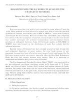
Figure 3. Morphology of PANC-1 cells under the control and following treatment with nicolaioidesin C (15, 1.5 μM) in NDM at 24 h and stained by
ethidium bromide (EB)/acridine orange (AO). Live cells were stained with AO and emitted a bright green fluorescence, while dead cells were stained
with EB and emitted a red fluorescence. Treatment with nicolaioidesin C (15) at 1.5 μM led to dramatic alteration of PANC-1 cell morphology and total
death of PANC-1 cells within 24 h.
ring, respectively, and were very different from those of 1−5. The
relative configuration of 6 was established from the coupling
constant data and the NOESY spectrum. The large coupling
constant between H-1′ and H-6′ (J = 11.4 Hz) indicated that
they have a trans-diaxial orientation, while the small coupling
constant between H-1′ and H-2′ (J = 5.2 Hz) is cis-oriented.
Furthermore, in the NOESY spectrum, correlations between H1′ and H-2′, H-1′ and H-5′α, H-6′ and H-2‴/H-6‴, H-6′ and H1″, and H-6′ and H-5′β (Figure 2) were observed, suggesting
their proximity, as in nicolaioidesin B (13). Therefore, the
structure of panduratin M (6) was concluded as shown.
Panduratins N (7) and O (8) were both obtained as yellowish,
amorphous solids, and they were found to possess the same
molecular formula, C26H30O4, as determined by HRESIMS and
HRFABMS, respectively. The 1H and 13C NMR data of 7 and 8
were similar to those of nicolaioidesin B (13) 15 and
isopanduratin A1 (14),17 respectively. Also apparent were the
methoxy groups at C-4 in 7 and at C-6 in 8, as confirmed by the
HSQC and HMBC spectra (Figure 1). However, 7 and 8 were
observed to differ from 13 and 14 from a variation in the
stereoconfiguration at C-2′ of the cyclohexenyl moiety. Both the
large coupling constants between H-1′ and H-6′ (J = 11.0−11.2
Hz) and between H-1′ and H-2′ (J = 10.9 Hz) indicated that they
are oriented in a trans-diaxial manner. This was also supported by
the NOESY correlations between H-1′ and H-1″, H-1′ and H5′α, H-2′ and H-2‴/H-6‴, H-2′ and H-6′, H-6′ and H-2‴/H-6‴,
andH-6′ and H-5′β (Figure 2). Therefore, the structures of
panduratins N (7) and O (8) were assigned as shown.
All isolated compounds were tested for their preferential
cytotoxic activity against the PANC-1 human pancreatic cancer
cell line, according to an antiausterity strategy.2 Their PC50 values
(the 50% preferential cell death in NDM without cytotoxicity in
DMEM) are listed in Table 4. Among the compounds tested,
isopanduratin A1 (14) and nicolaioidesin C (15) exhibited the
most potent preferential cytotoxicity, with PC50 values of 1.0 and
0.84 μM, respectively, which is comparable to that of arctigenin, a
positive control (PC50 value, 0.8 μM).
The activity of the isolates was found to greatly depend on the
nature of the substituents in the cyclohexene chalcone unit. In
general, compounds having a phenyl group at C-1′ and a benzoyl
substituent at C-6′ of the cyclohexenyl moiety were found to
have potent activity (6 > 4, 13 > 10 and 9, 14 > 11). Interestingly,
at C-1′ and C-2′ of the cyclohexenyl substituent, prenyl and
benzoyl groups or prenyl and phenyl groups on the same side of
the ring are more favorable than when on different sides (12 > 1,
13 > 7, 14 > 8). Moreover, at C-2′ and C-3′ of the cyclohexenyl
unit, it was observed that the position of the prenyl moiety or its
modified form leads to a change of activity (2 > 4 > 10 > 3, 15 >
Panduratin K (4) was isolated as a yellowish, amorphous solid,
and its molecular formula was found to be C26H30O5 by
HRESIMS. The IR spectrum of 4 displayed absorbances for
hydroxy, carbonyl, and phenyl groups. The 1H and 13C NMR
data of 4 also resembled analogous data for panduratin A (10).16
However, they differed in the signals due to the prenyl side chain.
The 1H NMR and HSQC spectra showed the signals of a pair of
trans-coupled double bond [δH 5.54 dd (J = 15.4 and 9.4 Hz); δC
126.1 (C-1″) and δH 5.37 d (J = 15.4 Hz); δC 142.0 (C-2″)], a
quaternary oxygenated carbon [δC 70.2 (C-3″)], and two tertiary
methyl groups [δH 1.18; δC 30.6 (C-4″) and δH 1.17; δC 30.6 (C5″)]. In the HMBC spectrum, the two H3-4″ and H3-5″ tertiary
methyl groups showed correlations with the C-3″ quaternary
oxygenated carbon and the C-2″ olefinic methine carbon,
suggesting the linkage of C-4″ and C-5″ with C-2″ of the double
bond via the C-3″ quaternary oxygenated carbon (Figure 1). The
relative configuration of the cyclohexenyl unit of 4 was found to
be the same as those of 2 and 3 based on the coupling constant
data and the NOESY spectroscopic analysis. Therefore, the
structure of panduratin K (4) was determined as shown.
Panduratin L (5) was obtained as a yellowish, amorphous
solid. It showed a sodiated molecular ion at m/z 459.2163 [M +
Na]+, corresponding to the molecular formula, C27H32O5Na, in
the HRESIMS. The 1H and 13C NMR data of 5 were similar to
those of compound 4, except for the appearance of one more
methoxy group (δH 3.00; δC 50.4), and two meta-coupled
aromatic proton signals at δH 6.05 and 5.85 (J = 1.9 Hz) instead
of the singlet signal of two magnetically equivalent aromatic
protons. The aromatic methoxy group was located at C-6 based
on the HMBC correlation between the methoxy proton at δH
3.99 and the C-6 oxygenated quaternary aromatic carbon at δC
164.0. The location of the aliphatic methoxy group was
determined to be at C-3″ based on the HMBC correlations
observed between the methoxy proton (δH 3.00) and the C-3″
quaternary oxygenated carbon (δC 75.1) (Figure 1). Analysis of
the NOESY correlations together with the coupling constants
indicated the relative configuration of the cyclohexenyl moiety to
be the same as in 2−4. Therefore, the structure of panduratin L
(5) was established as shown.
Panduratin M (6) was isolated as a yellowish, amorphous solid.
Its molecular formula was assigned as C26H30O5 by HRESIMS.
The 1H and 13C NMR spectra were similar to those of
nicolaioidesin B (13),15 a compound isolated from the same
plant extract. This compound showed the presence of a trans-3methyl-3-hydroxybutenyl group at C-2′ of the cyclohexenyl ring
instead of the prenyl side chain (Figure 1). On the basis of the
HMBC spectrum, the phenyl and 2,6-dihydroxy-4-methoxybenzoyl units were assigned at C-1′ and C-6′ of the cyclohexenyl
D
DOI: 10.1021/acs.jnatprod.6b00784
J. Nat. Prod. XXXX, XXX, XXX−XXX
Journal of Natural Products
Article
Table 1. 1H NMR Spectroscopic Data (500 MHz) of Compounds 1−5 in Acetone-d6 (δ in ppm, Multiplicities, J in Hz)
position
3
5
1′
2′
4′
5′α
5′β
6′
Me-3′
1″
2″
4″
5″
2‴, 6‴
3‴, 5‴
4‴
OH-2
4-OH-4
OH-2″
OH-4″
OMe-4
OMe-6
OMe-3″
1
2
3
4
5
5.74 s
5.74 s
4.85 dd (11.4, 10.2)
2.84 brd (10.2)
5.58 d (4.4)
2.31 dd (18.2, 11.3)
2.14 ddd (18.2, 4.6, 4.4)
3.06 ddd (11.4, 11.3, 4.6)
1.71 s
2.23 brd (16.7)
2.15 brd (16.7)
5.13 t (6.5)
1.51 s
5.95 s
5.95 s
4.84 dd (11.8, 4.6)
2.73 ddd (10.3, 5.0, 4.6)
5.43 br
2.13 dd (18.2, 11.2)
2.38 ddd (18.2, 6.4, 4.2)
3.45 ddd (11.8, 11.2, 6.4)
1.78 s
2.00 ddd (15.5, 6.9, 5.0)
2.34 ddd (15.5, 10.3, 6.9)
5.19 t (6.9)
3.74 d (5.8)
5.94 s
5.94 s
4.89 dd (11.7, 4.5)
2.98 ddd (10.5, 4.5, 4.3)
5.36 br
2.07 dd (18.0, 11.3)
2.32 ddd (18.0, 6.0, 4.6)
3.30 ddd (11.7, 11.3, 6.0)
1.80 s
1.34 ddd (17.6, 10.6, 4.3)
2.00 ddd (17.6, 10.5, 4.0)
3.50 ddd (10.6, 3.7, 3.0)
4.61 s
4.81 s
1.62 s
7.22 d (7.8)
7.17 dd (7.8, 7.5)
7.05 t (7.5)
11.90 s
5.94 s
5.94 s
4.93 dd (11.6, 5.0)
3.16 dd (9.4, 5.0)
5.51 br
2.07 dd (17.8, 11.5)
2.37 ddd (17.8, 4.6, 4.4)
3.40 ddd (11.6, 11.5, 4.6)
1.66 s
5.54 dd (15.4, 9.4)
5.85 d (1.9)
6.05 d (1.9)
4.71 dd (11.8, 4.9)
3.13 dd (9.0, 4.9)
5.55 br
2.08 dd (18.0, 11.6)
2.37 ddd (18.0, 5.1, 5.0)
3.40 ddd (11.8, 11.6, 5.1)
1.71 s
5.47 dd (15.7, 9.0)
5.37 d (15.4)
1.18 s
5.18 d (15.7)
1.15 s
1.41 s
7.25 d (7.8)
7.07 dd (7.8, 7.4)
6.97 t (7.4)
1.56 s
7.22 d (7.4)
7.18 dd (7.4, 7.2)
7.05 t (7.2)
11.86 s
9.08 s
1.12 s
7.22 d (7.4)
7.18 dd (7.4, 7.2)
7.06 t (7.2)
13.8 s
9.45 s
3.6 d (3.7)
3.25 t (5.8)
3.77 s
3.77 s
3.76 s
3.99 s
3.00 s
680 g of a dry extract. The MeOH extract was suspended in H2O (1.5 L)
and then partitioned successively with CHCl3 (3 × 1.5 L) and EtOAc (3
× 1.5 L) to give CHCl3 (470 g), EtOAc (10 g), and H2O (150 g)
extracts, respectively. A part of the CHCl3-soluble extract (450 g) was
subjected to silica gel column chromatography (9 × 120 cm), eluted
with EtOAc−n-hexane gradient mixtures (0−50%), to yield 15 fractions
(fr-1, 22.0 g; fr-2, 197.5 g; fr-3, 22.0 g; fr-4, 26.0 g; fr-5, 6.0 g; fr-6, 24.0 g;
fr-7, 15.0 g; fr-8, 20.0 g; fr-9, 22.0 g; fr-10, 21.0 g; fr-11, 11.0 g; fr-12, 18.0
g; fr-13, 14.0 g; fr-14, 10.0 g; fr-15, 22.0 g). Fraction 3 (22.0 g) was
subjected to further silica gel column chromatography (7.5 × 120 cm),
eluted with EtOAc−n-hexane gradient mixtures (0−80%), to give seven
subfractions (fr-3-1, 160 mg; fr-3-2, 2.2 g; fr-3-3, 8.1 g; fr-3-4, 6.3 g; fr-35, 3.3 g; fr-3-6, 1.5 g; fr-3-7, 1.3 g). Subfraction 3-2 was rechromatographed on silica gel with a CHCl3−n-hexane gradient system to yield
four subfractions, fr-3-2-1−4. Subfraction 3-2-1 (379 mg) was
chromatographed on ODS silica gel with MeOH−H2O gradient
mixtures (0−50%) to give 10 (300 mg), followed by normal-phase
preparative TLC with EtOAc−n-hexane (20:80) to afford 9 (6.1 mg).
Subfraction 3-2-3 (377 mg) was chromatographed on ODS silica gel
with MeOH−H2O gradient mixtures (0−50%) and then purified by
normal-phase preparative TLC with EtOAc−CHCl3 −n-hexane
(5:25:70) to give 7 (5.0 mg), 13 (5.0 mg), and 14 (6.6 mg). Subfraction
3-3 was dissolved in CHCl3−n-hexane and left overnight to give crystals
of 11 (6.0 g). Subfraction 3-6 was subjected to silica gel column
chromatography with an acetone−n-hexane gradient system, to yield
five subfractions, fr-3-6-1−5. Subfraction 3-6-4 (190 mg) was again
separated by silica gel column chromatography with a further acetone−
n-hexane gradient system, followed by reversed-phase preparative TLC
with MeOH−CH3CN−H2O (10:70:20), to afford 14 (10.0 mg).
Fraction 4 (26.0 g) was subjected to silica gel column (7.5 × 120 cm)
chromatography, eluted with an acetone−n-hexane gradient system, to
yield 14 subfractions (fr-4-1, 18 mg; fr-4-2, 113 mg; fr-4-3, 127 mg; fr-44, 199 mg; fr-4-5, 65 mg; fr-4-6, 34 mg; fr-4-7, 616 mg; fr-4-8, 20−23 g;
fr-4-9, 269 mg; fr-4-10, 32 mg; fr-4-11, 31 mg; fr-4-12, 850 mg; fr-4-13,
303 mg; fr-4-14, 2−8 g). Subfraction 4-13 was chromatographed by
silica gel column chromatography, with CHCl3−n-hexane gradient
mixtures (0−100%), to obtain 8 (6.5 mg). Fraction 6 (24.0 g) was
further separated by silica gel column (7.5 × 120 cm) chromatography,
13 > 6). Furthermore, the presence of a methoxy group at C-6 of
the benzoyl moiety was found to result in more potent activity
than when a methoxy group at C-4 or a hydroxy group at C-6 is
present (11 > 10, 14 > 13, 8 > 7, 11 > 12). At C-4, a hydroxy
group was favored over a methoxy group (1 > 9, 12 > 10).
Nicolaioidesin C (15) was studied further for its effects on the
morphological changes of PANC-1 using an ethidium bromide
and acridine orange (EB/AO) staining assay.12 Cells treated with
nicolaioidesin C (15, 1.5 μM) showed round morphology of
PANC-1 cells and emitted a red fluorescence of EB, indicative of
dead cells. In contrast, the control cells showed intact
morphology and gave a bright green fluorescence of AO,
suggestive of live cells (Figure 3).
■
1.17 s
7.23 d (7.4)
7.18 dd (7.4, 7.2)
7.06 t (7.2)
11.82 s
EXPERIMENTAL SECTION
General Experimental Procedures. Optical rotations were
recorded on a JASCO DIP-140 digital polarimeter. IR spectra were
measured with a Shimadzu IR-408 spectrophotometer in CHCl3
solution. NMR spectra were taken on a Bruker Advance III 500
spectrometer (Bruker Biospin) with tetramethylsilane as an internal
standard, and chemical shifts are expressed in δ values. HRESIMS and
HRFABMS measurements were carried out on a Bruker micrOTOFQII mass spectrometer and JEOLJMS-AX505HAD mass spectrometer,
respectively. Silica gel 60, 40−63 μm (230−400 mesh ASTM), for
column chromatography was purchased from Scharlau. Analytical and
preparative TLC was carried out on precoated Merck Kieselgel 60F254 or
RP-18F254 plates (0.25 or 0.5 mm thickness).
Plant Material. The rhizomes of Boesenbergia pandurata were
collected in Tinh Bien District of An Giang Province, Vietnam, in April
2013, and this species was identified by Ms. Hoang Viet, Faculty of
Biology, University of Science, Vietnam National University, Ho Chi
Minh City (VNU-HCM). A voucher specimen (MCE0043) has been
deposited at the Division of Medicinal Chemistry, Faculty of Chemistry,
University of Science, VNU-HCM.
Extraction and Isolation. Dried powdered rhizomes of B. pandurata (5.5 kg) were extracted with MeOH (15 L, reflux, 3 h × 3) to yield
E
DOI: 10.1021/acs.jnatprod.6b00784
J. Nat. Prod. XXXX, XXX, XXX−XXX
Journal of Natural Products
Article
Table 2. 1H NMR Spectroscopic Data (500 MHz) of Compounds 6−8 in Acetone-d6 (δ in ppm, multiplicities, J in Hz)
position
6
7
8
3
5
1′
2′
4′
5′α
5′β
6′
Me-3′
1″
5.95 s
5.95 s
3.53 dd (11.4, 5.2)
2.75 dd (5.2, 5.0)
5.56 br
2.12 dd (17.1, 11.2)
2.65 ddd (17.1, 5.4, 5.2)
4.92 ddd (11.4, 11.2, 5.4)
1.67 s
5.27 dd (15.0, 5.0)
5.84 s
5.84 s
3.14 dd (11.0, 10.9)
2.45 ddd (10.9, 8.2, 6.4)
5.66 d (4.5)
2.15 dd (16.5, 11.3)
2.45 ddd (16.5, 4.7, 4.5)
4.65 ddd (11.3, 11.0, 4.5)
1.72 s
1.94 ddd (16.2, 8.2, 5.0)
2.15 ddd (16.2, 6.4, 5.0)
5.05 t (5.0)
1.71 s
1.45 s
7.18 d (7.9)
7.14 dd (7.9, 7.2)
7.04 t (7.2)
11.71 s
3.72 s
5.74 d (2.0)
5.94 d (2.0)
3.11 dd (11.2, 10.9)
2.48 ddd (10.9, 7.9, 6.4)
5.67 d (4.8)
2.15 dd (16.7, 11.3)
2.36 ddd (16.7, 4.8, 4.5)
4.65 ddd (11.3, 11.2, 4.5)
1.72 s
1.93 ddd (16.1, 7.9, 4.9)
2.14 ddd (16.1, 6.4, 4.9)
5.05 t (4.9)
1.71 s
1.48 s
7.15 d (7.9)
7.18 dd (7.9, 7.2)
7.04 t (7.2)
13.58 s
2″
4″
5″
2‴, 6‴
3‴, 5‴
4‴
OH-2
OMe-4
OMe-6
5.27 d (15.0)
1.08 s
1.07 s
7.12 m
7.12 m
7.03 m
11.86 s
3.78 s
3.88 s
Table 3. 13C NMR Spectroscopic Data (125 MHz) of Compounds 1−8 in Acetone-d6
position
1
2
3
4
5
6
7
8
1
2
3
4
5
6
>CO
1′
2′
3′
4′
5′
6′
Me-3′
1″
2″
3″
4″
5″
1‴
2‴, 6‴
3‴, 5‴
4‴
OMe-4
OMe-6
OMe-3″
107.6
165.0
95.7
165.0
95.7
165.0
210.4
54.8
46.0
137.2
123.1
35.5
47.3
21.7
29.8
123.5
132.7
26.0
18.0
144.9
128.5
128.8
126.8
107.0
166.6
94.6
166.6
94.6
166.6
207.5
54.7
43.3
137.8
121.9
36.8
37.8
23.1
29.2
125.3
136.4
68.7
14.0
148.2
128.1
129.0
126.3
55.8
106.9
166.8
94.6
166.8
94.6
166.8
208.1
54.3
39.1
139.2
120.7
37.0
38.6
22.3
37.4
75.4
150.1
109.6
18.2
147.9
128.2
129.0
126.3
55.8
106.6
166.5
94.4
166.5
94.4
166.5
206.6
55.1
46.7
135.5
122.2
36.9
37.9
22.4
126.1
142.0
70.2
30.6
30.6
147.8
128.1
129.0
126.3
55.7
106.7
168.8
97.0
165.4
92.0
164.0
206.4
55.3
47.1
135.1
122.6
36.9
38.1
22.4
129.9
138.7
75.1
26.4
26.2
147.7
128.1
129.0
126.4
106.4
166.9
94.5
166.9
94.5
166.9
210.1
47.6
50.1
136.6
121.3
32.1
44.2
22.5
125.7
142.4
70.2
30.4
30.4
144.2
128.4
129.3
126.3
55.8
106.3
166.7
94.3
166.7
94.3
166.7
209.6
47.7
46.9
136.8
123.6
31.2
51.4
21.7
26.1
121.4
133.2
27.1
18.3
145.3
128.6
129.5
126.7
55.7
105.5
167.3
95.9
165.3
91.3
163.2
207.9
47.2
45.8
135.8
122.7
30.3
50.8
20.9
26.2
120.5
132.2
25.2
17.4
144.3
127.7
128.6
125.8
56.4
50.4
with a MeOH−CHCl3 gradient system, to yield 13 subfractions (fr-6-1,
464 mg; fr-6-2, 388 mg; fr-6-3, 1.6 g; fr-6-4, 3.8 g; fr-6-5, 5.0 g; fr-6-6, 576
mg; fr-6-7, 1.7 g; fr-6-8, 3.3 g; fr-6-9, 1.1 g; fr-6-10, 1.7 g; fr-6-11, 794 mg;
fr-6-12, 815 mg; fr-6-13, 442 mg). Subfraction 6-6 was also
chromatographed on silica gel with an acetone−n-hexane gradient
system, followed by normal-phase preparative TLC with acetone−nhexane (20:80), to give 3 (0.8 mg). Subfraction 6-7 was subjected to
silica gel chromatography, with an EtOAc−n-hexane gradient system, to
give four subfractions, fr-6-7-1−4. Of these, fr-6-7-2 (115 mg) was
chromatographed on ODS silica gel, with acetone−H2O gradient
mixtures (0−80%), and followed by normal-phase preparative TLC with
55.3
EtOAc−n-hexane (20:80), to afford 1 (18.3 mg). Subfraction 6-9 was
also chromatographed on silica gel with an EtOAc−n-hexane gradient
system to give three subfractions, fr-6-9-1−3, and then fr-6-9-2 was
dissolved in EtOAc−n-hexane and left overnight to give 12 (300.0 mg).
Fraction 8 (20.0 g) was chromatographed on silica gel (7.5 × 120 cm)
with MeOH−CHCl3 gradient mixtures (0−50%) to give 20 subfractions
(fr-8-1, 12 mg; fr-8-2, 30 mg; fr-8-3, 40 mg; fr-8-4, 22 mg; fr-8-5, 6.9 mg;
fr-8-6, 140 mg; fr-8-7, 4.2 g; fr-8-8, 4.2 g; fr-8-9, 402 mg; fr-8-10, 752 mg;
fr-8-11, 216 mg; fr-8-12, 2.43 g; fr-8-13, 4.43 g; fr-8-14, 538 mg; fr-8-15,
89 mg; fr-8-16, 119 mg; fr-8-17, 414 mg; fr-8-18, 639 mg; fr-8-19, 116
mg; fr-8-20, 158 mg). Subfraction 8-9 was subjected to silica gel column
F
DOI: 10.1021/acs.jnatprod.6b00784
J. Nat. Prod. XXXX, XXX, XXX−XXX
Journal of Natural Products
Article
chromatography, eluted with EtOAc−n-hexane gradient mixtures (0−
30%) and then acetone−n-hexane gradient mixtures (0−50%), to afford
2 (5.0 mg). Subfraction 8-10 was subjected to silica gel column
chromatography with acetone−n-hexane gradient mixtures (0−50%) to
yield three subfractions, fr-8-10-1−3. Both fr-8-10-1 (143 mg) and fr-810-3 (315 mg) were subjected to silica gel column chromatography,
eluted with acetone−n-hexane gradient mixtures (0−50%), to afford six
subfractions, fr-8-10-1-1−3 and fr-8-10-3-1−3, respectively. Subfraction
8-10-1-1 (36.3 mg) was chromatographed over ODS silica gel with
MeOH−H2O gradient mixtures (0−80%), followed by reversed-phase
preparative TLC with MeOH−H2O (20:80), to give 5 (6.7 mg).
Subfraction 8-10-3-2 (45.1 mg) was subjected to normal-phase
preparative TLC with EtOAc−n-hexane (4:96) to give two subfractions.
Of these, fr-8-10-3-2-1 (25.6 mg) was recrystallized with MeOH−
CHCl3 to afford 4 (13.1 mg), while fr-8-10-3-2-2 (9.3 mg) was purified
by reversed-phase preparative TLC with acetone−H2O (30:70) to
afford 6 (6.0 mg).
Compound 1: yellow, amorphous solid; [α]D25 +35.6 (c 1,
CH3COCH3); IR νmax (CHCl3) 3500, 1640, 1450, 1100 cm−1; 1H
and 13C NMR (acetone-d6, 500 MHz, see Tables 1 and 3); HRESIMS
m/z 415.1885 [M + Na]+ (calcd for C25H28O4Na, 415.1885).
purchased from the Riken BRC cell bank and maintained in standard
Dulbecco’s modified Eagle medium (DMEM) with 10% fetal bovine
serum supplemented and stored at 37 °C under a humidified
atmosphere of 5% CO2 and 95% air. Briefly, human pancreatic cancer
cells were seeded in 96-well plates (1.5 × 10⁴/well) and incubated in
fresh DMEM at 37 °C under 5% CO2 and 95% air for 24 h. After the cells
were washed twice with phosphate-buffered saline (PBS), the medium
was changed to serially diluted test samples in both nutrient-rich
medium (DMEM) and nutrient-deprived medium (NDM)2 with a
control and blank in each test plate. The composition of the NDM was
as follows: 265 mg/L CaCl2 (2 H2O), 0.1 mg/L Fe(NO3)3 (9 H2O), 400
mg/L KCl, 200 mg/L MgSO4 (7 H2O), 6400 mg/L NaCl, 700 mg/L
NaHCO3, 125 mg/L NaH2PO4, 15 mg/L phenol red, 25 mM/L HEPES
buffer (pH 7.4), and MEM vitamin solution (Life Technologies, Inc.,
Rockville, MD, USA); the final pH was adjusted to 7.4 with 10%
NaHCO3. Arctigenin, the positive control in this study, was isolated
from the seeds of Arctium lappa.2 After 24 h of incubation with each test
compound in DMEM and NDM, the cells were washed twice with PBS
and replaced with 100 μL of DMEM containing a 10% WST-8 cell
counting kit solution. After 3 h of incubation, the absorbance at 450 nm
was measured (PerkinElmer EnSpire multilabel reader). Cell viability
was calculated from the mean values of data from three wells by using the
following equation:
Table 4. Preferential Cytotoxicity of Compounds 1−15
against the PANC-1 Human Pancreatic Cancer Cell Line in
Nutrient-Deprived Medium (NDM)
compound
PC50, μMa
compound
PC50, μMa
1
2
3
4
5
6
7
8
5.8
6.3
18.1
6.6
10.5
3.3
7.8
7.4
9
10
11
12
13
14
15
arctigeninb
7.2
7.8
2.8
3.1
2.1
1.0
0.84
0.83
Cell viability (%) = [Abs(test sample) − Abs(blank)/Abs(control)
− Abs(blank)]× 100%
Morphological Assessment of Cancer Cells. PANC-1 cells were
seeded in 24-well plates (6 × 10⁴/well) and incubated in fresh DMEM at
37 °C under 5% CO2 and 95% air for 24 h. After the cells were washed
twice with PBS, the medium was changed to NDM (control) or
nicolaioidesin C (15, 1.5 μM) in NDM (treated). After a 24 h
incubation, 8 μL of EB/AO reagent was added to the each test well and
incubated for 5 min, and the morphology was captured using an EVOS
FL cell imaging system (20× objective) under fluorescent and phase
contrast mode.
■
a
Concentration at which 50% of cells were killed preferentially in
NDM. bPositive control.
ASSOCIATED CONTENT
S Supporting Information
*
The Supporting Information is available free of charge on the
ACS Publications website at DOI: 10.1021/acs.jnatprod.6b00784.
Copies of spectroscopic data for 1−8 (PDF)
Compound 2: yellow, amorphous solid; [α]D25 +31.4 (c 1,
CH3COCH3); IR νmax (CHCl3) 3600, 1640, 1460, 1090 cm−1; 1H
and 13C NMR (acetone-d6, 500 MHz, see Tables 1 and 3); HRESIMS
m/z 445.1991 [M + Na]+ (calcd for C26H30O5Na, 445.1976).
Compound 3: yellow, amorphous solid; [α]D25 +77.5 (c 1,
CH3COCH3); IR νmax (CHCl3) 3600, 1640, 1445, 1090 cm−1; 1H
and 13C NMR (acetone-d6, 500 MHz, see Tables 1 and 3); HRESIMS
m/z 445.1991 [M + Na]+ (calcd for C26H30O5Na, 445.1991).
Compound 4: yellow, amorphous solid; [α]D25 +27.9 (c 1,
CH3COCH3); IR νmax (CHCl3) 3600, 1640, 1450, 1100 cm−1; 1H
and 13C NMR (acetone-d6, 500 MHz, see Tables 1 and 3); HRESIMS
m/z 445.1997 [M + Na]+ (calcd for C26H30O5Na, 445.1991).
Compound 5: yellow, amorphous solid; [α]D25 +33.8 (c 1,
CH3COCH3); IR νmax (CHCl3) 3500, 1640, 1440, 1090 cm−1; 1H
and 13C NMR (acetone-d6, 500 MHz, see Tables 1 and 3); HRESIMS
m/z 459.2163 [M + Na]+ (calcd for C27H32O5Na, 459.2147).
Compound 6: yellow, amorphous solid; [α]D25 +27.5 (c 1,
CH3COCH3); IR νmax (CHCl3) 3600, 1650, 1450, 1100 cm−1; 1H
and 13C NMR (acetone-d6, 500 MHz, see Tables 2 and 3); HRESIMS
m/z 445.1980 [M + Na]+ (calcd for C26H30O5Na, 445.1991).
Compound 7: yellow, amorphous solid; [α]D25 +37.5 (c 1,
CH3COCH3); IR νmax (CHCl3) 3500, 1650, 1460, 1100 cm−1; 1H
and 13C NMR (acetone-d6, 500 MHz, see Tables 2 and 3); HRESIMS
m/z 405.2075 [M − H]− [calcd for C26H29O4, 405.2066].
Compound 8: yellow, amorphous solid; [α]D25 +23.6 (c 1,
CH3COCH3); IR νmax (CHCl3) 3500, 1650, 1450, 1090 cm−1; 1H
and 13C NMR (acetone-d6, 500 MHz, see Tables 2 and 3); HRFABMS
m/z 407.22253 [M + H]+ (calcd for C26H31O4, 401.22224).
Preferential Cytotoxicity Assay against PANC-1 Cells. The
PANC-1 (RBRC-RCB2095) human pancreatic cancer cell line was
■
AUTHOR INFORMATION
Corresponding Authors
*E-mail (N. T. Nguyen): Tel: +84-907426-331. Fax: +84-838-353-659.
*E-mail (S. Awale): Tel: +81-76434-7640. Fax: +81-76-434-7640.
ORCID
Nhan Trung Nguyen: 0000-0001-5142-4573
Notes
The authors declare no competing financial interest.
■
ACKNOWLEDGMENTS
This research was supported by a grant from Vietnam’s National
Foundation for Science and Technology Development (No.
104.01-2013.72) to N.T.N. and by a Grant in Aid for Scientific
Research (16K08319) from the Japan Society for the Promotion
of Science (JSPS), Japan, to S.A.
■
REFERENCES
(1) Izuishi, K.; Kato, K.; Ogura, T.; Kinoshita, T.; Esumi, H. Cancer Res.
2000, 60, 6201−6207.
G
DOI: 10.1021/acs.jnatprod.6b00784
J. Nat. Prod. XXXX, XXX, XXX−XXX
Journal of Natural Products
Article
(2) Awale, S.; Lu, J.; Kalauni, S. K.; Kurashima, Y.; Tezuka, Y.; Kadota,
S.; Esumi, H. Cancer Res. 2006, 66, 1751−1757.
(3) Awale, S.; Nakashima, E. M. N.; Kalauni, S. K.; Tezuka, Y.;
Kurashima, Y.; Lu, J.; Esumi, H.; Kadota, S. Bioorg. Med. Chem. Lett.
2006, 16, 581−583.
(4) Win, N. N.; Awale, S.; Esumi, H.; Tezuka, Y.; Kadota, S. J. Nat. Prod.
2007, 70, 1582−1587.
(5) Win, N. N.; Awale, S.; Esumi, H.; Tezuka, Y.; Kadota, S. Bioorg.
Med. Chem. 2008, 16, 8653−8660.
(6) Win, N. N.; Awale, S.; Esumi, H.; Tezuka, Y.; Kadota, S. Bioorg.
Med. Chem. Lett. 2008, 18, 4688−4691.
(7) Win, N. N.; Awale, S.; Esumi, H.; Tezuka, Y.; Kadota, S. Chem.
Pharm. Bull. 2008, 56, 491−496.
(8) Ueda, J.-Y.; Awale, S.; Athikomkulchai, S.; Miyatake, R.; Saiki, I.;
Esumi, H. Drug Des., Dev. Ther. 2014, 8, 39−47.
(9) Nguyen, H. X.; Nguyen, M. T. T.; Nguyen, T. A.; Nguyen, N. Y. T.;
Phan, D. A. T.; Thi, P. H.; Nguyen, T. H. P.; Dang, P. H.; Nguyen, N. T.;
Ueda, J.-Y.; Awale, S. Fitoterapia 2013, 91, 148−153.
(10) Nguyen, M. T. T.; Nguyen, N. T.; Nguyen, K. D. H.; Dau, H. T.
T.; Nguyen, H. X.; Dang, P. H.; Le, T. M.; Nguyen Phan, T. H.; Tran, A.
H.; Nguyen, B. D.; Ueda, J.-Y.; Awale, S. Planta Med. 2014, 80, 193−200.
(11) Nguyen, H. X.; Nguyen, N. T.; Dang, P. H.; Thi Ho, P.; Nguyen,
M. T. T.; Van Can, M.; Dibwe, D. F.; Ueda, J.-Y.; Awale, S.
Phytochemistry 2016, 122, 286−293.
(12) Nguyen, H. X.; Do, T. N. V.; Le, T. H.; Nguyen, M. T. T.; Nguyen,
N. T.; Esumi, H.; Awale, S. J. Nat. Prod. 2016, 79, 2053.
(13) Vo, V. C. An Giang Medicinal Plants; Science & Technology
Publisher: An Giang, Vietnam, 1991.
(14) Wiart, C. Medicinal Plants of China, Korea, and Japan: Bioresources
for Tomorrow’s Drugs and Cosmetics; CRC Press: London, UK, 2012.
(15) Gu, J.-Q.; Park, E. J.; Vigo, J. S.; Graham, J. G.; Fong, H. H. S.;
Pezzuto, J. M.; Kinghorn, A. D. J. Nat. Prod. 2002, 65, 1616−1620.
(16) Tuntiwachwuttikul, P.; Pancharoen, O.; Reutrakul, V.; Byrne, L.
T. Aust. J. Chem. 1984, 37, 449−453.
(17) Pandji, C.; Grimm, C.; Wray, V.; Witte, L.; Proksch, P.
Phytochemistry 1993, 34, 415−419.
(18) Trakoontivakorn, G.; Nakahara, K.; Shinmoto, H.; Takenaka, M.;
Onishi-Kameyama, M.; Ono, H.; Yoshida, M.; Nagata, T.; Tsushida, T. J.
Agric. Food Chem. 2001, 49, 3046−3050.
H
DOI: 10.1021/acs.jnatprod.6b00784
J. Nat. Prod. XXXX, XXX, XXX−XXX