26. Organ and tissue dependent effect of resveratrol and exercise on antioxidant defenses of old mice
Bạn đang xem bản rút gọn của tài liệu. Xem và tải ngay bản đầy đủ của tài liệu tại đây (889.36 KB, 9 trang )
Aging Clin Exp Res
DOI 10.1007/s40520-015-0366-8
ORIGINAL ARTICLE
Organ and tissue-dependent effect of resveratrol and exercise
on antioxidant defenses of old mice
Bui Thanh Tung1 • Elisabet Rodriguez-Bies2 • Hai Nguyen Thanh1 •
Huong Le-Thi-Thu1 • Pla´cido Navas2 • Virginia Motilva Sanchez3 •
Guillermo Lo´pez-Lluch2
Received: 28 October 2014 / Accepted: 20 April 2015
Ó Springer International Publishing Switzerland 2015
Abstract
Background Oxidative stress has been considered one of
the causes of aging. For this reason, treatments based on
antioxidants or those capable of increasing endogenous
antioxidant activity have been taken into consideration to
delay aging or age-related disease progression.
Aim In this paper, we determine if resveratrol and exercise have similar effect on the antioxidant capacity of
different organs in old mice.
Methods Resveratrol
(6 months)
and/or exercise
(1.5 months) was administered to old mice. Markers of
oxidative stress (lipid peroxidation and glutathione) and
activities and levels of antioxidant enzymes (SOD, catalase, glutathione peroxidase, glutathione reductase and
transferase and thioredoxin reductases, NADH cytochrome
B5-reductase and NAD(P)H-quinone acceptor oxidoreductase) were determined by spectrophotometry and
Western blotting in different organs: liver, kidney, skeletal
muscle, heart and brain.
Results Both interventions improved antioxidant activity
in the major organs of the mice. This induction was accompanied by a decrease in the level of lipid peroxidation
in the liver, heart and muscle of mice. Both resveratrol and
& Bui Thanh Tung
1
School of Medicine and Pharmacy, Vietnam National
University, Hanoi, Floor 5 Building Y1, 144 Xuan Thuy,
Cau Giay, Hanoi, Vietnam
2
Centro Andaluz de Biologı´a del Desarrollo, Universidad
Pablo de Olavide-CSIC, CIBERER, Instituto de Salud Carlos
III, Carretera de Utrera Km. 1, 41013 Seville, Spain
3
Departmento de Farmacologı´a, Facultad de Farmacia,
Universidad de Sevilla, 41012 Seville, Spain
exercise modulated several antioxidant activities and protein levels. However, the effect of resveratrol, exercise or
their combination was organ dependent, indicating that
different organs respond in different ways to the same
stimulus.
Conclusions Our data suggest that physical activity and
resveratrol may be of great importance for the prevention
of age-related diseases, but that their organ-dependent effect must be taken into consideration to design a better
intervention.
Keywords Antiaging Á Antioxidant Á Resveratrol Á
Exercise Á Old mice
Introduction
Imbalance in the activity of antioxidant enzymes and the
production of free radicals by metabolic activities, mainly
associated with mitochondria, have been associated with
the aging process by the free radical theory of aging [1, 2].
The main radicals in cells are derived from reactive oxygen
species (ROS) and have been considered active factors in
aging and aging research because of their potential involvement in many degenerative diseases. These ROS are
highly reactive and damage many biological macromolecules such as DNA, RNA, protein and lipids [3]. For
this, antioxidant enzymes constitute an important defense
system to clear up the harmful ROS in vivo and to prevent
oxidative damage of macromolecules.
Resveratrol (trans-3,40 ,5-trihydroxystilbene) (RSV) is a
naturally occurring phytoalexin found in red wine, berries
and peanuts. RSV has shown many positive effects on
biological systems ranging from cancer chemoprevention
[4], prevention of inflammation [5] and antioxidant
123
Aging Clin Exp Res
capacity [6]. Although its effect has been studied for more
than a decade, the molecular mechanisms of RSV remain
elusive [7]. Although a direct activation seems to be unlikely, the modulation of sirtuins by regulation of NAD?/
NADH ratio and the activation of the AMPK-dependent
pathway seem to be the main mechanisms involved in the
RSV effect on cells and organisms affecting longevity,
metabolism regulation, cancer, inflammation, etc. [7, 8].
During the last years, the activation of the DNA damagedependent pathway [9, 10] by activating ATM and the
regulation of the different pathways affected by this kinase
probably indicate a common mechanism of action for all
the effects of RSV on the cell and organisms. Furthermore,
several investigations have demonstrated the role and
protective effect of RSV against certain forms of oxidant
damage, through a hydrogen-electron donation from its
hydroxyl groups [11] or by increasing the expression of
antioxidant enzymes [12]. Therefore, RSV may be an important dietary factor to improve health and prolong the
average lifespan in animals [13, 14].
Physical activity is associated with better health mainly
by its effect on muscle strength and the cardiovascular
system. On the other hand, physical activity can also
positively affect physiological endogenous antioxidant
defenses in old subjects. It reduces the production of oxidants and oxidative damage, improves antioxidant defense
system and increases the resistance of organs and tissues
against the deleterious action of free radicals [15]. Furthermore, the physical activity level correlates closely with
antioxidant enzymatic activities, especially related to the
glutathione-dependent system in the liver and brain [16].
Furthermore, recently we have described the positive relationship of coenzyme Q10-dependent prevention of LDL
oxidation and physical activity in elderly people [17, 18].
The aim of the present study was to evaluate the possible antiaging properties of RSV and/or physical activity
in old mice by modulating endogenous antioxidant activities and enzyme levels in different organs. Thus, the
content of glutathione, sulfhydryl group and lipid damage
through malondialdehyde (MDA) and the activity and
levels of superoxide dismutase (SOD), catalase (CAT),
glutathione peroxidase (GPx), glutathione reductase (GR),
glutathione-S-transferase (GST), NAD(P)H-quinone acceptor oxidoreductase (NQO1), NADH cytochrome b5 reductase (CytB5Rase) and thioredoxin reductase (TrxR) in
different mice organs were determined. Furthermore, the
antioxidant ratio (R) indicated as the activity of SOD related to the sum of the activities of CAT and GPx, which
has been related to cell senescence [19, 20] was also determined. The different responses of organs to the same
stimulus and their relationship with the induction of endogenous antioxidant systems are discussed.
123
Materials and methods
Animals and feeding regimen
Male mice (C57BL/6 J) at the age of 18 months were used
for these experiments. The experiments were of duration
6 months until their killing. Thus, at the end of the study,
the mice were 24 months of age (old mice). A total of 16
mice were used, divided into four groups: Control no
trained (Control-NT), Control trained (Control-T),
Resveratrol no trained (RSV-NT) and Resveratrol trained
(RSV-T). The animals were fed with a basal diet (Teklad
Global Diet chow 2014S, Harlan) and kept in a thermostatically controlled cage holder at 22 °C with a 12 h
lighting cycle. All animals were maintained according to a
protocol approved by the Ethical Committee of the
University Pablo de Olavide and following the international rules for animal research.
Training consisted of running at a speed of 20 m/min,
20 min/day, 5 days/week, for all the time. The group
Control was fed a liquid containing ethanol in water
(180 lL ethanol/100 mL H2O) and the group RSV was fed
a liquid containing resveratrol [180 lL of a dilution of
55 mg/mL trans-resveratrol in ethanol in100 mL H2O,
reaching a concentration of 100 mg/L (0.01 % RSV)] in
opaque bottles to avoid light-dependent decomposition.
Drinking water was changed twice a week for both groups.
Taken into consideration an average drinking of
4–5 mL/day and the weight of the animals, the calculated
dose of RSV was around 500 lg/animal/day (16.67 mg/
kg/day). Animals were killed by cervical dislocation and
dissected. The brain, kidney heart, muscle and liver were
frozen in liquid nitrogen and stored at -80 °C until the
analysis.
All animals were maintained according to a protocol
approved by the Ethical Committee of the University Pablo
de Olavide of resolution 03/09 and following the international rules for animal research.
Body weight
Mice’s body weight was measured every 2 weeks to check
for a possible influence of physical performance and RSV.
Treatment of sample
Frozen tissue from the brain, kidney heart, muscle and liver
was homogenized in nine volumes of ice-cold tissue lysis
buffer containing 150 mM sodium chloride, 1.0 % NP-40,
50 mM Tris, pH 8.0, and 1 mM PMSF (phenylmethylsulfonyl fluoride) with protease inhibitors (Sigma). Homogenates were centrifuged at 10009g for 10 min at 4 °C.
Aging Clin Exp Res
Single-use aliquots of the homogenates were stored at
-80 °C before measurements. The protein concentration
was determined by the Bradford’s method.
Measurement of antioxidant activities and oxidative
damage
The SOD activity was spectrophotometrically measured
using the method developed by Marklund and Marklund
[21]. Briefly, SOD was detected on the basis of its ability to
inhibit superoxide-mediated oxidation of pyrogallol. One
unit was determined as the amount of enzyme that inhibited
oxidation of pyrogallol by 50 %.
CAT activity was measured by following the rate of
disappearance of H2O2 at 240 nm [22]. One unit of CAT
activity is defined as the amount of enzyme catalyzing the
degradation of 1 lmol H2O2 per min and specific activity
corresponding to transformation of H2O2 (in nmol) per min
per mg protein.
The whole amount of glutathione, reduced (GSH) plus
oxidized (GSSG) forms, was determined by the method
suggested by Anderson [23]. The amount of glutathione
was expressed as nmol per mg total protein.
The GPx activity was determined in a coupled assay
with glutathione reductase by measuring the rate of
NADPH oxidation at 340 nm using H2O2 as the substrate
[24]. GR activity was determined by following the oxidation of NADPH at 340 nm as described by Carlberg and
Mannervik [25]. GT activity was determined by Habig’s
methods [26] based on the conjugation of 1-chloro-2,4dinitrobenzene (CDNB) with reduced glutathione. Enzymatic activity was calculated by using the extinction coefficient of 9.6 mM-1 cm-1 for CDNB and expressed as
nmol/min/mg protein.
Total CytB5Rase activity was assayed by measuring the
rate of potassium ferricyanide reduction spectrophotometrically, according to the method of Strittmatter and Velick
[27]. The enzyme activity was calculated using the extinction coefficient of 6.22 mM-1 cm-1 for NADH and
expressed as nmol/min/mg protein.
NQO1 activity was determined spectrophotometrically
by monitoring the reduction of the standard electron acceptor, 2,6-dichlorophenol-indophenol (DCPIP) at 600 nm
as described by Benson et al. [28] in the absence or presence of dicoumarol. The dicoumarol-inhibitable part of
DCPIP’s reduction was calculated as NQO1 activity using
the extinction coefficient of 21.0 mM-1 cm-1 and expressed as nmol DCPIP reduced/min/mg protein.
TrxR activity was determined by the method of Hillet
et al. [29] and based on the reduction of 5,50 -dithiobis
(2-nitrobenzoic acid) (DTNB) determined by the increase
in absorbance at 412 nm. A unit of activity was defined as
1.0 nmol 5-thio-2-nitrobenzoic acid (TNB) formed/min/mg
protein.
Protein thiol (SH) groups were estimated by Ellman’s
method [30]. Briefly, 0.5 ml of sample homogenate was
added to a cuvette containing 0.5 ml phosphate buffer
(0.1 M, pH 7.4); 0.2 ml of 3 mM 5,5-dithiobis (2-nitrobenzoic acid) was then added to start the reaction.
After 10 min, absorbance was measured at 412 nm. The
amount of SH groups was calculated according to the
formula: mol SH/ml = [(Dsample/14,150)/dilution factor]/ml.
Lipid peroxidation assay was performed by determining
the reaction of malondialehyde with two molecules of
1-methyl-2-phenylindole at 45 °C as described by Ge´rardMonniern et al. [31]. Peroxidized lipids are expressed as
nmol MDA equivalents/mg protein.
All the biochemical analyses and enzyme activities were
determined in triplicate per sample. Activity or determination per sample was considered as the mean of these
three determinations. Data are the result of the mean of the
samples from four animals per treatment (n = 4).
Immunoblotting analysis
Homogenates of samples were separated by 10 % (v/v)
SDS-PAGE and then transferred to nitrocellulose membranes and subjected to immunoblot analyses using the
primary antibodies anti-Cu, Zn-SOD (SOD1), anti-CAT,
(Santa Cruz Biotechnology), anti-CytB5Rase (rabbit polyclonal antibody kindly provided by Dr. J. M. Villalba,
Universidad de Co´rdoba, Spain), anti-GPx1, anti-TrxR1,
anti-TrxR2 (Acris Antibodies, Germany) or anti-NQO1
(Abcam, Cambridge, UK) and secondary antibodies
horseradish peroxidase-conjugated goat anti-rabbit or antisheep antibodies (Calbiochem, Germany). Protein expression levels were corrected for whole protein loading determined by staining membrane with Ponceau S. Protein
expressions were visualized by the ChemiDocTM XRS?
System and compiled with Image LabTM 4.0.1 Software
(Bio-Rad Laboratories).
Statistical analysis
The results were analyzed by two-way ANOVA using
SigmaPlot 10.0 program (Systat Software Inc.). All values
were expressed as mean ± SE. The critical significance
level a was established at 0.05 and, then, statistical significance was defined as P \ 0.05.
123
Aging Clin Exp Res
Results
RSV and exercise decreased oxidative damage in old
mice
To evaluate the levels of some oxidative damage markers,
we determined the level of glutathione and MDA in different tissues. The results are shown in Table 1.
The highest levels of glutathione were found in the liver,
whereas other tissues showed similar lower levels around
6 nmol/mg proteins. In this case, exercise increased the
level in the heart, muscle and liver without any effect on
the brain and the kidney. On the other hand, RSV increased
the level in the brain, muscle and liver without any effect
on the kidney and the heart. Combined RSV with exercise
only had an effect on the liver and heart.
The lLevels of MDA, indicating lipid peroxidation, were
higher in the brain and lower in the heart, kidney, and liver.
Exercise decreased the level MDA in the liver, whereas
RSV decreased MDA only in muscles. A combination of
both produced a clear decrease in the heart, muscle and liver.
Antioxidant activities are improved by RSV and exercise
in old mice
We determined the activity of several antioxidant enzymes
in the different tissue (Tables 2, 3 and 4).
Kidney and liver showed the highest CAT activity,
whereas heart showed the lowest levels (Table 2). Interestingly, in those organs showing the highest activity,
kidney and liver, it was further increased by exercise. RSV
only induced activity in kidney, whereas the combination
of both increased the activity in both organs. A trend toward increase in muscle was found, but without reaching
statistical significance.
Table 1 Oxidative stress
markers in old mice
Control-NT
In the case of SOD, the response was different. Liver
showed the highest activity, but exercise or RSV decreased
it whereas their combination increased it. Heart activity
was also induced by both as well as its combination. Exercise combined with RSV slightly increased its activity in
brain. A trend toward an increase was also found in muscle,
but without significance. Other important enzymatic activity involved in eliminating H2O2, GPx, was higher in
kidney, muscle and liver and lower in heart and brain
(Table 3). Interestingly, GPx activity increased in all organs after RSV treatment or training. This increase was
significant in the liver, heart and kidney in the case of
training, whereas it was significant in the brain, heart and
liver in the case of RSV. The combination of both increased significantly the activity only in the kidney and
liver.
Interestingly, GR and GST activities did not respond to
exercise or RSV as GPx. GR was not affected, whereas
GST only increased in the heart and liver with exercise,
RSV and their combination.
Other interesting antioxidant activities linked to antioxidant protection in cell membranes were also affected
by exercise or RSV depending on the organ (Table 4).
CytB5Rase was induced by exercise in kidney and by the
combination of exercise and RSV in kidney and liver. No
effect was found with RSV alone. On the other hand,
NQO1 activity was strongly induced in liver by both exercise and RSV, whereas in the heart exercise induced
while RSV decreased it.
Thioredoxin reductase activity was only determined in
the kidney and liver. Exercise, RSV and its combination
increased significantly TrxR activity in kidney. However,
the contrary effect was found in the liver which showed a
trend toward a decrease with exercise or RSV that was
significant when both were combined.
Control-T
RSV-NT
RSV-T
Glutathione
Brain
5.67 ± 0.14
5.60 ± 0.46
6.97 ± 0.50*
6.16 ± 0.54
Heart
6.19 ± 0.22
8.44 ± 0.91*
7.49 ± 0.65
7.69 ± 0.39*
Kidney
5.85 ± 0.69
6.45 ± 0.43
6.34 ± 0.69
6.52 ± 0.55
Muscle
6.52 ± 0.44
10.27 ± 0.52*
8.70 ± 0.65*
7.75 ± 0.72
Liver
21.4 ± 5.8
37.9 ± 0.1*
35.3 ± 0.3*
38.5 ± 2.3*
Brain
4.66 ± 0.24
4.68 ± 0.53
4.82 ± 0.38
4.71 ± 0.29
Heart
0.47 ± 0.06
0.31 ± 0.11
0.34 ± 0.05
0.29 ± 0.06*
Kidney
0.51 ± 0.12
0.55 ± 0.03
0.50 ± 0.07
0.52 ± 0.03
Muscle
1.07 ± 0.05
1.14 ± 0.02
0.69 ± 0.06*
0.85 ± 0.05*
Liver
0.45 ± 0.11
0.32 ± 0.06*
0.38 ± 0.07
0.32 ± 0.07*
MDA
Values are the mean ± SE. * Significant difference vs. Control-NT levels, P \ 0.05. Glutathione (sum of
oxidized and reduced form) and MDA levels are indicated as nmol/mg protein
123
Aging Clin Exp Res
Table 2 Antioxidant CAT and
SOD activities in old mice
Control-NT
Control-T
RSV-NT
RSV-T
Brain
2.07 ± 0.09
2.04 ± 0.05
2.11 ± 0.02
2.21 ± 0.07
Heart
3.37 ± 0.43
3.14 ± 0.39
3.35 ± 0.29
2.88 ± 0.34
Kidney
195 ± 22
236 ± 21*
250 ± 13*
276 ± 18*
Muscle
44.4 ± 5.4
57.9 ± 5.2
56.3 ± 5.1
58.1 ± 2.6
77 ± 3
110 ± 13*
85 ± 9
104 ± 7*
Brain
2.34 ± 0.12
2.39 ± 0.08
2.55 ± 0.1
2.77 ± 0.09*
Heart
7.51 ± 0.35
9.25 ± 0.34*
8.62 ± 0.21*
Kidney
2.21 ± 0.25
2.41 ± 0.14
2.50 ± 0.08
CAT
Liver
SOD
10 ± 0.35*
2.35 ± 0.21
Muscle
19.4 ± 4.8
26.7 ± 4.2
27.7 ± 1.8
32.5 ± 1.3*
Liver
1542 ± 386
1298 ± 260
1470 ± 246
1761 ± 370
Values are the mean ± SE. * Significant difference vs. Control-NT levels, P \ 0.05. Activities are indicated as nmol/min/mg protein
Table 3 Antioxidant GPx, GR
and GST activities in old mice
Control-NT
Control-T
RSV-NT
RSV-T
GPx
Brain
17.4 ± 0.5
19.7 ± 0.6
21.5 ± 1.5*
19.7 ± 0.6
Heart
13.6 ± 2.4
16.1 ± 2.1*
16.1 ± 1.4*
14.7 ± 0.7
Kidney
30.3 ± 1.5
39.8 ± 3.8*
36.5 ± 1.4
39.5 ± 1.8*
Muscle
28.7 ± 0.5
30.5 ± 0.8
30.1 ± 1.3
28.5 ± 1.1
Liver
35.1 ± 12.8
79.2 ± 4.3*
69.9 ± 4.3*
85.7 ± 13.1*
GR
Brain
2.30 ± 0.18
2.09 ± 0.08
2.19 ± 0.09
1.99 ± 0.14
Heart
4.84 ± 1.09
5.39 ± 0.61
5.30 ± 0.28
4.09 ± 0.55
Kidney
24.1 ± 0.9
22.5 ± 0.3
22.5 ± 1.2
26.6 ± 1.4
Muscle
8.36 ± 1.12
5.84 ± 1.10
9.42 ± 1.15
8.15 ± 0.89
Liver
GST
28.2 ± 1.3
28.9 ± 1.6
27.0 ± 2.1
24.2 ± 0.5
Brain
90.7 ± 2.4
92.4 ± 1.4
97.4 ± 2.7
99.9 ± 4.7
Heart
18.4 ± 0.6
12.4 ± 0.6*
13.6 ± 0.5*
12.8 ± 0.8*
Kidney
35.1 ± 7.2
28.4 ± 1.0
31.5 ± 1.8
32.9 ± 1.9
Muscle
50.4 ± 7.6
54.2 ± 1.6
51.9 ± 3.7
54.5 ± 2.0
Liver
751 ± 212
1094 ± 106*
1130 ± 118*
897 ± 119
Values are the mean ± SE. * Significant difference vs. Control-NT levels, P \ 0.05. Activities are indicated as nmol/min/mg protein
Antioxidant protein levels are differentially affected
by RSV and/or exercise in old mice
Our experience shows that in many cases, increase of enzymatic activity is not accompanied by higher protein
levels and vice versa [12, 32, 33]. For this reason, we also
determined changes in the protein levels of antioxidant
enzymes as indicated in the tables (Fig. 1).
CAT levels were only induced by exercise and/or RSV
in muscle, whereas other organs did not respond to these
stimuli. However, SOD1 did show modifications at the
protein level in all the organs studied, with kidney and
muscle being the most affected. Interestingly, in brain and
liver, exercise induced SOD1 expression, but this effect
was avoided when combined with RSV. In the case of
GPx1, only the liver showed response to all the stimuli,
whereas the brain only showed higher levels when both
exercise and RSV were combined. In the case of GR, the
response varied from lower levels in the brain to higher
levels in the heart after exercise and RSV combination.
In the case of the NAD(P)H-depending enzymes,
CytB5Rase and NQO1, induction of activity found in kidney was accompanied by higher levels of the protein in this
organ, whereas in the heart this increase was found when
123
Aging Clin Exp Res
Table 4 Antioxidant CytB5Rase, NQO1 and TrxR activities in old
mice
Control-NT
Control-T
RSV-NT
RSV-T
CytB5Rase
Brain
245 ± 2
254 ± 5
258 ± 6
228 ± 1
Heart
772 ± 91
730 ± 72
715 ± 63
890 ± 69*
197 ± 21*
Kidney
148 ± 15
165 ± 13*
150 ± 8
Muscle
4.1 ± 0.5
5.3 ± 0.2
4.4 ± 1.0
5.7 ± 0.8
Liver
328 ± 33
351 ± 28
336 ± 26
381 ± 42*
NQO1
Brain
Heart
5.3 ± 0.1
5.7 ± 0.3
16.3 ± 3.5
19.0 ± 2.9*
7.4 ± 0.4*
6.8 ± 0.7*
12.3 ± 2.7*
13.5 ± 2.5*
Kidney
1.1 ± 0.2
2.4 ± 0.2*
1.6 ± 0.4
3.1 ± 0.9*
Muscle
0.5 ± 0.0
0.5 ± 0.0
0.7 ± 0.1
0.6 ± 0.1
Liver
5.4 ± 0.9
11.7 ± 0.5*
7.7 ± 1.5*
9.8 ± 1.7*
TrxR
Brain
Not determined
Heart
Not determined
Muscle
Not determined
Kidney
1.6 ± 0.4
2.3 ± 0.3*
3.0 ± 0.5*
3.0 ± 0.3*
Liver
5.2 ± 0.7
4.6 ± 0.3
4.4 ± 0.5
2.8 ± 1.0*
Values are the mean ± SE. * Significant difference vs. Control-NT
levels, P \ 0.05. Activities are expressed as nmol/min/mg protein
RSV and exercise were combined and was accompanied by
lower levels of protein. However, in the case of NQO1, the
decrease in the activity found in heart when RSV and exercise were combined, was accompanied by lower levels of
protein. However, the contrary was found in liver, higher
activity accompanied by lower amount of protein.
TRxR proteins showed a complex response to RSV and/
or exercise. Remarkably, in kidney, RSV or exercise seems
to decrease the levels of TRxR1, but when combined these
levels increased. In liver, this protein responded with
higher presence induced by exercise but not by RSV,
although the activity was inhibited by both interventions.
Discussion
It is widely considered that the deleterious and irreversible
changes produced by free radicals throughout the life of the
organism are one of the main factors involved in aging [1].
Thus, free oxygen radicals have been proposed as important causative agents of aging. For this reason, in the theory
of aging coined by Harman [34] in 1956, it is postulated
that aging is produced by oxidative reactions caused by a
higher production of free radicals or a lower capacity to
eliminate them or to repair their oxidative effect. Abundant
evidences show that a variety of ROS and other free radical
are truly involved in the occurrence of molecular damage,
123
which can lead to structural and functional disorders, diseases and death.
RSV has been shown to have potent antiaging and
health-promoting activities by modulating antioxidant activities in cells among other effects [12]. The same antiaging effect by modulating endogenous antioxidant
capacity has been also associated with caloric restriction
that is considered to be mimicked by RSV [32, 35]. On the
other hand, physical activity ameliorates age-related impairments by reducing the oxidative damage and also improving antioxidant defense systems. Many antioxidants
and antioxidant activities are involved in the protection
against oxidative damage. These endogenous enzymatic
antioxidant defenses include CAT, SOD, CytB5Rase,
NQO1, glutathione, GPx, GR, GST and TrxR.
Lipid peroxidation is one of the main events induced by
oxidative stress and is particularly active in biomembranes
like mitochondria. Polyunsaturated fatty acids (PUFAs) are
one family of the most important components of cell
membranes in living systems. Free radicals attack PUFAs
leading to the formation of highly reactive electrophilic
aldehydes, including MDA, 4-hydroxy-2-nonenal (HNE),
and the most abundant products. Nohl’s study has reported
accumulation of lipid peroxidation products during aging
[36]. Furthermore, we have found that the antiaging effect
of CR is more effective when the source of fat is rich in
monounsaturated and saturated fatty acids than when rich
in PUFAs [37–39]. In accordance with these studies, we
have found that MDA levels increase with aging in mice
liver. In this study, we also found that RSV and/or exercise
can protect these membranes in the muscle, heart and liver
in old mice indicating an induction of the endogenous
antioxidant systems in these organs by mainly affecting
SOD and GPx activities and levels of glutathione
(Table 1).
In this study, we show that RSV and/or exercise can
affect endogenous antioxidant activities in different ways
depending on the organ. Taking into consideration the
different roles and locations of each enzyme, this effect can
reflect the adaptive mechanisms of these organs against a
mild oxidative stress induced by exercise or regulated by
RSV. Our results agree with previous work by Wong et al.
[40] which showed that long-term RSV intake attenuates
oxidative damage in tissues specially affected during aging
such as liver, heart or kidney. Moreover, the previous study
of Thirunavukkarasu and coworkers [41] showed that exercise increases glutathione-dependent activities. Similarly,
in the present study, administration of RSV and exercise
improved the activity of GPx in old mice.
However, the complex relationship between antioxidant
activities can induce wrong conclusions from activity or
protein levels. We show that in many cases, activity is not
accompanied by similar changes at the protein levels.
Aging Clin Exp Res
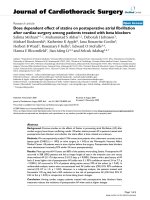
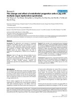
Fig. 1 Protein expression of
antioxidant enzymes in different
organs in old mice. Indicated
organs were processed as
indicated in ‘‘Materials and
Methods’’ and the presence of
CAT, SOD1, GPX1, GR,
CytB5Rase, NQO1, TRxR1 and
TRxR2 proteins determined by
Western blotting. Quantification
was performed considering
Ponceau Red staining as loading
control. Results refer to the
levels found in each organ of the
control group. *Significant
differences vs. control levels in
each organ. P \ 0.05
Furthermore, the relationship between antioxidant activities must also be taken into consideration. In fact, it was
proposed that the imbalance in the ratio of SOD to CAT
and GPx results in the accumulation of H2O2 that through
the Fenton reaction results in the formation of hydroxyl
radicals which are highly reactive and damage
macromolecules such as DNA, protein and lipids. For this
reason, the balance in the activity of these enzymes has
been directly related to cell senescence [19, 42]. Interestingly, a previous work demonstrated that the R ratio
[R = SOD/(CAT ? GPx) in activities] increases in liver
along with age [12]. The results shown in this manuscript
123
Aging Clin Exp Res
demonstrate that both exercise and RSV decrease this ratio,
indicating a higher protective effect in the liver. However,
R was not affected in the other organs.
In conclusion, our results indicate that both RSV and
exercise improve in different manner the activities of endogenous antioxidant enzymes such as CAT, SOD1, GPx,
GR, GST, NQO1 in old mice, and in some cases preventing
the decrease of these activities associated with aging.
Consequently, RSV supplementation and a higher physical
activity should be strongly encouraged in older people, not
only to improve physical function, avoid sarcopenia and
maintain higher independence, but also to attenuate oxidative damage caused by aging. However, we cannot extrapolate the effects of these interventions in one or few
organs to the whole organism. A deeper study of the
regulation of antioxidant enzymatic activities and expression in relationship with aging is needed.
Acknowledgments We thank Almudena Velazquez Dorado and
Ana Sanchez Cuesta for their technical support. The group was financed by the Andalusian Government as the BIO177 Group through
FEDER funds (European Commission). The research was financed by
the Spanish Government Grant DEP2012-39985 (Spanish Ministry of
Economy and Competitiveness). Tung Bui Thanh received a fellowship from the AECID program (Spanish Ministry of Foreing Affair).
ERB, PN and GLL are also members of the Centro de Investigacio´n
Biome´dica en Red de Enfermedades Raras (CIBERER), Instituto
Carlos III.
Conflict of interest On behalf of all authors, the corresponding
author states that there is no conflict of interest.
Human and Animal Rights All animals were maintained according to a protocol approved by the Ethical Committee of the University
Pablo de Olavide of resolution 03/09 and following the international
rules for animal research. This article does not contain any studies
with humans performed by any of the authors.
References
1. Harman D (1956) Aging: a theory based on free radical and
radiation chemistry. J Gerontol 11(3):298–300
2. Marzetti E, Calvani R, Cesari M et al (2013) Mitochondrial
dysfunction and sarcopenia of aging: from signaling pathways to
clinical trials. Int J Biochem Cell Biol 45(10):2288–2301. doi:10.
1016/j.biocel.2013.06.024
3. Dai DF, Chiao YA, Marcinek DJ (2014) Mitochondrial oxidative
stress in aging and healthspan. Longev Healthspan 3:6. doi:10.
1186/2046-2395-3-6
4. Wolter F, Ulrich S, Stein J (2004) Molecular mechanisms of
the chemopreventive effects of resveratrol and its analogs in
colorectal cancer: key role of polyamines? J Nutr 134(12):
3219–3222
5. Donnelly LE, Newton R, Kennedy GE (2004) Anti-inflammatory
effects of resveratrol in lung epithelial cells: molecular mechanisms. Am J Physiol Lung Cell Mol Physiol 287(4):L774–L783.
doi:10.1152/ajplung.00110.2004
6. Cai YJ, Fang JG, Ma LP (2003) Inhibition of free radical-induced
peroxidation of rat liver microsomes by resveratrol and its analogues. Biochim Biophys Acta 1637(1):31–38
123
7. Kulkarni SS, Canto C (2014) The molecular targets of resveratrol.
Biochim Biophys Acta. doi:10.1016/j.bbadis.2014.10.005
8. Burkewitz K, Zhang Y, Mair WB (2014) AMPK at the nexus of
energetics and aging. Cell Metab 20(1):10–25. doi:10.1016/j.
cmet.2014.03.002
9. Fox JT, Sakamuru S, Huang R (2012) High-throughput genotoxicity assay identifies antioxidants as inducers of DNA damage
response and cell death. Proc Natl Acad Sci USA 109(14):
5423–5428. doi:10.1073/pnas.1114278109
10. Tyagi A, Gu M, Takahata T (2011) Resveratrol selectively induces DNA Damage, independent of Smad4 expression, in its
efficacy against human head and neck squamous cell carcinoma.
Clin Cancer Res 17(16):5402–5411. doi:10.1158/1078-0432.
CCR-11-1072
11. Lopez M, Martinez F, Del Valle C (2003) Study of phenolic
compounds as natural antioxidants by a fluorescence method.
Talanta 60(2–3):609–616. doi:10.1016/S0039-9140(03)00191-7
12. Tung BT, Rodriguez-Bies E, Ballesteros-Simarro M (2014)
Modulation of endogenous antioxidant activity by resveratrol and
exercise in mouse liver is age dependent. J Gerontol A Biol Sci
Med Sci 69(4):398–409. doi:10.1093/gerona/glt102
13. Mercken EM, Carboneau BA, Krzysik-Walker SM (2012) Of
mice and men: the benefits of caloric restriction, exercise, and
mimetics. Ageing Res Rev 11(3):390–398. doi:10.1016/j.arr.
2011.11.005
14. Pallauf K, Giller K, Huebbe P (2013) Nutrition and healthy
ageing: calorie restriction or polyphenol-rich ‘‘MediterrAsian’’
diet? Oxid Med Cell Longev 2013:707421. doi:10.1155/2013/
707421
15. Polidori MC, Mecocci P, Cherubini A (2000) Physical activity
and oxidative stress during aging. Int J Sports Med
21(3):154–157. doi:10.1055/s-2000-8881
16. Yamamoto T, Ohkuwa T, Itoh H (2003) Relation between voluntary physical activity and oxidant/antioxidant status in rats.
Comp Biochem Physiol C Toxicol Pharmacol 135(2):163–168
17. Del Pozo-Cruz J, Rodriguez-Bies E, Ballesteros-Simarro M (2014)
Physical activity affects plasma coenzyme Q10 levels differently
in young and old humans. Biogerontology 15(2):199–211. doi:10.
1007/s10522-013-9491-y
18. Del Pozo-Cruz J, Rodriguez-Bies E, Navas-Enamorado I (2014)
Relationship between functional capacity and body mass index
with plasma coenzyme Q10 and oxidative damage in communitydwelling elderly-people. Exp Gerontol 52:46–54. doi:10.1016/j.
exger.2014.01.026
19. de Haan JB, Cristiano F, Iannello R (1996) Elevation in the ratio
of Cu/Zn-superoxide dismutase to glutathione peroxidase activity
induces features of cellular senescence and this effect is mediated
by hydrogen peroxide. Hum Mol Genet 5(2):283–292
20. Powers SK, Criswell D, Lawler J (1994) Influence of exercise and
fiber type on antioxidant enzyme activity in rat skeletal muscle.
Am J Physiol 266(2 Pt 2):R375–R380
21. Marklund S, Marklund G (1974) Involvement of the superoxide
anion radical in the autoxidation of pyrogallol and a convenient
assay for superoxide dismutase. Eur J Biochem 47(3):469–474
22. Aebi H (1984) Catalase in vitro. Methods Enzymol 105:121–126
23. Anderson ME (1985) Determination of glutathione and glutathione disulfide in biological samples. Methods Enzymol
113:548–555
24. Lawrence RA, Burk RF (1976) Glutathione peroxidase activity in
selenium-deficient rat liver. Biochem Biophys Res Commun
71(4):952–958
25. Carlberg I, Mannervik B (1985) Glutathione reductase. Methods
Enzymol 113:484–490
26. Habig WH, Pabst MJ, Jakoby WB (1974) Glutathione S-transferases. The first enzymatic step in mercapturic acid formation.
J Biol Chem 249(22):7130–7139
Aging Clin Exp Res
27. Strittmatter P, Velick SF (1957) The purification and properties
of microsomal cytochrome reductase. J Biol Chem 228(2):
785–799
28. Benson AM, Hunkeler MJ, Talalay P (1980) Increase of
NAD(P)H:quinone reductase by dietary antioxidants: possible
role in protection against carcinogenesis and toxicity. Proc Natl
Acad Sci USA 77(9):5216–5220
29. Hill KE, McCollum GW, Burk RF (1997) Determination of
thioredoxin reductase activity in rat liver supernatant. Anal
Biochem 253(1):123–125. doi:10.1006/abio.1997.2373
30. Ellman GL (1959) Tissue sulfhydryl groups. Arch Biochem
Biophys 82(1):70–77
31. Gerard-Monnier D, Erdelmeier I, Regnard K (1998) Reactions of
1-methyl-2-phenylindole with malondialdehyde and 4-hydroxyalkenals. Analytical applications to a colorimetric assay of lipid
peroxidation. Chem Res Toxicol 11(10):1176–1183. doi:10.1021/
tx9701790
32. Rodriguez-Bies E, Navas P, Lopez-Lluch G (2015) Age-dependent effect of every-other-day feeding and aerobic exercise in
ubiquinone levels and related antioxidant activities in mice
muscle. J Gerontol A Biol Sci Med Sci 70(1):33–43. doi:10.1093/
gerona/glu002
33. Rodriguez-Bies E, Santa-Cruz Calvo S, Fontan-Lozano A (2010)
Muscle physiology changes induced by every other day feeding
and endurance exercise in mice: effects on physical performance.
PLoS One 5(11):e13900. doi:10.1371/journal.pone.0013900
34. Harman D (1960) The free radical theory of aging: the effect of
age on serum mercaptan levels. J Gerontol 15:38–40
35. Lopez-Lluch G, Rios M, Lane MA (2005) Mouse liver plasma
membrane redox system activity is altered by aging and
modulated by calorie restriction. Age (Dordr) 27(2):153–160.
doi:10.1007/s11357-005-2726-3
36. Nohl H (1993) Involvement of free radicals in ageing: a consequence or cause of senescence. Br Med Bull 49(3):653–667
37. Lopez-Dominguez JA, Khraiwesh H, Gonzalez-Reyes JA (2013)
Dietary fat modifies mitochondrial and plasma membrane apoptotic signaling in skeletal muscle of calorie-restricted mice. Age
(Dordr) 35(6):2027–2044. doi:10.1007/s11357-012-9492-9
38. Lopez-Dominguez JA, Khraiwesh H, Gonzalez-Reyes JA (2014)
Dietary fat and aging modulate apoptotic signaling in liver of
calorie-restricted mice. J Gerontol A Biol Sci Med Sci. doi:10.
1093/gerona/glu045
39. Lopez-Dominguez JA, Ramsey JJ, Tran D (2014) The influence
of dietary fat source on life span in calorie restricted mice.
J Gerontol A Biol Sci Med Sci. doi:10.1093/gerona/glu177
40. Wong YT, Gruber J, Jenner AM (2009) Elevation of oxidativedamage biomarkers during aging in F2 hybrid mice: protection by
chronic oral intake of resveratrol. Free Radic Biol Med
46(6):799–809. doi:10.1016/j.freeradbiomed.2008.12.016
41. Thirunavukkarasu V, Balakrishnan SD, Ravichandran MK (2003)
Influence of 6-week exercise training on erythrocyte and liver
antioxidant defense in hyperinsulinemic rats. Comp Biochem
Physiol C Toxicol Pharmacol 135(1):31–37
42. de Haan JB, Cristiano F, Iannello RC (1995) Cu/Zn-superoxide
dismutase and glutathione peroxidase during aging. Biochem Mol
Biol Int 35(6):1281–1297
123