DSpace at VNU: Good manufacturing practice-compliant isolation and culture of human umbilical cord blood-derived mesenchymal stem cells
Bạn đang xem bản rút gọn của tài liệu. Xem và tải ngay bản đầy đủ của tài liệu tại đây (900.24 KB, 10 trang )
Pham et al. Journal of Translational Medicine 2014, 12:56
/>
RESEARCH
Open Access
Good manufacturing practice-compliant isolation
and culture of human umbilical cord blood-derived
mesenchymal stem cells
Phuc Van Pham*, Ngoc Bich Vu, Vuong Minh Pham, Nhung Hai Truong, Truc Le-Buu Pham, Loan Thi-Tung Dang,
Tam Thanh Nguyen, Anh Nguyen-Tu Bui and Ngoc Kim Phan
Abstract
Background: Mesenchymal stem cells (MSCs) are an attractive source of stem cells for clinical applications. These
cells exhibit a multilineage differentiation potential and strong capacity for immune modulation. Thus, MSCs are
widely used in cell therapy, tissue engineering, and immunotherapy. Because of important advantages, umbilical
cord blood-derived MSCs (UCB-MSCs) have attracted interest for some time. However, the applications of
UCB-MSCs are limited by the small number of recoverable UCB-MSCs and fetal bovine serum (FBS)-dependent
expansion methods. Hence, this study aimed to establish a xenogenic and allogeneic supplement-free expansion
protocol.
Methods: UCB was collected to prepare activated platelet-rich plasma (aPRP) and mononuclear cells (MNCs).
aPRP was applied as a supplement in Iscove modified Dulbecco medium (IMDM) together with antibiotics. MNCs
were cultured in complete IMDM with four concentrations of aPRP (2, 5, 7, or 10%) or 10% FBS as the control.
The efficiency of the protocols was evaluated in terms of the number of adherent cells and their expansion, the
percentage of successfully isolated cells in the primary culture, surface marker expression, and in vitro differentiation
potential following expansion.
Results: The results showed that primary cultures with complete medium containing 10% aPRP exhibited the
highest success, whereas expansion in complete medium containing 5% aPRP was suitable. UCB-MSCs isolated
using this protocol maintained their immunophenotypes, multilineage differentiation potential, and did not form
tumors when injected at a high dose into athymic nude mice.
Conclusion: This technique provides a method to obtain UCB-MSCs compliant with good manufacturing practices
for clinical application.
Keywords: Mesenchymal stem cells, Platelet-rich plasma, Umbilical cord blood, Good manufacturing practice,
Clinical application
Introduction
Mesenchymal stem cells (MSCs) are one of the most
studied and applied types of stem cells to date. These
cells were first described by Friedenstein et al. as a cell
population similar to fibroblasts [1], which can differentiate into multiple cell types such as osteoblasts, adipocytes, and chondrocytes [2]. MSCs have been isolated
from many tissues including bone marrow [3,4], adipose
* Correspondence:
Laboratory of Stem Cell Research and Application, University of Science,
Vietnam National University, Ho Chi Minh city, Vietnam
tissue [5-7], peripheral blood, umbilical cord blood (UCB)
[8-10], banked UCB [11-14], umbilical cords [15,16],
placenta [17], amniotic fluid [18], dental pulp [19], and
menstrual blood [20].
Compared with other stem cell sources, UCB-MSCs
have advantages such as non-invasive recovery, the
abundance of MSCs, and well-known characteristics. In
both pre-clinical and clinical settings, MSCs have been
studied to treat a various diseases. Pre-clinically, UCBMSCs have been used to treat neonatal brain injury [21],
fibrocartilaginous embolic myelopathy [22], spinal cord
© 2014 Pham et al.; licensee BioMed Central Ltd. This is an Open Access article distributed under the terms of the Creative
Commons Attribution License ( which permits unrestricted use, distribution, and
reproduction in any medium, provided the original work is properly credited. The Creative Commons Public Domain
Dedication waiver ( applies to the data made available in this article,
unless otherwise stated.
Pham et al. Journal of Translational Medicine 2014, 12:56
/>
injury [23,24], diabetic renal injury [25,26], bone loss
[27], ischemia [28,29], hearing loss [30], damaged corneal endothelium [31], Alzheimer’s disease [32], graftversus-host disease (GVHD) [33], acute hepatic necrosis
[34], diabetes mellitus [35], and liver cirrhosis [36]. Clinically, UCB-MSCs have been transplanted for treatment
of autism [37], hereditary spinocerebellar ataxia [38], foot
disease in patients with type 2 diabetes mellitus [39], and
basilar artery dissection [40]. Clinical trials (retrieved from
clinicaltrial.gov) include mesenchymal stem cell transplantation for engraftment of unrelated hematopoietic stem
cell transplantation (NCT00823316), treatment of steroidrefractory acute or GVHD (NCT01549665), articular cartilage defect treatment (NCT01733186), and hematologic
malignancy treatment (NCT01854567).
The main concern in UCB-MSC applications is in vitro
expansion that is mostly affected by the culture medium.
For production protocols of UCB-MSCs under clinical
conditions, it is essential to include sterility controls, analysis for viral markers, and genetic testing such as karyotyping. Currently, UCB-MSCs can be produced at a GMP
(good manufacturing practice) grade by automated processing protocols and some novel protocols. Procedures
have been developed to isolate mononuclear cells (MNCs)
in closed systems such as the SEPAX device [41,42]. Other
systems can also be used to expand MSCs such as the
Cell Stack System [43]. However, almost all of these
methods require fetal bovine serum (FBS) for culture. FBSbased medium has some limitations associated with clinical
application, especially prion and viral transmission or adverse
immunological reactions against xenogenic components.
Some novel methods use human serum for MSC culture, especially platelet-rich plasma (PRP). Recent studies have used PRP from peripheral blood [44-48] and
UCB [49-52], which showed that PRP from peripheral
blood or UCB significantly stimulates the proliferation
of MSC from bone marrow [45,50], UCB [49,53], or adipose tissue [44,54]. More importantly, MSCs cultured in
medium supplemented with PRP exhibit a normal phenotype and characteristics [49-52], and maintain their multipotency for differentiation into adipocytes, osteoblasts,
and chondrocytes. Taken together, these studies show that
PRP can replace FBS for in vitro MSC expansion.
All of these previous protocols have used allogeneic
PRP. The use of PRP allows MSCs to avoid xenogenic
immunological reactions, and prion and viral transmission, but MSCs may encounter human viral transmission
and immunological reactions induced by allogeneic components. According to the European Medicines Agency
and regulation No. [EC] 1394/2007 of the European
Commission, MSC are considered as medicinal products
[55] and must be produced in compliance with GMP.
The GMP standards ensure that cells are produced with
the highest standards of sterility, quality control, and
Page 2 of 10
documentation following a standard operating procedure. Therefore, in this study, we aimed to establish an
UCB-MSC isolation protocol using autologous PRP from
the same umbilical blood sample. This protocol is GMP
compliant and can be used for clinical applications.
Materials and methods
UCB collection and sample selection for study
UCB was collected from the umbilical cord vein with informed consent of the mother. The collection was performed in accordance with the ethical standards of the
local ethics committee. To eliminate differences between
UCB samples, the stem cell quantity was enumerated
based on the number of hematopoietic stem cells (HSCs)
using an Enumeration Pro-Count Kit (BD Bioscience)
following the manufacturer’s guidelines. Only samples
with ≥1 × 10⁶ HSCs/ml were used in experiments.
MNC isolation and activated PRP preparation
First, blood samples were centrifuged at 2000 rpm for
15 min. The cell pellet was kept to isolate MNCs and
the plasma was collected and centrifuged at 3500 rpm
for 10 min. To prepare activated PRP (aPRP), a third of
the plasma volume and the platelet pellet was collected
and resuspended, and then 100 μL CaCl2 per 1 mL of
PRP was added to activate growth factor release. The
samples were then incubated at 37°C for 30 min or until
the occurrence of clotting. The centrifuged blood cells
were diluted at a ratio of 1:1 with phosphate buffered solution (PBS) and then applied to density centrifugation
using Ficoll Hypaque (1.077 g/mL; Sigma-Aldrich, St
Louis, MO). The collected MNCs were washed twice
with PBS and then applied to experiments.
Primary culture
Twenty UCB samples were used for primary culture.
MNCs were cultured in Iscove modified Dulbecco medium
(IMDM) containing 1% antibiotic-mycotic (Sigma-Aldrich,
Louis St, MO), 10 ng/mL epidermal growth factor (EGF),
10 ng/mL basic fibroblast growth factor (bFGF), and various concentrations of aPRP (2, 5, 7, or 10%) or 10% fetal
bovine serum (FBS) for the control. The cells were plated
at 5 × 10⁴cells/mL in T-75 flasks (Corning) and incubated
at 37°C with 5% CO2. After 3 days of incubation, 6 mL of
fresh media were added to each flask. After 7 days, the
media were replaced with fresh media. Then, the media
was replaced every 4 days until the cells reached 70–80%
confluence. The efficiency of the media was evaluated by
the time required for adherent cells to appear and then
reach 70–80% confluence for the first subculture.
Secondary culture
After successful primary culture, the samples were subcultured to evaluate the effects of the various media.
Pham et al. Journal of Translational Medicine 2014, 12:56
/>
The proliferation rate was evaluated by the eXCELLIgence system (Roche Applied Science, Indianapolis, IN).
A total of 1 × 10³ cells were seeded into each well of a
96-well E-plate in triplicate. The culture plates were
placed into the eXCELLIgence system and incubated at
37°C with 5% CO2. Cell proliferation was monitored for
300 h with fresh medium changes every third day. Both
the cell doubling time and slope value were determined
by the software of the eXCELLIgence system.
Flow cytometry
Cell markers were analyzed following a previously published protocol [11]. Briefly, cells were washed twice in
PBS containing 1% bovine serum albumin (Sigma-Aldrich).
The cells were then stained with anti-CD13-FITC, antiCD14-FITC, anti-CD34-FITC, anti-CD44-PE, anti-CD45FITC, anti-CD73-FITC, anti-CD90-PE, anti-CD105-FITC,
anti-CD106-PE, anti-CD166-PE, or anti-HLA-DR-FITC
antibodies (all purchased from BD Biosciences, San Jose,
CA). Stained cells were analyzed by a FACSCalibur flow
cytometer (BD Biosciences). Isotype controls were used in
all analyses.
In vitro differentiation
For differentiation into adipogenic cells, UCB-MSCs
were differentiated as described previously [9]. Briefly,
passage 5 cells were plated at 1 × 10⁴ cells/well in 24well plates. At 70% confluence, the cells were cultured
for 21 days in IMDM containing 0.5 mmol/L 3-isobutyl1-methyl-xanthine, 1 nmol/L dexamethasone, 0.1 mmol/L
indo-methacin, and 10% FBS (all purchased from SigmaAldrich). Adipogenic differentiation was evaluated by
observing lipid droplets in cells under a microscope.
For differentiation into osteogenic cells, UCB-MSCs
were plated at 1 × 10⁴ cells/well in 24-well plates. At
70% confluence, the cells were cultured for 21 days in
IMDM containing 10% FBS, 10-7 mol/L dexamethasone,
50 μmol/L ascorbic acid-2 phosphate, and 10 mmol/L
β-glycerol phosphate (all purchased from Sigma-Aldrich)
[9]. Osteogenic differentiation was confirmed by Alizarin
red staining.
Page 3 of 10
For differentiation into chondrogenic cells, UCB-MSCs
were induced to differentiate by a commercial medium for
chondrogenesis (StemPro Chondrogenesis Differentiation
Kit, A10071-01; Life Technologies). UCB-MSCs were differentiated in pellet form according to the manufacturer’s
guidelines. After 21 days, the cell pellets were stained with
an anti-aggrecan monoclonal antibody (BD Biosciences).
Tumorigenicity assay
The tumorigenicity of UCB-MSCs was examined in athymic nude mice. All manipulations of mice were approved
by the Local Ethics Committee of Stem Cell Research and
Application, University of Science (Ho Chi Minh city,
Vietnam). Each mouse was injected subcutaneously with
5 × 10⁶ cells (three mice per group). As a positive control,
the mice were also injected with breast cancer cells at a
different site. Tumor formation in mice was followed up
for 3 months.
Statistical analysis
The significance of differences between mean values was
assessed by t-tests and analysis of variance. A P-value of
less than 0.05 was considered to be significant. All data
were analyzed by Prism 6 software.
Results
Primary cell culture
We collected 30 UCB samples of which 20 were applied
to experiments. For primary culture, MNCs from the
same sample were divided and cultured in five different
media containing 10% FBS or 2, 5, 7, or 10% aPRP. There
were differences in the time needed for MNCs to adhere
and exhibit a particular shape in the various media. For
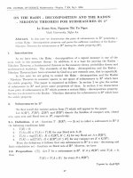
example, at 72 h post-plating, there were clear differences in the number of adhered and fibroblast-like
cells (Figure 1). The trend among the various media indicated that the number of cells gradually increased in 2,
5, 7, or 10% aPRP or 10% FBS in that order. The data
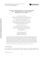
from the 20 samples are presented in Figure 2.
As shown in Figure 2A, fibroblast-like cells appeared
the most rapidly in 10% aPRP (46.20 ± 8.94 h), which
Figure 1 UCB-MSC candidates adhered and exhibited a particular shape. After 72 h of culture, adherent UCB-MSC candidates appeared in
all media. However, the highest numbers of cells appeared in 10% FBS (A) and 10% aPRP (E), and the number of adherent cells gradually
decreased in 7% (D), 5% (C) and 2% aPRP (B).
Pham et al. Journal of Translational Medicine 2014, 12:56
/>
Page 4 of 10
Figure 2 Timing of adherence and confluence of primary cultured cells. In 10% aPRP, MNCs adhered the soonest, and the adherence time
gradually increased as the concentration of aPRP was decreased gradually (A). Consequently, the time needed to reach 70–80% confluence in
10% aPRP was the shortest, which gradually increased as the concentration of aPRP decreased gradually (B).
was significantly sooner than that in 7% aPRP (52.80 ±
10.59 h), 5% aPRP (60.60 ± 10.64 h), 2% aPRP (61.80 ±
10.50 h), and 10% FBS (52.80 ± 12.56 h). These results
showed that increases of the aPRP concentration led to
decreases in the time needed for MNCs to adhere and
exhibit a fibroblastic shape, indicating that components
of aPRP were important for adherence and proliferation
of UCB-MSCs.
These times also dictated the number of days until
the first subculture (Figure 2B). As a result, 70–80%
confluence was reached at 23.35 ± 4.73 days in 10%
aPRP, whereas 25.50 ± 4.62, 30.40 ± 5.05, 30.40 ± 5.05,
and 26.55 ± 7.05 days were required in 7, 5, or 2%
aPRP, or 10% FBS, respectively.
Cell proliferation rates in the various media
After subculture, five samples were used to evaluate the
effects of the various media on UCB-MSC proliferation.
The results are presented in Figure 3. The proliferation
rates of the cells in the various media were recorded
from 0 to 300 h. At 0–130 h, the proliferation rates
among the media were not significantly different. However, from 130 to 260 h, the proliferation rates were significantly different in 5, 7, or 10% aPRP, or 10% FBS compared
with that in 2% aPRP. From 260 to 300 h, cells in all the
various media underwent contact inhibition and death. As
shown in Figure 3, the proliferation rate of cells in 10%
aPRP was higher than that in the other media but the
difference was not significant.
We also compared the cell doubling times and slope
values (Figure 4). The doubling time is the time needed
for the cell population to double in number. There were
no differences in the doubling times for the whole
period from 0 to 300 h. However, there were significant
changes at each period from 0 to 130 h and 130 to
260 h. In the early stage (0–130 h), the doubling times
were similar between the media (24.9 ± 6.11, 22.6 ± 4.9,
28.1 ± 5.71, 27.6 ± 5.52, and 25.5 ± 6.48 h in 10% FBS or
2, 5, 7, or 10% aPRP, respectively) (P < 0.05). In the next
period (from 130 to 260 h), cells in all the various media
proliferated with increasing doubling times. At this
stage, the doubling times were similar in 5, 7, or 10%
aPRP (P < 0.05) (131 ± 2.51, 134 ± 3, and 134 ± 2.49 h in
5, 7, or 10% aPRP, respectively). In contrast, the doubling
time in 2% aPRP suddenly increased to 148 ± 3.63 h. In
10% FBS, the doubling time was 139 ± 2.82 h which was
lower than that in 2% aPRP but higher than that in 5, 7,
or 10% aPRP, but the difference was not significant.
These data further confirmed that the proliferation
rate of UCB-MSCs in 5, 7, or 10% aPRP were similar to
that in 10% FBS, but higher than that in 2% aPRP. The
slope values also supported these results. In the early
stage (0–130 h), there were no differences in the slope
values among the media (0.021 ± 0.001, 0.018 ± 0.001,
0.020 ± 0.001, 0.021 ± 0.001, and 0.021 ± 0.001 for 10%
FBS or 2, 5, 7, or 10% aPRP, respectively). In the next
stage (130–260 h), the slope values exhibited significant differences between 10% FBS or 5, 7, or 10% aPRP
Figure 3 Cell proliferation in the various media as recorded by
the exCelligence method. The results showed that the proliferation
rates of cells in 5, 7, or 10% aPRP, or 10% FBS were not significantly
different, while there was a significant difference in 2% aPRP.
Pham et al. Journal of Translational Medicine 2014, 12:56
/>
Page 5 of 10
Figure 4 Doubling times and slope values in the various media. The values were obtained at three stages, 0–130 h, 130–260 h, and 260–300 h,
and the whole proliferation curve at 0–300 h. There were no significant differences between the doubling times (A) and slope values (B) in 5, 7, or
10% aPRP, or 10% FBS, but a significant difference in 2% aPRP.
Figure 5 Immunophenotypes of MSCs in the various media. The results include flow cytometric analysis of CD13, CD14, CD34, CD44, CD45,
CD73, CD90, CD105, CD106, CD166, and HLA-DR (A). Data were analyzed and presented in a graph (B).
Pham et al. Journal of Translational Medicine 2014, 12:56
/>
(0.023 ± 0.001, 0.023 ± 0.001, 0.023 ± 0.001, and 0.024 ±
0.001, respectively) compared with that in 2% aPRP.
Phenotypes of MSCs
MSC-specific marker expression of UCB-MSCs cultured
in the various media were evaluated and compared as
presented in Figure 5. The results showed that UCBMSCs in all the media exhibited a MSC-specific marker
profile, positivity for CD13, CD44, CD73, CD90, CD105,
CD106, and CD166, and negativity for CD14, CD34,
CD45, and HLA-DR. The expression levels of positive
markers in MSCs were similar among the media.
UCB-MSCs cultured in the various media were examined for their capacity to differentiate into adipogenic,
osteogenic, and chondrogenic lineages. The results of
differentiation assays are presented in Figures 6 and 7.
In all media, the cells successfully differentiated into adipocytes, osteoblasts, and chondrocytes. Compared with
cells prior to induction (Figure 6A–E), cells in all media
accumulated lipid droplets in their cytoplasm after induction with adipocyte differentiation medium (Figure 6F–K).
Following induction with osteoblast differentiation medium,
cells in all media accumulated Ca2+ and Mg2+ in the cytoplasm and extracellular matrix, which were stained with
Alizarin red (Figure 6I–P). After 21 days of chondrogenic
differentiation, cells in all media expressed aggrecan,
a cartilage-specific proteoglycan core protein (Figure 7).
Tumorigenicity of UCB-MSCs
UCB-MSCs cultured in the various media were injected
into athymic nude mice. Breast cancer cells were also
Page 6 of 10
injected at a different location as a positive control. Injection of UCB-MSCs resulted in no tumor formation,
whereas injection of breast cancer cells resulted in
tumor formation in all mice (Figure 8).
Discussion
The aim of this study was to establish a GMP-compliant
protocol for isolation of UCB-MSC for clinical application.
Therefore, we eliminated xenogenic and allogeneic components that can cause immunological reactions and viral
transmission. Our approach replaced FBS in the medium
with autologous aPRP that was isolated from the same
UCB sample used to isolate MNCs.
Because the source of autologous aPRP was limited
and the necessary number of MSCs for clinical application is high, we evaluated four concentrations of aPRP in
complete medium, including 2, 5, 7, and 10%, and 10%
FBS as a control. The effects of aPRP on MSC proliferation was evaluated in primary and secondary cultures. In
primary culture, medium containing 10% aPRP significantly stimulated MSC proliferation compared with that
of the other aPRP concentrations and 10% FBS. In
medium containing 10% aPRP, MSCs adhered quickly and
proliferated rapidly. These observations demonstrated that
aPRP contains all the essential components similar to those
in FBS for support of cell attachment and proliferation. In
fact, aPRP contains high amounts of attachment proteins
such as fibrin, fibronectin, vitronectin, and thrombospondin [49,56]. In addition, aPRP contains several growth
factors that stimulate cell proliferation, such as EGF,
acidic fibroblast growth factor, keratinocyte growth factor,
Figure 6 MSCs cultured in the various media maintain their potential for differentiation into adipocytes and osteoblasts. Compared with
the control (A, B, C, D, and E are 10% FBS or 2, 5, 7, or 10% aPRP, respectively), MSCs accumulated lipid droplets in their cytoplasm after 21 days of
induction (F, G, H, I, and K are 10% FBS or 2, 5, 7, or 10% aPRP, respectively). Following induction with osteoblast differentiation medium, the cells
differentiated into osteoblasts that were positive for Alizarin red staining (L, M, N, O, and P are 10% FBS or 2, 5, 7, or 10% aPRP, respectively).
Pham et al. Journal of Translational Medicine 2014, 12:56
/>
Page 7 of 10
Figure 7 MSCs cultured in the various media can differentiate into chondrocytes. Differentiated cells (A, E, I, R, and N are 10% FBS or 2, 5, 7,
or 10% aPRP, respectively) were stained with Hoescht 33342 B, F, K, O, and S are 10% FBS or 2, 5, 7, or 10% aPRP, respectively) and an anti-aggrecan
monoclonal antibody (C, G, L, P and T are 10% FBS or 2, 5, 7, or 10% aPRP, respectively). Merged images with brightfield as shown in D, H, M, Q,
and U for 10% FBS or 2, 5, 7, or 10% aPRP, respectively.
vascular endothelial growth factor, platelet-derived growth
factor, hepatocyte growth factor, and bFGF [49,52,57].
Compared with bovine growth factors in FBS, aPRP can
be obtained from humans, allowing better interactions between growth factors and cell receptors. In primary culture, the time needed to reach confluency in 10% aPRP
indicated that this concentration of aPRP induced stronger MSC proliferation than that of FBS.
In expansion culture, the effects of the four concentrations of aPRP were also evaluated alongside the 10% FBS
control. The results showed that there were no differences
between 5, 7, or 10% aPRP supplementation compared with
10% FBS, but these aPRP concentrations showed significantly different effects than those of 2% aPRP. In fact, we
confirmed these effects by the proliferation curve, doubling time, and slope value for proliferation. Based on proliferation curves, we could easily recognize differences in
the proliferation rates between 2% aPRP and the other
aPRP concentrations. However, the proliferation rates in
5, 7, or 10% aPRP or 10% FBS were not increased significantly. These data indicated the differences in the growth
factor concentrations at 5, 7 and 10% aPRP did not cause
Pham et al. Journal of Translational Medicine 2014, 12:56
/>
Page 8 of 10
Figure 8 Tumorigenicity of UCB-MSCs in athymic nude mice. MSCs from all groups (10% FBS (A) or 2 (B), 5 (C), 7 (D), or 10% aPRP (E)) could
not cause tumors in the athymic nude mice while breast cancer cells easily caused tumors when injected in the same mice. MSCs were injected
in the left breast; and breast cancer cells were injected in the left breast.
any significant difference in the proliferation of UCBMSCs.
Other important properties that we evaluated were
the effects of aPRP-containing medium on surface marker
expression and multilineage differentiation of UCB-MSCs.
We used the marker profile of positive and negative
markers suggested by Domicini et al. [58]. The results
showed that UCB-MSCs maintained their marker expression in aPRP-containing media compared with that in
medium supplemented with FBS. UCB-MSCs cultured
in aPRP- and FBS-containing media did not express
hematopoietic markers, such as CD14, CD34 and CD45,
or HLA-DR, while they expressed stromal cell markers
such as CD13, CD44, CD73, CD90, CD105, and CD106.
These results completely agreed with other studies of UCBMSCs cultured in FBS-supplemented medium [8,9,59-61],
human peripheral blood-derived PRP [62], and UCBderived PRP [49-51]. UCB-MSCs cultured in the various
media also exhibited multilineage differentiation to adipocytes, osteoblasts, and chondrocytes. These results
were similar to those of UCB-MSCs isolated in serumsupplemented medium [8,9,59-61].
Some previous studies have shown that PRP has some
effects on MSCs. In addition to PRP strongly stimulating
MSC proliferation, PRP also triggers differentiation. However, these effects of PRP are different between the various types of MSCs. PRP induces UCB-MSCs and bone
marrow-derived MSCs to differentiate into osteoblasts
[46], [53,63] and adipose-derived stem cells to differentiate
into chondrocytes [7]. In this study, we did not evaluate
the effects of PRP on UCB-MSC differentiation. However,
we found that MSCs cultured in medium containing 2, 5,
7, or 10% aPRP maintained their potential for differentiation into adipocytes, osteoblasts, and chondrocytes. This
result indicated that aPRP cultured UCB-MSCs had not
become mature cells such as osteoblasts or chondrocytes.
In fact, in previous studies, although MSCs have been proposed to differentiate into osteoblasts and chondrocytes,
they also maintain their differentiation capacity for adipocytes, osteoblasts, and chondrocytes [62,64]. In our final
analysis, UCB-MSCs cultured in the various media were
examined for tumorigenicity in athymic nude mice. The
results showed that all mice injected with UCB-MSCs cultured in the various media showed no tumor formation at
the injection site, while cancer cells caused tumor formation in all mice at their injection site.
In summary, we successfully established a protocol for
isolation of GMP-compliant UCB-MSCs. For primary
culture, IMDM plus 10% aPRP is appropriate. For expansion, culture medium plus 5% aPRP is suitable. This
protocol complies with GMP because of its xenogenicand allogeneic-free medium components.
Conclusion
UCB is a rich source of MSCs. UCB-MSCs can be isolated
with xenogenic and allogeneic component-free medium. In
this study, we successfully established a GMP-compliant
UCB-MSC isolation protocol. Autologous aPRP can be
used to replace FBS. Both aPRP and MNCs can be isolated
from the same blood sample. In primary culture, MNCs
should be cultured in IMDM plus 10% aPRP and 1%
antibiotic-mycotic. However, in expansion culture, MSCs
should be cultured in IMDM plus 5% aPRP and 1%
antibiotic-mycotic. MSCs isolated by this protocol proliferate similarly as those in 10% FBS, maintain MSC phenotypes such as expression of CD13, CD44, CD73, CD90,
CD105, CD106, and CD166, and do not express CD14,
CD34, CD45, or HLA-DR. They also maintain their multilineage differentiation potential for adipocytes, osteoblast,
and chondrocytes. In particular, the isolated MSCs do not
form tumors at a high dose in athymic nude mice. This
promising protocol is suitable for clinical applications of
UCB-MSCs in the near future.
Competing interests
The authors declare that they have no competing interests.
Authors’ contributions
PVP, NBV conceived the study, performed PRP preparation, evaluated the
effects of PRP on mesenchymal stem cell proliferation. VMP, NHT primarily
cultured mesenchymal stem cells from mononuclear cells; TLBP, TTN
collected umbilical cord blood, isolated mononuclear cells from umbilical
Pham et al. Journal of Translational Medicine 2014, 12:56
/>
cord blood; LTTD, ANTB carried out the differentiation assays; NKP evaluated
the tumorigenecity of MSCs in mice model. All authors read and approved
the final manuscript.
Acknowledgements
This work was funded by grants from Vietnam National University, Ho Chi
Minh city, Vietnam.
Received: 6 November 2013 Accepted: 19 February 2014
Published: 24 February 2014
References
1. Friedenstein AJ, Piatetzky S II, Petrakova KV: Osteogenesis in transplants of
bone marrow cells. J Embryol Exp Morphol 1966, 16:381–390.
2. Friedenstein AJ, Deriglasova UF, Kulagina NN, Panasuk AF, Rudakowa SF,
Luria EA, Ruadkow IA: Precursors for fibroblasts in different populations
of hematopoietic cells as detected by the in vitro colony assay method.
Exp Hematol 1974, 2:83–92.
3. Prockop DJ, Sekiya I, Colter DC: Isolation and characterization of rapidly
self-renewing stem cells from cultures of human marrow stromal cells.
Cytotherapy 2001, 3:393–396.
4. Jones EA, Kinsey SE, English A, Jones RA, Straszynski L, Meredith DM,
Markham AF, Jack A, Emery P, McGonagle D: Isolation and characterization
of bone marrow multipotential mesenchymal progenitor cells.
Arthritis Rheum 2002, 46:3349–3360.
5. Bunnell BA, Flaat M, Gagliardi C, Patel B, Ripoll C: Adipose-derived stem
cells: isolation, expansion and differentiation. Methods 2008, 45:115–120.
6. Boquest AC, Shahdadfar A, Brinchmann JE, Collas P: Isolation of stromal
stem cells from human adipose tissue. Methods Mol Biol 2006, 325:35–46.
7. Van Pham P, Bui KH, Ngo DQ, Vu NB, Truong NH, Phan NL, Le DM, Duong
TD, Nguyen TD, Le VT, Phan NK: Activated platelet-rich plasma improves
adipose-derived stem cell transplantation efficiency in injured articular
cartilage. Stem Cell Res Ther 2013, 4:91.
8. Bieback K, Kern S, Kluter H, Eichler H: Critical parameters for the isolation
of mesenchymal stem cells from umbilical cord blood. Stem Cells 2004,
22:625–634.
9. Lee OK, Kuo TK, Chen WM, Lee KD, Hsieh SL, Chen TH: Isolation of
multipotent mesenchymal stem cells from umbilical cord blood.
Blood 2004, 103:1669–1675.
10. Mareschi K, Biasin E, Piacibello W, Aglietta M, Madon E, Fagioli F: Isolation
of human mesenchymal stem cells: bone marrow versus umbilical cord
blood. Haematologica 2001, 86:1099–1100.
11. Phuc PV, Nhung TH, Loan DT, Chung DC, Ngoc PK: Differentiating of
banked human umbilical cord blood-derived mesenchymal stem cells
into insulin-secreting cells. In Vitro Cell Dev Biol Anim 2011, 47:54–63.
12. Phuc PV, Ngoc VB, Lam DH, Tam NT, Viet PQ, Ngoc PK: Isolation of three
important types of stem cells from the same samples of banked
umbilical cord blood. Cell Tissue Bank 2012, 13:341–351.
13. Gong W, Han Z, Zhao H, Wang Y, Wang J, Zhong J, Wang B, Wang S, Wang Y,
Sun L, Han Z: Banking human umbilical cord-derived mesenchymal stromal
cells for clinical use. Cell Transplant 2012, 21:207–216.
14. Matsuo A, Yamazaki Y, Takase C, Aoyagi K, Uchinuma E: Osteogenic
potential of cryopreserved human bone marrow-derived mesenchymal
stem cells cultured with autologous serum. J Craniofac Surg 2008,
19:693–700.
15. Romanov YA, Svintsitskaya VA, Smirnov VN: Searching for alternative
sources of postnatal human mesenchymal stem cells: candidate
MSC-like cells from umbilical cord. Stem Cells 2003, 21:105–110.
16. Wang HS, Hung SC, Peng ST, Huang CC, Wei HM, Guo YJ, Fu YS, Lai MC,
Chen CC: Mesenchymal stem cells in the Wharton's jelly of the human
umbilical cord. Stem Cells 2004, 22:1330–1337.
17. In 't Anker PS, Scherjon SA, Kleijburg-van der Keur C, de Groot-Swings GM,
Claas FH, Fibbe WE, Kanhai HH: Isolation of mesenchymal stem cells of
fetal or maternal origin from human placenta. Stem Cells 2004,
22:1338–1345.
18. Roubelakis MG, Pappa KI, Bitsika V, Zagoura D, Vlahou A, Papadaki HA,
Antsaklis A, Anagnou NP: Molecular and proteomic characterization of
human mesenchymal stem cells derived from amniotic fluid:
comparison to bone marrow mesenchymal stem cells. Stem Cells Dev
2007, 16:931–952.
Page 9 of 10
19. Perry BC, Zhou D, Wu X, Yang FC, Byers MA, Chu TM, Hockema JJ, Woods EJ,
Goebel WS: Collection, cryopreservation, and characterization of human
dental pulp-derived mesenchymal stem cells for banking and clinical use.
Tissue Eng Part C Methods 2008, 14:149–156.
20. Ulrich D, Muralitharan R, Gargett CE: Toward the use of endometrial and
menstrual blood mesenchymal stem cells for cell-based therapies.
Expert Opin Biol Ther 2013, 13:1387–1400.
21. Verina T, Fatemi A, Johnston MV, Comi AM: Pluripotent possibilities:
human umbilical cord blood cell treatment after neonatal brain injury.
Pediatr Neurol 2013, 48:346–354.
22. Chung WH, Park SA, Lee JH, Chung DJ, Choi CB, Kim DH, Han H, Kim HY:
Percutaneous transplantation of human umbilical cord-derived
mesenchymal stem cells in a dog with suspected fibrocartilaginous
embolic myelopathy. J Vet Sci 2013, 14(4):495–497.
23. Lee JH, Chang HS, Kang EH, Chung DJ, Choi CB, Lee JH, Hwang SH, Han H,
Kim HY: Percutaneous transplantation of human umbilical cord bloodderived multipotent stem cells in a canine model of spinal cord injury.
J Neurosurg Spine 2009, 11:749–757.
24. Roh DH, Seo MS, Choi HS, Park SB, Han HJ, Beitz AJ, Kang KS, Lee JH:
Transplantation of human umbilical cord blood or amniotic epithelial
stem cells alleviates mechanical allodynia after spinal cord injury in rats.
Cell Transplant 2013, 22:1577–1590.
25. Park JH, Hwang I, Hwang SH, Han H, Ha H: Human umbilical cord bloodderived mesenchymal stem cells prevent diabetic renal injury through
paracrine action. Diabetes Res Clin Pract 2012, 98:465–473.
26. Park JH, Park J, Hwang SH, Han H, Ha H: Delayed treatment with human
umbilical cord blood-derived stem cells attenuates diabetic renal injury.
Transplant Proc 2012, 44:1123–1126.
27. An JH, Park H, Song JA, Ki KH, Yang JY, Choi HJ, Cho SW, Kim SW, Kim SY,
Yoo JJ, Baek WY, Kim JE, Choi SJ, Oh W, Shin CS: Transplantation of human
umbilical cord blood-derived mesenchymal stem cells or their conditioned
medium prevents bone loss in ovariectomized nude mice. Tissue Eng Part A
2013, 19:685–696.
28. Roura S, Bago JR, Soler-Botija C, Pujal JM, Galvez-Monton C, Prat-Vidal C,
Llucia-Valldeperas A, Blanco J, Bayes-Genis A: Human umbilical cord bloodderived mesenchymal stem cells promote vascular growth in vivo. PLoS
One 2012, 7:e49447.
29. Lim JY, Jeong CH, Jun JA, Kim SM, Ryu CH, Hou Y, Oh W, Chang JW,
Jeun SS: Therapeutic effects of human umbilical cord blood-derived
mesenchymal stem cells after intrathecal administration by lumbar
puncture in a rat model of cerebral ischemia. Stem Cell Res Ther 2011,
2:38.
30. Choi MY, Yeo SW, Park KH: Hearing restoration in a deaf animal model
with intravenous transplantation of mesenchymal stem cells derived
from human umbilical cord blood. Biochem Biophys Res Commun 2012,
427:629–636.
31. Joyce NC, Harris DL, Markov V, Zhang Z, Saitta B: Potential of human
umbilical cord blood mesenchymal stem cells to heal damaged corneal
endothelium. Mol Vis 2012, 18:547–564.
32. Kim JY, Kim DH, Kim JH, Lee D, Jeon HB, Kwon SJ, Kim SM, Yoo YJ, Lee EH,
Choi SJ, Seo SW, Lee JI, Na DL, Yang YS, Oh W, Chang JW: Soluble
intracellular adhesion molecule-1 secreted by human umbilical cord
blood-derived mesenchymal stem cell reduces amyloid-beta plaques.
Cell Death Differ 2012, 19:680–691.
33. Gregoire-Gauthier J, Selleri S, Fontaine F, Dieng MM, Patey N, Despars G,
Beausejour CM, Haddad E: Therapeutic efficacy of cord blood-derived
mesenchymal stromal cells for the prevention of acute graft-versus-host
disease in a xenogenic mouse model. Stem Cells Dev 2012, 21:1616–1626.
34. Shi LL, Liu FP, Wang DW: Transplantation of human umbilical cord blood
mesenchymal stem cells improves survival rates in a rat model of acute
hepatic necrosis. Am J Med Sci 2011, 342:212–217.
35. Ngoc PK, Phuc PV, Nhung TH, Thuy DT, Nguyet NT: Improving the efficacy
of type 1 diabetes therapy by transplantation of immunoisolated
insulin-producing cells. Hum Cell 2011, 24:86–95.
36. Jung KH, Uhm YK, Lim YJ, Yim SV: Human umbilical cord blood-derived
mesenchymal stem cells improve glucose homeostasis in rats with liver
cirrhosis. Int J Oncol 2011, 39:137–143.
37. Lv YT, Zhang Y, Liu M, Qiuwaxi JN, Ashwood P, Cho SC, Huan Y, Ge RC,
Chen XW, Wang ZJ, Kim BJ, Hu X: Transplantation of human cord blood
mononuclear cells and umbilical cord-derived mesenchymal stem cells
in autism. J Transl Med 2013, 11:196.
Pham et al. Journal of Translational Medicine 2014, 12:56
/>
38. Jin JL, Liu Z, Lu ZJ, Guan DN, Wang C, Chen ZB, Zhang J, Zhang WY, Wu JY,
Xu Y: Safety and efficacy of umbilical cord mesenchymal stem cell
therapy in hereditary spinocerebellar ataxia. Curr Neurovasc Res 2013,
10:11–20.
39. Li XY, Zheng ZH, Li XY, Guo J, Zhang Y, Li H, Wang YW, Ren J, Wu ZB:
Treatment of foot disease in patients with type 2 diabetes mellitus using
human umbilical cord blood mesenchymal stem cells: response and
correction of immunological anomalies. Curr Pharm Des 2013,
19:4893–4899.
40. Han H, Chang SK, Chang JJ, Hwang SH, Han SH, Chun BH: Intrathecal
injection of human umbilical cord blood-derived mesenchymal stem
cells for the treatment of basilar artery dissection: a case report. J Med
Case Rep 2011, 5:562.
41. Dazey B, Duchez P, Letellier C, Vezon G, Ivanovic Z: Cord blood processing
by using a standard manual technique and automated closed system
“Sepax” (Kit CS-530). Stem Cells Dev 2005, 14:6–10.
42. Solves P, Planelles D, Mirabet V, Blanquer A, Carbonell-Uberos F: Qualitative
and quantitative cell recovery in umbilical cord blood processed by two
automated devices in routine cord blood banking: a comparative study.
Blood Transfus 2013, 11:405–411.
43. Aktas M, Buchheiser A, Houben A, Reimann V, Radke T, Jeltsch K, Maier P,
Zeller WJ, Kogler G: Good manufacturing practice-grade production of
unrestricted somatic stem cell from fresh cord blood. Cytotherapy 2010,
12:338–348.
44. Kocaoemer A, Kern S, Kluter H, Bieback K: Human AB serum and thrombinactivated platelet-rich plasma are suitable alternatives to fetal calf serum
for the expansion of mesenchymal stem cells from adipose tissue.
Stem Cells 2007, 25:1270–1278.
45. Bieback K, Hecker A, Kocaomer A, Lannert H, Schallmoser K, Strunk D,
Kluter H: Human alternatives to fetal bovine serum for the expansion
of mesenchymal stromal cells from bone marrow. Stem Cells 2009,
27:2331–2341.
46. Jonsdottir-Buch SM, Lieder R, Sigurjonsson OE: Platelet lysates produced
from expired platelet concentrates support growth and osteogenic
differentiation of mesenchymal stem cells. PLoS One 2013, 8:e68984.
47. Rauch C, Feifel E, Amann EM, Spotl HP, Schennach H, Pfaller W,
Gstraunthaler G: Alternatives to the use of fetal bovine serum: human
platelet lysates as a serum substitute in cell culture media. ALTEX 2011,
28:305–316.
48. Blande IS, Bassaneze V, Lavini-Ramos C, Fae KC, Kalil J, Miyakawa AA,
Schettert IT, Krieger JE: Adipose tissue mesenchymal stem cell expansion
in animal serum-free medium supplemented with autologous human
platelet lysate. Transfusion 2009, 49:2680–2685.
49. Ding Y, Yang H, Feng JB, Qiu Y, Li DS, Zeng Y: Human umbilical cord-derived
MSC culture: the replacement of animal sera with human cord blood plasma.
In Vitro Cell Dev Biol Anim 2013, 49(10):771–777.
50. Shetty P, Bharucha K, Tanavde V: Human umbilical cord blood serum can
replace fetal bovine serum in the culture of mesenchymal stem cells.
Cell Biol Int 2007, 31:293–298.
51. Ma HY, Yao L, Yu YQ, Li L, Ma L, Wei WJ, Lu XM, Du LL, Jin YN: An effective
and safe supplement for stem cells expansion ex vivo: cord blood
serum. Cell Transplant 2012, 21:857–869.
52. Murphy MB, Blashki D, Buchanan RM, Yazdi IK, Ferrari M, Simmons PJ,
Tasciotti E: Adult and umbilical cord blood-derived platelet-rich plasma
for mesenchymal stem cell proliferation, chemotaxis, and cryopreservation. Biomaterials 2012, 33:5308–5316.
53. Baba K, Yamazaki Y, Ishiguro M, Kumazawa K, Aoyagi K, Ikemoto S, Takeda
A, Uchinuma E: Osteogenic potential of human umbilical cord-derived
mesenchymal stromal cells cultured with umbilical cord blood-derived
fibrin: A preliminary study. J Craniomaxillofac Surg 2013, 41(8):775–782.
54. Escobedo-Lucea C, Bellver C, Gandia C, Sanz-Garcia A, Esteban FJ, Mirabet V,
Forte G, Moreno I, Lezameta M, Ayuso-Sacido A, Garcia-Verdugo JM:
A xenogeneic-free protocol for isolation and expansion of human
adipose stem cells for clinical uses. PLoS One 2013, 8:e67870.
55. Fekete N, Rojewski MT, Furst D, Kreja L, Ignatius A, Dausend J,
Schrezenmeier H: GMP-compliant isolation and large-scale expansion of
bone marrow-derived MSC. PLoS One 2012, 7:e43255.
56. Marx RE: Platelet-rich plasma: evidence to support its use. J Oral
Maxillofac Surg 2004, 62:489–496.
57. Lee JY, Nam H, Park YJ, Lee SJ, Chung CP, Han SB, Lee G: The effects of
platelet-rich plasma derived from human umbilical cord blood on the
Page 10 of 10
58.
59.
60.
61.
62.
63.
64.
osteogenic differentiation of human dental stem cells. In Vitro Cell Dev
Biol Anim 2011, 47:157–164.
Dominici M, Le Blanc K, Mueller I, Slaper-Cortenbach I, Marini F, Krause D,
Deans R, Keating A, Prockop D, Horwitz E: Minimal criteria for defining
multipotent mesenchymal stromal cells. The international society for
cellular therapy position statement. Cytotherapy 2006, 8:315–317.
Hua J, Gong J, Meng H, Xu B, Yao L, Qian M, He Z, Zou S, Zhou B, Song Z:
Comparison of different methods for the isolation of mesenchymal stem
cells from umbilical cord matrix: proliferation and multilineage
differentiation as compared to mesenchymal stem cells from umbilical
cord blood and bone marrow. Cell Biol Int 2013: [Epub ahead of print].
Lee MW, Yang MS, Park JS, Kim HC, Kim YJ, Choi J: Isolation of
mesenchymal stem cells from cryopreserved human umbilical cord
blood. Int J Hematol 2005, 81:126–130.
Laitinen A, Laine J: Isolation of mesenchymal stem cells from human cord
blood. Curr Protoc Stem Cell Biol 2007, Chapter 2:Unit 2A 3.
Prins HJ, Rozemuller H, Vonk-Griffioen S, Verweij VG, Dhert WJ, SlaperCortenbach IC, Martens AC: Bone-forming capacity of mesenchymal
stromal cells when cultured in the presence of human platelet lysate as
substitute for fetal bovine serum. Tissue Eng Part A 2009, 15:3741–3751.
Jung J, Moon N, Ahn JY, Oh EJ, Kim M, Cho CS, Shin JC, Oh IH:
Mesenchymal stromal cells expanded in human allogenic cord blood
serum display higher self-renewal and enhanced osteogenic potential.
Stem Cells Dev 2009, 18:559–571.
Vogel JP, Szalay K, Geiger F, Kramer M, Richter W, Kasten P: Platelet-rich
plasma improves expansion of human mesenchymal stem cells and
retains differentiation capacity and in vivo bone formation in calcium
phosphate ceramics. Platelets 2006, 17:462–469.
doi:10.1186/1479-5876-12-56
Cite this article as: Pham et al.: Good manufacturing practice-compliant
isolation and culture of human umbilical cord blood-derived mesenchymal
stem cells. Journal of Translational Medicine 2014 12:56.
Submit your next manuscript to BioMed Central
and take full advantage of:
• Convenient online submission
• Thorough peer review
• No space constraints or color figure charges
• Immediate publication on acceptance
• Inclusion in PubMed, CAS, Scopus and Google Scholar
• Research which is freely available for redistribution
Submit your manuscript at
www.biomedcentral.com/submit