DSpace at VNU: The production of β-glucosidases by Fusarium proliferatum NBRC109045 isolated from Vietnamese forest
Bạn đang xem bản rút gọn của tài liệu. Xem và tải ngay bản đầy đủ của tài liệu tại đây (583.67 KB, 13 trang )
Gao et al. AMB Express 2012, 2:49
/>
ORIGINAL ARTICLE
Open Access
The production of β-glucosidases by
Fusarium proliferatum NBRC109045 isolated
from Vietnamese forest
Ziqing Gao1, Duong Van Hop2, Le Thi Hoang Yen2, Katsuhiko Ando3, Shuichi Hiyamuta4 and Ryuichiro Kondo1*
Abstract
Fusarium proliferatum NBRC109045 is a filamentous fungus isolated from Vietnamese forest due to high production
of β-glucosidases. Production of the enzyme was studied on varied carbon source based mediums. The highest
activity was obtained in medium containing 1% corn stover + 1% wheat bran (3.31 ± 0.14 U/ml). It is interesting to
note that glucose (0.69 ± 0.02 U/ml) gave higher activity and just followed by cellobiose among the di- and
mono-saccharides, which is generally regarded as a universal repressor of hydrolases. We improved the zymogram
method to prove that in response to various carbon sources, F. proliferatum could express various β-glucosidases.
One of the β-glucosidases produced by F. proliferatum growing in corn stover + wheat bran based medium was
partially purified and proved to have high catalytic ability.
Keywords: Fusarium proliferatum, β-glucosidases, Differential expression, The translation elongation factor 1-α
Introduction
Biofuels derived from lignocellulosic biomass are emerging as promising alternatives to fossil fuels to meet the
increasing global energy demands (Ragauskas et al.
2006). One of the key steps in bioconversion process is
the enzymatic hydrolysis of the cellulose polymers in the
biomass to monomeric sugars that are subsequently fermented to ethanol (Percival et al. 2006; Adsul et al.
2007). The three main categories of players in cellulose
hydrolysis are cellobiohydrolases (or exo-1, 4-β-glucanases) (EC 3.2.1.91), endo-1, 4-β-glucanases (EC 3.2.1.4),
and β-glucosidases (EC 3.2.1.21) (Beguin and Aubert
1994). The endo-1, 4-β-glucanases randomly attack cellulose in amorphous zones and release oligomers. The cellobiohydrolases liberate cellobiose from reducing and
non-reducing ends. And finally β-glucosidases hydrolyze
the cellobiose and in some cases the cellooligosaccharides to glucose (Ryu 1980; Wood 1985). Cellulose polymers are degraded to glucose through sequential and
cooperative actions of these enzymes. Cellobiohydrolases
* Correspondence:
1
Department of Agro-Environmental Sciences, Faculty of Agriculture, Kyushu
University, Fukuoka, Japan
Full list of author information is available at the end of the article
and endoglucanases are often inhibited by cellobiose,
making β-glucosidases important in terms of avoiding
decreased hydrolysis rates of cellulose over time due to
cellobiose accumulation (Workman and Day 1982). Low
efficiency and high costs associated with the enzymatic
hydrolysis process present a major bottleneck in the production of ethanol from lignocellulosic feedstocks (Banerjee et al. 2010). For the enzymatic conversion of
biomass to fermentative sugar on a commercial scale, it
is necessary to have all cellulolytic components at the optimal level. Since β-glucosidases activity is low in many
microbial preparations used usually for the saccharification process (Enari 1983). It is necessary to supply additional β-glucosidases to such reaction. In order to
optimize the use of different biomasses, it is important to
identify new β-glucosidases with improved abilities on
the specific biomasses as well as with improved abilities
such as stability and high conversion rates. β-Glucosidases
have potential roles in various fields such as the food,
pharmacology and cosmetic industries and also in the valorisation of some products, due to the properties of this
enzyme to convert and to synthesize biomolecules of high
added value (Esen 1993). There are hundreds of different
β-glucosidic flavor precursors in plants, and their hydrolysis often enhances the quality of the beverages and foods
© 2012 Gao et al.; licensee Springer. This is an Open Access article distributed under the terms of the Creative Commons
Attribution License ( which permits unrestricted use, distribution, and reproduction
in any medium, provided the original work is properly cited.
Gao et al. AMB Express 2012, 2:49
/>
produced from them (Gϋnata 2003; Esen 2003). Aside
from flavor enhancement, foods, feeds, and beverages may
be improved nutritionally by release of vitamins, antioxidants, and other beneficial compounds from their glycosides (Opassiri et al. 2004). Indeed, β-glucosidase can
either degrade or synthesize small carbohydrate polymers,
depending on particular experimental conditions (Crout
and Vic 1998). The β-glucosidases can be arranged in
three groups related to localization: intracellular, cell wall
associated, and extracellular. Primarily the extracellular
β-glucosidases are of industrial interest (Soewnsen 2010).
The number of fungal species on earth is estimated to
1.5 million of which as little as approximately 5% are
known (Hawksworth 1991; 2001). So there is a statement
that calls for all-out effort to unravel the potential of
unknown species found in nature. The identification
and characterization of new fungal species are often
encountered in literature. Cuc Phuong Park and Ba Be
Park is the old national one in Vietnam and boasts an
engaging cultural and wildlife heritage and enchanting
scenery. Covered in a dense forest, these landscapes
are rich and diverse tropical and subtropical species of
microorganisms for wood and plant degradation. In the
present study, a potential β-glucosidases-producing
fungus NBRC109045 was isolated from Ba Be national
park and identified as Fusarium proliferatum. Under optimized conditions, F. proliferatum produces β-glucosidases
with an activity of 3.3 U/ml based on pNPG as substrate
and an activity of 426 U/ml based on cellobiose as substrate. In this paper, we described ways that (a) isolating
and screening microbes to produce considerable quantities
of β-glucosidases; (b) modifying the method of zymogram
to prove that different carbon sources direct varied
β-glucosidases expression in F. proliferatum; (c) assaying
partial purification to prove high catalytic efficiency of
β-glucosidase produced by F. proliferatum growing in corn
stover + wheat bran based medium.
Materials and methods
Materials
Unless specified otherwise, all chemicals were of analytical grade. Solubilized crystalline cellulose was obtained
from Kyokuto Seiyaku Co., Ltd, Japan. Avicel [(R) RH101], 4-methylumbelliferyl-β-D-glucoside (MUG) and
carboxymethyl cellulose (CMC) were products of Sigma
Chemical Co., (St. Louis, Mo, USA). Cellobiose, xylose,
glucose, sucrose, galactose and maltose were purchased
from Wako Pure Chemical Industries, Ltd, Japan.
4-Nitrophenyl-β-D-glucopyranoside monohydrate (pNPG)
was purchased from Tokyo Chemical Industry Co., Ltd,
Japan. Corn stover was collected from Yingkou city, Liaoning Province in China. Wheat bran and bagasse were
obtained from private companies.
Page 2 of 13
Strains isolation
Wood chip of Jatropha carcass, branch and leaves of
J. carcass, wood chip of Manihot esculenta, branch and
leaves of M. esculenta, coconut shell, sugarcane, and rice
straw were used as lignocellulosic sources for degradation in Vietnamese National Park (Ba Be and Cuc
Phuong). One month later, lignocellulosic sources were
dug up. All strains that would be screened were isolated
from degraded biomass samples and washed soil collected. Isolated strains were inoculated on solubilized
crystalline cellulose (CC) plates and CMC plates to cultivate for two weeks (Deguchi et al. 2007). The microbes
that could grow on CC and CMC were picked up and
inoculated onto malt extract agar (MEA).
Screening of β-glucosidases-producing strains
The first step of screening
For primary screening, strains from MEA were plated
on potato dextrose agar (PDA) medium in a 9-cm diameter Petri dish and incubated at 30°C for 5 days. Then
the colonies were inoculated on β-glucosidases (EC
3.2.1.21) screening agar containing 1% of CMC, 0.5% of
MUG, 1.5% of agar, and Mandels salts (Daenen et al.
2008). The cultures were incubated at 30°C for 3 days.
Then the plates were observed under UV light. Colonies
which showed fluorescence were sorted out. It is because
methylumbelliferyl (MU) which was released from MUG
by β-glucosidases can emit fluorescence when induced
by UV light.
The second step of screening
For secondary screening, the mycelium of the βglucosidases-producing isolates obtained from the primary screening was transferred to a new PDA medium
in a 9-cm diameter Petri dish and incubated at 30°C.
Once the fungus covered most of the PDA plate, agar
plates with mycelium were transferred to a sterile
blender containing 25 ml of sterile water and homogenized for 30 s. Ten ml of the fungal homogenate was
used to inoculate into β-glucosidases secondary screening medium containing 1% corn stover + 1% wheat bran
in 100 ml, pH 5.0 Mandels salts medium with KH2PO4
2 g l-1, (NH4)2SO4 1.4 g l-1, urea 0.69 g l-1, CaCl2Á2H2O
0.3 g l-1, MgSO4Á7H2O 0.3 g l-1, and 1 ml trace elements
solution composing of MnSO4 1.6 g l-1, ZnSO4 2 g l-1,
CuSO4 0.5 g l-1, CoSO4 0.5 g l-1 (Saibi et al. 2011) then
incubated at 30°C, 150 rpm for 5 days. Crude enzyme
extract was obtained by centrifuging the liquid medium
at 20 000 g, 4°C for 20 min and collecting the supernatant for confirming the β-glucosidases activity.
Enzyme assay
β-Glucosidases activity towards p-nitrophenyl-β-D-glucopyranoside (pNPG) was measured with use of amount
Gao et al. AMB Express 2012, 2:49
/>
of p-nitrophenol (pNP) liberated from pNPG by using a
calibration curve at 410 nm (Cai et al. 1998). The reaction mixture contained 0.5 ml, 2 mM pNPG in 50 mM
sodium acetate buffer (pH 5.0) and an appropriately
diluted enzyme solution 0.125 ml. After incubation at
45°C for 10 min, the reaction was stopped after adding
1.25 mL, 1 M Na2CO3, and the color that formed as a
result of pNP liberation was measured at 410 nm. One
unit of β-glucosidases activity was defined as the amount
of enzyme required to liberate 1 μmol of pNP per minute under the assay conditions. Specific activity is
defined as the number of units per milligram of protein.
Cellobiase activity was assayed using cellobiose as substrate. The enzymatic reaction mixtures (1 ml) containing 0.25 ml of enzyme solution and 0.75 ml of 0.5%
cellobiose in 50 mM sodium acetate buffer (pH 5.0) were
incubated for 30 min at 50 C. And then the mixtures
were heated at 100 C for 5 min to stop the reaction. The
amount of glucose released was measured by Bio-sensor
(Oji Scientific Instruments Co., Itd). One enzyme unit
was defined as the amount of enzyme that produced
1 μmol of glucose per minute.
Protein concentration determination
Protein concentrations in the enzyme preparations were
determined with application of the method of Bradford
(Bradford 1976) with reference to a standard calibration
curve for bovine serum albumin (BSA).
Strain identification
DNA extraction and PCR amplification from cultures
Mycelia cultured on malt extract agar were harvested
with a spatula, and DNA was extracted with use of a
PrepManW Ultra Reagent (Life Technologies, Carlsbad,
California, USA). ITS-5.8S rDNA (ITS) and the D1/D2
regions of LSU rDNA (LSU) were amplified with the
KOD FX (Toyobo, Osaka, Japan), and with primers ITS5
(GGAAGTAAAAGTCGTAACAAGG) and NL4 (GGTC
CGTGTTTCAAGACGG) (O'Donnell 1993; White et al.
1990). The mixture was processed by following the manufacturer’s instructions of kit. The DNA fragments were
amplified in a T-gradient thermal-cycler (Biometra, Göttingen, Germany). Thermal-cycling program for LSU
and ITS was: initial denaturation at 94°C for 2 min,
30 cycles of denaturation at 98°C for 10 s, annealing at
56°C for 30 s, extension at 68°C for 1 min and a 4°C
soak. Amplified DNA was purified with use of the AgencourtW AMPureW Kit (Agencourt Bioscience, Beverly,
Massachusetts, USA).
DNA sequencing
Sequencing reactions were performed with the BigDyeW
Terminator 3.1 Cycle Sequencing Kit (Applied Biosystems, Foster City, California, USA), and with primers
Page 3 of 13
NL1 (GCATATCAATAAGCGGAGGAAAAG) and NL4
(GGTCCGTGTTTCAAGACGG) for LSU on the Tgradient thermal-cycler (Biometra). This thermal-cycler
program was employed: initial denaturation at 96°C for
1.5 min, 35 cycles of denaturation at 96°C for 10 s,
annealing at 50°C for 5 s, extension at 60°C for 1.5 min
and a 4°C soak. Sequencing reaction products were purified with the AgencourtW CleanSEQW Kit (Agencourt
Bioscience) and sequenced with the ABI PRISMW 3730
Genetic Analyzer (Applied Biosystems). Contiguous
sequences were assembled with ATGC software (Genetyx, Tokyo, Japan).
Phylogenetic analysis
DNA was analyzed with use of CLUSTAL W (Thompson et al. 1994). Based on the EF-1α sequence of Fusarium genus (O'Donnell et al. 2012), phylogenetic tree was
generated with use of the neighbor-joining algorithm in
the MEGA ver5.0. Concordance of the EF-1a gene datasets was evaluated with the partition-homogeneity test
implemented with MEGA (Tamura et al. 2011), using 1
000 random repartitions. The fungus was determined to
be most closely related to Fusarium proliferatum by
comparing it with related strains in GenBank. And the
NBRC deposition number is NBRC109045.
Effect of different carbon sources on β-glucosidases
production by F. proliferatum
The mycelium stored on PDA medium was transferred
to new PDA medium in 9-cm diameter Petri dish and
incubated at 30°C for 5 days. Once the fungus covered
most of the PDA plate, agar plates with mycelium were
transferred to a sterile blender containing 25 ml of sterile water and then homogenized for 30 s. Ten ml of the
fungal homogenate was used to inoculate 100 ml of liquid pre-cultures, pH 7.0. Liquid pre-cultures were
made according to the modified Mandels medium with
and without 0.69 g L-1 urea supplemented with 0.1% of
yeast extract and 1% of glucose (Saibi et al. 2011). After
3 days, the mycelium homogenate made by a sterile
blender was used to inoculate the modified Mandels
medium which containing 2% carbon source with and
without urea as following, wheat bran, corn stover, 1%
wheat bran + 1% corn stover, bagasse, CMC, Avicel cellulose, sucrose, cellobiose, glucose, xylose, galactose and
maltose. β-Glucosidases production by F. proliferatum in
shaking flask batch cultures was carried out at 30°C and
150 rpm. Samples were withdrawn at different times
during 12 days, and then centrifuged at 20 000 g for
20 min. Supernatants as crude enzyme were assayed for
β-glucosidases activity, determined for pH, and analyzed
by zymogram. Each culture was carried out in triplicate.
Gao et al. AMB Express 2012, 2:49
/>
Page 4 of 13
Electrophoresis and zymogram
Zymography is an electrophoretic technique for detection
of purified or partly purified β-glucosidase. Zymography is
based on SDS-PAGE that includes a substrate such as
MUG or pNPG, which can be degraded by β-glucosidases.
The degradation product emits fluorescence or produces
change of color during the reaction period. However, this
is not a practical method to assay β-glucosidases existing
in the crude enzyme because various β-glucosidases existing in the crude enzyme caused overlapping fluorescence
bands. A modified method that combines effective isolation with identification was developed to overcome the
limitation of zymogram in the application on crude
enzyme.
Step1: add the loading buffer for SDS-PAGE to the
crude enzyme solution that was produced by
incubating F. proliferatum in corn sotver + wheat bran
based medium and glucose based medium, but the mix
was not heated at a temperature of 100°C (Laemmli
1970). The mix of the crude enzyme and loading buffer
was injected into the gel. Each sample was injected into
four different wells and then the electrophoresis was
applied.
Step2: After the electrophoresis, the first column of
each sample was cut out of the gel and then treated
with Coomassie Brilliant Blue (CBB) staining. The
remaining gel was soaked in 20 mM, pH8.5 Tris–HCl
buffer for two hours in order to remove SDS, so that
the activity can be regained. The buffer was replaced
every 30 min.
Step3: The first column that had been treated with
CBB staining was used as a marker to cut the protein
bands of the second column. The protein bands cut out
of the second column were soaked in 20 mM pNPG for
10 min at a temperature of 45°C with the aim of active
staining, and then 1.25 ml of 1 M Na2CO3 solution
were added. If the color of the bands changes from
colorlessness to yellow, it means that β-glucosidases
exist in the bands.
Step4: Corresponding bands were cut out of the third
and the fourth column based on positions of active
bands of the second column. The cuts containing
β-glucosidases were soaked in acetate buffer (0.05 M,
pH5.0), and were crushed and separated by
centrifugation. The supernatant was taken out and
mixed with the same volume of loading buffer and
then was analyzed with SDS-PAGE. Protein was stained
with silver stainIIkit (Wako Pure Chemical Industries,
Ltd, Japan).
Partial purification of β-glucosidase
Fine and dried powder of ammonium sulfate was added,
over ice, into the crude extract enzyme to 50% saturation.
Table 1 Screening of microorganism with β-glucosidases production
No.
Serial number
81
SIID11445
Cuc Phuong National Park
Manihot esculenta wood chip
+++
82
SIID11446
Cuc Phuong National Park
Rice straw
+
83
SIID11447
Ba Be National Park
Manihot esculenta wood chip
+
84
SIID11448
Cuc Phuong National Park
Soil around plant chip
+
85
SIID11449
Cuc Phuong National Park
Soil around plant chip
++
86
SIID11450
Cuc Phuong National Park
Soil around plant chip
++
87
SIID11451
Cuc Phuong National Park
Manihot esculenta wood chip
+
88
SIID11452
Cuc Phuong National Park
Jatropha carcass wood chip
+
89
SIID11453
Ba Be National Park
Jatropha carcass wood chip
+
90
SIID11454
Ba Be National Park
Jatropha carcass stems and leaves
+++
91
SIID11455
Cuc Phuong National Park
Jatropha carcass stems and leaves
++
92
SIID11456
Ba Be National Park
Soil around plant chip
+++
93
SIID11457
Ba Be National Park
Jatropha carcass wood chip
+
94
SIID11458
Ba Be National Park
Jatropha carcass wood chip
++
95
SIID11459
Ba Be National Park
Rice straw
+
96
SIID11460
Ba Be National Park
Coconut
+++
97
SIID11461
Ba Be National Park
Rice straw
+
98
SIID11462
Cuc Phuong National Park
Coconut
+
+++: with the brightest fluorescence.
++: with brighter fluorescence.
+: with bright fluorescence.
Sample site
Source
Fluorescence
remarks
Gao et al. AMB Express 2012, 2:49
/>
And then the mix was still stirring at 4°C for 30 min. After
centrifugation (42 500 g, 60 min), supernatant was decanted and the precipitate was discarded. Ammonium sulfate was added to bring the supernatant to 80% saturation.
The latter was stirred overnight at 4°C and then centrifuged again. The precipitate was dissolved and dialyzed
against 20 mM Tris–HCl buffer (pH 8.5). The dialyzed enzyme solution was centrifuged to remove the insoluble
component and applied on the DEAE sepharose CL-6B
column (1.5*20 cm) equilibrated with 20 mM Tris–HCl
buffer (pH 8.5). The nonadsorbed protein fraction was
eluted from the column with starting buffer (100 mL), and
the adsorbed enzyme was collected through 5-stepwise
elution chromatography (sodium chloride concentration:
0.1 M, 0.15 M, 0.2 M, 0.25 M and 0.3 M in the same buffer). There are two active peaks eluted from DEAESepharose CL-6B at about 0.15 M and 0.25 M NaCl. The
active fractions (0.15 M NaCl) were pooled and concentrated by a Centrifugal Filter Devices (Millipore Corporation Billerica, MA, USA), and then chromatographed
separately on a superdex 75 column (1.5*60 cm) equilibrated with 20 mM Tris–HCl buffer (pH 8.5). The proteins were eluted with the same buffer at a flow rate of
1 mL min-1.
Page 5 of 13
Results
Screening of β-glucosidases-producing strain
MUG released MU when MUG was catalyzed by β-glucosidases, and MU emitted fluorescence. In order to screen
the best strain for β-glucosidases production, firstly the
potential strains were cultivated in medium that contained
MUG. Of these potential strains, 4 strains showed the
brightest fluorescence (Table 1). Next, these 4 strains were
prepared in a medium that contained 1% of corn stover
and 1% of wheat bran for five days. Of these 4 strains,
SIID 11460 showed the highest activity of β-glucosidases.
Therefore, SIID 11460 was selected for further research.
Strain identification
The ITS1-5.8-ITS2 ribosomal RNA gene of SIID11460
was amplified with PCR for identification. However,
amplification showed no significant differences among
the sequences of the PCR products generated with the
internal transcribed spacer (ITS) primers. Due to many
fusaria within the Gibberella clade possess nonorthologous copies of ITS2, it can lead to incorrect
phylogenetic inferences with use of ITS sequence identification (O'Donnell and Cigelnil 1997; O'Donnell et al.
1998). Therefore, the elongation factor 1α (EF-1α) was
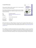
Figure 1 Phylogenetic tree based on EF-1α sequences of isolated strain SIID 11460 and other related species obtained from NCBI. The
phylogenetic tree was constructed by the neighbor-joining method using CLUSTAL W and MEGA ver5.0. Levels of bootstrap support were
indicated at nodes. The scale bar represents 0.005 nucleotide substitution per position.
Gao et al. AMB Express 2012, 2:49
/>
Page 6 of 13
Table 2 The activity of β-glucosidases produced by
F. proliferatum growing on different carbon sources
Carbon sources
With urea (U/ml)
Without urea (U/ml)
Agricultural by-products
Corn stover
0.90 ± 0.05
0.28 ± 0.04
Wheat bran
2.09 ± 0.13
1.26 ± 0.07
Baggase
1.32 ± 0.08
1.28 ± 0.08
Corn stover + wheat bran
3.31 ± 0.14
2.78 ± 0.10
0.37 ± 0.01
0.38 ± 0.08
Polysaccharides
CMC
Avicel cellulsoe
0.2 ± 0.004
0.10 ± 0.01
Disaccharides
Sucrose
0.24 ± 0.04
0
Cellobiose
0.90 ± 0.03
0
Glucose
0.69 ± 0.02
0
Xylose
0.28 ± 0.08
0
Galactose
0.02 ± 0.001
0
Maltose
0.02 ± 0.002
0
Monosaccharides
β-Glucosidases activity was determined based on pNPG as the substrate. The
different carbon sources were used at the concentration of 2% in modified
Mandels culture medium. Values are means ± SD of triplicate samples.
used for the identification of SIID11460. The EF-1α gene
of SIID11460 was successfully amplified by PCR. The fungal EF-1α gene was amplified from genomic DNA, and
then purified, sequenced and analyzed with the BLAST
program from NBRC. The strain showed the highest identity (99.3 ~ 100%) with Gibberella intermedia (Fusarium
proliferatum). Based on the EF-1α sequence of Fusarium
genus (O'Donnell et al. 2012), phylogenetic tree was built
up. Phylogenetic analysis indicated that SIID11460 and
Gibberella intermedia NRRL 25103, Gibberella intermedia
NRR52687 and Fusarium proliferatum NRRL 43545 belong to the same clade (Figure 1). Based on the comparison of the EF-1α gene sequences and the location of clade
in the species complex of Gibberella fujikuroi (O'Donnell
et al. 1998; Nirenberg and O'Donnell 1998), the strain
SIID11460 was identified as a strain of F. proliferatum that
belongs to Liseola section of the Fusarium genus (Nelson
et al. 1983) and its teleomorph is Gibberella intermedia.
SIID11460 was named as F. proliferatumNBRC109045.
β-glucosidases production by F. proliferatum in various
carbon sources
Various carbon sources, not only agricultural byproducts and polysaccharides but also mono- and disaccharides were tested for β-glucosidases production
by F. proliferatum with and without urea for 10-day
cultivation (Table 2). All substances with urea addition
induced β-glucosidases production at different levels.
When pNPG was used as substrate to measure activity
of β-glucosidases, the activity reached the highest level
of 3.31 ± 0.14 U/ml with use of corn stover + wheat
bran as carbon source. The activity level was still as
high as 2.09 ± 0.13 U/ml when wheat bran was used as
carbon source. An activity of 0.69 ± 0.02 U/ml was
assayed when the glucose was used as carbon source
even though glucose is regarded as a universal repressor of hydrolases. The activity level produced with use
of glucose as carbon source was a little bit below the
activity level produced with use of cellobiose as carbon
source.
When disaccharides and monosaccharides were used
as the sole source of carbon at pH 7.0 without urea, no
activity of β-glucosidase was detected even extending the
period of cultivation to 25 days. Only agricultural byproducts and polysaccharides at pH 7.0 without urea
addition induced β-glucosidases production. The variation of pH before and after culturing was expressed in
Table 3. Before cultivation of F. proliferatum, the pH of
mediums was adjusted to 7.0. Ten days later, the pH
values of glucose or cellobiose based mediums without
urea addition dropped to approximately 2.5; the pH
values of glucose or cellobiose based mediums with urea
addition hardly changed; the pH values of corn stover +
wheat bran based mediums with and without urea
addition were 7.1 and 6.0, respectively, after 10-day cultivation. It is reported that the biosynthesis of
β-glucosidases is greatly influenced by pH (Tangnu et al.
1981; Desrochers et al. 1981). For F. proliferatum in this
study, low pH of the glucose or cellobiose based mediums cut production of β-glucosidases. But addition of
urea halted reduction in pH of glucose or cellobiose
based mediums. When F. proliferatum grew in corn
stover + wheat bran based medium, the pH decreased
slightly. Therefore, whether adding urea to corn stover +
Table 3 The pH of mediums in which F. proliferatum grew for 10 days
With urea addition
Glucose
Without urea addition
Before
cultivation pH
After cultivation
pH
BGL
activity (U/ml)
Before
cultivation pH
After cultivation
pH
BGL Activity
(U/ml)
7.0
6.5 ± 0.2
0.69 ± 0.02
7.0
2.5 ± 0.2
0
cellobiose
7.0
6.5 ± 0.2
0.90 ± 0.03
7.0
2.6 ± 0.3
0
Corn stover + wheat bran
7.0
7.1 ± 0.1
3.31 ± 0.14
7.0
6.0 ± 0.1
2.78 ± 0.02
BGL: β-glucosidases.
Gao et al. AMB Express 2012, 2:49
/>
Page 7 of 13
wheat bran based medium did not affect production of
β-glucosidases evidently. Thus, the addition of urea
might have the ability to promote the production of
β-glucosidases, especially in mono and disaccharides. To
make sure of the function of urea, a comparative test
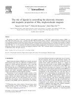
was carried out. Figure 2 indicated the time course of
β-glucosidases production by F. proliferatum using different carbon sources with and without addition of urea.
F. proliferatum started to produce β-glucosidases on the
8th day after incubating in glucose or cellobiose based
medium with urea addition (Figure 2-a). According to
the time course for β-glucosidases production, the same
amount of urea was added to the glucose and cellobiose
based mediums on the 8th day after incubating, respectively. Then the samples were taken out every 2 days to
determine the activity of β-glucosidases and pH. However, F. proliferatum did not produce β-glucosidases and
the pH of the mediums was kept at about 2.5. The results
indicated that there was no relationship between addition
of urea and halting reductions in pH of glucose or cellobiose based medium.
β-glucosidase activity (U/ml)
a
4
3.5
3
2.5
2
1.5
1
0.5
0
0
4
6
Time (day)
8
10
12
3.5
100
3
90
2.5
80
Activity retention (%)
β-glucosidase activity (U/ml)
b
2
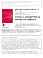
Figure 3 indicated that the glucose tolerance of the
β-glucosidases produced by F. proliferatum growing in
varied carbon sources based mediums. Supplementation
of glucose in the substrate resulted in severe reductions in
β-glucosidases activity. On the other hand, β-glucosidases
produced by F. proliferatum growing in corn stover +
wheat bran based medium had higher tolerance to glucose
compared to that in glucose or cellobisoe based medium.
β-Glucosidases produced with use of different carbon
sources have different level of tolerance to the glucose.
β-Glucosidases may be classified into three groups on
the basis of substrate specificity. (1) Aryl β-glucosidases exclusively hydrolysing or showing a great preference towards aryl β-glucosides; (2) cellobiases hydrolysing
cellobiose and small oligosaccharides and finally (3) the
members of the third group, termed as broad-specificity
β-glucosidases, that act on both substrates (aryl-β-glucosides, cellobiose and cellooligosaccharides) and are the
most commonly observed group in cellulolytic microbes
(Patchett et al. 1987). The hydrolysis capacity of
β-glucosidases produced by F. proliferatum growing in
corn stover + wheat bran based medium and glucose
based medium were tested on cellobiose (0.5%). After
30 min, aliquots were taken out and their glucose contents
were determined by Bio-sensor. Based on the substrate of
cellobiose, the activities of β-glucosidases produced by F.
proliferatum growing in corn stover + wheat bran based
medium and glucose based medium were 426 U/ml and
187 U/ml, respectively. According to the results mentioned
above and those in Table 2, β-glucosidases produced by F.
proliferatum grew in corn stover + wheat bran based
medium and glucose based medium belongs to the third
group of β-glucosidases, due to the capacity of
β-glucosidases to hydrolyze cellobiose and pNPG.
2
1.5
1
0.5
0
0
2
4
6
Time (day)
8
10
12
Figure 2 Time course of β-glucosidases production by
F. proliferatum using different carbon sources a: with addition
of urea. b: without addition of urea. Corn stover + wheat bran (⋄),
bagasse(□), CMC(5), cellobiose(Χ), glucose(*), and xylose (○) were
used individually, at the concentration of 2% in the modified
Mandels medium. Samples were withdrawn every two days during
12 days
70
60
50
40
30
20
10
0
0
50
100
150
200
250
Glucose concentration (mM)
300
Figure 3 The glucose tolerance of the β-glucosidases produced
by F. proliferatum growing in varied carbon sources based
mediums. Corn stover + wheat bran (5), cellobiose (□), and
glucose (○). Values are means ± SD of triplicate samples.
Gao et al. AMB Express 2012, 2:49
/>
Differential expression of β-glucosidases in response to
carbon sources
Zymogram analysis was used to assay the β-glucosidases
produced by F. proliferatum that grew in corn stover +
wheat bran based medium and glucose based medium.
Figure 4 (See legend on next page.)
Page 8 of 13
When zymogram analysis was used to detect different
β-glucosidases existing in the crude enzyme, the exact
number of the fluorescence bands could not be identified because the fluorescence bands overlapped each
other, and it was also difficult to get clear pictures.
Gao et al. AMB Express 2012, 2:49
/>
Page 9 of 13
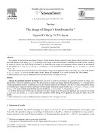
(See figure on previous page.)
Figure 4 Schematic of the modified zymogram. a: add the mix of loading buffer and crude enzyme solution to the gel. But the mix was not
heated at 100°C. Blue: crude enzyme from glucose based medium; Green: crude enzyme from corn stover + wheat bran based medium; Red:
loading buffer only. b: after electrophoresis, the first column of each sample was cut out. c: the first column of each sample was stained with
CBB. d: the remaining gel was soaked in Tris–HCl buffer to remove SDS. e: the first column after CBB staining was used as a marker to cut the
protein bank of the second column. f: the protein bank cut of the second column was soaked in pNPG for active staining. g: after adding
Na2CO3, the band coming from the second column kept colorlessness. h: the color of the band from the second column changed from
colorlessness to yellow following addition of Na2CO3. i: according to the position of active band of the second column, cut the corresponding
bands of the third and fourth column. j: protein coming from the bands of the third and fourth column was injected to the gel for SDS-PAGE
following a series of treatments.
Therefore, we modified the zymogram method and usefully applied the modified method to prove a differential
expression pattern of β-glucosidases produced by F. proliferatum that grew in the carbon sources (Figure 4).
After the electrophoresis, the first column of each sample was cut out of the gel and then treated with Coomassie Brilliant Blue (CBB) staining. Figure 5-a shows 8
bands of proteins that existed in the crude enzyme
growing in glucose based medium and 6 bands of proteins that existed in the crude enzyme growing in corn
stover + wheat bran based medium. Based on the stained
bands of the first column, the correspondent gel bands
on the second column of the same sample were cut as
narrow as possible and these cuts were separately incubated in pNPG for 10 min. Actually, bands Glu2,Glu3,
Glu4,Glu7,CW2,CW4,CW5 changed to yellow. That
proved existence of β-glucosidase activity. Among these
stained bands, colors of band Glu7 and CW2 were the
most visible. The position of band Glu2 at the gel corresponded to that of band CW2, band Glu3 matched with
band CW4, and band Glu4 was corresponding to band
CW5. Corresponding band of Glu7 was not found at the
CW gel. Band CW7 was cut out of CW gel based on the
position of band Glu7 on the assumption that the same
β-glucosidases would be produced by F. proliferatum
that grows in different carbon sources. The cut was treated for activity staining but no change of color was
observed. It indicated that the cut did not contain any
β-glucosidase. Subsequently, the bands with β-glucosidase
activity on the second column were used as markers to
cut the corresponding bands out of the third and fourth
column of the same sample as narrow as possible. The
cuts were soaked in acetate buffer (0.05 M, pH5.0) to recover the protein of β-glucosidase containing in the gel
and treated with SDS-PAGE and then with silver staining.
Figure 5-b indicates the results of the SDS-PAGE. However, the amount of proteins existing in bands Glu2,Glu3,
Glu4,CW4 and CW5 was too low to be visible after the
SDS-PAGE. In all, at least four different β-glucosidases
were produced by F. proliferatum growing in glucose
Figure 5 Zymogram demonstrated that F. proliferatum expressed differentially in response to various carbon source at 2% (w/v) a:
Coomassie staining of SDS-PAGE of crude enzyme. b: Silver staining of the SDS–PAGE. Glu, glucose; CW, corn stover + wheat bran; M,
molecular weight marker (kDa); B, loading buffer only.
Gao et al. AMB Express 2012, 2:49
/>
Page 10 of 13
based medium and at least three different β-glucosidases
were produced by F. proliferatum growing in medium of
corn stover + whea bran. β-Glucosidase with the molecular weight of approximate 46 was produced in glucose
based medium only. Therefore, we came to a conclusion
that different β-glucosidases can be produced by that
grows in different carbon based mediums.
Partial purification of β-glucosidase
The partial purification process was summarized in
Table 4. In the initial step of purification with ammonium
sulfate fractionation, about 70% of total β-glucosidase
activities could be recovered in the fraction of 50–70%
ammonium sulfate saturation with a purification of 3.3
times. In the second step, ion-exchange chromatography
on DEAE-Sepharose CL-6B was performed using five
concentration of sodium chloride for elution. In this step,
greater purity was realized since most of the contaminating protein was removed. β-glucosidase was eluted from
the ion exchanger at the sodium chloride concentration
of 0.15 M, as one broad peak. About 32% of total
β-glucosidase activities could be recovered. Accordingly,
β-glucosidase was purified 9.2 times. In the third step,
active fraction (0.15 M NaCl) gained from DEAESepharose CL-6B was applied on Superdex 75 column.
About 16% of total β-glucosidase activities could be
recovered. As a result, β-glucosidase was purified 18.0
times. After all these steps, we got β-glucosidase that had
a specific activity of 287.7 U/mg based on pNPG and 6
400 U/mg based on cellobiose. The results pointed out
that β-glucosidase produced by F. proliferatum that grows
in corn stover + wheat bran based medium has high catalytic efficiency (Table 5). There were two major bands on
the SDS-PAGE of the active peak from Superdex 75.
Compared the location band of CW2 that came from the
modified zymogram and active peak from superdex75
(Figure 6-c), we can get the conclusion that the band on
the top of lane 2 on Figure 6-c is the β-glucosidase we
need to purify.
Discussion
Cellobiose was considered as an inducer of cellulase
which includes β-glucosidases (Mandels and Reese
1957). However, the amount of β-glucosidases when
F. proliferatum grew in cellobiose based medium was
Table 5 Specific activity of purified β-glucosidase from
various sources
Strain
Specific activity
(U/mg)
Reference
Rhizomucor miehei (NRRL 5282)
62
(Krisch et al. 2012)
Candida peltata (NRRL Y-6888)
108
(Saha and Bothast 1996)
Daldinia eschscholzii
78
(Karnchanatat et al. 2007)
Stachybotrys microspora
20
(Saibi and Gargouri 2011)
Thymepkilic anaerobic
bacterium
149
(Patchett et al. 1987)
Aspergillus niger (NIAB 280)
42
(Rashid and
Siddiqui 1997)
Xylaria regalis
23
(Wei et al. 1996)
Trichoderma sp.
214
(Fadda et al. 1994)
Aspergillus niger
(CCRC 31494)
199
(Yan and Lin 1997).
Fusarium proliferatum
(NBRC 109045)
288*
This study
*partially purified.
less than that in corn stover + wheat bran based
medium. When compared the yield of β-glucosidases in
cellobiose based medium with that in corn stover +
wheat bran based medium, F. proliferatum grew in cellobiose faster than that in corn stover + wheat bran (data
not shown). This proved that cellobiose is an excellent
growth substance for and is rapidly consumed, whereas
corn stover + wheat bran is a relatively poor growth substance and is slowly consumed. The same phenomenon
was observed by (Mandels and Reese 1960). They held
the opinion that the inhibitory effect of cellobiose on
β-glucosidases production seems to be related to rapid
metabolism of the cellobiose.
Wheat bran that contains significant quantities of
starch, protein and so on is a rich source of nutrients and
could promote growth and enzyme production of fungus.
Corn stover that is mainly composed of lignocellulose is
a very common and cost-free agricultural product. Supplementation of the mixture of wheat bran and corn
stover resulted in a significant increase in β-glucosidases
activity when compared to individual application. The
likely reasons for the result were that wheat bran provided F. proliferatum with adequate nutrition at the early
growth stage and made the strain grow fast. After nutrition contained in wheat bran ran out, F. proliferatum
Table 4 Summary of the purification steps of the β-glucosidase produced by F. proliferatum growing in corn stover +
wheat bran based medium
Step
Total activity (U)
Total protein (mg)
Specific activity (U/mg)
Purification factor
Crude extract
150
9.38
16.0
1.0
Recovery (%)
100
(NH4)2SO4
106
1.98
53.5
3.3
70.4
DEAE Sepharose CL-6B
48.6
0.33
148
9.2
32.3
Superdex75
23.8
0.08
288
18
15.8
Gao et al. AMB Express 2012, 2:49
/>
Figure 6 SDS-PAGE analysis (10% polyacrylamide) of protein
sample from each step of the partial purification. a-(1),
molecular weight markers; a-(2), crude protein extract; b-(1), the
active peak from DEAE sepharose CL-6B; b-(2), molecular weight
markers; c-1, the protein from CW2 band of the modified zymogram;
c-(2), the protein from the active peak of Superdex75; c-(3)
molecular weight markers; c-(4), loading buffer only. The protein
showed on A and B was stained with CBB R-250. The protein
showed on C was with silver staining.
could hardly get required nutrition from corn stover and
became starve and then produced a huge amount of
β-glucosidases.
F. proliferatum did not produce β-glucosidases in glucose or cellobiose based medium without addition of
urea (Table 2). And it has been proved that there was no
direct relationship between addition of urea and halting
reduction in the pH of glucose or cellobiose based
medium. Emergence of the phenomenon prompted us
to ponder a problem that how the addition of urea contributed to β-glucosidase production in glucose or cellobiose based medium. This is probably due to the
metabolites produced by F. proliferatum growing in glucose or cellobiose based medium or the derivatives of
the components containing in the glucose or cellobiose
Page 11 of 13
based medium. The metabolites or derivatives produced
by the fungus would reduce the pH of the glucose or
cellobiose based medium. Low pH of the glucose or
cellobiose based medium, in turn, cut the production of
β-glucosidases. But addition of urea at the beginning of
cultivation can cut the production of the metabolites or
derivatives. That, in turn, halted reduction in pH of glucose or cellobiose based medium. However, adding urea
to the glucose or cellobiose based medium after 8-day
cultivation cannot damage the metabolites or derivatives
produced in large quantities during incubation. In
this case β-glucosidases still cannot be produced by F.
proliferatum even addition of urea. The possible reasons
for slight decrease in pH of the corn stover + wheat bran
based medium are because the metabolites or derivatives
were not produced by F. proliferatum, or only tiny
amount of the metabolites or derivatives was produced.
Therefore, urea addition did not affect the production of
β-glucosidases produced by F. proliferatum growing in
corn stover + wheat bran based medium significantly.
β-Glucosidases produced by F. proliferatum in different carbon sources based mediums expressed varied glucose tolerance (Figure 3). (Isorna et al. 2007) purified a
β-glucosidase, named as BglB, produced by P. polymyxa
and obtained the crystallographic structure of the BglB
with glucose. In this structure, the ring of glucose
resided in the active site, through the interactions with
nine amino acids of BglB. Of the nine residues, seven
were involved in intermediate binding to glucose directly,
while the other two, Trp412 and His181, indirectly binding to glucose. The seven directly interacting residues
were found to conserve among different β-glucosidases
belonging to GH1, whereas Trp412 and His181 in BglB
are fairly variable. The two variable residues were
assumed to play important roles in glucose tolerance. It
has been proved that the 184th residue of β-glucosidase
BglB plays an important role in glucose tolerance (Liu
et al. 2011). Glucose acts as an inhibitor by competing with
the substrate in binding to the enzyme (Fang et al. 2010).
But the mechanism of β-glucosidase tolerance to glucose
is still unclear. Presumably the cause is that there are
variable special residuals on active site of β-glucosidases by
F. proliferatum in different carbon sources based mediums.
These residuals are not only the binding site of glucose but
the binding site of the substrate, so changes of the special
residuals cause difference in degree of bond to glucose.
That, in turn, makes difference in level of tolerance to
glucose.
We reported the modified zymogram method that is a
combination of the technology zymogram, gel purification and SDS-PAGE to prove that F. proliferatum could
express varied β-glucosidases in respond to varied carbon sources. The approach described in the paper overcomes the disadvantages of applying crude enzyme on
Gao et al. AMB Express 2012, 2:49
/>
zymogram and combines effective isolation with identification assay.
Competing interests
The authors declare that they have no competing interests.
Author details
1
Department of Agro-Environmental Sciences, Faculty of Agriculture, Kyushu
University, Fukuoka, Japan. 2Center of Biotechnology, Vietnam National
University, Hanoi, Vietnam. 3Department of Biotechnology, National Institute
of Technology and Evaluation, Tokyo, Japan. 4Advanced Technology
Laboratories IDEMITSU KOSAN Co., Ltd, Chiba, Japan.
Received: 23 August 2012 Accepted: 29 August 2012
Published: 14 September 2012
References
Adsul MG, Bastawde KB, Varma AJ, Gokhale DV (2007) Strain improvement of
Penicillium janthinellum NCIM1171 for increased cellulase production.
Bioresour Technol 98:1467–1473
Banerjee S, Mudliar S, Sen R, Giri B, Satpute D, Chakrabarti T, Pandey RA (2010)
Commercializing lignocellulosic bioethanol: technology bottlenecks and
possible remedies. Biofuels Bioprod Bioref 4:77–93
Beguin P, Aubert JP (1994) The biological degradation of cellulose. FEMS
Microbiol Rev 13:25–28
Bradford MM (1976) A rapid and sensitive method for the quantitation of
microgram quantities of protein utilizing the principle of protein-dye
binding. Anal Biochem 72:248–254
Cai YJ, Buswell JA, Chang ST (1998) β-Glucosidase component of the cellulolytic
system of the edible straw mushroom, Volvariella volvacea. Enzyme Microb
Technol 22:122–129
Crout DH, Vic G (1998) Glycosidases and glycosynthetases in glycoside and
oligosaccharide synthesis. Curr Opin Chem Biol 2:98–111
Daenen L, Saison D, Sterckx F, Delvaux FR, Verachtert H, Derdelinckx G (2008)
Screening and evaluation of the glucoside hydrolase activity in
Saccharomyces and Brettanomyces brewing yeasts. J Appl Microbiol
104:478–488
Deguchi S, Tsudome M, Shen Y, Konishi S, Tsujii K, Ito S, Horikoshi K (2007)
Preparation and characterisation of nanofibrous cellulose plate as a new
solid support for microbial culture. Soft Matter 3:1170–1175
Desrochers M, Jurasek L, Paice MG (1981) High production of β-glucosidase in
Schizophyllum commune: isolation of the enzyme and effect of the culture
filtrate on cellulose hydrolysis. Appl Environ Microbiol 41:222–228
Enari TM (1983) Microbial cellulase. In: Fogarty WM, Kelly CT (eds) Microbial
enzymes and biotechnology. Applied Science Publishers, London-New York,
p p183
Esen A (1993) β-Glycosidases: overview. In: Esen A (ed) β-Glucosidases
biochemistry and molecular biology, vol 533, ACS Symposium. American
Chemical Society, Washington, DC, pp 1–14
Esen A (2003) β-Glucosidases. In: Whitaker JR, Voragen AGJ, Wong DWS (eds)
Handbook of food enzymology. Marcel Dekker Inc, New York, p 791
Fadda MB, Curreli N, Pompei R, Rescigno A, Runaldi A, Sandust E (1994) A highly
active fungal β-glucosidase. Appl Biochem Biotechnol 44:263–270
Fang ZM, Fang W, Liu JJ, Hong YZ, Peng H, Zhang XC, Sun BL, Xiao YZ (2010)
Cloning and characterization of a β-glucosidase from marine microbial
metagenome with excellent glucose tolerance. J microbial Biotechnol
20:1351–1358
Gϋnata Z (2003) Flavor enhancement in fruit juices and derived beverages by
exogenous glycosidases and consequences of the use of enzyme
preparations. In: Whitaker JR, Voragen AGJ, Wong DWS (eds) Handbook of
food enzymology., New York, p 303
Hawksworth DL (1991) The fungal dimension of biodiversity: magnitude,
significance, and conservation. Mycol Res 95:641–655
Hawksworth DL (2001) The magnitude of fungal diversity: the 1.5 million species
estimate revisited. Mycol Res 105:1422–1432
Isorna P, Polaina J, Latorre-García L, Cañada FJ, González B, Sanz-Aparicio J (2007)
Crystal structures of Paenibacillus polymyxa β-glucosidase B complexes reveal
the molecular basis of substrate specificity and give new insights in
to the catalytic machinery of family 1 glycosidases. J Mol Biol
371:1204–1218
Page 12 of 13
Karnchanatat A, Petsom A, Sangvanich P, Piaphukiew J, Whalley AJS, Reynolds
CD, Sihanonth P (2007) Purification and biochemical characterization of an
extracellular β-glucosidase from the wood-decaying fungus Daldinia
eschscholzii (Ehrenb.:Fr.) Rehm. FEMS Microbiol Lett 270:162–170
Krisch J, Bencsik O, Papp T, Vagvolgyi C, Tako M (2012) Characterization of a
β-glucosidase with transgalactosylation capacity from the zygomycete
Rhizomucor miehei. Bioresource Technology 114:555–560
Laemmli UK (1970) Cleavage of structural proteins during the assembly of the
head of bacteriophage T4. Nature 227:680–685
Liu J, Zhang X, Fang Z, Fang W, Peng H, Xiao Y (2011) The 184th residue of
β-glucosidase BglB plays an important role in glucose tolerance. J Biosci and
Bioeng 112:447–450
Mandels M, Reese ET (1957) Induction of cellulase in Trichoderma viride as
influenced by carbon source and metals. J Bacteriol 73:269–278
Mandels M, Reese ET (1960) Induction of cellulase in fungi by cellobiose.
J Bacteriol 79:816–826
Nelson PE, Toussoun TA, Marasas WFO (1983) Fusarium species: An illustrated
manual for identification. Centre County, Pennsylvania
Nirenberg HI, O'Donnell K (1998) New Fusarium species and combination with
the Gibberella fujikuroi species complex. Mycologia 90:434–458
O'Donnell K (1993) Fusarium and its near relatives. In: Reynolds DR, Taylor JW
(eds) The fungal holomorph: mitotic, meiotic and pleomorphic speciation in
fungal systenatics., Wallingford, p 225
O'Donnell K, Cigelnil E (1997) Two divergent intragenomic rDNA ITS2 types
within a monophyletic lineage of the fungus Fusarium are nonorthologous.
Mol Phyl Evol 7:103–116
O'Donnell K, Cigelnik E, Nirenberg HI (1998) Molecular systematics and
phylogeography of the Gibberella fujikuroi species complex of Fusarium.
Mycologia 90:465–493
O'Donnell K, Humber RA, Geiser DM, Kang S, Park B, Robert VARG, Crous PW,
Johnston PR, Aoki T, Rooney AP, Rehner SA (2012) Phylogenetic diversity of
insecticolous fusaria inferred from multilocus DNA sequence data and their
molecular identification via FUSARIUM-ID and FUSARIUM MLST. Mycologia
104:427–445
Opassiri R, Hua Y, Wara-Aswapati O, Akiyama T, Svasti J, Esen A, Ketudat Cairns JR
(2004) β-Glucosidase, exo-β-glucanase and pyridoxine transglucosylase
activities of rice BGlu1. Biochem J 379:125–131
Patchett ML, Daniel RM, Morgan HW (1987) Purification and properties of a stable
beta-glucosidase from an extremely thermophilic anaerobic bacterium.
Biochem J 243:779–787
Percival ZYH, Himmel ME, Mielenz JR (2006) Outlook for cellulase improvement:
screening and selection strategies. Biotechnol Adv 24:452–481
Ragauskas AJ, Williams CK, Davison BH, Britovsek G, Cairney J, Eckert CA (2006)
The path forward for biofuels and biomaterials. Science
311:484–489
Rashid MH, Siddiqui KS (1997) Purification and characterization of a β-glucosidase
from Aspergillus niger. Folia Microbiol 42:544–550
Ryu DDY (1980) Cellulases: biosynthesis and applications. Enzyme Microb Technol
2:91–102
Saha BC, Bothast RJ (1996) Production, purification, and characterization of a
highly glucose tolerant novel β-glucosidase from Candida peltata. Appl
Environ Microbiol 62:3165–3170
Saibi W, Gargouri A (2011) Purification and biochemical characterization of an
atypical β-glucosidase from Stachybotrys microspore. J Mol Catal B Enzym
72:107–115
Saibi W, Abdeljalil S, Gargouri A (2011) Carbon source directs the differential
expression of β-glucosidases in Stachybotrys microspore. World J Microbiol
Biotechnol 27:1765–1774
Soewnsen A (2010) A new highly efficient beta-glucosidase from the novel
species Aspergillus Saccharolyticus. Dissertation, Aalborg University
Tamura K, Peterson D, Peterson N, Stecher G, Nei M, Kumar S (2011) MEGA5:
Molecular evolutionary genetics analysis using maximum likelihood,
evolutionary distance, and maximum parsimony methods. Mol Biol Evol
28:2731–2739
Tangnu SK, Blanch HW, Wilke CR (1981) Enhanced production of cellulase,
hemicellulase, and β-glucosidase by Trichoderma reesei (Rut C-30). Biotechnol
Bioeng 23:1837–1849
Thompson JD, Higgins DG, Gibson TJ, CLUSTAL W (1994) Improving the
sensitivity of progressive multiple sequence alignment through sequence
weighting, position-specific gap penalties and weight matrix choice. Nucleic
Acids Res 22:4673–4680
Gao et al. AMB Express 2012, 2:49
/>
Page 13 of 13
Wei DL, Kirimura K, Usami S, Lin TH (1996) Purification and characterization of an
extracellular β-glucosidase from the wood- grown fungus Xylaria regalis.
Curr Microbiol 33:297–301
White TJ, Bruns T, Lee S, Taylor J (1990) Amplification and direct sequencing of
fungal ribosomal RNA genes for phylogenetics. In: Innis MA, Gelfand DH,
Sninsky JJ, White TJ (eds) PCR Protocols: a guide to methods and
applications., San Diego, pp 315–322
Wood TM (1985) Properties of cellulolytic enzyme systems. Biochem Soc Trans
13:407–410
Workman WE, Day DF (1982) Purification and properties of β-glucosidase from
Aspergillus terrus. Appl Environ Microbiol 44:1289–1295
Yan TR, Lin CL (1997) Purification and characterization of a glucose-tolerant
β-glucosidase from Aspergillus niger CCRC 31494. Biosci Biotech Biochem
61:965–970
doi:10.1186/2191-0855-2-49
Cite this article as: Gao et al.: The production of β-glucosidases by
Fusarium proliferatum NBRC109045 isolated from Vietnamese forest.
AMB Express 2012 2:49.
Submit your manuscript to a
journal and benefit from:
7 Convenient online submission
7 Rigorous peer review
7 Immediate publication on acceptance
7 Open access: articles freely available online
7 High visibility within the field
7 Retaining the copyright to your article
Submit your next manuscript at 7 springeropen.com