DSpace at VNU: Development and In Vitro Evaluation of Liposomes Using Soy Lecithin to Encapsulate Paclitaxel
Bạn đang xem bản rút gọn của tài liệu. Xem và tải ngay bản đầy đủ của tài liệu tại đây (3.33 MB, 8 trang )
Hindawi
International Journal of Biomaterials
Volume 2017, Article ID 8234712, 7 pages
/>
Research Article
Development and In Vitro Evaluation of Liposomes Using
Soy Lecithin to Encapsulate Paclitaxel
Thi Lan Nguyen,1,2 Thi Hiep Nguyen,3 and Dai Hai Nguyen1
1
Institute of Applied Materials Science, Vietnam Academy of Science and Technology, 01 TL29, District 12, Ho Chi Minh City, Vietnam
Can Tho University, 3/2 Street, Ninh Kieu District, Can Tho City, Vietnam
3
Tissue Engineering and Regenerative Medicine Group, Department of Biomedical Engineering, International University,
Vietnam National University-HCMC (VNU-HCMC), Ho Chi Minh City 70000, Vietnam
2
Correspondence should be addressed to Dai Hai Nguyen;
Received 3 January 2017; Revised 7 February 2017; Accepted 9 February 2017; Published 26 February 2017
Academic Editor: Fahima Dilnawaz
Copyright © 2017 Thi Lan Nguyen et al. This is an open access article distributed under the Creative Commons Attribution License,
which permits unrestricted use, distribution, and reproduction in any medium, provided the original work is properly cited.
The formulation of a potential delivery system based on liposomes (Lips) formulated from soy lecithin (SL) for paclitaxel (PTX) was
achieved (PTX-Lips). At first, PTX-Lips were prepared by thin film method using SL and cholesterol and then were characterized
for their physiochemical properties (particle size, polydispersity index, zeta potential, and morphology). The results indicated
that PTX-Lips were spherical in shape with a dynamic light scattering (DLS) particle size of 131 ± 30.5 nm. Besides, PTX was
efficiently encapsulated in Lips, 94.5 ± 3.2% for drug loading efficiency, and slowly released up to 96 h, compared with free PTX.
More importantly, cell proliferation kit I (MTT) assay data showed that Lips were biocompatible nanocarriers, and in addition the
incorporation of PTX into Lips has been proven successful in reducing the toxicity of PTX. As a result, development of Lips using
SL may offer a stable delivery system and promising properties for loading and sustained release of PTX in cancer therapy.
1. Introduction
Discovered in 1962, paclitaxel (PTX) is one of the most
powerful anticancer drugs for various types of solid tumors,
especially for breast cancer and advanced ovarian carcinoma.
However, PTX has several disadvantages such as poor water
solubility, high toxicity, and low bioavailability, which limit
its potential clinical application [1–4]. Among approaches to
overcome these drawbacks, drug delivery system is suggested
to be a promising candidate owing to the knowledge that
nanocarriers can efficiently control the pharmacokinetic
characteristics of drugs [5, 6]. This method can deliver
medication within desired therapeutic range to abnormal
cells without affecting normal cells while maintaining the
systemic levels of drugs [7–13].
Considering the variety of nanocarriers, liposomes (Lips),
spherical vesicles consisting of at least one phospholipid
bilayer, have been investigated as potential carriers for drug
delivery applications due to their high biocompatibility,
complete biodegradability, low toxicity, and ability to entrap
both water- and lipid-soluble functional compounds and
simplify specific drug delivery to tumor site. Furthermore,
the stability of the functional components encapsulated in
Lips can be increased, therefore maintaining their activities
in environments that typically result in rapid degradation.
In addition, Lips properties differ considerably with regard
to lipid composition, particle size, surface charge, and the
method of Lips preparation. The rigidity/fluidity and the
charge of the bilayer were strongly influenced by the choice
of bilayer components, for instance, saturated or unsaturated
phospholipids from natural sources such as egg or soybean
phosphatidylcholine [5, 14, 15]. Among these choices, the
use of soy lecithin (SL), a naturally occurring phospholipid
derived from soybeans, not only provides much more permeable Lips but also facilitates large-scale industrial production
because of the reduction of production costs as compared
with saturated phospholipids [16]. Several studies have been
conducted on the benefits of using SL to obtain desired Lips.
2
International Journal of Biomaterials
2.2. Methods
+
PTX
Thin film
fabrication
Time
technique
SL
and cholesterol
PTX-Lips
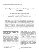
Figure 1: Schematic illustration of the formation of PTX-Lips and
the release of PTX from Lips over time.
Madrigal-Carballo et al. prepared multilayered biopolymercoated Lips formulated from SL as a novel system for ellagic
acid delivery. They successfully achieved monodispered and
stable spherical Lips with a diameter of 386.5 ± 25.9 nm
and a surface charge of −30.66 ± 1.55 nm by thin film
fabrication technique for the liposomal system coated with
four biopolymer layers. These results indicated that the
biopolymer-coated Lips offer good features for loading into
their liposomal core and slow release of ellagic acid [17].
Additionally, in a study conducted by Mura and coworkers,
Lips made from SL and alkyl polyglucosides (OrNS10) were
formulated and characterized for the purpose of designing
suitable drug delivery systems for their potential uses. The
stability of Lips was also studied by checking average particle
size and zeta potential value variation of different liposomal
formulations during 4 weeks. The results showed that the
addition of OrNS10 to SL has the ability to improve Lips
stability [18].
Herein, we developed Lips formulated from SL for PTX
delivery (PTX-Lips). The formation of PTX-Lips was prepared according to the thin film method (Figure 1) and later
these PTX-Lips were characterized by dynamic light scattering (DLS), zeta potential measurement, and transmission
electron microscopy (TEM). Either drug loading or drug
release behavior of PTX-Lips was also evaluated. Particularly,
cell proliferation kit I (MTT) assay was used to determine the
ability of PTX-Lips to minimize the toxicity to HeLa cells of
PTX. This study is expected to improve the stability of Lips
which was synthesized by eco-friendly SL for PTX delivery
in cancer therapy.
2. Materials and Methods
2.1. Materials. PTX was supplied by Samyang Corporation
(Seoul, Korea). Lecithin from soybean (CAS number 800243-5) and Tween 80 (polyoxyethylene sorbitan monooleate,
CAS number 900 5-65-6) were purchased from Tokyo Chemistry Industry Co., Ltd. (Kita-ku, Tokyo, Japan). Cholesterol
was obtained from Sigma-Aldrich (St Louis, MO, USA).
Cetyltrimethylammonium bromide (CTAB) was purchased
from Merck (Darmstadt, Germany). All chemicals and solvents were of highest analytical grade and used without
further purification.
2.2.1. Preparation of PTX-Lips. PTX-Lips were prepared by
conventional thin film technique using SL and cholesterol.
Briefly, SL (500 mg), cholesterol (56 mg), CTAB (5 mg), and
5% PTX (32 mg) were dissolved in chloroform-methanol
(2 : 1 v/v) at room temperature. The mixture was evaporated in
a rotary evaporator (B¨uchi Rotavapor R-114, Essen, Germany)
at 45∘ C for 4 h, resulting in a formation of thin lipid film. The
obtained thin films were hydrated with 15 mL of deionized
water (deH2 O) containing 80 mg of Tween 80 under constant
stirring at 60∘ C. The suspension was further homogenized
(EmulsiFlex-05 homogenizer, Avestin Inc., Ottawa, Canada)
at 800 bar for 5 cycles, followed by centrifugation at 5500 rpm
for 30 min to remove nonencapsulated PTX. The resulting
sample was then lyophilized using 10% mannitol as cryoprotectants and stored at 2–8∘ C.
2.2.2. Characterization. The particle size and polydispersity
index of PTX-Lips were measured by DLS using a Zetasizer
Nano ZS (ZEN 3600, Malvern Instruments Ltd., Malvern,
Worcestershire, UK). A helium-neon (He-Ne) ion laser at
633 nm was used as the incident beam. The detection angle
and the temperature were 90∘ and 25∘ C, respectively. All
samples (1 mg/mL) were sonicated for 15 min, filtered (pore
size = 0.45 𝜇m), and carried out at 37∘ C. The zeta potential
of PTX-Lips was also measured using a Zetasizer Nano
ZS ZEN 3600. All measurements were made in triplicate
for each sample. The size and morphology of PTX-Lips
were confirmed by TEM using JEM-1400 (300 kV; JEOL,
Tokyo, Japan). The samples were prepared by placing a
drop of solution in deH2 O (1 mg/mL) onto a carbon-copper
grid (300-mesh, Ted Pella, Inc., USA) and air-drying for
10 min.
2.2.3. PTX Loading Contents and In Vitro PTX Release. In
order to determine the PTX loading contents in Lips, PTXLips were first mixed with 1% Triton X100, incubated for
1 h, and centrifuged at 6000 rpm for 30 min at 25∘ C to
separate PTX from Lips. The total PTX contents in Lips were
measured using a Shimadzu LC-20A Prominence System
(Shimadzu, Kyoto, Japan). The injected volume was 10 𝜇L
and the mobile phase (acetonitrile : water = 50 : 50 v/v) was
delivered at 1.00 mL/min. A reverse-phase Fortis C18 column
(150 4.6 mm i.d., pore size 5 𝜇m; Fortis Technologies Ltd.,
Cheshire, UK) was used, and column effluent was monitored
with a UV detector at 227 nm. The calibration curve for
quantification of PTX in Lips was found to be linear over the
standard PTX concentration range of 0–50,000 ng/mL with
a high correlation coefficient of 𝑅2 = 0.998. The following
equations were used to calculate the drug loading efficiency
(DLE) and drug loading content (DLC):
DLE (%) =
weight of PTX in Lips
× 100,
weight of PTX feed initially
(1)
weight of PTX inLips
DLC (%) =
× 100.
weight of Lips and PTX
International Journal of Biomaterials
3
The in vitro PTX release experiments were performed in
PBS buffer (0.01 M, pH 7.4) at 37∘ C using dialysis method.
Initially, 1 mL of PTX-Lips suspended in PBS containing 2%
Tween 80 was transferred to a dialysis bag (MWCO 6–8 kDa,
Spectrum Laboratories, Inc., Rancho Dominguez, CA, USA)
and immersed into 20 mL of the release medium in vials
at 37∘ C. The vials were then placed in an orbital shaker
bath, which was maintained at 37∘ C and shaken horizontally
at 100 rpm. At specific time intervals, 2 mL of the release
medium was collected and an equal volume of fresh medium
was added. The collected samples were filtered (pore size
= 0.22 𝜇m) before high performance liquid chromatography
analysis.
2.2.4. MTT Viability Test. The MTT assay was used to
evaluate cytotoxicity of PTX-Lips on Hela cells. The cells
were seeded in a 96-well plate at a density of 1 × 10⁴
cells/well in 130 𝜇L of Dulbecco’s Modified Eagle’s medium
(DMEM) supplemented with 10% FBS and 1% penicillinstreptomycin and cultured 1 day at 37∘ C. Then, the medium
was removed and the cells were incubated with samples. The
cells were incubated for 48 h, followed by removing medium
and washing twice with PBS. MTT solution (25 𝜇L) and
culture medium (130 𝜇L) were added to each well and the cells
were cultured for 3 h. DMSO (130 𝜇L) was added to each well
to dissolve the precipitate. The cells cultured with medium
only were used as a control and assigned to 100% survival.
The absorbance was measured at 570 nm using a multiplate
reader (SpectraMax M2e, Molecular Devices Co., USA). Cell
viability of all other groups was calculated by normalization
of its absorbance intensity to that of “Ctrl” group with the
following equation:
Cell viability (%) =
([Abs]sample − [Abs]blank )
([Abs]control − [Abs]blank )
(2)
× 100%.
3. Results and Discussion
3.1. Characterization of PTX-Lips. Two of the most important
properties for in vivo integrity and biological fate of nanoparticles (NPs) are particle size and surface charge. In other
words, development of carriers with appropriate size and
charge plays a crucial role in the field of drug delivery [19, 20].
Several early studies have reported that the cellular uptake
efficiency of NPs decreases when increasing the particle size.
It is stated that NPs in the range of 100–200 nm have the highest potential to extend circulation time in the bloodstream
because they are small enough to avoid mechanical filtration
by the spleen, but large enough to avoid selective uptake in
the liver. Small size permits NPs passively targeting tumor
cells through the enhanced permeability and retention (EPR)
effect, improving intracellular accumulation and localization
of NPs in tumor area [21, 22]. Another important parameter
that controls the stability of NPs in physiological condition
is zeta potential. Not only does negative charge in particular
improve the physical stability of Lips by preventing them
from fusion and aggregation but also the negatively charged
NPs are phagocytized significantly less than positive ones
[23]. Therefore, particle size and zeta potential are the two
key parameters, which have been proven effective for drug
delivery applications.
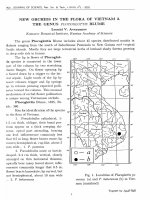
As shown in Figures 2(b) and 2(d), the DLS particle
size of Lips and PTX-Lips and their population standard
deviation were 167 ± 39.1 nm and 131 ± 30.5 nm, whereas the
polydispersity index values were 0.286 ± 0.01 and 0.339 ± 0.02,
respectively. These results indicated that the particle sizes of
Lips and PTX-Lips were not significantly different and their
distributions were quite narrow, respectively. Furthermore,
the corresponding TEM images (Figures 2(a) and 2(c))
showed that Lips and PTX-Lips were spherical in shape
with diameter range of <200 nm and without aggregation
or fusion, which were correlated with the values of DLS
measurement. Besides particle size, zeta potential values of
Lips were approximately −41.7 mV and showed an increase in
the surface charge intensity upon inclusion of PTX, −54.3 mV,
which may be caused by the PTX interaction with lipid
bilayers (Figure 3). Taken together, PTX-Lips might serve as
stable spherical nanocarriers with long term circulation in the
bloodstream.
3.2. Loading and In Vitro Release of PTX. DLE is an important
property in drug-loaded nanocarriers and directly affects the
therapeutic effect of the system. The higher the encapsulation
capacity NPs have, the larger the number of drugs released
at the tumor site [24, 25]. In this study, the DLE and DLC
of PTX-Lips were found to be 94.5 ± 3.2% and 4.48 ±
0.47%, respectively. In comparison with other studies, Jiang
et al. developed novel dual-functional Lips, PTX-loaded
1,2-distearoyl-sn-glycero-3-phosphoethanolamine- (DSPE-)
peptide D [KLAKLAK]2 (KLA) 2,3-dimethylmaleic anhydride (DMA) Lips (DKD/PTX-Lips), to overcome multidrug
resistance. The results showed that the DLE of DKD/PTXLips was 81.8 ± 0.7% [26]. In another previous study by
Zhou et al., the mitochondrial targeting d-a-tocopheryl polyethylene glycol 1000 succinate- (TPGS1000 -) triphenylphosphine (TPP) conjugate (TPGS1000 –TPP) was synthesized and
surface-modified onto PTX-Lips. The targeting PTX-Lips
were successfully prepared with DLE of 86.27 ± 3.15% [27].
These results demonstrated that the prepared Lips with the
high DLE have the potential to be delivered more efficiently
to tumor tissues.
In vitro release profiles of free PTX and PTX from
PTX-Lips were performed in order to evaluate the stability
and release behavior of PTX-Lips. As shown in Figure 4,
the prepared Lips showed a long term stable drug release
profile up to 96 h. The cumulative release amount of PTX
in first 2 h was around 11% as compared with 60% of free
PTX. The initial release of PTX could be explained by
the PTX molecules, which were absorbed into the outer
phospholipid bilayers of Lips. Zhou et al. reported that the
initial release of PTX from the targeting PTX-Lips was less
than 30% during the first 2 h [27]. Moreover, total release
amount of PTX was 56% after 96 h, compared with 97%
of free PTX; in other words, the release behavior of free
4
International Journal of Biomaterials
25
Intensity (%)
20
15
10
5
200 nm
0
10
100
1000
Size (d·nm)
(a)
(b)
25
Intensity (%)
20
15
10
5
200 nm
0
10
(c)
100
Size (d·nm)
1000
(d)
Figure 2: (a, c) TEM image and (b, d) particle size distribution by DLS of Lips and PTX-Lips, respectively.
PTX was significantly faster than PTX in the prepared Lips.
This means that the release rate of PTX was dependent on
the presence of Lips. Thus, PTX-Lips may serve as stable
NPs, therefore increasing drug accumulation into tumor
sites.
3.3. In Vitro Cytotoxicity. Biocompatibility of a material is an
important factor for its success in biomedical applications.
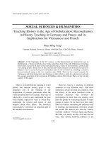
In this study, MTT assay was carried out to suggest the biocompatibility of PTX-Lips. Figure 5 illustrates the inhibitory
effects of free PTX, PTX-Lips, and free PTX of Lips on
HeLa cells. The blank Lips showed no obvious cytotoxicity
towards HeLa cells. Almost 100% of cells were still viable
at 500 𝜇g/mL of samples for 2 days, indicating that Lips
are biocompatible. On the other hand, the cells growth was
significantly inhibited when they were treated with PTXLips. A dose-dependent cytotoxicity was observed when
individually incubating various doses of free PTX and PTXLips with HeLa cells. The majority of cells were killed when
they were treated with PTX at concentration of 10 𝜇g/mL
for 2 days. The inhibitory effects of Lips and PTX-Lips were
consistent with previous researches. It is expected that the
toxicity of PTX would be reduced after being encapsulated
in Lips. As shown in Figure 5(b)-(A), the percentage of
viable cells of PTX-Lips at equivalent PTX concentration of
10 𝜇g/mL was around 64% as compared with that only around
12% of free PTX. These results clearly confirmed that PTXLips have the great inhibitory effect against HeLa cells and
could be safely used as drug delivery vehicles for in vivo
applications.
International Journal of Biomaterials
5
3.0
2.5
Total counts
2.0
1.5
1.0
0.5
0.0
−150
−100
−50
Zeta potential (mV)
0
50
Figure 3: Nanoparticle surface charge via zeta potential of Lips (dashed line) and PTX-Lips (solid line).
100
PTX release (%)
80
60
40
20
0
0
20
40
60
80
100
Time (h)
Figure 4: In vitro release profiles of free PTX (circle) and PTX from PTX-Lips (square).
4. Conclusion
Liposomal delivery systems for PTX have been successfully
developed by thin film technique. The prepared PTX-Lips
were spherical in shape with a diameter around 131 nm, which
would be suitable for in vivo drug release. The NPs had
DLE and DLC of 94.5 ± 3.2% and 4.48 ± 0.47% and, in
addition, the release profile showed sustained release of PTX,
respectively. Particularly, it was clear that PTX-Lips could
reduce the toxicity of PTX determined by MTT assay. Our
results suggest that the Lips made from natural SL have the
potential as stable, biocompatible, and efficient PTX delivery
systems for the treatment of cancer.
Competing Interests
The authors declare that there is no conflict of interests
regarding the publication of this paper.
6
International Journal of Biomaterials
(A)
500 g
250 g
100 g
50 g
0 g
20 g
10 g
5 g
1 g
0.1 g
(B)
(C)
120
80
100
Cell viability (%)
Cell viability (%)
(a)
100
60
40
80
60
40
20
20
0
0
5
10
PTX conc. (g/ml)
15
20
0
100
200
300
400
Free PTX of Lips (g/ml)
500
Free PTX
PTX-Lips
(A)
(B)
(b)
Figure 5: (a) Images of HeLa cells incubated with (A) Lips at different concentrations, (B) PTX-Lips, and (C) free PTX at different PTX doses
observed under microscope for 48 h (scale bar = 80 𝜇m) and (b) viability of HeLa cells incubated with (A) free PTX, PTX-Lips at different
PTX doses, and (B) free PTX of Lips at different concentrations for 48 h. The cells were exposed to the samples for the indicated times. The
data represent the mean values ± the standard deviation (SD) (𝑛 = 4).
International Journal of Biomaterials
Acknowledgments
This research was funded by the Development of Science and
Technology (DOST) under Grant no. 69/2016, date: 18 July
2016.
References
[1] T. Zhao, H. Chen, Y. Dong et al., “Paclitaxel-loaded poly(Glycolide-co-𝜀caprolactone)-b-d-𝛼-tocopheryl polyethylene glycol 2000 succinate nanoparticles for lung cancer therapy,”
International Journal of Nanomedicine, vol. 8, pp. 1947–1957,
2013.
[2] R. M. Taylor and L. O. Sillerud, “Paclitaxel-loaded iron platinum
stealth immunomicelles are potent MRI imaging agents that
prevent prostate cancer growth in a PSMA-dependent manner,”
International Journal of Nanomedicine, vol. 7, pp. 4341–4352,
2012.
[3] T. Konno, J. Watanabe, and K. Ishihara, “Enhanced solubility of paclitaxel using water-soluble and biocompatible 2methacryloyloxyethyl phosphorylcholine polymers,” Journal of
Biomedical Materials Research, vol. 65, no. 2, pp. 209–214, 2003.
[4] D. Q. Hoang, T. V. Tran, N. Q. Tran et al., “Functionalization
of Fe3 O4 nanoparticles with biodegradable chitosan-graftedmPEG for paclitaxel delivery,” Green Processing and Synthesis,
vol. 5, no. 5, pp. 459–466, 2016.
[5] D. H. Nguyen, J. S. Lee, J. H. Choi et al., “Heparin nanogelcontaining liposomes for intracellular RNase delivery,” Macromolecular Research, vol. 23, no. 8, pp. 765–769, 2015.
[6] H. D. Nguyen, T. D. Nguyen, D. H. Nguyen, and P. T. Nguyen,
“Magnetic properties of Cr doped Fe3 O4 porous nanoparticles
prepared through a co-precipitation method using surfactant,”
Advances in Natural Sciences: Nanoscience and Nanotechnology,
vol. 5, no. 3, 2014.
[7] M. Goldberg, R. Langer, and X. Jia, “Nanostructured materials
for applications in drug delivery and tissue engineering,” Journal of Biomaterials Science, Polymer Edition, vol. 18, no. 3, pp.
241–268, 2007.
[8] D. H. Nguyen, J. S. Lee, J. H. Choi, K. M. Park, Y. Lee, and K.
D. Park, “Hierarchical self-assembly of magnetic nanoclusters
for theranostics: tunable size, enhanced magnetic resonance
imagability, and controlled and targeted drug delivery,” Acta
Biomaterialia, vol. 35, pp. 109–117, 2016.
[9] T. T. Nguyen Thi, T. V. Tran, N. Q. Tran, C. K. Nguyen, and
D. H. Nguyen, “Hierarchical self-assembly of heparin-PEG
end-capped porous silica as a redox sensitive nanocarrier for
doxorubicin delivery,” Materials Science and Engineering C, vol.
70, part 2, pp. 947–954, 2017.
[10] D. H. Nguyen, J. Hoon Choi, Y. Ki Joung, and K. Dong Park,
“Disulfide-crosslinked heparin-pluronic nanogels as a redoxsensitive nanocarrier for intracellular protein delivery,” Journal
of Bioactive and Compatible Polymers, vol. 26, no. 3, pp. 287–300,
2011.
[11] D. H. Nguyen, Y. K. Joung, J. H. Choi, H. T. Moon, and K.
D. Park, “Targeting ligand-functionalized and redox-sensitive
heparin-Pluronic nanogels for intracellular protein delivery,”
Biomedical Materials, vol. 6, no. 5, Article ID 055004, 2011.
[12] D. H. Nguyen, J. W. Bae, J. H. Choi, J. S. Lee, and K.
D. Park, “Bioreducible cross-linked Pluronic micelles: PHtriggered release of doxorubicin and folate-mediated cellular
uptake,” Journal of Bioactive and Compatible Polymers, vol. 28,
no. 4, pp. 341–354, 2013.
7
[13] T. D. van, N. Q. Tran, D. H. Nguyen, C. K. Nguyen, D. L. Tran,
and P. T. Nguyen, “Injectable hydrogel composite based gelatinPEG and biphasic calcium phosphate nanoparticles for bone
regeneration,” Journal of Electronic Materials, vol. 45, no. 5, pp.
2415–2422, 2016.
[14] A. Akbarzadeh, R. Rezaei-Sadabady, S. Davaran et al., “Liposome: classification, preparation, and applications,” Nanoscale
Research Letters, vol. 8, article no. 102, 2013.
[15] S. K. Sahoo and V. Labhasetwar, “Nanotech approaches to drug
delivery and imaging,” Drug Discovery Today, vol. 8, no. 24, pp.
1112–1120, 2003.
[16] P. P. Thi and D. H. Nguyen, “Gelatin as an ecofriendly natural
polymer for preparing colloidal silver@gold nanobranches,”
Green Processing and Synthesis, vol. 5, no. 5, pp. 467–472, 2016.
[17] S. Madrigal-Carballo, S. Lim, G. Rodriguez et al., “Biopolymer
coating of soybean lecithin liposomes via layer-by-layer selfassembly as novel delivery system for ellagic acid,” Journal of
Functional Foods, vol. 2, no. 2, pp. 99–106, 2010.
[18] S. Mura, M. Manconi, S. Madrigal-Carballo et al., “Composite soy lecithin-decylpolyglucoside vesicles: a theoretical and
experimental study,” Colloids and Surfaces A: Physicochemical
and Engineering Aspects, vol. 323, no. 1–3, pp. 175–179, 2008.
[19] Y. Yoshizawa, Y. Kono, K.-I. Ogawara, T. Kimura, and K.
Higaki, “PEG liposomalization of paclitaxel improved its in vivo
disposition and anti-tumor efficacy,” International Journal of
Pharmaceutics, vol. 412, no. 1-2, pp. 132–141, 2011.
[20] P. P. N. Thi, M. T. Nguyen, and D. H. Nguyen, “Role of
collagen concentration in stability of Star-shaped Silver@Gold
nanoparticles,” Journal of Nano Research, vol. 40, pp. 113–119,
2016.
[21] B. Aslan, B. Ozpolat, A. K. Sood, and G. Lopez-Berestein,
“Nanotechnology in cancer therapy,” Journal of Drug Targeting,
vol. 21, no. 10, pp. 904–913, 2013.
[22] D. H. Nguyen, J. S. Lee, J. W. Bae et al., “Targeted doxorubicin
nanotherapy strongly suppressing growth of multidrug resistant
tumor in mice,” International Journal of Pharmaceutics, vol. 495,
no. 1, pp. 329–335, 2015.
[23] Y. Sheng, L. Chang, T. Kuang, and J. Hu, “PEG/heparindecorated lipid–polymer hybrid nanoparticles for longcirculating drug delivery,” RSC Advances, vol. 6, no. 28, pp.
23279–23287, 2016.
[24] H. Li, J. Z. Zhang, Q. Tang, M. Du, J. Hu, and D. Yang,
“Reduction-responsive drug delivery based on mesoporous
silica nanoparticle core with crosslinked poly(acrylic acid)
shell,” Materials Science and Engineering C, vol. 33, no. 6, pp.
3426–3431, 2013.
[25] T. U. Ly, N. Q. Tran, T. K. D. Hoang, K. N. Phan, H. N.
Truong, and C. K. Nguyen, “Pegylated dendrimer and its effect
in fluorouracil loading and release for enhancing antitumor
activity,” Journal of Biomedical Nanotechnology, vol. 9, no. 2, pp.
213–220, 2013.
[26] L. Jiang, L. Li, X. He et al., “Overcoming drug-resistant lung
cancer by paclitaxel loaded dual-functional liposomes with
mitochondria targeting and pH-response,” Biomaterials, vol. 52,
no. 1, pp. 126–139, 2015.
[27] J. Zhou, W.-Y. Zhao, X. Ma et al., “The anticancer efficacy
of paclitaxel liposomes modified with mitochondrial targeting
conjugate in resistant lung cancer,” Biomaterials, vol. 34, no. 14,
pp. 3626–3638, 2013.
Journal of
Nanotechnology
Hindawi Publishing Corporation
Volume 2014
International Journal of
International Journal of
Corrosion
Hindawi Publishing Corporation
Polymer Science
Volume 2014
Hindawi Publishing Corporation
Volume 2014
Smart Materials
Research
Hindawi Publishing Corporation
Journal of
Composites
Volume 2014
Hindawi Publishing Corporation
Volume 2014
Journal of
Metallurgy
BioMed
Research International
Hindawi Publishing Corporation
Volume 2014
Nanomaterials
Hindawi Publishing Corporation
Volume 2014
Submit your manuscripts at
Journal of
Materials
Hindawi Publishing Corporation
Volume 2014
Journal of
Nanoparticles
Hindawi Publishing Corporation
Volume 2014
Nanomaterials
Journal of
Advances in
Materials Science and Engineering
Hindawi Publishing Corporation
Volume 2014
Journal of
Hindawi Publishing Corporation
Volume 2014
Journal of
Nanoscience
Hindawi Publishing Corporation
Scientifica
Hindawi Publishing Corporation
Volume 2014
Journal of
Coatings
Volume 2014
Hindawi Publishing Corporation
Crystallography
Volume 2014
Hindawi Publishing Corporation
Volume 2014
The Scientific
World Journal
Hindawi Publishing Corporation
Volume 2014
Hindawi Publishing Corporation
Volume 2014
Journal of
Journal of
Textiles
Ceramics
Hindawi Publishing Corporation
International Journal of
Biomaterials
Volume 2014
Hindawi Publishing Corporation
Volume 2014