DSpace at VNU: Preparation and anti-cancer activity of polymer-encapsulated curcumin nanoparticles
Bạn đang xem bản rút gọn của tài liệu. Xem và tải ngay bản đầy đủ của tài liệu tại đây (1.66 MB, 8 trang )
Home
Search
Collections
Journals
About
Contact us
My IOPscience
Preparation and anti-cancer activity of polymer-encapsulated curcumin nanoparticles
This content has been downloaded from IOPscience. Please scroll down to see the full text.
2012 Adv. Nat. Sci: Nanosci. Nanotechnol. 3 035002
( />View the table of contents for this issue, or go to the journal homepage for more
Download details:
IP Address: 128.153.5.49
This content was downloaded on 04/10/2013 at 13:17
Please note that terms and conditions apply.
IOP PUBLISHING
ADVANCES IN NATURAL SCIENCES: NANOSCIENCE AND NANOTECHNOLOGY
Adv. Nat. Sci.: Nanosci. Nanotechnol. 3 (2012) 035002 (7pp)
doi:10.1088/2043-6262/3/3/035002
Preparation and anti-cancer activity of
polymer-encapsulated curcumin
nanoparticles
Phuong Thu Ha1 , Mai Huong Le2 , Thi My Nhung Hoang3 , Thi Thu
Huong Le4 , Tuan Quang Duong5 , Thi Hong Ha Tran2 , Dai Lam Tran1
and Xuan Phuc Nguyen1
1
Institute of Materials Science (IMS), Vietnam Academy of Science and Technology (VAST),
18 Hoang Quoc Viet, Hanoi, Vietnam
2
Institute of Natural Products Chemistry, Vietnam Academy of Science and Technology (VAST),
18 Hoang Quoc Viet, Hanoi, Vietnam
3
Hanoi University of Science, Vietnam National University, 334 Nguyen Trai, Thanh Xuan, Hanoi,
Vietnam
4
Hanoi University of Agriculture, Trau Quy, Gia Lam, Hanoi, Vietnam
5
Department of Chemistry, Hue University, 34 Le Loi, Hue City, Vietnam
E-mail: and
Received 14 February 2012
Accepted for publication 28 February 2012
Published 29 May 2012
Online at stacks.iop.org/ANSN/3/035002
Abstract
Curcumin (Cur) is a yellow compound isolated from rhizome of the herb curcuma longa.
Curcumin possesses antioxidant, anti-inflammatory, anti-carcinogenic and antimicrobial
properties, and suppresses proliferation of many tumor cells. However, the clinical application
of curcumin in cancer treatment is considerably limited due to its serious poor delivery
characteristics. In order to increase the hydrophilicity and drug delivery capability, we
encapsulated curcumin into copolymer PLA-TPGS, 1,3-beta-glucan (Glu), O-carboxymethyl
chitosan (OCMCs) and folate-conjugated OCMCs (OCMCs-Fol). These
polymer-encapsulated curcumin nanoparticles (Cur-PLA-TPGS, Cur-Glu, Cur-OCMCs and
Cur-OCMCs-Fol) were characterized by infrared (IR), fluorescence (FL), photoluminescence
(PL) spectra, field emission scanning electron microscopy (FE-SEM), and found to be
spherical particles with an average size of 50–100 nm, being suitable for drug delivery
applications. They were much more soluble in water than not only free curcumin but also
other biodegradable polymer-encapsulated curcumin nanoparticles. The anti-tumor promoting
assay was carried out, showing the positive effects of Cur-Glu and Cur-PLA-TPGS on tumor
promotion of Hep-G2 cell line in vitro. Confocal microscopy revealed that the nano-sized
curcumin encapsulated by polymers OCMCs and OCMCs-Fol significantly enhanced the
cellular uptake (cancer cell HT29 and HeLa).
Keywords: curcumin, nanoparticles, anti-cancer activity, tumor promotion, cellular uptake
Classification numbers: 2.05, 5.09
chemotherapy, agents such as cisplatin, mitoxantrone,
estramustine, doxorubicin, etoposide, vinblastine, paclitaxel,
vinorelbine, or a combination drugs have been widely
used in cancer treatment and they ultimately improve
quality of life [1–3]. However, these agents also show
unexpected toxicity to normal organs and the patients
suffer from serious side effects. Furthermore, most of the
1. Introduction
Cancer as a leading cause of death worldwide is of great
concern, not only among the scientific community, especially
pharmacists, biologists and chemists, but increasingly
among the general population. The common treatments
of cancer are surgery, radiation and chemotherapy. For
2043-6262/12/035002+07$33.00
1
© 2012 Vietnam Academy of Science & Technology
Adv. Nat. Sci.: Nanosci. Nanotechnol. 3 (2012) 035002
P T Ha et al
chemotherapeutic agents may not kill all cancer cells and their
repeated administration develops drug resistance or androgen
refractory stage which is most difficult to cure [4].
Therefore, there is an urgent need to develop therapeutic
modalities with no or minimal side effects to normal organs.
In this regard, a variety of natural dietary compounds
have been investigated. As a potential candidate, curcumin
[1,7-Bis(4-hydroxy-3-medithoxyphenyl)-1,6-heptadiene-3, 5dione], a yellow compound isolated from rhizome of the herb
curcuma longa, has been receiving considerable attention
because of its putative cancer prevention and anti-cancer
activities which are mediated through influencing multiple
signaling pathways [5, 6].
Although curcumin proves to be remarkably non-toxic
and has promising anti-cancer activities, its application in
anti-cancer therapies is limited due to its low aqueous
solubility and poor bioavailability. To deal with this obstacle,
a variety of methods including the incorporation of curcumin
into liposomes and into phospholipid vesicles are being
studied [7, 8]. More recently, the approach of biodegradable
polymer nanoparticles has been developed [9–11]. This
offers promising therapeutic performance of anti-cancer drugs
by increasing their bioavailability, solubility and retention
time [12]. These drug formulations are superior to traditional
medicines with respect to control release, targeted delivery
and therapeutic impact.
Polymeric nanoparticles act as nanocarriers with many
advantages, such as low toxicity and high stability.
Several drugs formulated in polymeric micelles are used
in clinical trial development for the treatment of various
cancers [13]. As indicated in [14], nanocurcumin particles
less than 100 nm in size could be synthesized using
a cross-linked and random copolymer of N-isopropyl
crylamid (NIPAAM) with N-vinyl-2-pyrrolidone (VP) and
poly(ethyleneglicol)monoacrylate (PEG-A), which demonstrate superior efficacy compared to free (bulk) curcumin in
human cancer cell line models. Polymeric nanoparticles have
attracted significant attention in the study of drug delivery
systems as they offer a means for localized or targeted delivery
systems of a drug to specific tissue/organ sites of interest with
an optimal release rate [15].
The above-mentioned drug delivery systems are usually
restricted by the poor biocompatibility of the polymeric
matrix material and the surfactant used in the formulation
process. For formalization of curcumin nanosystem we
consider in our study three polymer materials derived
from natural product. Firstly, we aimed at synthesis of
an amphiphilic copolymer, which comprises polylactide
(PLA)—often used in studies of drug delivery due to its
very low toxicity and D-α-tocopheryl polyethylene glycol
succinate (TPGS) —a safe and effective form of vitamin E due
to its good oral bioavailability. The PLA–TPGS copolymer
has many other potential applications, such as solubilizer,
absorption enhancer and as a vehicle for lipid-based drug
delivery formulations as well as enhancement of cytotoxicity
of anticancer agents such as doxorubicin, vinblastine,
paclitaxel and curcumin [16]. Secondly, Hericium erinaceus,
a traditional edible mushroom, was chosen for investigation
because of its biological activities [17]. Hericium erinaceus
was also reported to have cytotoxic effects on cancer cell
lines thanks to its polysaccharide 1,3-β-glucan [18]. The
third polymer was O-carboxymethyl chitosan (OCMCs)—an
amphiprotic ether exhibiting non-toxicity, biodegradability,
biocompatibility and strong bioactivity. It has therefore
stimulated increasing interest in biomedical applications.
More interestingly, OCMCs can load hydrophobic anticancer
drugs effectively [19–21] and also immobilize a targeting
agent such as folic acid (Fol). Several studies have recently
reported that OCMCs-Fol is a potential targeted drug delivery
system [22–25].
In this paper, we not only present the procedures for
the encapsulation of curcumin by copolymer PLA–TPGS,
polysaccharide Glu, OCMCs and OCMCs-Fol, but also
indicate the improvements of the solubility and anti-cancer
activity of the fabricated nanosystems.
2. Experimental
2.1. Materials
Lactide (3,6-dimethyl-1,4-dioxane, C6 H8 O4 ), stannous
octoate (Sn (OOCC7 H15 )2 ), O-carboxymethyl chitosan
and folic acid, ethanol ( 99.5%), chloroform ( 99.5%),
dimethylsulfoxide (DMSO) ( 99.9%), triethylamine (TEA),
N-hydroxysuccinimide (NHS) and 1-[3-dimethylamino)
propyl]-3-ethylcarbodiimide hydrochloride (EDC), tris
base, trichloroacetic (TCA), sulforhodamine B (SRB),
acetic acid, fetal bovine serum (FBS), fetal bovine serum
minimum essential medium (FBS-MEM), phosphate
buffered saline (PBS), agar, agarose, cell culture media
like Dulbecco’s modified eagle medium, Roswell Park
Memorial Institute (RPMI) 1640 medium, and tumor
initiator N-methyl-N -nitro-N-nitrosoguanidine (MNNG)
were purchased from Sigma-Aldrich. Vitamin E TPGS
(d-α-tocopheryl polyethylene glycol 1000 succinate) and
C33 O5 H54 (CH2 CH2 O)23 were from Merck. Curcumin
( 95% purity, (E,E)-1,7-bis(4-hydroxy-3-methoxyphenyl)1,6-heptadiene-3,5-dione) was purchased from Mumbai,
India. 1,3-β-Glucan was isolated from medicinal mushroom
Hericium erinaceus SH. Anti-tumor promotion assay in vitro
on human hepatocellular carcinoma cell line (HepG2) (the cell
line obtained from National Institute of Hygienic Epidemy—
NIHE) has been performed at Experimental Biology
Lab—Institute of Natural Products Chemistry, Vietnam
Academy of Science and Technology. Human hepatocellular
carcinoma cell lines HT29, HeLa were obtained from
Department of Biology, Hanoi University of Science. All
chemicals were used as received without further purification.
2.2. Preparation of polymers
PLA-TPGS copolymer was synthesized by ring-opening
bulk polymerization of lactide monomer (3,6-dimethyl1,4-dioxane, C6 H8 O4 ) with vitamin E TPGS in the presence
of stannous octoate as catalyst [26].
1,3-β-glucan with short chain and molecular weight of
990 was obtained from polysaccharides isolated from the
mushroom Hericium erinaceus. Amylase enzyme was used
to break down the long chain of the polysaccharides and
eliminate 1,4-α-glucan [27].
2
Adv. Nat. Sci.: Nanosci. Nanotechnol. 3 (2012) 035002
P T Ha et al
Figure 1. Solubility of (a) Cur nanoparticles and (b) Cur in water.
Figure 2. FTIR spectra of Cur, Cur-OCMCs and Cur-OCMCs-Fol.
2.3. Encapsulation of curcumin
Nanoprecipitation technique was used to prepare the
polymer-encapsulated curcumin. Polymers were first
dissolved in double distilled water. Curcumin dissolved
in absolute ethanol was added into solutions of polymer.
The resulting solutions were then stirred or ultrasonically
vibrated for hours. This dispersion of nanoparticles was
vacuum evaporated to eliminate the organic solvent
completely. Larger aggregates and free polymers were
removed by centrifugation at 5000 rpm for 15 min. The
supernatant containing curcumin-encapsulated nanoparticles
was recovered by ultra-centrifugation at 30 000 rpm.
Folate was attached to the surface amino groups of
OCMCs via a carbodiimide reaction [22,23]. Briefly, folic
acid was dissolved into a mixture of anhydrous DMSO, TEA
and activated by equal amounts of EDC and NHS under
nitrogen anhydrous conditions for 2 h at room temperature.
The OCMCs were dissolved in distilled water, and stirred until
the solutions were optically transparent. Then activated folic
acid was added dropwise to OCMCs solution. The resulting
mixture was stirred at room temperature for about 24 h under
nitrogen atmosphere to let folic acid conjugate onto OCMCs
molecules, and then titrated to pH 9.0 with 0.1 M NH3
solution to terminate the reaction. The solution was dialysed
first against phosphate buffer saline (PBS, pH = 7.4) for 3
days to remove excess of unreacted substrates and then against
distilled water for 3 days to obtain OCMCs-Fol solution.
Curcumin was then encapsulated to OCMCs-Fol solution
to form Cur-OCMCs-Fol in a similar way of preparation to
Cur-OCMCs.
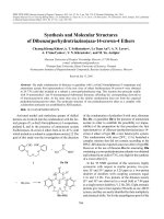
Figure 3. Fluorescence spectra of Cur, Cur-OCMCs and
Cur-OCMCs-Fol.
3. Results and discussion
3.1. Encapsulation efficiency
The polymer-encapsulated curcumin nanoparticles show
enormous improvements in aqueous solubility characteristics.
While free curcumin immediately precipitates in aqueous
medium due to very low solubility (∼20 µg ml−1 ), the
absolute concentration of curcumin in filtered 1,3-β-glucan
solution was found to be 4 mg ml−1 , which is 220-folds
compared with the solubility of curcumin encapsulated by
hydrophobically modified starch (HMS) [28]. The aqueous
solubility of Cur-Glu was 2-folds compared with that of
Cur-PLA-TPGS (2 mg ml−1 ) and 4-folds compared with that
of Cur-OCMCs or Cur-OCMCs-Fol (1 mg ml−1 ). The higher
solubility characteristics of Cur-Glu may result from better
compatibility to curcumin of 1,3-β-Glucan due to its short
chain.
Lyophilized Cur-Glu, Cur-PLA-TPGS, Cur-OCMCs and
Cur-OCMCs-Fol powder were reconstituted with water. These
powders dissolved back into clear solution very quickly and
easily, with no noticeable curcumin precipitates (figure 1).
The results suggested that curcumin was indeed trapped in the
micelles and the complex of polymers and curcumin could
resist against freeze-drying.
2.4. Characterization
Infrared spectra were recorded with a Fourier transform
infrared (FTIR) spectrometer SHIMADZU, using KBr pellets,
in the region of 400–4000 cm−1 . Field emission scanning
electron microscope (FE-SEM) images were taken by a
Hitachi S-4800. Fluorescence spectra were recorded by using
a Jobin-Yvon FL3-22. Photoluminsescence spectra were
taken with a 442 nm excitation line. Encapsulated curcumin
were estimated using the calibration curve of curcumin
solution in acetone or ethanol.
3
Adv. Nat. Sci.: Nanosci. Nanotechnol. 3 (2012) 035002
P T Ha et al
(a)
(b)
(c)
(d)
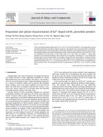
Figure 4. FE-SEM images of Cur-PLA-TPGS (a), Cur-Glu (b), Cur-OCMCs (c) and Cur-OCMCs-Fol (d).
3.2. FTIR spectra
3.3. Fluorescence spectra
The fluorescence spectra of curcumin and polymerencapsulated curcumin are shown in figure 3. Curcumin
in ethanolic solution exhibits an absorption peak at 540 nm,
while the solutions of Cur-Glu, Cur-PLA-TPGS, Cur-OCMCs
and Cur-OCMCs-Fol show peaks at 529, 530, 491 and
525 nm, respectively. The blue-shifts in the fluorescence
are likely due to the intermolecular hydrogen bonding
between curcumin and polymers. Especially, the appearance
of a weak peak at 435 nm in the fluorescence spectrum of
Cur-OCMCs-Fol might be explained by the presence of folate
on the nanoparticles.
While the fluorescence intensity of Cur-OCMCs-Fol
is slightly lower than that of Curcumin itself, the much
higher fluorescence intensity of Cur-PLA-TPGS, Cur-Glu,
Cur-OCMCs suggests that curcumin is encapsulated in the
hydrophobic core of micelle of PLA-TPGS and curcumin is
present in the Cur-Glu and Cur-OCMCs.
All FTIR spectra of Cur-Glu, Cur-PLA-TPGS, Cur-OCMCs
and Cur-OCMCs-Fol have several shifts as compared to those
of free curcumin or polymers. This indicates the formation of
polymer-encapsulated curcumin nanoparticles. For example,
compared with that of pure 1,3-β-Glucan, the IR spectrum
of Cur-Glu showed a band shift from 3400 to 3417 cm−1 ,
which is probably due to the hydrogen bonding between–OH
groups in curcumin and 1,3-β-Glucan (spectra omitted for
brevity). The FTIR spectrum of OCMCs showed broad bank
at 3420 cm−1 due to the stretching vibration of hydroxyl
group. A peak at 1634 cm−1 corresponds to the stretching
vibrations of carbonyl. Comparing OCMCs and Cur-OCMCs,
peak shifts were observed from 3420 to 3261 cm−1 and
1634 to 1625 cm−1 . The result confirmed the presence of
curcumin in the Cur-OCMCs. The characteristic absorption
band of OCMCs that appeared at 1598 cm−1 was assigned
to the N–H banding vibration of the primary amine. In the
case of Cur-OCMCs-Fol this peak is shifted to 1635 cm−1 .
The increased absorption of amide band may be due to the
formation of the amide linkage between the amino acid group
on the OCMCs and the carboxyl group of folic acid (figure 2).
3.4. Surface morphology
The size and morphology of the polymer-encapsulated
curcumin nanoparticles were confirmed by FE-SEM imaging.
Figure 4 shows FE-SEM images of the curcumin encapsulated
4
Adv. Nat. Sci.: Nanosci. Nanotechnol. 3 (2012) 035002
P T Ha et al
by PLA-TPGS (a), Glu (b), OCMCs (c) and OCMCs-Fol
(d). It is shown that the particles have an average size of
50–100 nm, which lies in the optimal size range (below
200 nm) suitable for drug delivery applications. There is
a significant decrease in size of curcumin nanoparticles
compared to that of curcumin. This is probably because the
hydrophilic polymers prevent the aggregation of hydrophobic
curcumin.
A PL image of free curcumin dispersed in ethanolic
solution is shown in figure 5(a). The spherical shape of the
particles is seen with a size of 1–10 µm. Figures 5(b) and (c)
show PL images of the polymer-encapsulated curcumin with
a large range of sizes similar to that of curcumin. Curcumin
in the form of nanoparticles is a strong PL substance, thus
when used to treat cancer it could also act as a labeling
material. Hence we can determine the efficiency of the
drug transport process in different conditions. PL images of
Cur-PLA-TPGS (figure 5(b)) and Cur-Glu (figure 5(c)) show
that both nanoparticles are highly fluorescent, implying that
these materials can be used not only for cancer treatment but
also for biolabeling.
(a)
3.5. Colony assays in soft agar
Cell survival cytotoxicity experiments using sulforhodamine
B method were performed in order to determine the maximal
doses of test materials for anti-tumor-promoting activity
assays. Soft agar colony assay anti-tumor-promoting activity
was estimated based on the inhibition of soft agar colony
induction in the Hep-G2 cell line. The cells were cultured
in 10% FBS-MEM medium at 36.5 ◦ C in an incubator with
5% CO2 and 95% air. Cells growing logarithmically in a
monolayer culture were trypsinized and suspended in 0.33%
agar medium containing 10% FBS with or without samples at
the concentrations of 25 µg ml−1 . For anti-tumor promoting
assay, in duplicate 6-well plate, 500 µl of the suspension (1 ×
104 cells) was poured onto an agar layer containing the same
concentration of sample (10 µg ml−1 ) in 5% DMSO. Soft agar
colonies of cells were investigated after 2 weeks’ incubation
under an inverted microscope with camera to compare the
visual cell in their tumor formation, the tumor size and
morphology. The inhibitory activities were the average of two
independent experiments and expressed as a percentage of that
of the control.
The results showed that there were no distinct differences
of cell survival in cytotoxicity assay, and the ratio of tumor
promotion in anti-tumor promoting assay with the Cur,
Glu, PLA-TPGS alone was comparable to the control, but
there were clear changes in size and morphology of tumor
between the control and all the tested samples, especially
curcumin encapsulated with glucan copolymer. In the control
wells, the tumor size was much larger and their surface was
very rough in comparison to the tumor on the tested wells
(figure 6). It was obvious that encapsulated curcumin had
positive effects on tumor promotion of Hep-G2 cell line
in vitro.
(b)
(c)
Figure 5. Fluorescent images of Cur (a), Cur-PLA-TPGS (b) and
Cur-Glu (c).
Hela and HT29 cells at 4 and 12 h. Hela, HT29 cells
were maintained 24 h and then incubated with Cur-OCMCs
and Cur-OCMCs-Fol within 4 and 12 h. Immunofluorescent
stains were processed and cells were visualized in confocal
laser scanning microscopy LSM-510. Fluorescence intensities
in specimens were compared to evaluate the quantity of
curcumin within cancer cells.
3.6. Intracellular uptake of nanoparticles
To study the uptake of the nanoparticles Cur-OCMCs
and OCMCs-Cur-Fol, confocal imaging was performed on
5
Adv. Nat. Sci.: Nanosci. Nanotechnol. 3 (2012) 035002
P T Ha et al
(a)
(a)
(b)
(b)
(c)
Figure 7. Fluorescent Image of HT29 after 4 h incubating with
control (a), Cur-OCMCs (b) and Cur-OCMCs-Fol (c).
that folate-conjugated carboxymethyl chitosan may be very
effective carrier to use as delivery system for targeted
anticancer drug.
The rate of Cur-OCMCs and Cur-OCMCS-Fol in
HT29 and HeLa indicates a different level of uptake of
curcumin. This is consistent with the level of expression
of folate receptor on cell surface: HT29-overexpression and
Hela-mediated expression.
Fluorescence intensity of Cur-OCMCs-Fol inside
the cell at 12 h (figure 8(b)) shows a lower content than
that at 4 h (figure 8(a)). This can be explained by the
degradation of curcumin to form smaller molecules such as
trans - 6-(4-hydroxy-3-methoxyphenul)-2, 4-dioxo - 5-hexenal,
vaniline, ferulic acid, feruloy methane which can no longer
remain auto-fluorescence like curumin [29]. However,
fluorescence intensity of Cur-OCMCs at 12 h shows a higher
content than that at 4 h.
(c)
Figure 6. Anti-tumor-promoting effects of the curcumin
encapsulated by copolymer in Hep-G2 cell lines after two weeks
of cell growth on agar: control (a), Cur (b) and Cur-PLA-TPGS
(c) under inverted microscope ×100.
From the confocal microscope images (figure 7) the
folate-conjugated nanoparticles were found to be distributed
in the zone of nucleus, indicating cellular uptake instead
of adhesion to the surface, and that the nanoparticles
preferentially targeted the cancer cells and were internalized.
This internalization might be due to the folate receptor
mediated endocytosis [23]. This observation clearly infers
6
Adv. Nat. Sci.: Nanosci. Nanotechnol. 3 (2012) 035002
P T Ha et al
Acknowledgments
This work was financially supported by an IMS
research grant, the National Foundation for Science and
Technology Development of Vietnam NAFOSTED grant
No 106.99-2010.42 (HPT), No 106.03.84.09 (MHL) and
the Ministry of Science and Technology grant (No 04/ 02
/742/2009/HD-DTDL). The authors are thankful to Professor,
Academician Nguyen Van Hieu for his encouragement
and interest in this research. The authors would like to
acknowledge all members of IMS-VAST Key Laboratory for
providing laboratory facilities.
References
(a)
[1]
[2]
[3]
[4]
[5]
[6]
[7]
[8]
[9]
[10]
[11]
[12]
[13]
(b)
Figure 8. Comparison of cellular uptake between Cur-OCMCS and
Cur-OCMCS-Fol on HT29 and Hela cell lines at 4 h (a) and 12 h (b).
[14]
[15]
[16]
[17]
The key difference may come from the presence of folic
acid, which actively leads the nanosystem to the cancer cells
with expression of folate receptor on its surface. In that case
curcumin can be transferred to the cancer cells more quickly
and efficiently.
[18]
[19]
4. Conclusion
[20]
[21]
In the present studies copolymer PLA-TPGS, 1,3-βGlucan, O-carboxymethyl chitosan and folate-conjugated
O-carboxymethyl chitosan-encapsulated curcumin nanoparticles were prepared successfully by nanoprecipitation
technique. It was found that these particles have a good
solubility in water. As spherical particles with an average
size from 50 to 100 nm, they are also believed to be
suitable for drug delivery applications. Confocal microscopy
revealed that folate enhances the uptake of curcumin into
cancer cells expressing folate receptor. Besides, the anti-tumor
promoting assay also shows strong positive effects of
Cur-PLA-TPGS and Cur-Glu on tumor promotion of Hep-G2
cell line in vitro. With all these good features that have
been found, Cur-PLA-TPGS, Cur-Glu, Cur-OCMCs and
Cur-OCMCS-Fol, could be used toward cancer therapy.
[22]
[23]
[24]
[25]
[26]
[27]
[28]
[29]
7
Berry W R 2005 Urology 65 2
Oh W K, Tay M H and Huang J 2007 Cancer 109 477
Petrylak D P 2005 Urology 65 8
Urakami S, Shiina H, Sumura M, Honda S, Wake K, Hiraoka
T, Inoue S, Ishikawa N and Igawa M 2008 Int. Urol.
Nephrol. 40 365
Karmakar S, Banik N L, Patel S J and Ray S K 2006 Neurosci.
Lett. 407 53
Anand P, Sundaram C, Jhurani S, Kunnumakkara A B and
Aggarwal B B 2008 Cancer Lett. 267 133
Li L, Braiteh F S and Kurzrock R 2005 Cancer 104 1322
Sou K, Inenaga S, Takeoka S and Tsuchida E 2008 Int. J.
Pharm. 325 287
Yallapu M M, Jaggi M and Chauhan S C 2010 Colloids Surf. B
79 113
Anand P, Nair H B, Sung B, Kunnumakkara A B, Yadav V R,
Tekmal R R and Aggarwal B B 2010 Biochem. Pharmacol.
79 330
Tran D L et al 2010 Colloids Surf. A 371 104
Shenoy D B and Amiji M M 2005 Int. J. Pharm 293 261
Sahu A, Bora U, Kasoju N and Goswami P 2008 Acta
Biomater. 4 1752
Bisht S, Feldmann G, Soni S, Ravi R, Karikar C, Amornath M
and Aniirban M 2007 J. Nanobiotechnol. 5 3
Mu L and Seow P H 2006 Colloids Surf. B 47 90
Pan J and Feng S S 2008 Biomaterials 29 2663
Nakatsugawa M, Takahashi H, Takezawa C, Nakajima K,
Harada K, Sugawara Y, Kobayashi S, Kondo T and Abe S
2003 Int. Med. 42 1219
Mizuno T, Wasa T, Ito H, Suzuki C and Ukai N 1992 Biosci.
Biotechnol. Biochem. 56 347
Zhu A P, Liu J H and Ye W H 2006 Carbohydr. Polym.
63 89
Ha P T et al 2011 Chem. Lett. 40 1264
Anitha A, Maya S, Deepa N, Chennazhi K P, Nair S V, Tamura
H and Jayakumar R 2011 Carbohydr. Polym. 83 452
Wang F, Zhang D, Duan C, Jia L, Feng F, Liu Y, Wang Y, Hao
L and Zhang Q 2011 Carbohydr. Polym. 84 1192
Sahu S K, Mallick S K, Santra S, Maiti T K, Ghosh S K and
Pramanik P 2010 J. Mater. Sci.: Mater. Med. 21 1587
Yang S J, Lin F H, Tsai H M, Lin C F, Chin H C, Wong J M
and Shieh M J 2011 Biomaterials 32 2174
Low P S and Kularatne S A 2009 Curr. Opin. Chem. Biol.
13 256
Ha P T, Tran T M N, Pham H D, Nguyen Q H and Nguyen X P
2010 Adv. Nature Sci.: Nanosci. Nanotechnol. 1 015012
Le M H, Ha P T, Nguyen T B T, Tran T H H, Ha T M T, Mai
T T, Tran T N H, Do H N, Nguyen X P and Duong T Q
2011 Chem. Lett. 40 846
Yu H and Huang Q 2010 Food Chem. 11 669
Aggarwal B B, Surh Y J and Shishodia S 2007 Adv. Exp. Med.
Biol. 595 269