DSpace at VNU: A Simple Approach to the Fabrication of Graphene-Carbon Nanotube Hybrid Films on Copper Substrate by Chemical Vapor Deposition
Bạn đang xem bản rút gọn của tài liệu. Xem và tải ngay bản đầy đủ của tài liệu tại đây (802.79 KB, 12 trang )
Accepted Manuscript
A Simple Approach to the Fabrication of Graphene-Carbon Nanotube Hybrid Films on
Copper Substrate by Chemical Vapor Deposition
Nguyen Van Chuc, Cao Thi Thanh, Nguyen Van Tu, Vuong TQ. Phuong, Pham Viet
Thang, Ngo Thi Thanh Tam
PII:
S1005-0302(15)00070-5
DOI:
10.1016/j.jmst.2014.11.027
Reference:
JMST 496
To appear in:
Journal of Materials Science & Technology
Received Date: 19 September 2014
Revised Date:
14 November 2014
Accepted Date: 19 November 2014
Please cite this article as: N. Van Chuc, C.T. Thanh, N. Van Tu, V.T. Phuong, P.V. Thang, N.T. Thanh
Tam, A Simple Approach to the Fabrication of Graphene-Carbon Nanotube Hybrid Films on Copper
Substrate by Chemical Vapor Deposition, Journal of Materials Science & Technology (2015), doi:
10.1016/j.jmst.2014.11.027.
This is a PDF file of an unedited manuscript that has been accepted for publication. As a service to
our customers we are providing this early version of the manuscript. The manuscript will undergo
copyediting, typesetting, and review of the resulting proof before it is published in its final form. Please
note that during the production process errors may be discovered which could affect the content, and all
legal disclaimers that apply to the journal pertain.
ACCEPTED MANUSCRIPT
A Simple Approach to the Fabrication of Graphene-Carbon
Nanotube Hybrid Films on Copper Substrate by Chemical Vapor
Deposition
Nguyen Van Chuc1,*, Cao Thi Thanh1, Nguyen Van Tu1, Vuong TQ Phuong2, Pham Viet
Thang2, Ngo Thi Thanh Tam1
Institute of Materials Science, Vietnam Academy of Science and Technology, 18 Hoang Quoc
RI
PT
1
Viet, Cau Giay, Hanoi, Vietnam
2
Hanoi University of Science, Vietnam National University, Hanoi, Vietnam
SC
[Received 19 September 2014; Received in revised form 14 November 2014; Accepted 19 November 2014]
* Corresponding author. Ph.D.; Tel.: +84 3 7565763; Fax: +84 3 8360705.
M
AN
U
E-mail address: (N.V. Chuc).
In this study, graphene-carbon nanotube (CNT) hybrid films were directly synthesized on
polycrystalline copper (Cu) substrates by themal chemical vapor deposition (CVD) method.
Graphene films were synthesized on Cu substrate at 1000 oC in mixture of gases: argon (Ar),
TE
D
hydrogen (H2), and methane (CH4). Then, carbon nanotubes (CNTs) were grown uniformly on
the surface of graphene/Cu films at 750 oC in mixture of Ar, H2, and acetylene (C2H2) gases.
Ferric salt FeCl3 solution deposited onto the surface of graphene/Cu substrate by spin coating
method was used as precursor for the growth of the CNTs. The density and quality of the
EP
CNTs on the surface of graphene/Cu films can be controlled by varying the concentration of
FeCl3 salt catalyst.
AC
C
Key words: Graphene; Carbon nanotube (CNT); Hybrid films; Copper substrate;
Chemical vapor deposition (CVD)
1. Introduction
Owing to its unique electrical, mechanical and optical properties, graphene with
two-dimensional (2D) carbon nanostructure has emerged as a new class of promising materials
attractive for various applications, such as transparent electrodes[1–3], field-effect transistors[4,5],
supercapacitors[6], composites[7], energy storage materials[8–11], chemical and biosensing[12–15].
The combination of 2D graphene of high charge density and one- dimensional (1D) carbon
nanotube (CNT) of large surface area generates a flexible three-dimensional (3D)
1
ACCEPTED MANUSCRIPT
graphene-CNT hybrid network with outstanding properties. Studies proved that graphene-CNT
hybrid nanomaterials exhibit higher electrical conductivities, large specific area and catalytic
properties compared with either pristine CNT or graphene[16–18]. Graphene-CNT hybrid
material has also been applied in many applications including electronics (such as transparent
conductors[2,19,20], electron field emitters[21], field effect transistors[20]), supercapacitors[6],
Li-ion batters[22,23], sensors[24,25] and biosensors[26–28].
RI
PT
The hybrids were prepared by several approaches including sonication method[16,17],
chemical vapor deposition (CVD) method[2,18,29,30], and electrostatic spray technique[31]. In
these methods, CVD method has emerged as an appropiate approach to synthesize
graphene-CNT hybrid materials. Different transition metal (e.g, iron (Fe), cobalt (Co), nickel
SC
(Ni), copper (Cu), gold (Au), palladium (Pd), platinum (Pt), and ruthenium (Ru)[32,33]) have
been used to grow graphene and CNTs. Transition metals such as Fe, Co, Ni and Cu are of
M
AN
U
particular interest, due to their low cost and availability. However, Fe, Co and Ni are not
preferred for mono or bilayer grephene growth due to their higher-than-desirable capability to
decompose hydrocarbons. On the other hand, the lower decomposition rate of methane on Cu
(since Cu cannot form carbide with carbon thereby resulting in low solubility of carbon in Cu)
allows the possibility of controlling the number of graphene layers[34], and wet etchant
TE
D
selectivity to graphene[35]. Using a rapid heating and cooling CVD system, Nguyen et al.[2]
synthesized thin networks of CNTs with different densities that are controlled by varying the
thickness of an iron film sputtered on the graphene/copper substrates. However, this method
requires the sputtering equipment to produce iron catalyst on the surface of graphene/copper.
EP
In this study, we present a simple approach to fabricate graphene-CNT hybrid films on
polycrystalline Cu substrate. By performing CVD method, graphene layer was synthesized on
AC
C
Cu substrate in mixture of gases Ar, H2 and CH4. CNTs were subsequently produced on the
surface of graphene/Cu film in mixture of gases Ar, H2 and C2H2. The density and quality of
the CNTs in the hybrid materials can be controlled by varying the concentration of FeCl3 salt
catalyst.
2. Experimental
2.1. Synthesis of graphene film
The graphene films were synthesized by thermal CVD method of high temperature of
1000 °C in Ar environment (1000 sccm). The polycrystalline Cu with a thickness of 35 µm and
a size of 1.0 cm × 1.0 cm were used as substrate for graphene-films synthesis process. To
2
ACCEPTED MANUSCRIPT
reduce the native copper oxide and to facilitate Cu grain growth on the Cu substrate surface,
the samples were annealed at CVD temperature for 30 min in a flow of Ar/hydrogen (1000/ 300
sccm). After 30 min, a flow of methane (CH4, 20 sccm) was introduced for growth process. The
time for the CVD process was 30 min. After a preset graphene growth time, the samples were
cooled rapidly under a flow of Ar (1000 sccm).
RI
PT
2.2. Synthesis of graphene-CNT hybrid film
In this work, the ferric salt FeCl3 was used as precursor for the formation of catalytic iron
nanoparticles. Firstly, it was dissolved in deionized water. The resultant solution was
SC
subsequently deposited on the graphene/Cu substrate. The sample was dried naturally at room
temperature to prepare for the growth of CNTs.
The CNTs growth process was performed via “fast heating” CVD method, using C2H2 as
M
AN
U
carbon source. The graphene/Cu substrates initially placed outside the heating zone were
subsequently transferred into the center of CVD chamber when the temperature of the whole
system reached to 750 °C in Ar (30 sccm). This step was carried out by moving the CVD
chamber to the proper position. The mixture of C2H2 (5 sccm), Ar (30 sccm), and H2 (30 sccm)
was simultaneously passed through the tube reactor for 30 min. The whole process was finally
TE
D
followed by cooling the CVD system in Ar to room temperature.
In the growth process, catalyst iron nanoparticles play a crucial role in controlling the
structure of CNTs. As mentioned in many previous articles[2,36,37], it is widely accepted that the
diameter, which is the main parameter to determine the quality of CNTs, is defined by the size
EP
of the catalyst nanoparticles. For that reason, the concentration of the catalyst precursor and its
deposition techniques onto the substrates are the key issues for monitoring the shape and size
AC
C
of nanoparticles. To investigate the influence of the formation of surfactant catalytic iron
nanoparticle on the growth density and quality of CNTs, the FeCl3 solution was used with four
initial different concentrations (0.001, 0.005, 0.01, and 0.05 mol/L). The deposition process of
FeCl3 solution onto the graphene/Cu substrate was conducted via spin coating at 5000 r/min for
1 min.
2.3. Sample characterizations
The scanning electron microscopy (SEM) and transmission electron microscopy (TEM)
images of the samples were obtained by using Hitachi S-4800 and Jeol JEM-1010 instruments,
respectively. The graphene layer structure was studied using a high resolution transmission
3
ACCEPTED MANUSCRIPT
electron microscopy (HRTEM, FEI TECNAI G20). The concentrations of the chemical
elements were determined from the energy dispersive X-ray spectroscopy (EDX, JED-2300
Analysis Station). Raman spectra were accquired using LAMBRAM-1B under ambient
condition with excitation laser of He–Ne (wavelength: 632.8 nm). Atomic force microscopy
(AFM) image was accquired in the tapping mode using Agilent PicoScan 2500.
RI
PT
3. Results and Discussion
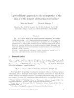
Fig. 1(a) and (b) shows the photographs of Cu substrate (with area of 1.0 cm × 1.0 cm)
before and after graphene growth, respectively. Homogeneous growth of graphene film all over
SC
the area was observed. Luster of the Cu substrate was changed to grayish shade after graphene
growth. Graphene on Cu was grown by surface adsorption process[35]. Fig. 1(c) shows a typical
M
AN
U
SEM image of Cu substrate surface after CVD process. The white lines observed on graphene
film (Fig. 1(c)) are graphene wrinkles. The wrinkled feature of the graphene films is believed
to be due to accommodation of the differences in the thermal expansion coefficients between
graphene or graphite and Cu substrate (αgraphene = –6 × 10⁻⁶/K at 27 °C; αgraphite = 0.9 × 10⁻⁶/K
between 600–800 °C; αCu = 24 × 10–6/K[38]). This is indicative of graphene continuity, since
TE
D
these wrinkles span the Cu grain boundaries[39,40]. Fig. 1(d) shows the typical EDX
characterization of the graphene film grown on the surface of Cu substrate. The calculated
results from the EDX characterizations measured at four different points (with each point area
EP
of 0.6 mm × 0.6 mm) showed that the concentration of C and Cu were 2.91 ± 0.37 and 97.07 ±
1.35 wt% , respectively.
To estimate clearly the thickness of the graphene film, we transferred the graphene film
AC
C
from the Cu substrate to SiO2 substrate. Fig. 2(a) shows the AFM image of the graphene film
after transferring from the Cu substrate to SiO2 substrate. A uniform color contrast in the AFM
image indicates uniform graphene thicknesses, although bright lines in the graphene films were
formed during the transfer process and due to the difference of thermal expansion between
graphene and copper during CVD process. The height profiles of the cross-section lines
associated with the AFM images were used to indicate the thickness of the graphene films.
AFM image indicates that the thickness of the graphene film is about 1 nm.
High-resolution transmission electron microscopy (HRTEM) image can provide direct
evidence of the number of graphene layers. Fig. 2(b) indicates an HRTEM image of the
graphene film after transferring from the Cu substrate to a copper grid for TEM examination.
4
ACCEPTED MANUSCRIPT
HRTEM image indicates that the number of graphene layers is two layers (0.68 nm thick). This
result is consistent with the thickness of the transferred film as analyzed by AFM.
Raman spectroscopy is a powerful, yet relatively simple method to characterize the
thickness and crystalline quality of graphene layers. Raman spectroscopy was performed under
excitation laser (λ = 632.8 nm) on the CVD grown graphene. Raman spectrum of graphene film
RI
PT
(Fig. 2(c)) reveals that the CVD-grown graphene films exhibit a graphitic G at 1580 cm–1 and
second order resonance double-resonance 2D peaks at 2710 cm–1, whilst there is no apparent
defect related to D peak in the synthesized graphene film. It is known that I2D/IG is a sensitive
probe of the number of graphene layers[39,41]. The ratio I2D/IG of about 2–3 is for monolayer
SC
graphene, 2>I2D/IG>1 for bilayer and I2D/IG<1 for multilayers[39,42]. Based on ratio I2D/IG = 1.18
we can conclude that the graphene film grown on the Cu substrate contains more than one
layer.
M
AN
U
Beside the exceptional properties that contribute to the overall characteristic of hybrid
material, graphene has an important role in the growth of CNTs. It is considered as a barrier
that prevents the diffusion of catalytic iron particles formed under high temperature condition
of CVD process into the copper substrate that could lead to the deactivation of the iron
catalyst[2]. To evaluate clearly the role of graphene during growth, CNTs were synthesized on
TE
D
two different copper substrates (with and without graphene layers on the surface) under the
same CVD conditions. Structural properties of the bare Cu substrate and graphene-coated Cu
substrate after the growth of CNTs were analyzed by SEM. As shown in Fig. 3(a), the sample
without graphene is partly coated by a thin network of CNTs with large diameter. Moreover, a
EP
certain amount of amorphous carbon deposited around the CNTs can be observed either.
Meanwhile, after growth, the higher density of CNTs with high purity covered uniformly the
AC
C
entire surface of graphene-coated Cu substrate (Fig. 3(b)).
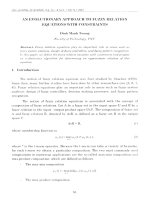
Fig. 4(a–d) shows typical FESEM images obtained from graphene/copper substrates
coated with concentration of FeCl3: 0.001, 0.005, 0.01, and 0.05 mol/L after the CNT growth
process, respectively. With low concentration of FeCl3 (0.001 mol/L), a thin network of CNTs
is distributed non-uniformly on the graphene surface, as shown in Fig. 4(a). As the
concentration of FeCl3 is increased to 0.005 and 0.01 mol/L, the graphene surfaces are covered
by a denser CNT network due to the provision of more catalyzing nanoparticles, as indicated in
Fig. 4(b) and (c), respectively. The diameter of CNTs is uniform and there are no amorphous
carbon deposited around the CNTs. However, when the substrate is coated with higher
concentration of FeCl3 (0.05 mol/L), a certain amount of amorphous carbon deposited around
5
ACCEPTED MANUSCRIPT
the CNTs can also be observed. The Fe nanoparticles are probably difficult to form due to the
cohesive agglomeration. That is why we have observed low density and large diameter CNTs,
as indicated in Fig. 4(d). Insets in Fig. 4(a–d) show their corresponding high resolution FESEM
images where the quality of CNTs can be observed clearly.
Comparison of the CNT Raman spectrum shown in Fig. 5(a) to the graphene spectrum
(Fig. 2(b)) clearly indicates the presence of CNTs with the appearance of D peak at ~ 1376
RI
PT
cm–1, and the broadening and decreased intensity of the 2D peak at ~ 2713 cm–1. To observe
clearly the morphology, quality of the graphene-CNTs hybrid film, the sample was transferred
from Cu substrate onto Ni grid for TEM analyst. Fig. 5(b) indicates the typical TEM image of
graphene-CNTs hybrid film grown with concentration of FeCl3 solution of 0.01 mol/L. The
SC
TEM image demonstrates that grain boundaries of the graphene films are continuous. The
M
AN
U
CNTs exhibits bamboo-like structure. The diameter of CNTs is about 30–40 nm.
4. Conclusion
By “fast-heating” CVD technique in atmospheric pressure, the CNTs network was
synthesized onto the surface of the graphene/copper substrate. The quality and density of the
CNTs were controlled via the concentration of FeCl3 catalyst. Optimized FeCl3 catalyst
TE
D
concentration for the growth of CNTs on the surface of graphene/copper was 0.01 mol/L. The
CNTs synthesized had bamboo-like structure and uniform diameters in the range of 30–40 nm.
These results open highly applicable for using graphene-CNTs hybrid film in electrochemical
Acknowledgements
EP
biosensor and field effect transistor biosensor.
AC
C
Funding of this work was supported mainly by the National Foundation for Science and
Technology Development (No. 103.99-2012.15). A part of work was supported by VAST
03.06/14-15. Besides, a part of the work was done with the help about devices of the IMS Key
Lab.
References
[1] R. Wang, J. Sun, L. Gao, C. Xu, J. Zhang, Y. Liu, Nanoscale 3 (2011) 904–906.
[2] D.D. Nguyen, N.H. Tai, S.Y. Chen, Y.L. Chueh, Nanoscale 4 (2012) 632–638.
[3] K. Rana, J. Singh, J.H. Ahn, J. Mater. Chem. C 2 (2014) 2646–2656.
[4] B. Zhan, C. Li, J. Yang, G. Jenkins, W. Huang, X. Dong, Small 10 (2014) 4042–4065.
[5] Y.H. Kwak, D.S. Choi, Y.N. Kim, H. Kim, D.H. Yoon, S.S. Ahn, J.W. Yang, W.S. Yang, S.
6
ACCEPTED MANUSCRIPT
Seo, Biosens. Bioelectron. 37 (2012) 82–87.
[6] Y.S. Kim, K. Kumar, F.T. Fisher, E.H. Yang, Nanotechnology 23 (2012) 015301.
[7] X.Y. Qi, K.Y. Pu, H. Li, X.Z. Zhou, S.X. Wu, Q.L. Fan, B. Liu, F. Boey, W. Huang, H.
Zhang, Angew. Chem. Int. Ed. 49 (2010) 9426–9429.
[8] H. Wang, L.F. Cui, Y. Yang, H.S. Casalongue, J.T. Robinson, Y. Liang, Y. Cui, H. Dai, J.
Am. Chem. Soc. 132 (2010) 13978–13980.
RI
PT
[9] G. Wang, X. Shen, J. Yao, J. Park, Carbon 47 (2009) 2049–2053.
[10] Z.S. Wu, G. Zhou, L.C. Yin, W. Ren, F. Li, H.M. Cheng, Nano Energy 1 (2012) 107–131.
[11] D.A.C. Brownson, D.K. Kampouris, C.E. Banks, J. Power Sources 196 (2011) 4873–4885.
[12] J.L. Her, T.M. Pan, W.Y. Lin, K.S. Wang, L.J. Li, Sens. Actuators B-Chem. 182 (2013)
SC
396–400.
[13] T.M.B.F. Oliveira, M.F. Barroso, S. Morais, M. Araújo, C. Freire, P. de Lima-Neto, A.N.
M
AN
U
Correia, M.B.P.P. Oliveira, C.D. Matos, Biosens. Bioelectron. 47 (2013) 292–299.
[14] H.B. Nguyen, V.C. Nguyen, V.T. Nguyen, H.D. Le, V.Q. Nguyen, T.T.T. Ngo, Q.P. Do,
X.N. Nguyen, N.M. Phan, D.L. Tran, Adv. Nat. Sci-Nanosci. Nanotechnol. 4 (2013) 015013
[15] V. Mani, B. Devadas, S.M. Chen, Biosens. Bioelectron. 41 (2013) 309–315.
[16] B. Devadas, V. Mani, S.M. Chen, Int. J. Electrochem. Sci. 7 (2012) 8064–8075.
TE
D
[17] M.Y. Yen, M.C. Hsiao, S.H. Liao, P.I. Liu, H.M. Tsai, C.C.M. Ma, N.W. Pu, M.D. Ger,
Carbon 49 (2011) 3597–3606.
[18] R.K. Sahoo, P. Jeyapandiarajan, K. Devi Chandrasekhar, B.S.S. Baniel, A. Venimadhav,
S.B. Sant, C. Jacob, J. Alloy. Compd. 5 (2014) 348–354.
EP
[19] D.D. Nguyen, R.N. Tiwari, Y. Matsuoka, G. Hashimoto, E. Rokuta, Y.Z. Chen, Y.L. Chueh,
M. Yoshimura, ACS Appl. Mater. Interfaces 6 (2014) 9071–9077.
AC
C
[20] S.H. Kim, W. Song, M.W. Jung, M.A. Kang, K. Kim, S.J. Chang, S.S. Lee, J. Lim, J.
Hwang, S. Myung, K.S. An, Adv. Mater. 26 (2014) 4247–4252.
[21] J.H. Deng, R.T. Zheng, Y. Zhao, G.A. Cheng, ACS Nano 6 (2012) 3727–3733.
[22] S. Chen, W. Yeoh, Q. Liu, G. Wang, Carbon 50 (2012) 4557–4565.
[23] Y. Hu, X. Li, J. Wang, R. Li, X. Sun, J. Power Sources 237 (2013) 41–46.
[24] B. Unnikrishnan, V. Mani, S.M. Chen, Sens. Actuators B-Chem. 173 (2012) 274–280.
[25] X. Chen, J. Zhu, Q. Xi, W. Yang, Sens. Actuators B-Chem. 161 (2012) 648–654.
[26] J. Chen, X. Zheng, F. Miao, J. Zhang, X. Cui, W. Zheng, J. Appl. Electrochem. 42 (2012)
875–881.
[27] X. Dong, Y. Ma, G. Zhu, Y. Huang, J. Wang, M.B.C. Park, L. Wang, W. Huang, P. Chen, J.
Mater. Chem. 22 (2012) 17044–17048.
7
ACCEPTED MANUSCRIPT
[28] X. Sun, Z. Gong, Y. Cao, X. Wang, Nano-Micro Lett. 5 (2013) 47–56.
[29] X. Dong, B. Li, A. Wei, X. Cao, M.B.C. Park, H. Zhang, L.J. Li, W. Huang, P. Chen,
Carbon 49 (2011) 2944–2949.
[30] M. Ghazinejad, S. Guo, W. Wang, M. Ozkan, C.S. Ozkan, J. Mater. Res. 28 (2013)
958–968.
[31] P. Han, Y. Yue, Z. Liu, W. Xu, L. Zhang, H. Xu, S. Dong, G. Cui, Energy Environ. Sci. 4
RI
PT
(2011) 4710–4717.
[32] C. Mattevi, H. Kim, M. Chhowalla, J. Mater. Chem. 21 (2011) 3324–3334.
[33] T. Oznuluer, E. Pince, E.O. Polat, O. Balci, O. Salihoglu, C. Kocabas, Appl. Phys. Lett. 98
(2011) 183101.
SC
[34] W. Liu, H. Li, C. Xu, Y. Khatami, K. Banerjee, Carbon 49 (2011) 4122–4130.
[35] X. Li, W. Cai, L. Colombo, R.S. Ruoff, Nano Lett. 9 (2009) 4268–4272.
M
AN
U
[36] C.J. Lee, S.C. Lyu, Y.R. Cho, J.H. Lee, K.I. Cho, Chem. Phys. Lett. 341 (2001) 245–249.
[37] M.H. Rümmeli, C. Kramberger, F. Schäffel, E. Borowiak-Palen, T. Gemming, B.
Rellinghaus, O. Jost, M. Löffler, P. Ayala, T. Pichler, R.J. Kalenczuk, Phys. Stat. Sol. (b)
244 (2007) 3911–3915.
[38] C. Mattevi, H. Kim, M. Chhowalla, J. Mater. Chem. 21 (2011) 3324–3334.
S. Kim, J. Nah, D. Yang, R. Piner, A. Velamakanni, I. Jung, E.
TE
D
[39] X. Li, W. Cai, J. An,
Tutuc, S.K. Banerjee, L. Colombo, R.S. Ruoff, Science 324 (2009) 1312–1314.
[40] A. Guermoune, T. Chari, F. Popescu, S.S. Sabri, J. Guillemette, H.S. Skulason, T. Szkopek,
M. Siaj, Carbon 49 (2011) 4204–4210.
EP
[41] K.S. Kim, Y. Zhao, H. Jang, S.Y. Lee, J.M. Kim, J.H. Ahn, P. Kim, J.Y. Choi, B.H. Hong,
Nature 457 (2009) 706–710.
AC
C
[42] W. Wu, Z. Liu, L.A. Jauregui, Q. Yu, R. Pillai, H. Cao, J. Bao, Y.P. Chen, S.S. Pei, Sens.
Actuators B-Chem. 150 (2010) 296–300.
Figure captions
Fig. 1 Photographs of Cu substrate before (a) and after (b) CVD process, typical SEM image (c)
and EDX spectrum (d) of Cu substrate after CVD process.
Fig. 2 (a) AFM image of graphene layer after transferring from the Cu substrate to SiO2 substrate, (b)
HRTEM image of graphene layer after transferring from the Cu substrate to copper grid and (c) Raman
8
ACCEPTED MANUSCRIPT
spectrum of the graphene film grown on Cu substrate.
Fig. 3 SEM images of CNTs grown onto the surface of: (a) Cu substrate, (b) graphene/Cu
substrate. Insets in (a) and (b) are their corresponding TEM images.
Fig. 4 SEM images of CNTs/graphene/Cu grown with various concentration of FeCl3 solution:
RI
PT
(a) 0.001 mol/L, (b) 0.005 mol/L, (c) 0.01 mol/L and (d) 0.05 mol/L. Insets in (a–d) are their
corresponding images at high magnifications.
Fig. 5 (a) Raman spectrum and (b) TEM image of graphene-CNTs hybrid film grown with
AC
C
EP
TE
D
M
AN
U
Figure list
SC
concentration of FeCl3 solution of 0.01 mol/L. Inset in (b) is its corresponding TEM image.
Fig. 1
9
Fig. 3
AC
C
EP
TE
D
M
AN
U
SC
Fig. 2
RI
PT
ACCEPTED MANUSCRIPT
10
M
AN
U
SC
RI
PT
ACCEPTED MANUSCRIPT
AC
C
EP
TE
D
Fig. 4
Fig. 5
11