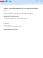
DSpace at VNU: Integrin alpha llb beta 3-Dependent ERK Signaling Is Regulated by Src and Rho Kinases in Both Leu33 and Pro33 Polymorphic Isoforms
Bạn đang xem bản rút gọn của tài liệu. Xem và tải ngay bản đầy đủ của tài liệu tại đây (248.04 KB, 7 trang )
Original Paper
Acta Haematol 2017;137:44–50
DOI: 10.1159/000450783
Received: June 27, 2016
Accepted after revision: September 8, 2016
Published online: December 7, 2016
Integrin αIIbβ3-Dependent ERK Signaling Is
Regulated by Src and Rho Kinases in Both Leu33
and Pro33 Polymorphic Isoforms
Khon C. Huynh a, c Thi-Hiep Nguyen a Dinh Chuong Pham b
Huong T.T. Nguyen c Toi Van Vo a Marianna Gyenes c Volker R. Stoldt c
a
Biomedical Engineering Department, International University, Vietnam National University, Ho Chi Minh City,
Vietnam; b Faculty of Applied Sciences, Ton Duc Thang University, Ho Chi Minh City, Vietnam; c Department of
Hemostasis, Hemotherapy, and Transfusion Medicine, Heinrich Heine University Medical Center, Düsseldorf, Germany
Abstract
Platelet integrin αIIbβ3 possesses a Leu/Pro polymorphism
at residue 33 (Leu33/HPA-1a or Pro33/HPA-1b). The Pro33
isoform has been suggested to exhibit prothrombotic features. αIIbβ3-expressing CHO (Chinese hamster ovary) cells
on immobilized fibrinogen show activation of the MAP kinase family member ERK2, with an enhanced ERK2 activity in
Pro33 cells compared to Leu33 cells. In our present work, we
examined how the Leu/Pro polymorphism modulates the
ERK2 activation stimulated by 2 differently triggered outside-in signalings. We either treated the CHO cells with Mn2+
or allowed them to adhere to fibrinogen. Moreover, we studied which signaling cascades are involved in ERK2 activation.
In contrast to immobilized fibrinogen, Mn2+ did not significantly increase ERK2 activation. However, Mn2+ had a synergistic effect on ERK2 phosphorylation when combined with
immobilized fibrinogen. Pro33 cells adherent to fibrinogen
exhibited a significantly greater ERK2 activity than Leu33
cells in the presence of Mn2+, which peaked after 10 min of
© 2016 S. Karger AG, Basel
www.karger.com/aha
adhesion. Our data showed that Src family and rho kinases
play a crucial role in the integrin αIIbβ3-dependent outsidein signaling to ERK2.
© 2016 S. Karger AG, Basel
Introduction
The major platelet integrin, the fibrinogen receptor
αIIbβ3, interacts with numerous plasma and extracellular
matrix proteins and thus plays an important role in platelet adhesion and aggregation during hemostasis and
thrombosis. Upon ADP or thrombin activation of the
platelets, integrin αIIbβ3 becomes activated (inside-out
signaling), and it can bind soluble fibrinogen, which in
turn induces the activation of various cellular responses
such as spreading and aggregation (outside-in signaling)
[1–3]. The β3 subunit of αIIbβ3 is polymorphic at residue
33, and these alleles encode either Leu (HPA-1a) or Pro
(HPA-1b). Platelets expressing the Pro33 phenotype show
an increased αIIbβ3 function, e.g., enhanced aggregation,
shorter bleeding times, and a greater affinity on immobilized fibrinogen [4–7]. The possible clinical aspects of this
polymorphism have been published in several studies
Khon C. Huynh
Biomedical Engineering Department
International University, Vietnam National University
Ho Chi Minh City (Vietnam)
E-Mail hckhon @ hcmiu.edu.vn
Downloaded by:
Rutgers University Alexander Library
128.6.218.72 - 2/20/2017 12:55:45 PM
Keywords
αIIbβ3 · ERK signaling · Leu33 · Pro33 · Polymorphisms ·
Rho kinase · Src signaling
Materials and Methods
Antibodies and Reagents
Anti-Src pY418 was from Invitrogen (Darmstadt, Germany),
anti-v-Src from Calbiochem (Darmstadt, Germany), phosphorylated ERK1/2 from Cell Signaling Technology (Danvers, MA,
USA), and anti-ERK1/2 from Promega (Mannheim, Germany).
Nonconjugated IgG mouse was from Sigma (Taufkirchen, Germany), secondary antibody rabbit HRG and mouse HRG from GE
Healthcare (Munich, Germany), FITC-conjugated clone P2 antibody and clone SZ21 antibody from Immunotech (Krefeld, Germany), and FITC-conjugated nonspecific mouse IgG from Becton-Dickinson (Heidelberg, Germany). Alfazyme was from PAA
αIIbβ3-Dependent ERK Signaling Is
Regulated by Src and Rho Kinases
Laboratories GmbH (Pasching, Germany), PP1 from Biomol
(Hamburg, Germany), PP3 from Merck (Darmstadt, Germany),
and the staining kit from Bio-Rad (Munich, Germany). Protease
and phosphatase inhibitors, apyrase, PGE1, human fibrinogen,
and all other reagents were from Sigma.
Flow Cytometry
Two CHO cell clones stably expressing αIIbβ3 isoforms Leu33
and Pro33 were obtained from the Department of Hemostasis,
Hemotherapy, and Transfusion Medicine, Heinrich Heine University Medical Center, Düsseldorf, Germany [22]. To check the
expression of αIIbβ3 isoforms, cells were resuspended in PBS, incubated with either FITC-conjugated CD-41 clone P2 antibody (1:
10) or FITC-conjugated HPA-1a-specific antibody (CD61 clone
SZ21, 1:10) in 50 μL end volume for 15 min at room temperature.
As a control, nonspecific mouse IgG-FITC was used. The labeled
cells were analyzed on a FACScalibur flow cytometer (Becton
Dickinson).
Cell Adhesion to Immobilized Fibrinogen
CHO cells were grown to 70–80% confluence, detached by Alfazyme for 5 min at 37 ° C, collected by centrifugation at 300 g for
7 min at room temperature, and resuspended in Tyrode’s buffer
(137 mM NaCl, 2.8 mM KCl, 12 mM NaHCO3, 0.4 mM NaH2PO4,
1 mM MgCl2, 1 mM CaCl2, and 5.5 mM glucose, pH 7.4). Six-well
tissue plates were coated with 500 μL (100 μg/mL) fibrinogen or
1% heat-denaturated BSA in PBS. Approximately 1.5 × 10⁵ cells
were added to each well and incubated for the indicated time periods at 37 ° C in a 5% CO2 incubator. For Western blotting, adherent cells were lysed on the plates with ice-cold lysis buffer (10 mM
Tris, 100 mM NaCl, 1 mM EDTA, 1 mM EGTA, 1 mM NaF, 20 mM
Na4P2O7, 2 mM Na3VO4, 1% Triton X-100, 10% glycerol, and 0.5%
sodium deoxycholate; pH 7.4) supplied with 250 μg/mL AEBSF, 15
μg/mL pepstatin, chymostatin, antipain, 55 μg/mL leupeptin, and a
phosphatase inhibitor mixture. For immunoprecipitation, we used
a lysis buffer containing 1% Triton X-100, 20 mM Tris, 136 mM
NaCl, 1 mM Na3VO4, 1 mM NaF, 2 mM EDTA, and 20 mM Na4P2O7
(pH 7.4) supplied with protease and phosphatase inhibitors. A nonadherent cell suspension was added to ice-cold 2× lysis buffer. The
lysates were chilled for 30 min on ice and clarified by centrifugation
at 13,200 rpm for 30 min at 4 ° C in a microcentrifuge. Protein concentration was determined by the Bradford method.
Gel Electrophoresis and Western Blotting
Equal amounts of protein were subjected to electrophoresis,
and all samples were electrophoresed either in 8% (for Src) or 10%
(for ERK) acrylamide gel for SDS-PAGE, transferred onto PVDF
membranes, and subjected to immunoreaction. The signals were
densitometrically visualized with a chemiluminescence ECL (Amersham Biosciences) system and quantified using an Azure c300
Imaging System (Azure Biosystems).
Results
Expression of αIIbβ3 Isoforms in CHO Cells
We obtained αIIbβ3-transfected CHO cells with the
appropriate αIIbβ3 isoforms (Leu33 or Pro33) [22]. The
Acta Haematol 2017;137:44–50
DOI: 10.1159/000450783
45
Downloaded by:
Rutgers University Alexander Library
128.6.218.72 - 2/20/2017 12:55:45 PM
demonstrating a potential association between these
symptoms, acute coronary syndromes [8], and a premature myocardial infarction by patients with coronary artery disease who are carriers of HPA-1b/1b [9].
Although the activation of integrin αIIbβ3 mostly occurs via inside-out signaling, adhesive ligand occupation
(i.e., immobilized fibrinogen/fibronectin) to the integrin
can also generate the active conformation of the integrin
leading to outside-in signaling [2]. Divalent Mn2+ cations
have also been reported to induce an active conformational state of αIIbβ3 [10] and to generate a subsequent
outside-in signaling [11, 12]. Src tyrosine kinase is associated with the cytoplasmic tail of the β3 subunit and has
been reported to play a crucial role in the integrin-mediated outside-in signaling [13, 14]. A number of other signal molecules and pathways have also been identified to
participate in the integrin-mediated outside-in signaling,
among others the mitogen-activated protein kinase
(MAPK) family member ERK2, whose Tyr/Thr phosphorylation regulates various cellular processes, including the release of stored Ca2+ in platelets [15], cell adhesion, and spreading [16]. Via its substrate, the myosin
light chain kinase (MLCK), ERK2 modulates the myosin
function and thereby the cytoskeletal clustering of integrins, shape changes, and secretion in platelets [17–19].
Previously, it has been reported that the substitution of
Leu to Pro at residue 33 enhances signaling to ERK2,
MLCK, and the extent of the phosphorylated state of the
regulatory subunit in the myosin phosphatase [3, 20, 21].
As these signal proteins are essential for cytoskeletal rearrangement, adhesion, and spreading, these results correlate well with the increased αIIbβ3 activity observed in the
HPA-1b/1b isoform. In our work, we examined how the
Leu33Pro polymorphism modulates ERK2 activation in
outside-in signaling. In addition, we studied which signaling pathways are involved in αIIbβ3-mediated ERK
activation.
46
Acta Haematol 2017;137:44–50
DOI: 10.1159/000450783
Counts
HPA-1a (Leu33)
HPA-1b (Pro33)
0
100
101
a
102
103
104
Fluorescence intensity
20
HPA-1b
(Pro33)
HPA-1a
(Leu33)
0
b
100
101
102
103
Fluorescence intensity
104
Fig. 1. Characterization of stable αIIbβ3 expression of a CHO cell
line; expression levels in HPA-1a (Leu33) and HPA-1b (Pro33)
CHO cells were determined by flow-cytometric analysis with the
FITC conjugated αIIbβ3-specific P2 antibody (a). To identify the
HPA-1a variant, antibody SZ21, a HPA-1a-specific antibody, was
utilized (b).
Src Family Kinases and Rho Kinases in ERK Signaling
ERK2 activation is mediated by dual phosphorylation
on threonine 185 and tyrosine 187 residues [23]. Therefore, Src tyrosine kinase and Rho kinase (ROCK) are suggested to be involved in ERK activation. Following fibrinogen engagement, Src pY418 activity was enhanced in
both Leu33 and Pro33 cells (Fig. 4a). To examine the role
of Src tyrosine kinases in ERK2 signaling, we incubated
Leu33 and Pro33 cells with the selective Src family kinase
inhibitor PP1 and subsequently allowed them to adhere
to 100 μg/mL fibrinogen. Cell suspensions over BSA surfaces were used as a control. As shown in Figure 4, PP1
completely blocked the ERK2 phosphorylation in both
isoforms indicating an Src kinase-dependent ERK2 actiHuynh/Nguyen/Pham/Nguyen/Van Vo/
Gyenes/Stoldt
Downloaded by:
Rutgers University Alexander Library
128.6.218.72 - 2/20/2017 12:55:45 PM
pERK2 Activity in αIIbβ3-Transfected CHO Cells
To study the modulation of the polymorphism onto
the integrin-mediated outside-in signaling, we investigated the activation of ERK in αIIbβ3-expressing CHO cells
on immobilized fibrinogen. After placing cells onto 100
μg/mL immobilized fibrinogen, we allowed them to adhere for 10 min followed by ERK activation analysis.
Pro33 cells exhibit higher ERK activation than Leu33 cells
(Fig. 2).
Mn2+ is also known to induce integrin activation via
shifting the receptor conformation from an inactive to an
active state [4]. In the next part of our work, we studied
the influence of Mn2+ on the ERK2 activation in fibrinogen-adherent CHO cells. Moreover, we assessed how the
Leu33/Pro33 polymorphism modulates this effect. To analyze whether Mn2+ alone induces outside-in signaling to
ERK2, we examined the effect of 0.5 mM Mn2+ on ERK2
activation in both Leu33 and Pro33 CHO cell suspensions
over BSA surface. As shown in Figure 2, Mn2+ slightly
stimulated ERK2 activation, but the extent of the stimulation was considerably less than in cells adhering to 100
μg/mL immobilized fibrinogen. Higher concentrations of
Mn2+ (1 or 2 mM) exhibited a similar effect as 0.5 mM
Mn2+ (data not shown). The combination of Mn2+ and
immobilized fibrinogen resulted in a synergism of ERK
activation. Mn2+ concentrations of 0.5 and 1 mM induced
significantly greater ERK2 phosphorylation in Pro33 cells
than in Leu33 cells. Using a concentration of 2 mM Mn2+,
both HPA-1 isoforms showed approximately equal ERK
activation (Fig. 2).
To analyze the kinetics of the ERK2 phosphorylation
as a consequence of the immobilized fibrinogen-Mn2+
combination, we allowed Leu33 and Pro33 CHO cells to
adhere to fibrinogen surfaces in the presence of 0.5 mM
Mn2+ for various periods of time. As shown in Figure 3,
both Leu33 and Pro33 cells exhibited a maximal ERK2
activity after 10 min of incubation with a subsequent decrease after 20 min adhesion.
64
Counts
2 clones were confirmed for an equivalent expression level of Leu33 and Pro33 isoforms prior to adhesion experiments. Flow-cytometric analysis with FITC-conjugated
anti-αIIbβ3 antibody P2 demonstrated equivalent receptor expression in the cell lines generated with the Leu33
(HPA-1a)- and Pro33 (HPA-1b)-containing αIIbβ3 isoforms, respectively. SZ21, a specific monoclonal antibody
to the HPA-1a isoform, presented a substantially lower
affinity for the Pro33 cells than for the Leu33 cells (Fig. 1).
pERK2
Total ERK1/2
Fig. 2. pERK2 activity in αIIbβ3-expressing
*
1.0
0.5
0
+ 0.5 mM
Mn2+
Fibrinogen
vation. PP3, an inactive analogue of PP1, did not exhibit
an inhibitory effect.
To examine the potential role of ROCK in the αIIbβ3dependent ERK2 activation, we performed adhesion experiments on fibrinogen in the presence of either Y27632
or HA1077, 2 pharmacologically distinct specific inhibitors of ROCK. Both inhibitors completely blocked the
ERK activation (Fig. 5). These observations suggest an involvement of the Src family kinases and ROCK in the fibrinogen-mediated αIIbβ3 outside-in signaling to ERK2.
+ 2 mM
Mn2+
BSA + 0.5 mM
Mn2+
Total ERK1/2
2.0
*
1.5
0.5
Leu33
Pro33
*
*
1.0
0
The platelet integrin αIIbβ3 plays a crucial role in
platelet aggregation and thrombus formation by binding
to fibrinogen initiating fibrinogen-dependent plateletcrosslinking [24, 25]. The fibrinogen engagement of the
integrin activates a great variety of outside-in signals
leading to elevated intracellular Ca2+ flux and cytoskeletal
rearrangement [2, 10]. Several polymorphisms in the integrin β3 subunit have been associated with platelet dysfunction. Among them, the Leu33Pro substitution of
αIIbβ3 has been reported to exhibit prothrombotic characteristics in several works [4, 7, 20].
ERK1 and ERK2 are involved in cell growth, proliferation, and adhesion, megakaryocyte differentiation, proplatelet formation [26, 27], and the release of stored Ca2+
+ 1 mM
Mn2+
pERK2
Discussion
αIIbβ3-Dependent ERK Signaling Is
Regulated by Src and Rho Kinases
*
*
2.5
5
10
Adhesion time, min
20
Fig. 3. Activation of ERK2 in αIIbβ3-expressing CHO cells adhering to immobilized fibrinogen in the presence of 0.5 mM Mn2+.
CHO cells were incubated in the presence of 0.5 mM Mn2+ for 3
min and subsequently allowed to adhere to 100 μg/mL fibrinogen
or maintained in suspension over 1% BSA for 2.5, 5, 10, and 20 min
at 37 ° C. After the incubation time, cells were solubilized, and the
lysates were analyzed for pERK2 activity. The enhanced ERK2 activation in Pro33 compared to Leu33 cells was significant at 5, 10,
and 20 min. Results are representative of 4 experiments. * p < 0.05,
evaluated by unpaired t test.
Acta Haematol 2017;137:44–50
DOI: 10.1159/000450783
47
Downloaded by:
Rutgers University Alexander Library
128.6.218.72 - 2/20/2017 12:55:45 PM
Leu33
Pro33
*
1.5
pERK2/total ERK1/2
2.0
pERK2/total ERK1/2
CHO cells adhering to immobilized fibrinogen in the presence of various concentrations of Mn2+. CHO cells were incubated in
the absence or presence of the indicated
concentrations of Mn2+ for 3 min and subsequently allowed to adhere to 100 μg/mL
fibrinogen or maintained in suspension
over 1% BSA. After a 10-min incubation at
37 ° C, cells were solubilized, and the lysates
were processed as described in Materials
and Methods. The enhanced ERK2 activation in Pro33 compared to Leu33 cells was
significant at concentrations of 0, 0.5, and
1 mM Mn2+. Results are representative of
3 experiments. * p < 0.05, evaluated by unpaired t test.
BSA
Fibrinogen
BSA
Fibrinogen
+ 10 μM + 10 μM
PP1
PP3
Fibrinogen
Src pY418
Total ERK1/2
3.0
2.5
*
pERK2/total ERK1/2
Src pY418/total Src
BSA
pERK2
Total Src
2.0
1.5
1.0
*
0.5
a
Fibrinogen
+ 10 μM + 10 μM
PP1
PP3
BSA
0
HPA-1a (Leu33)
b
HPA-1b (Pro33)
*
2.0
1.8
1.6
1.4
1.2
1.0
0.8
0.6
0.4
0.2
0
Fig. 4. Src pY418 activity (a) and effect of PP1 on ERK2 activity (b)
in αIIbβ3-expressing CHO cells adhering to immobilized fibrinogen. CHO cells were maintained in suspension over 1% BSA or
allowed to adhere to 100 μg/mL fibrinogen. To study the effect of
PP1 on ERK2 activity, cells were preincubated with 10 μM PP1 for
30 min and subsequently allowed to adhere to fibrinogen. After 10
*
*
*
HPA-1b (Pro33)
HPA-1a (Leu33)
min of incubation, cells were solubilized, and equal aliquots of
samples containing 50 μg of protein were separated by 10% SDSPAGE gel. The blots were probed with anti-Src pY418, anti-v-Src,
anti-pERK2, or anti-ERK antibodies and quantified by densitometry. * p < 0.05, evaluated by unpaired t test. Results are representative of 3 experiments.
20 μM
Fibrinogen 10 μM
Y27632 HA1077
BSA
Fibrinogen
10 μM
Y27632
20 μM
HA1077
BSA
pERK2
Total ERK1/2
1.2
Fig. 5. Effect of Rho kinase inhibition on
48
Acta Haematol 2017;137:44–50
DOI: 10.1159/000450783
0.8
0.6
*
0.4
0.2
0
HPA-1a (Leu33)
HPA-1b (Pro33)
Huynh/Nguyen/Pham/Nguyen/Van Vo/
Gyenes/Stoldt
Downloaded by:
Rutgers University Alexander Library
128.6.218.72 - 2/20/2017 12:55:45 PM
*
1.0
pERK2/total ERK1/2
ERK2 activity in αIIbβ3-expressing CHO
cells adhering to immobilized fibrinogen
CHO cells preincubated either with PBS or
with 10 μM Y27632 or 20 μM HA1077 for
30 min at 37 ° C and subsequently allowed
to adhere to 100 μg/mL fibrinogen. After 10
min of incubation at 37 ° C, cells were solubilized, and equal aliquots of samples containing 50 μg of protein were separated by
10% SDS-PAGE gel. The blots were probed
with antiphospho-antibody (pERK2) or
anti-ERK antibody and quantified by densitometry (ratio of pERK2 to total ERK in
arbitrary units). * p < 0.05, evaluated by unpaired t test. Results are representative of 2
experiments.
in platelets [15]. Fibrinogen-adherent Pro33 CHO cells
exhibit enhanced αIIbβ3-mediated outside-in signaling
to ERK2 and MLC [18], suggesting a role of ERK2 signaling in prothrombotic characteristics of this isoform in
platelets. Our aim was to further assess which signaling
pathways are involved in ERK activation.
One of the possibilities to activate integrins is triggering an active conformation in their extracellular domains
by divalent cations [28, 29]. We raised the following question: to what extent do Mn2+ ions alone regulate αIIbβ3mediated outside-in signaling in comparison to ligand
engagement? To analyze how this distinct manner of activation is reflected in receptor signaling, we examined
outside-in signaling induced by immobilized fibrinogen
or Mn2+ alone and by a combination of both. Furthermore, we analyzed how these processes are modified by
the Leu33/Pro33 polymorphism. Our observations that
Mn2+ cations alone elevate the ERK2 activity only to a
small extent when compared to immobilized fibrinogen
suggest a less important role of Mn2+ in regulating ERK2
signaling (Fig. 2). Previous studies have shown that Mn2+
increases the binding affinity of αIIbβ3 to ligands, but this
activation is not maximal and depends on the integrin
isoform type as well as the context [30]. Based on previous
studies and our data, it is hypothesized that Mn2+ alone
cannot induce ERK signaling of αIIbβ3. In contrast, when
combined with immobilized fibrinogen, Mn2+ induces a
synergistic effect leading to maximal ERK activity after 10
min of adhesion. In general, upon the whole incubation
time, Pro33 cells exhibited a significantly higher ERK activity than Leu33 cells.
In our work, we showed that the Leu33/Pro33 polymorphism modulates the αIIbβ3-mediated outside-in
signaling to Src (Fig. 4a). This tyrosine kinase plays an
essential role in integrin signaling and is directly associated with αIIbβ3 integrin [13, 14, 31]. Src kinase has been
reported to play a central role in the regulation of various
pathways, including the MAP kinase cascade [32]. On the
one hand, it has been shown that in adherent chick embryo fibroblast cells phosphorylated ERK is targeted after
integrin engagement or upon v-Src activation to newly
forming cell-matrix adhesion [33]. On the other hand, in
thrombin-activated human platelets, the Src kinase inhibitor PP1 did not block ERK activation [34], indicating
Src-independent ERK signaling. It seems that integrinmediated ERK activation can occur through several, from
each other independent, signaling cascades. Therefore,
we raised the question whether Src kinases participate in
the regulation of ERK2 signaling in fibrinogen-adherent
CHO cells. Our observation that the Src kinase family in-
hibitor PP1 entirely blocked ERK2 activation in both isoforms (Fig. 4) provides evidence that the ERK2 activation
in fibrinogen-adhering CHO cells is mediated via Src kinases. It has been reported that Src family kinases are also
involved in the regulation of the small GTPases [32].
These signal proteins are essential for cytoskeleton reorganization, and ROCK is an effector protein of the Rho
GTPase with a regulatory function. Moreover, ROCK is
proposed to be included in MLC phosphorylation [35].
As the Thr696 phosphorylation of the PP1-myosine
phosphatase regulatory subunit is modulated by the
Leu33/Pro33 polymorphism in thrombin-treated platelets [21] and this phosphorylation is regulated by ROCK,
we investigated the role of ROCK in the αIIbβ3-mediated
outside-in signaling to ERK2. Both Y27632 and HA1077,
2 pharmacologically distinct, specific inhibitors of ROCK,
completely blocked ERK2 activation, indicating an essential role of ROCK in αIIbβ3-mediated outside-in signaling to ERK2 (Fig. 5).
In conclusion, we provided evidence that the αIIbβ3associated outside-in signaling to ERK is mediated via the
Src kinase-ROCK signaling pathway in fibrinogen-adherent CHO cells. Although Mn2+ alone only slightly activates ERK, it synergizes the effect of adhesive fibrinogen
on ERK activation in both genotypes, showing a significantly higher ERK2 activation in the Pro33 isoform.
αIIbβ3-Dependent ERK Signaling Is
Regulated by Src and Rho Kinases
Acta Haematol 2017;137:44–50
DOI: 10.1159/000450783
Acknowledgments
We are grateful to Mrs. Bianka Maaßen-Weingart und to Mrs.
Elisabeth Kirchhoff for their excellent technical assistance. This
work was supported by the Deutsche Forschungsgemeinschaft,
Sonderforschungsbereich 612, TP B2, and grant No. 1161/QĐĐHQG-KHCN of the Vietnam National Universities Ho Chi
Minh City.
1 Shattil SJ, Newman PJ: Integrins: dynamic
scaffolds for adhesion and signaling in platelets. Blood 2004;104:1606–1615.
2 Stegner D, Nieswandt B: Platelet receptor signaling in thrombus formation. J Mol Med
(Berl) 2011;89:109–121.
3 Varga-Szabo D, Pleines I, Nieswandt B: Cell
adhesion mechanisms in platelets. Arterioscler Thromb Vasc Biol 2008;28:403–412.
4 Michelson AD, Furman MI, GoldschmidtClermont P, Mascelli MA, Hendrix C, Coleman L, Hamlington J, Barnard MR, Kickler T,
Christie DJ, Kundu S, Bray PF: Platelet GP
IIIa PlA polymorphisms display different sensitivities to agonists. Circulation 2000; 101:
1013–1018.
49
Downloaded by:
Rutgers University Alexander Library
128.6.218.72 - 2/20/2017 12:55:45 PM
References
50
14 Arias-Salgado EG, Lizano S, Sarkar S, Brugge
JS, Ginsberg MH, Shattil SJ: Src kinase activation by direct interaction with the integrin
beta cytoplasmic domain. Proc Natl Acad Sci
USA 2003;100:13298–13302.
15 Rosado JA, Sage SO: Phosphoinositides are
required for store-mediated calcium entry in
human platelets. J Biol Chem 2000;275:9110–
9113.
16 Zhu X, Assoian RK: Integrin-dependent activation of MAP kinase: a link to shape-dependent cell proliferation. Mol Biol Cell 1995; 6:
273–282.
17 Klemke RL, Cai S, Giannini AL, Gallagher PJ,
de Lanerolle P, Cheresh DA: Regulation of
cell motility by mitogen-activated protein kinase. J Cell Biol 1997;137:481–492.
18 Vijayan KV, Liu Y, Dong JF, Bray PF: Enhanced activation of mitogen-activated protein kinase and myosin light chain kinase by
the Pro33 polymorphism of integrin beta 3. J
Biol Chem 2003;278:3860–3867.
19 Kamm KE, Stull JT: Dedicated myosin light
chain kinases with diverse cellular functions.
J Biol Chem 2001;276:4527–4530.
20 Vijayan KV, Bray PF: Molecular mechanisms
of prothrombotic risk due to genetic variations in platelet genes: enhanced outside-in
signaling through the Pro33 variant of integrin β3. Exp Biol Med (Maywood) 2006; 231:
505–513.
21 Vijayan KV, Liu Y, Sun W, Ito M, Bray PF:
The Pro33 isoform of integrin β3 enhances
outside-in signaling in human platelets by
regulating the activation of serine/threonine
phosphatases. J Biol Chem 2005; 280: 21756–
21762.
22 Stoldt VR, Berendes S, Scharf RE: The HPA1b (Pro33) variant integrin αIIbβ3 increases
the resistance of adherent platelets and transfected CHO cells upon exposure to shear
stress. 54th Annu Meet Soc Thromb Hemost,
Nuremberg, 2010, A93.
23 Buscà R, Pouyssegur J, Lenormand P: ERK1
and ERK2 map kinases: specific roles or functional redundancy? Front Cell Dev Biol 2016;
4:53.
24 Calvete JJ: Clues for understanding the structure and function of a prototypic human integrin: the platelet glycoprotein IIb/IIIa complex. Thromb Haemost 1994;72:1–15.
Acta Haematol 2017;137:44–50
DOI: 10.1159/000450783
25 Ruggeri ZM: Platelets in atherothrombosis.
Nat Med 2002;8:1227–1234.
26 Whalen AM, Galasinski SC, Shapiro PS, Nahreini TS, Ahn NG: Megakaryocytic differentiation induced by constitutive activation of
mitogen-activated protein kinase kinase. Mol
Cell Biol 1997;17:1947–1958.
27 Jiang F, Jia Y, Cohen I: Fibronectin- and protein kinase C-mediated activation of ERK/
MAPK are essential for proplateletlike formation. Blood 2002;99:3579–3584.
28 Bazzoni G, Hemler ME: Are changes in integrin affinity and conformation overemphasized? Trends Biochem Sci 1998;23:30–34.
29 Plow EF, Haas TA, Zhang L, Loftus J, Smith
JW: Ligand binding to integrins. J Biol Chem
2000;275:21785–21788.
30 Smith JW, Piotrowicz RS, Mathis D: A mechanism for divalent cation regulation of beta
3-integrins. J Biol Chem 1994;269:960–967.
31 Obergfell A, Eto K, Mocsai A, Buensuceso C,
Moores SL, Brugge JS, Lowell CA, Shattil SJ:
Coordinate interactions of Csk, Src, and Syk
kinases with αIIbβ3 initiate integrin signaling
to the cytoskeleton. J Cell Biol 2002;157:265–
275.
32 Lee JW, Juliano R: Mitogenic signal transduction by integrin- and growth factor receptormediated pathways. Mol Cells 2004; 17: 188–
202.
33 Fincham VJ, James M, Frame MC, Winder SJ:
Active ERK/MAP kinase is targeted to newly
forming cell-matrix adhesions by integrin engagement and v-Src. EMBO J 2000; 19: 2911–
2923.
34 Tulasne D, Bori T, Watson SP: Regulation of
RAS in human platelets. Evidence that activation of RAS is not sufficient to lead to ERK1-2
phosphorylation. Eur J Biochem 2002; 269:
1511–1517.
35 Schoenwaelder SM, Hughan SC, Boniface K,
Fernando S, Holdsworth M, Thompson PE,
Salem HH, Jackson SP: RhoA sustains integrin αIIbβ3 adhesion contacts under high shear.
J Biol Chem 2002;277:14738–14746.
Huynh/Nguyen/Pham/Nguyen/Van Vo/
Gyenes/Stoldt
Downloaded by:
Rutgers University Alexander Library
128.6.218.72 - 2/20/2017 12:55:45 PM
5 Feng D, Lindpaintner K, Larson MG, Rao VS,
O’Donnell CJ, Lipinska I, Schmitz C, Sutherland PA, Silbershatz H, D’Agostino RB,
Muller JE, Myers RH, Levy D, Tofler GH: Increased platelet aggregability associated with
platelet GPIIIa PlA2 polymorphism: the
Framingham Offspring Study. Arterioscler
Thromb Vasc Biol 1999;19:1142–1147.
6 Vijayan KV, Goldschmidt-Clermont PJ, Roos
C, Bray PF: The PlA2 polymorphism of integrin beta3 enhances outside-in signaling and
adhesive functions. J Clin Invest 2000; 105:
793–802.
7 Loncar R, Stoldt V, Hellmig S, Zotz RB, Mihalj
M, Scharf RE: HPA-1 polymorphism of
αIIbβ3 modulates platelet adhesion onto immobilized fibrinogen in an in-vitro flow system. Thromb J 2007;5:2.
8 Williams MS, Bray PF: Genetics of arterial
prothrombotic risk states. Exp Biol Med
(Maywood) 2001;226:409–419.
9 Zotz RB, Winkelmann BR, Müller C, Boehm
BO, März W, Scharf RE: Association of polymorphisms of platelet membrane integrins
αIIbβ3 (HPA-1b/PlA2) and α2β1 (α2807TT)
with premature myocardial infarction. J
Thromb Haemost 2005;3:1522–1529.
10 Litvinov RI, Nagaswami C, Vilaire G, Shuman
H, Bennett JS, Weisel JW: Functional and
structural correlations of individual αIIbβ3
molecules. Blood 2004;104:3979–3985.
11 Petrich BG, Fogelstrand P, Partridge AW,
Yousefi N, Ablooglu AJ, Shattil SJ, Ginsberg
MH: The antithrombotic potential of selective blockade of talin-dependent integrin
αIIbβ3 (platelet GPIIb–IIIa) activation. J Clin
Invest 2007;117:2250–2259.
12 Shattil SJ: Signaling through platelet integrin
αIIbβ3: inside-out, outside-in, and sideways.
Thromb Haemost 1999;82:318–325.
13 Shattil SJ: Integrins and Src: dynamic duo of
adhesion signaling. Trends Cell Biol 2005;15:
399–403.