DSpace at VNU: Premilinary Study in the Cause of Color in Zircon from Krông Năng Mining Area in Đắk Lắk Province
Bạn đang xem bản rút gọn của tài liệu. Xem và tải ngay bản đầy đủ của tài liệu tại đây (637.62 KB, 8 trang )
VNU Journal of Science: Earth and Environmental Sciences, Vol. 31, No. 3 (2015) 60-66
Premilinary Study in the Cause of Color in Zircon from
Krông Năng Mining Area in Đắk Lắk Province
Bùi Thị Sinh Vương, Lê Thị Thu Hương*
Faculty of Geology, VNU University of Science, 334 Nguyễn Trãi, Hanoi, Vietnam
Received 29 September 2014
Revised 16 October 2014; Accepted 26 August 2015
Abstract: Zircon occurs in many colors including various shades of pink, red, purple, yellow,
orange, brown as well as less common shades of green, and blue. Generally, the colors of zircon
are caused by the trace element composition (transition metals, lanthanides, actinides and REEs)
and radiation damage (radiation induced color centers) [1]. The color centers of zircon are
complex and the details surrounding the color-inducing mechanisms are still debated. The authors
collected some zircon samples from Krong Nang mining, Central Highlandss, using UV-Vis-NIR
and FTIR techniques to determine the causes of their color. The UV-VIS-NIR absorption spectra
of these samples show continuous increase absorption from around 600 nm toward the UV region
occasionally with shoulder at around 500 nm, which are identified as structural defect color center
due to the radiation damage by radioactive elements such as U and Th. The OH- hydrous species
was detected in all FTIR absorption spectra confirm a slight radiation damage by radioactive
elements of zircon samples.
Keywords: Zircon, UV-Vis-NIR, FTIR.
1. Introduction∗
yellow series that ranges between pale yellow,
straw, honey, brown (crystalline to moderately
radiation-damaged zircon samples). Normally,
the trace element composition (transition
metals, lanthanides, actinides and REE) and
radiation damage (radiation induced color
centers) contribute to the color of this gem. For
example, Red zircon has radiation-induced
color centers in which Nb4+ substitutes for Zr4+
[3]. Blue zircon is attributed to the presence of
U4+ [3]. No spectral features attributed to these
color centers have been observed in this study.
Zircon is a zirconium silicate that
crystallizes in the tetragonal crystal system:
I41/amd and Z=4 [2]. The ideal structure
consists of a chain of alternating, edge-sharing
SiO4
tetrahedra
and
ZrO8
triangular
dodecahedra
extending
parallel
to
crystallographic axis. A common empirical
formula showing some of the range of
substitution in zircon is (Zr1–y, REEy)(SiO4)1–
x(OH)4x–y. Zircon comes in a variety of colors
and most zircons fall into two general color
series of increasing radiation damage: 1/a
common pink series that ranges between pink,
rose, red, purple (“hyacinth"), and black (highly
metamict zircon samples); 2/a less common
The zircon samples from Krong Nang
mining area have been studied with a FTIR and
UV-Vis-NIR techniques. These techniques are
based on different physical phenomena, such as
transitions between spin states of nuclei and
electrons, energetic transitions of valence
electrons, intra-molecular vibrations, or
_______
∗
Corresponding author. Tel.: 84-912201167.
Email:
60
B.T.S. Vương, L.T.T. Hương / VNU Journal of Science: Earth and Environmental Sciences, Vol. 31, No. 3 (2015) 60-66
vibrations of atoms and molecular units in the
lattice. All of the diverse spectroscopic
techniques, however, have in common that they
probe energy differences between a ground and
excited states, mostly upon interaction of the
mineral with incident radiation. Such
interactions are not only determined by the
excited elementary particles or molecules
themselves but depend greatly on their local
environments. Spectroscopic techniques are
thus sensitive to the local structure and provide
information on the short-range order.
This study brings a short communication of
the applications of spectroscopic analytical
techniques
to
the
investigation
and
characterization of zircon from studied area.
The analyzed results state that these zircon from
Central Highlands are little bit radiation damage
61
by radioactive elements with the cause of color
being structural defect color center due to the
radiation damage by radioactive elements such
as U and Th.
2. Materials and methods
The majority of the samples used for this
study was purchased or collected by the authors
during different field trips to the mine (circled
area in figure 1).Totally, there are 36 selected
samples including cutting samples and rough
samples, of which 30 ones used for observing
the appearance features and 6 ones (3 cutting
and 3 rough)(figure 2) used for studying the
spectroscopic characteristics of zircon in study
area.
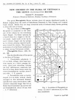
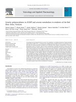
Figure 1. This map shows Vietnam’s 14 most important gem provinces and the major geologic environments.
The main sources for zircon are also shown in the map in which the studied area Krong Nang in Dak Lak
province is pointed out with arrow.
62
B.T.S. Vương, L.T.T. Hương / VNU Journal of Science: Earth and Environmental Sciences, Vol. 31, No. 3 (2015) 60-66
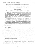
Zr-tn-c 01
Zr-tn-c 02
Zr-tn-r 03
Zr-tn-r 06
Zr-tn-c 03
Zr-tn-r 08
Figure 2. Representative zircon samples showing orange to reddish - brown color (Photo by B.T.S Vuong).
The UV-Vis-NIR spectra of zircon were
obtained from a Perkin Elmer Lambda 900
spectrophotometer. The absorption spectra were
recorded over the range of 200 to 1600 nm in
absorbance mode at a scan speed of 300
nm/minutes and a slit width of 2.5 millimeters.
The data were complied by Perkin Elmer
Spectrum V.5.0.1 program.
The FTIR spectrum were obtained from a
Thermo Scientific, Nicolet Model 6700 brands
which uses a He-Ne Laser by the study of wave
in
numbers
between
400-7000
cm-1
transmittance mode and scan 128 seconds. The
standard resolution of the Nicolet 6700
spectrometer is 0.09 cm-1. The data were
compiled by OMNIC software program.
3. Results and discussion
3.1 UV-Vis-NIR absorption spectrum
The absorption spectra of 6 natural zircon
show similar patterns with a little variation
except for the relative intensities of the peaks
that can be correlated qualitatively with the
depth of the body color and size of the
specimens. Each spectrum was recorded from
200-1600 nm. They consist of bands and peaks
in four regions:
(1) The appearance of an increasing
absorption toward the ultraviolet gives rise to
the brown component of the color. This may be
considered as a result from a color center that
produces a broad absorption band in the
B.T.S. Vương, L.T.T. Hương / VNU Journal of Science: Earth and Environmental Sciences, Vol. 31, No. 3 (2015) 60-66
ultraviolet with an absorption "tail" extending
into the visible.
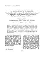
(2) A broad region of absorption in the
range of 400-600nm with the shoulder at
around 500 nm was recorded (figure 3). This
absorption pattern is likely to be due to the
defect in crystal structure caused by the
radiation damage from radioactive elements
such as U and Th.
(3) A series of weak but sharp bands such
as 590, 652, 689 nm were observed in some
darker samples(Zr-tn-r 03, Zr-tn-r 06D and Zrtn-r 08) that had no influence on the color and
were attributed to trace amounts of uranium (as
U4+). It can be stated that the darker one contain
the higher concentration of Uranium than other
brighter.
(4) A weak band centered at 760 nm
presented only in the spectrum recorded parallel
to the optic axis (Zr-tn-r 06D) [4].
Besides, some spectrum also reveal
prominent absorption peaks at 1114 nm and
1505 nm probably due to U5+ [5]. The weak
sharp bands attributed to uranium were present
in each spectrum but with slight variations in
intensity. It can be seen from figure 4 that the
samples with darker color (Zr-tn-r 08, Zr-tn-r
06 D and Zr-tn-r 03) are characterized with the
peaks of higher intensity. The intensity of the
peaks depends on the concentration of the ion.
This observation, again, confirms the above
mention and leads to the understanding that the
concentration of U ion in darker zircon is higher
as compared to brighter one.
5
UV-Vis-NIR absorption spectrum
Absorbance
4
Zr-tn-r 03
3
2
1
0
400
450
500
550
63
600
650
700
750
Wavelength (nm)
Figure 3. UV-Vis-NIR absorption spectrum of a reprentative zircon sample (Zr-tn-r 03)
in the range 400-700 nm
64
B.T.S. Vương, L.T.T. Hương / VNU Journal of Science: Earth and Environmental Sciences, Vol. 31, No. 3 (2015) 60-66
Zr-tn-c 01
Zr-tn-c 02
Zr-tn-c 03
6
Absorption band
Zr-tn-r 06D
Zr-tn-r 06L
Zr-tn-r 03
Absorbance
5+
1114 (U )
Zr-tn-r 08
5+
1505 (U
4
)
4+
652 (U
)
2
0
200
400
600
800
1000
1200
1400
1600
Wavelength (nm)
Figure 4. UV-Vis-NIR absorption spectrum of reprentative zircon samples in the full range 200-1600 nm.
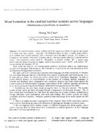
3.2. FTIR absorption spectrum
Various bands consistent with those
typically seen in Zircon were observed in the
FTIR spectra of the Dak Lak zircon (figure 5)
such as some strong absorptions bands at 2334,
2501, 2761, 2856, 2918, 3196cm-1. The
particular attention is paid to the peak at 3196
cm-1 which is the evidence of OH-stretching
characteristic. Besides, the presence of peak at
6663 cm-1 indicates that a small number of U
ions are in the pentavalent state (U5+
amorphous) in ZrSiO4 [6]. An absorption band
in the 1,400-2,000 cm-1 interval is probably
related to Si-O stretching which still indicate a
well crystalline zircon [6]. Moreover, some
spectra indicate two very weak bands located
near 4078 and 4268cm-1 which may be
attributed to the combination of OH stretching
and the vibrations of the framework [7].
The details behind the incorporation of OHand H2O into various structural sites of zircon
remain controversial. Like titanite, an increase
in metamictization results in an increase in OHconcentration. Well-crystallized zircon exhibit
sharp, anisotropic IR peaks associated with OH,
whereas the IR spectra of damaged crystals
usually display an additional peak associated
with the presence of H2O molecules [8]. In this
study, FTIR spectra confirm the presence of
two peaks centered at 3417 cm-1 and 3383 cm-1
associated with Si occupied tetrahedrons or
with OH- defects in crystalline Zircon [9]. All
these indicate these samples are crystalline
zircon with a little bit radiation damage by
radioactive elements [6].
B.T.S. Vương, L.T.T. Hương / VNU Journal of Science: Earth and Environmental Sciences, Vol. 31, No. 3 (2015) 60-66
6663
5+
U
4078
4268
2501
80
3417
60
40
Absorption band
1400-2000
20
Si-O Stretching
2334
3196
OH-Stretching
2856
2918
Transmittance
FTIR absorption spectra
Zr-tn-c 01
Zr-tn-r 06
3385
100
65
0
1000
2000
3000
4000
5000
6000
7000
-1
Wavenumber (cm )
Figure 5. FTIR absorption spectrum of zircon from Dak Lak showed a band at 3196 cm-1 that is associated with
OH-stretching characteristic and a band at 6663 cm-1 that is due to U ion is in the pentavalent state.
4. Conclusions
Study in zircon crystals from Dak Lak
province using FTIR and UV-Vis-NIR
spectroscopic techniques lead to the
understanding of internal structures and the
causes of color of the samples. The UV-VisNIR absorption spectrum indicates that the
causes of orange-brown color components are
due to structural defect color center by the
radiation damage from radioactive elements
such as U and Th. Besides, this also mentioned
its color depends on the concentration of U ion,
the darker zircon has higher content of this ion
than brighter one. In addition, the presence of
OH-stretching in zircon structure which is
related to structure damage by radioactive
elements was indicated by FTIR spectroscopy
(peak at 3197 cm-1). They exhibit no evidence
of H2O molecules, thus, these samples can be
evaluated at being or becoming metamict and,
more importantly, are not detectably
radioactive. This locality is likely to be a
commercial source of gem zircon as well as
other gem materials in the future.
5. Acknowledgment
Special thanks are given to Dr. Somruedee
Satitkune, Faculty of Science, Kasetsart University
(Thailand) for the discussion and advices.
References
[1] Anderson B. W., Payne C. J. (1940) Recent
investigations
of
Zircon.
IV.
The
absorption spectrum. Gemmologist, Vol. 9,
pp. 1-5.
[2] Hazen R. M., and Finger L. W. (1979) Crystal
structure and compressibility of zircon at high
66
B.T.S. Vương, L.T.T. Hương / VNU Journal of Science: Earth and Environmental Sciences, Vol. 31, No. 3 (2015) 60-66
pressure, American Mineralogist, Vol. 64, pp.
196.
[3] Fritsch E., G. R. Rossman (1988) An update on
color in gems, Part 2: Colors involving
multiple atoms and color centers. Gems and
Gemology, Vol.24, No.1, pp. 3-15.
[4] Maxwell J. Faulkner and James E. Shigley
(1989) Zircon from the harts range, northern
territory, Australia. Gems & Gemology, Vol.
254, No. 4, pp. 207.
[5] Benjawan Klinkaew (2008), Heat treatment of
Zircon from Cambodia, A report submitted in
partial fulfillment of the requirement for the
degree of the bachelor of Science department of
Geology Chulalongkorn University, pp. 22-23
[6] Woodhead J. A., Rossman G. R., Silver L. T.
(1991), The metamictization of zircon: radiation
dose-dependent
structural
characteristics,
American Mineralogist Vol.76, pp. 74.
[7] Richman I., Kisliuk P.and Wong E. Y. (1967)
Absorption spectrum of U4+ in zircon (ZrSiO4).
Physical Review, Vol. 155, p. 262.
[8] Beran A. and Libowitzky E. (2003) IR
spectroscopic characterization of OH defects in
mineral phases. Phase transitions, Vol. 76, No.
1-2, pp. 1-15.
[9] Dawson P., Hargreave M. M. and Wilkison G.
R. (1971)The vibrational spectrum of zircon
(ZrSiO4). Journal of physics C: Solid State
Physics, Vol. 4, pp. 240.
Nghiên cứu nguyên nhân tạo màu của Zircon
huyện Krông Năng, tỉnh Đắk Lắk
Bùi Thị Sinh Vương, Lê Thị Thu Hương
Khoa Địa chất, Trường Đại học Khoa học Tự nhiên, ĐHQGHN, 334 Nguyễn Trãi, Hà Nội, Việt Nam
Tóm tắt: Zircon hình thành với nhiều màu sắc bao gồm các sắc thái khác nhau từ hồng, đỏ tới tím,
vàng, cam, nâu; ngoài ra còn có màu ít phổ biến hơn như xanh lá cây và xanh dương. Nhìn chung, màu
sắc của zircon được gây ra bởi các thành phần nguyên tố vi lượng (kim loại chuyển tiếp, nguyên tố
nhóm Lantan, actinides và đất hiếm) và do sự phá hủy phóng xạ (bức xạ gây ra các tâm màu). Tâm
màu zircon rất phức tạp và những nghiên cứu chi tiết xung quanh vấn đề cơ chế tạo màu này vẫn còn
gây nhiều tranh cãi. Trong nghiên cứu này, tác giả đã thu thập một số mẫu zircon từ mỏ Krông Năng,
Đắk Lắk, Tây Nguyên, sử dụng phương pháp phổ hấp thụ UV-Vis-NIR và quang phổ FTIR để xác
định nguyên nhân gây màu của chúng. Phổ hấp thụ của các mẫu cho thấy một sự hấp thụ tăng liên tục
từ khoảng 600 nm về phía cực tím, với đỉnh hấp thụ vào khoảng 500 nm, điều này được xác định gây
bởi sự sai hỏng trong cấu trúc với hiệu ứng tâm màu do sự phá hủy phóng xạ của các nguyên tố phóng
xạ như U và Th. Bên cạnh đó, nhóm OH xuất hiện trong tất cả các phổ hấp thụ hồng ngoại trong khi
H2O lại vắng mặt hoàn toàn, điều này chỉ ra rằng zircon vùng Đắk Lắk thuộc loại zircon kết tinh có
mức độ metamict thấp.
Từ khóa: Zircon, UV-Vis-NIR, FTIR.
B.T.S. Vương, L.T.T. Hương / VNU Journal of Science: Earth and Environmental Sciences, Vol. 31, No. 3 (2015) 60-66
_______
67