DSpace at VNU: Microfluidic Impedance Biosensors For Monitoring A Single And Multiple Cancer Cells In Anticancer Drug Treatments
Bạn đang xem bản rút gọn của tài liệu. Xem và tải ngay bản đầy đủ của tài liệu tại đây (538.32 KB, 5 trang )
Seediscussions,stats,andauthorprofilesforthispublicationat: />
MicrofluidicImpedanceBiosensorsFor
MonitoringASingleAndMultipleCancerCellsIn
AnticancerDrugTreatments
ConferencePaper·June2016
READS
20
7authors,including:
Duc-TanTran
ChiHieuLe
VietnamNationalUniversity,Hanoi
UniversityofGreenwich
127PUBLICATIONS118CITATIONS
38PUBLICATIONS189CITATIONS
SEEPROFILE
Allin-textreferencesunderlinedinbluearelinkedtopublicationsonResearchGate,
lettingyouaccessandreadthemimmediately.
SEEPROFILE
Availablefrom:Duc-TanTran
Retrievedon:11July2016
12
Microfluidic Impedance Biosensors For Monitoring A Single And Multiple Cancer
Cells In Anticancer Drug Treatments
T. A. Nguyen1, Tien V. Nguyen1, D.T. Tran2, Toan V. Nguyen1, C.H. Le3, V.B. Nguyen4 and H.Q. Le5
1
Le Quy Don Technical University, Ha Noi, Viet Nam
VNU University of Engineering and Technology, Ha Noi, Vietnam
3
Faculty of Engineering and Science, University of Greenwich, Kent, United Kingdom
4
College of Engineering and Technology, University of Derby, Derby, United Kingdom
5
Saigon Hi-Tech Park - SHTP, Ho Chi Minh, Vietnam
2
Abstract— In this work, we present a novel microfluidic
impedance biosensor chip for trapping both a single and
multiple cancer cells and monitoring their response to
the anticancer drug treatment. By designing different
sizes of working microelectrodes together with the Vshaped cell capture structures, a single or multiple cells
are trapped on the microelectrodes surfaces. In addition,
by utilizing the passive pumping method, cells can be
trapped and positioned inside the microchannels without
the need of using the outer micro pump or syringe. The
impedance change induced by the response of cells to the
anticancer drug Cisplatin treatment was successfully
recorded. The proposed biosensor chip has a great potential for applications in cancer cell research, drug
screening, and quantification of cancer cells from various tumor stages. The results of this study open potential
research collaborations about development of costeffective devices and lab-on-chips for early disease detection, studies of cancerous cells and their response to
anti-cancer drugs to optimize cancer treatments, characterisation of mechanical properties of cells, new drug
delivery mechanisms, and micro and nano manufacturing.
Keywords— Microfluidic, Biosensor, Impedance, Single cell,
Cancer, Anticancer drug treatment.
limited capabilities to trap and control a single cell [2, 6].
Therefore, the average measurement is assumed to represent
the behavior of a typical cell within a cell population. This
might lead to the inaccuracy or misleading results [7].
Therefore, there has been an emerging demand to develop
innovative and smart devices which are able to be used to
study the behaviors and signals from a single cell.
Recently, the fast advancements of microfluidic techniques as well as micro and nano manufacturing brought
many advantages for single cell studies [8, 9]. By owning
unique features such as a small size, laminar flow, and small
volumes of samples and reagents, chip-based microfluidics
have attracted the growing attention about a single cell
monitoring and analysis. The proposed microfluidic sensor
chip in this work can capture both a single and multiple
breast cancer MCF-7 cells on the microelectrode surface
for monitoring the sequential cellular behaviors and
testing their response to the anticancer drug. The rest of the
paper is organized as follows. Section II presents design and
fabrication of a proposed sensor chip and experiments.
Section III discusses the main results and challenges of
using microfluidics for single cell studies. Finally, Section
IV presents conclusions and addresses the potential collaborations in Biomedical Engineering (BME), especially
among research institutions in Vietnam and UK.
II.
I. INTRODUCTION
Cell-based impedance biosensors have been recognized
as valuable and powerful tools for detecting biochemical
effects such as cellular physiological changes [1], pharmaceutical effects [2], and environmental toxicities [3]. They
can be used to study various cellular activities in a realtime, label-free and nondestructive manner, including cell
spreading, growth, and motility; this is done via monitoring
the electrical alternations at the interfaces between the cell
and electrode [4,5]. For conventional cell-based sensors, a
large cell population is normally used and randomly positioned on the top of big-sized electrodes; this is due to their
BME2016 in Vietnam, IFMBE Proceedings, 2016
MATERIALS AND METHOD
A. Microfluidic chip design and fabrication process
The microfluidic impedance biosensor comprises three
main parts, including the sensing, the cell capture structures,
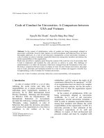
and the microfluidic. Fig. 1 presents a packaged chip with
the sensing and cell trap structures. By utilizing the electrical cell-substrate impedance sensing technique [10], the
sensing part composes of Microelectrode Arrays (MEAs)
which are patterned in two identical channels (see Fig.1
(b)). In each channel, the MEAs comprises four columns of
a working microelectrode (WE) which are located symmetrically on two sides of a rectangular large counter electrode
(CE, 350 500 µm2). Two columns of the WEs on one side
12
of the CE are shifted by 40 μm in order to increase the cell
trapping effectiveness from the cell suspension flow. The
WEs are in a square of 25x25 µm and 60x60µm to host
single cells and multiple cells respectively.
The cell capture structures are designed and arranged
corresponding to the each working microelectrode. Each
cell-trap composes of two identical blocks placing closely
together to form a V-shaped recess. There are small gaps
between these two blocks to allow the hydrodynamic flow
passing through and avoiding the captured cells. The novel
design of the V-shaped cell-trap in this work leads to the
higher cell trapping efficiency. In addition, it can overcome
the limitations of conventional hydrodynamic cell trapping
methods [11].
(a)
(b)
Fig.1 (a): An image of a packaged chip. (b): A micrograph of a Microelectrode Array with the 3D cell capture structures
There are two different size of trap corresponding to the
size of the WEs which aim to capture single or multiple
cells on the top of microelectrodes. The V-shaped traps on
two sides of the CE are placed oppositely to each other and
their recesses are arranged toward the inlets for trapping
cells from the cell suspension flow. As a result, the microelectrodes on one side of the sensor are able to capture cells
and serve as WEs, while the microelectrodes on the other
side without capturing cells serve as reference microelectrodes. The microelectrode arrays are integrated inside a
microfluidic channel.
The sensor was fabricated on the Pyrex wafers based on
the standard micro fabrication techniques. The V-shaped
cell-capture arrays are made of SU-8 of 20 μm in height and
fabricated in the same layer with the microchanel walls. The
microfluidic part of the sensor chip consists of (1) microchannel walls with the same height as the cell capture and
(2) a PDMS cover which was fabricated separately using
micro moulding method [12]. Two microchannels with the
height of 20 μm were created inside the cover. Four holes
with a diameter of 1 mm were punched through the PDMS
layer to produce the inlets and outlets. Two microchannels
of 40 μm in height were formed by a natural adhesion of the
PDMS cover onto the microchannel walls. The design and
fabrication of a proposed sensor chip can be found in details
in our previous works [11, 13].
After dicing, the total size of one chip is only 11 ×
BME2016 in Vietnam, IFMBE Proceedings, 2016
5.3mm2. The chip was glued into a double-sided printed
circuit board (PCB) and wire bonding for the impedance
measurement. By perforating a window through the PCB
together with the use of a thin gold microelectrode and
PDMS cover, the sensor chip is total “transparent”. Therefore, it is ideal for monitoring cells inside the microchannels
during experiments.
B. Cell culture
Human breast cancer cells MCF-7 were cultivated in the
DMEM medium supplemented with 10% fetal bovine serum (FBS) under the standard conditions (37°C, 5% CO2)
inside an incubator. Cells were detached from the culture
flasks by a treatment with trypsin-EDTA for 2 min. After a
detachment, they were resuspended in the DMEM to inactivate any remaining trypsin activities. After a centrifugation
for 10 min, they were resuspended in the CO2 independent
medium which is supplemented with 4 mM L-glutamine to
the final concentration of 106 cells/ml.
C. Impedance measurement
The spectrum measurement was carried out by using
Solartron impedance analyser 1260 (SI 1260). The SI 1260
delivered an alternating voltage of 10 mV amplitude over a
frequency range from 102 to 106 Hz. For the real-time
measurement, the SI 1260 was set to deliver an alternating
voltage of 10 mV at 4 kHz.
D. Experiment procedure
Firstly, the sensor chip is wire bonded, cleaned, and
modified. Then, a complete chip is formed by aligning the
PDMS cover on to the microchannel walls. Next, the cell
suspended medium is injected into the channel by placing a
small drop on the inlet. Single and multiple cells are captured on the top of microelectrodes. Finally, the further
tasks of an experiment are able to be carried out. The detail
of an experiment procedure can be found in our previous
works [11].
III.
RESULTS
A. Hydrodynamic trapping single and multiple cells
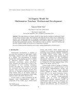
Figure 2 presents a photo of a chip used in the experiment (a) and a micrograph of two microelectrodes with a
single and multiple cells on the top (b). Only one microchanel was used in each experiment. The chip can be reused
several times by removing the PDMS cover and cleaning
the chip surface. After the cells were trapped on the top of
the working microelectrodes, two drops of a cell culture
126
medium was placed on the inlet and the outlet. Then, the
chip was placed into an incubator to culture cells. As shown
in Fig. 2 (a), two working microelectrodes with cells were
trapped and incubated. Two cells on the big-size microelectrode did spread; the change of their shapes is clearly seen;
and the shape of a single cell on the smaller one did not
change; this may be may be a dead cell.
Two cells did spread:
Changes of a shape
C. Real-time monitoring the response of cells to anticancer
drug treatment
The real-time monitoring of the response of the MCF-7
cells to the anticancer drug Cisplatin treatment is illustrated
in Fig. 4. After a cell trapping process, the cells were cultured for 8 hours, so that the cells stabilise, attach, and
spread on the microelectrode surface. Then, the medium
inside the chip was replaced by Cisplatin to expose the cells
to physiological conditions. The impedance measurement
was performed for 6 hours, and the impedance magnitude
decrease approximately 15%.
A single cell did not spread:
No change of a shape
Fig.2 Right: A photo of a chip used in the experiment. Left: A micrograph of two microelectrodes with a single and multiple cells on the top.
B. Monitoring the response of cell to anticancer drug
Im p e d a n c e m a g n itu d e (O h m )
Im p e d a n c e m a g n itu d e d e c re a s e d a fe r tre a tin g b y C is p la tin
1 ,8 x 1 0 5
1 ,7 5 x 1 0 5
1 ,7 x 1 0 5
1 ,6 5 x 1 0 5
1 ,6 x 1 0 5
1 ,5 5 x 1 0 5
1 ,5 x 1 0 5
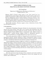
Figure 3 describes the response of a single MCF-7 cell to
the well-known anti-cancer drug-Cisplatin with a concentration of 100 µM after exposing for 6 hours.
0
1
2
3
4
5
6
7
T im e (h )
Fig. 4 A real-time monitoring of the response of the MCF-7 cell to the
anticancer drug Cisplatin treatment.
7
10
IV.
After replaced medium by drug medium
After exposed for 6h
6
Impedance magnitude (Ohm)
10
5
10
4
10
3
10
2
10
3
10
4
10
5
10
6
10
Frequency (Hz)
Fig.3 A Bode plot to monitor responses of a single cancer cell in the
Cisplatin treatment: Concentration of 100 µM after exposing for 6 hours.
After trapping the cells on the top of the microelectrodes,
the cells were cultured inside an incubator for 8 hours.
Then, the medium inside the microchannels was replaced by
physiological conditions. As shown in Fig.3, the impedance
magnitude after exposing for 6 hours (RED line) sharply
decreases, in comparison with the impedance magnitude,
just after replacing the medium inside the microchannel by
a drug medium (BLACK line).
BME2016 in Vietnam, IFMBE Proceedings, 2016
DISCUSSIONS AND CONCLUSIONS
A successful development of a novel microfludic impedance biosensor which is suitable for cell-based experiments and studies is presented in this paper. The chip can
trap a single or multiple cells on the surface of microelectrodes with a high efficiency for subsequent investigations.
After capturing and culturing inside the microchannels, cell
behaviors and their response to a surrounding environment
or anticancer drug treatments can be evaluated. The chip
can provide the information of a single cell in comparison
with multiple cells. However, the influence on cell behaviors due to the miniature environment and the behaviors of
cells in the long-term requires further tests. The results of
this study have potentials for applications in the cancer cell
research, drug screening, and quantification of cancer cells
from various tumor stages, especially for the further research and development of biosensors and lab-on-chips
which are based on behaviors and signals from cells.
Cancer is a global problem that accounts for almost 13%
of deaths worldwide; and more than half of all cancer cases
and nearly 2/3rd of global cancer deaths occur in developing countries; concretely 12.7 million new cancers were
diagnosed worldwide in 2008, and 7 million of which were
in developing countries [14, 15, 17]. By 2020, there will be
12
between 15 and 17 million new cases of cancer every year,
60-70% new cases of cancer and nearly 70% of cancer
deaths will be in economically disadvantaged countries
[17]. Annually, there are nearly 125 thousand people which
diagnosed with cancers in Viet Nam. However, cancer is
potentially the most preventable disease; with current resources, one-third of tumors could be preventable; and onethird of newly diagnosed cancer patients could experience
increased survival or early-stage detection [17]. There is an
urgent need for a multidisciplinary approach to improve
cancer care and reduce the rates of cancer deaths in resource-poor countries in which there exist a lack of access
to cancer therapy, poor early detections of cancers and
screening services, unfriendly health care and delivery systems, poor organization of supportive-care facilities [16,
17]. Developments of the cost-effective solutions, including
smart biosensors and lab-on-chips, for early detections of
cancers are therefore important and necessary for developing countries.
The total healthcare spending in Vietnam was
US$12.90 billion in 2014; it is estimated this to reach
US$27.48 billion in 2020 at a compound annual growth rate
of 13.4 % [18]. With the strong support and investments
from the government via research and technology
development (RTD) funding agencies and projects such as
National Foundation for Science and Technology
Development (NAFOSTED) and Fostering Innovation
through Research, Science and Technology (FIRST), in
collaborations with Newton Fund (UK), there are potentials
for collaborations in Biomedical Engineering and related
areas, especially among research institutions in Vietnam and
UK, to develop innovative products and cost-effective
solutions for screening, early detections, diagnosis and
treatments of cancers for developing countries, including
Vietnam.
In conclusions, we presented a microfluidic impedance
biosensor which can be used for a real-time monitoring of a
single or multiple tumor cells and their response to the
anticancer drug treatment. The results of this study open
potential research collaborations about development of costeffective devices and lab-on-chips for early disease
detection, studies of cancerous cells and their response to
anti-cancer drugs to optimize cancer treatments,
characterisation of mechanical properties of cells, new drug
delivery mechanisms, and micro &nano manufacturing.
These potential research collaborations may benefit from
the currently available RTD resources in micro & nano
manufacturing, nano-materials, and BME, in both Vietnam
and UK. For the short and medium term collaborations,
based on the successful preliminary results, the results of
this study can be expanded to the further test and studies
about the effects of the microenvironment on the vital of
BME2016 in Vietnam, IFMBE Proceedings, 2016
cells. In addition, we also aim at innovative developments
of low-cost microfluidic platforms for developing and
testing new antimicrobials on the artificial cells.
ACKNOWLEDGMENT
British Council – Newton Fund is acknowledged for their
support.
REFERENCES
1
. Giaever and C. R. Keese,(1984) Monitoring fibroblast behavior in
tissue culture with an applied electric field. Proceedings of the National Academy of Sciences; 81 (12): 3761–3764
2 Q. Liu,et al. (2014) Cell-based biosensors and their application in
biomedicine. Chemical Reviews, 114 (12), 6423–6461
3 F. Asphahani et al. (2007) Cellular impedance biosensors for drug
screening and toxin detection. Analyst, 2007: 132: 835-841.
4 I. Giaever and C. R. Keese, (1991) Micromotion of mammalian cells
measured electrically. PNAS;: 88: 7896-7900
5 J. H. Luong et al. (2011) Monitoring motility, spreading, and mortality
of adherent insect cells using an impedance sensor. Analytical chemistry; 73 (8): 1844–1848.
6 Q. Liu et al. (2009) Impedance studies of bio-behavior and
chemosensitivity of cancer cells by micro-electrode arrays. Biosensors
and Bioelectronics; 24 (5): 1305-1310
7 D. Di Carlo (2006) Dynamic single-cell analysis for quantitative
biology. Analytical Chemistry; 78 (23): 7918-7925
8 E. K. Sackmann at al. (2014) The present and future role of microfluidics in biomedical research. Nature, 2014: 507 (7491) 181-189
9 H. Yin and D. Marshall (2012) Microfluidics for single cell analysis.
Current opinion in biotechnology; 23 (1): 110-119
10 J. Wegener et al. (2000) Electric cell-substrate impedance sensing
(ECIS) as a noninvasive means to monitor the kinetics of cell spreading to artificial surfaces. Experimental Cell Research; 259 (1): 158-166.
11 T.A. Nguyen et al. (2013) Microfluidic chip with integrated electrical
cell-impedance sensing for monitoring single cancer cell migration in
three-dimentional matrixes. Analytical Chemistry; 85 (22): 1106811076
12 B.H. Jo et al. (2000) Three-dimensional micro-channel fabrication in
polydimethylsiloxane
(PDMS)
elastomer.
Journal
of
Microelectromechanical Systems; 9 (1): 76-81
13 T. Anh-Nguyen et al. (2016) An impedance biosensor for monitoring
cancer cell attachment, spreading and drug-induced apoptosis. Sensors
and Actuators A: Physical; 241: 231-237
14 J. Ferlay et al. (2012) Cancer incidence and mortality worldwide:
sources, methods and major patterns in GLOBOCAN 2012. Int. J.
Cancer 136 (5): e359–e386
15. Jemal A. et al. (2011) Global cancer statistics. CA: A Cancer Journal
for Clinicians; 61(2):69–90
16. Ramaiah V. K. et al. (2015) Health-care related supportive-care factors
may be responsible for poorer survival of cancer patients in developing
countries. Journal of Cancer Policy; 5:31–47
17. Miriam L.G. et al. (2013) Cancer in developing countries: The next
most preventable pandemic. The Global problem of cancer. Critical
Reviews in Oncology/Hematology; 88:117–122
18. Vietnam Healthcare Outlook: www.frost.com [Access: April 2016]
Author: Tien Anh Nguyen
Institute: Le Quy Don Technical University
Address: 236 Hoang Quoc Viet Street, Bac Tu Liem District
City:
Ha Noi
Country: Viet Nam
Email: or