CHAPTER 9 – IMPORT OF SOLUTES BY ABC TRANSPORTERS – THE MALTOSE AND OTHER SYSTEMS
Bạn đang xem bản rút gọn của tài liệu. Xem và tải ngay bản đầy đủ của tài liệu tại đây (3.81 MB, 29 trang )
157
9
CHAPTER
IMPORT OF SOLUTES BY ABC
TRANSPORTERS – THE MALTOSE
AND OTHER SYSTEMS
ERWIN SCHNEIDER
This article is dedicated to
Professor Dr Karlheinz Altendorf
on the occasion of his 60th birthday.
INTRODUCTION AND
GENERAL OVERVIEW
Starch is one of the major sources of carbon and
energy available to heterotrophic bacteria and
archaea. For example, microorganisms living
in soil and aquatic environments readily gain
access to starch derived from decomposing plant
material, while those that colonize the gastrointestinal tract of humans can feed on starch that
escaped digestion in the small bowel. The latter
is estimated to lie in the range of 10% of intake
in subjects on Western diets (Cummings and
Macfarlane, 1991). Since polysaccharides cannot
penetrate the cell membrane, a wide variety of
microorganisms secrete amylases that produce
maltose and maltodextrins (oligosaccharides of
two or more – up to seven – ␣-1,4 linked glucose
units) as major degradation products of starch.
The uptake of the latter is usually mediated by
an ABC transport system that belongs to a subclass of ABC importers recently designated as the
CUT1 (carbohydrate uptake transporter) or OSP
(oligosaccharides and polyols) family by Saier
(2000; />transport/3_A_1.html) and Dassa and Bouige
(2001; />pmtg/abc/index.html), respectively (see also
Chapter 1).
Members of the family transport a variety of
di- and oligosaccharides, glycerol-phosphate
and polyols (Table 9.1)1 and are composed of
the extracellular substrate-binding protein,
which mainly determines the specificity of the
transporter, two integral membrane proteins,
each usually spanning the membrane six times,
and two copies of an ATPase subunit (also
referred to as ABC protein/domain from here
on) (reviewed in Schneider, 2001). The
hydrophobic subunits contain the cytoplasmic
‘EAA’ sequence motif (consensus: EAA-X3-GX9-I-X-LP) typically shared by all membranespanning subunits of prokaryotic ABC
importers. The ATPase subunit is recognized
by the characteristic set of Walker A and B
boxes and by the ABC signature sequence
(‘LSGGQ’ motif) (reviewed in Schneider and
Hunke, 1998). However, this differs from a
classical consensus ABC domain, having a
carboxy-terminal extension of approximately
120 to 150 amino acid residues. In the Escherichia
coli and Salmonella typhimurium maltose transporter, the C-terminal domain is involved in
regulatory activities (reviewed in Boos and
1
It should be noted that in case of the archaeon Sulfolobus solfataricus, the ABC importer for maltose shows sequence
homology to the subfamily of oligo/dipeptide transporters rather than to the CUT1/OSP cluster (Elferink et al., 2001).
Thus, functional classification of ABC transporters solely based on computer-aided analysis should be taken with caution.
ABC Proteins: From Bacteria to Man
ISBN 0-12-352551-9
Copyright 2003 Elsevier Science Ltd
All rights of reproduction in any form reserved
158
ABC PROTEINS: FROM BACTERIA TO MAN
Shuman, 1998; see also Box 9.1). Several short
sequence motifs and conserved amino acid
residues within this peptide fragment can
serve as signatures, together with a conserved
sequence motif in the binding protein (Tam and
Saier, 1993), to identify new members of the
CUT1 family (Figure. 9.1).
Some ABC domains of the CUT1 family are
functionally exchangeable, thereby strengthening the above classification. For example, UgpC
of E. coli and LacK of Agrobacterium radiobacter
were both demonstrated to substitute for MalK
in maltose transport in E. coli (Hekstra and
Tommassen, 1993; Wilken et al., 1996).
TABLE 9.1. REPRESENTATIVE MEMBERS OF CUT1/OSP
FAMILY OF ABC IMPORTERS
Substrate(s) transported
Protein components
Representative organism(s)
Maltose/maltodextrins
Maltose/trehalose
Lactose
Melibiose, raffinose, sucrose
Glycerol-phosphate
Polyols
Cyclodextrins
MalEFGK
MalEFGK
LacEFGK
MsmEFGK
UgpAEBC
SmoEFGK
CymEFGD
E. coli, S. typhimurium
Thermococcus litoralis
Agrobacterium radiobacter
Streptococcus mutans
E. coli
Rhodobacter sphaeroides
Cellobiose/cellotriose
Maltose/sucrose/trehalose
Alginate
CebEFGMsiK
AglEFGK
AlgSM1M2Q1(?)Q2(?)
Klebsiella oxytoca
Streptomyces reticuli
Sinorhizobium meliloti
Sphingomonas sp.
Binding proteins are underlined and bold characters denote ABC proteins. Only those systems for which all components
were clearly identified by sequence alignment and/or biochemical evidence are considered. For data bank accession
numbers, see legend to Figure 9.1. Modified from Schneider (2001).
BOX 9.1. REGULATORY ACTIVITIES OF THE MALTOSE TRANSPORTER
The maltose transporter of E. coli/S. typhimurium is directly involved in transcriptional regulation of the maltose regulon,
most probably by interaction of the MalK subunits with the positive regulator protein MalT. MalT–MalK interaction has
been demonstrated in vitro (Panagiotidis et al., 1998). Activation of MalT is achieved by binding of ATP and maltotriose,
resulting in a conformational change and subsequent oligomerization of the protein, a prerequisite for the interaction
with its DNA binding sites (Danot, 2001; Schreiber and Richet, 1999). Binding of MalT to assembled MalK interferes with
this process, thereby repressing maltose-regulated gene expression (Boos and Böhm, 2000). Mutations in MalK that
diminish or abolish its inhibitory effect on MalT action, W267G and G346S, map in the C-terminal extension of the protein
(Kühnau et al., 1991). In the case of W267G, the mutation did not affect binding to MalT in vitro (Panagiotidis et al., 1998),
indicating that mere physical interaction is insufficient to antagonize MalT activity. Interestingly, MalK variants carrying
mutations in the ABC signature motif that cause loss of ATPase activity but still allow binding of ATP (G137A/V/T,
Q140K/N/L) act as super-repressors (Kühnau et al., 1991; Panagiotidis et al., 1998; Schmees et al., 1999b). Possibly, in this
case local conformational changes in the ATPase domain of the mutant proteins affect the affinity of the C-terminal
domain for its target, MalT. These findings led to the notion that substrate availability is sensed through the transporter,
which, in the idling mode, binds MalT and thereby represses mal gene transcription. In the presence of substrate,
however, transport activity is switched on, i.e. ATP is hydrolyzed at the MalK subunits, thus causing release of MalT
and subsequent induction of maltose-regulated gene expression (Boos and Böhm, 2000).
The maltose transporter is also involved in a second regulatory process called ‘inducer exclusion’, which is part of
the global carbon regulation in enteric bacteria. Here, in the presence of the preferred carbon source, glucose, the
transport of inducer molecules for alternative metabolic pathways is prevented. This is achieved by inhibition of the
respective transport systems via a component of the glucose transporter, the dephosphorylated enzyme IIAGlc of the
phosphoenolpyruvate phosphotransferase system (PTS) (Postma et al., 1996). In the case of the maltose transporter,
enzyme IIAGlc binds to the MalK subunits, thereby inhibiting ATP hydrolysis (Dean et al., 1990; Landmesser et al., 2002).
Again, mutations that render MalK insensitive to inhibition by enzyme IIAGlc predominantly affect residues in the
C-terminal domain (Dean et al., 1990; Kühnau et al., 1991) (Table 9.2).
159
Figure 9.1. Sequence alignment of ABC proteins of the CUT1/OSP family. The proteins considered are:
MALK_ST (Salmonella typhimurium; acc.no. spP19566), LACK_AR (Agrobacterium radiobacter; acc. no.
spQ01937), SMOK_RS (Rhodobacter sphaeroides; acc. no. spP54933), AGLK_SIM (Sinorhizobium meliloti;
spQ9Z3R8), MSMK_SM (Streptococcus mutans; acc.no. spQ00752), CYMD_KO (Klebsiella oxytoca;
spQ48394), ALGS_SSP (Sphingomonas sp.; acc. no. gbABO11415), UGPC_EC (Escherichia coli; acc. no.
spP10907), MSIK_SC (Streptomyces coelicolor; acc. no. gbAL160331), MALK_TL (Thermococcus litoralis;
acc. no. gbAF121946). Conserved sequence motifs and amino acid residues are boxed. Those that are
conserved throughout the ABC superfamily are highlighted in yellow, while motifs and single residues
confined to CUT1/OSP subfamily members are shown in pink. See also text for details.
160
ABC PROTEINS: FROM BACTERIA TO MAN
TABLE 9.2. MUTATIONS ANALYZED IN THE MALK PROTEINS
OF E. COLI/S. TYPHIMURIUM
Mutation
Transport
activity
in vivo
ATP
bindinga
Ϫ
nd
ϩ
nd
Walter et al. (1992b)
Ϫ
Ϫ
ϩ
ϩ
Ϫ
Ϫ
nd
nd
nd
ϩ
Ϯ
Ϯ
nd
nd
ϩ
nd
nd
Ϫ
nd
nd
ϩ
nd
nd
Ϫ
Ϫ
ϩ
ϩ
nd
Ϫ
nd
nd
nd
E74G
Lid
M79insert
Q82K
Q82E
A85C
A85M
ϩ
nd
nd
nd
Kühnau et al. (1991)
Kühnau et al. (1991)
Hunke and Schneider (1999)
Panagiotidis et al. (1993)
Panagiotidis et al. (1993)
Panagiotidis et al. (1993)
Wilken (1997)
Davidson and Sharma (1997)
Schneider et al. (1994)
Walter and Schneider,
unpubl.
Stein et al. (1997)
Ϫ
Ϯ
Ϯ
Ϯ
ϩ
nd
nd
nd
nd
nd
nd
Ϯ
ϩ
nd
nd
nd
nd
nd
nd
nd
Lippincott and Traxler (1997)
Walter et al. (1992b)
Walter et al. (1992b)
Hunke et al. (2000b)
Mourez et al. (1997a)
L86F
H89insert
E94Q, V
F98L, Y
delF98
K106C
V114C
V114M
Ϫ
Ϫ
ϩ
ϩ
ϩ
ϩ
ϩ
Ϯ
ϩ
nd
nd
ϩ
ϩ
nd
nd
nd
ϩ
nd
nd
nd
nd
nd
nd
nd
Ϫ
nd
nd
nd
nd
nd
nd
nd
V117C
V117M
ϩ
ϩ
nd
nd
nd
nd
nd
nd
E119K
ϩ
nd
nd
nd
L123F
Ϯ
nd
nd
nd
A124T
ϩ
nd
nd
nd
ABC signature
G137A,V,T
Ϫ
Ϯ
Ϫ
Ϫ
G137insert
Ϫ
nd
nd
nd
delS3V4
Walker A box
G36 ϩ R
delG36 P37A
C40S
C40G
K42I,Q,E
K42N
K42R
S43T
ATPase activity
Other properties
References
In soluble In transport
variant
complex
Suppressor of EAA
loop mutations in
MalFG
Affects interaction
with MalFG
Suppressor of EAA
loop mutations in
MalFG
Abolishes inducer
exclusion
Affects interaction
with MalFG
Abolishes inducer
exclusion
Super-repressors
of mal gene
regulation
Hunke et al. (2000a)
Lippincott and Traxler (1997)
Stein et al. (1997)
Panagiotidis et al. (1993)
Panagiotidis et al. (1993)
Hunke et al. (2000b)
Hunke et al. (2000b)
Scheffel and Schneider,
unpublished
Hunke et al. (2000b)
Mourez et al. (1997a)
Kühnau et al. (1991)
Scheffel and Schneider,
unpublished
Dean et al. (1990)
Schmees et al. (1999b)
Kühnau et al. (1991)
Panagiotidis et al. (1993)
Lippincott and Traxler (1997)
(continued)
IMPORT OF SOLUTES BY ABC TRANSPORTERS – THE MALTOSE SYSTEM
TABLE 9.2. (continued)
Mutation
Transport
activity
in vivo
ATP
bindinga
ATPase activity
References
In soluble In transport
variant
complex
delQR140–141 Ϫ
Ϯ
nd
nd
Q140L
Ϫ
Ϯ
Ϫ
Ϫ
Q140K,N
Ϫ
ϩ
ϩ
Ϫ
G145S
Ϫ
nd
nd
nd
T147insert
V149M,I
Ϫ
ϩ
nd
nd
nd
nd
nd
nd
P152L,Q
V154I
ϩ
ϩ
nd
nd
Ϯ
nd
nd
nd
Walker B box
D158N
Ϫ
Ϯ
nd
nd
E159G
P160L
D165N
A167insert
L179R
L172Q
M187I
Ϫ
Ϫ
Ϫ
Ϫ
Ϯ
ϩ
ϩ
nd
Ϯ
Ϯ
nd
nd
nd
nd
nd
Ϫ
Ϫ
nd
ϩ
ϩ
nd
nd
Ϫ
nd
nd
nd
nd
nd
Ϫ
nd
Ϫ
Ϫ
R211insert
ϩ
nd
nd
nd
R228C
ϩ
nd
nd
nd
F241I
ϩ
nd
nd
nd
W267G
ϩ
nd
nd
nd
V275insert
ϩ
nd
nd
nd
G278P
ϩ
nd
nd
nd
Switch
H192R,L
Other properties
Super-repressor
of mal gene
regulation
Super-repressor
of mal gene
expression
Affects interaction
with MalFG
Suppresses EAA
loop mutations
in MalG
Suppresses EAA
loop mutations
in MalFG
Suppresses EAA
loop mutations
in MalFG
Abolishes inducer
exclusion
Abolishes inducer
exclusion
Abolishes inducer
exclusion
Eliminates mal
gene repression
Eliminates mal
gene repression
Abolishes inducer
exclusion
Kühnau et al. (1991)
Panagiotidis et al. (1993)
Schmees et al. (1999b)
Schmees et al. (1999b)
Brinkmann and Schneider,
unpublished
Lippincott and Traxler (1997)
Mourez et al. (1997a)
Walter et al. (1992b)
Mourez et al. (1997a)
Kühnau et al. (1991)
Panagiotidis et al. (1993)
Stein and Schneider, unpubl.
Hunke et al. (2000a)
Hunke et al. (2000a)
Lippincott and Traxler (1997)
Walter et al. (1992b)
Walter et al. (1992b)
Mourez et al. (1997a)
Davidson and Sharma (1997)
Walter et al. (1992b)
Landmesser et al. (2002)
Lippincott and Traxler (1997)
Kühnau et al. (1991)
Dean et al. (1990)
Kühnau et al. (1991)
Lippincott and Traxler (1997)
Dean et al. (1990)
(continued)
161
162
ABC PROTEINS: FROM BACTERIA TO MAN
TABLE 9.2. (continued)
Mutation
Transport
activity
in vivo
ATP
bindinga
ATPase activity
S282L
ϩ
nd
nd
nd
L291insert
ϩ
nd
nd
nd
G302D
ϩ
nd
nd
nd
E306K (St)
S322F
Ϫ
ϩ
nd
nd
Ϯ
nd
Ϫ
nd
G346S
ϩ
nd
nd
nd
G346insert
ϩ
nd
nd
nd
C350S (St)
C360S (St)
R364insert
ϩ
ϩ
ϩ
nd
nd
nd
nd
nd
nd
nd
nd
nd
Other properties
References
Abolishes inducer
exclusion
Eliminates mal
gene repression
Abolishes inducer
exclusion
Kühnau et al. (1991)
In soluble In transport
variant
complex
Abolishes inducer
exclusion
Eliminates mal
gene repression
Eliminates mal
gene repression
Eliminates mal
gene repression,
abolishes inducer
exclusion
Lippincott and Traxler (1997)
Kühnau et al. (1991)
Hunke et al. (2000a)
Kühnau et al. (1991)
Kühnau et al. (1991)
Lippincott and Traxler (1997)
Hunke and Schneider (1999)
Hunke and Schneider (1999)
Lippincott and Traxler (1997)
a
Analyzed by photo-crosslinking with 8-azido-ATP in membrane vesicles or with purified soluble variants; del, deletion;
insert, insertion of peptide linkers; St, numbering according to S. typhimurium MalK; ϩ, indicates activities between
80 and 100% of control; Ϯ, indicates activities Ͻ80 and Ͼ20% of control; Ϫ, indicates activities Ͻ20% of control.
Biochemical and genetic evidence, as well as
computational analysis of complete microbial
genomes that became available within recent
years, revealed that ABC uptake systems, specific for maltose and/or maltodextrins, are
widespread among Gram-negative and Grampositive bacteria, including pathogens such as
S. typhimurium (Schneider et al., 1989), Yersinia
enterocolitica (Brzostek et al., 1993), Streptococcus
pneumoniae (Puyet and Espinosa, 1993), Vibrio
cholerae (Heidelberg et al., 2000), Aeromonas
hydrophila (Höner zu Bentrup et al., 1994),
Mycobacterium tuberculosis and Mycobacterium
leprae (Borich et al., 2000), to name just a few.
Homologous transporters were also identified
in archaea, such as Thermococcus litoralis
(Horlacher et al., 1998), Pyrococcus furiosus
(DiRuggiero et al., 2000) and Sulfolobus solfataricus (Elferink et al., 2001).
The maltose transporter is composed of
the extracellular (periplasmic) receptor, the
maltose-binding protein (MBP or MalE), and
the membrane-bound complex comprising the
hydrophobic subunits, MalF and MalG, and
two copies of the ATPase (ABC) subunit, MalK
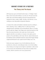
(Davidson and Nikaido, 1991) (Figure 9.2).
Interaction of the substrate-loaded binding
protein triggers conformational changes that
result in ATP hydrolysis at the MalK subunits
and eventually in substrate translocation
(Davidson et al., 1992). In Gram-negative
bacteria, an additional protein component,
maltoporin or LamB, is required in the outer
membrane to facilitate the diffusion of maltose
(at low concentrations) and maltodextrins into
the periplasm (Boos and Shuman, 1998; see
also Box 9.2). In Gram-positive bacteria, which
lack a periplasmic space, and in some archaea,
maltose-binding proteins are lipoproteins that
are anchored to the cytoplasmic membrane via
fatty acids covalently coupled to an N-terminal
cysteine residue (Horlacher et al., 1998; Sutcliffe
and Russel, 1995). In other archaea, attachment to the external side of the membrane is
achieved by a carboxy-terminal transmembrane segment (Elferink et al., 2001).
The genes encoding the transport components are usually clustered in one or two closely
linked operons (Boos and Shuman, 1998;
Heidelberg et al., 2000). These, however, as
IMPORT OF SOLUTES BY ABC TRANSPORTERS – THE MALTOSE SYSTEM
CH2OH
O
CH2OH
O
HO
OH
H,OH
OH
OH
O
OH
Maltose
Maltoporin
OM
MalE
MalE
MalE
MalF
ATP
MalG
MalE
CM
MalF
ATP
ATP
CM
ATP
MalK MalK
MalK MalK
MalT
MalG
EIIA
Gram-negative bacteria
(E. coli, S. typhimurium)
Gram-positive bacteria
Archaea
Figure 9.2. Schematic organization of components
involved in maltose transport. See text for details.
MalE, extracellular maltose-binding protein; MalF,
MalG, hydrophobic, membrane integral subunits,
presumably forming the translocation pore; MalK,
ATP-hydrolyzing subunit, ABC domain. MalE can
reside in an open and closed conformation. The
latter is stabilized by substrate binding. In
Gram-negative bacteria, the binding protein is
located freely in the periplasmic space between
outer and inner membrane. In Gram-positives and
in some archaea, MalE is attached to the
cytoplasmic membrane via an N-terminal lipid
anchor. In other archaea, a transmembrane segment
of the protein is used instead. In E. coli/S.
typhimurium and probably other closely related
bacteria, the maltose transporter is engaged in
regulatory processes that involve interactions of the
MalK subunits with the positive transcriptional
regulator of the mal regulon, MalT, and the
dephosphorylated form of enzyme IIA of the glucose
transporter (PTS). Whether similar activities exist
in other Gram-negative bacteria is unknown.
often found in Gram-positive bacteria and
archaea, may lack the gene encoding the ABC
protein (Greller et al., 1999; Hülsmann et al.,
2000; Puyet and Espinosa, 1993; Quentin et al.,
1999; van Wezel et al., 1997). This finding gave
rise to the notion that a single ATPase protein
could serve several transporters. Evidence in
favor of this view was recently presented in the
case of Streptomyces. Here, the ABC protein
2
MsiK assists in the uptake of maltose and cellobiose, which is mediated by two different
transporters (Schlösser et al., 1997).
The ABC importer for maltose/maltodextrins of E. coli and S. typhimurium (Boos and
Shuman, 1998) is by far the best-studied member of the CUT1 family. This, together with the
histidine transport system of S. typhimurium
(Doige and Ames, 1993; Liu et al., 1997; P.-Q.
Liu and Ames, 1998; Nikaido and Ames, 1999;
Nikaido et al., 1997), can serve as a model for
ABC transporters in general. This chapter summarizes the current knowledge on this system,
including relevant data for other members of
the CUT1 family. Where appropriate, a comparative analysis with the properties of the histidine transporter is also provided. The latter is
composed of the soluble substrate-binding protein HisJ and the membrane-bound complex,
comprising two membrane-spanning subunits,
HisQ and HisM, and two copies of the ABC
subunit HisP (Kerppola et al., 1991).
THE MALTOSE/
MALTODEXTRIN
TRANSPORT SYSTEM OF
E. COLI AND
S. TYPHIMURIUM
The proteins constituting the ABC transporter for maltose in E. coli and S. typhimurium
share Ͼ90% identical amino acid residues. Moreover, the components have been demonstrated
to be fully exchangeable (Hunke et al., 2000b).
Consequently, the data summarized below will
not in each case be specified with respect to the
original organism of the transporter for which
they have been obtained.
GENETIC ORGANIZATION AND
REGULATION
The genes encoding the transport proteins for
maltose are organized in two divergently transcribed operons at 91.4 min in the malB region
of the chromosome: malE malF malG, and malK
lamB malM.2 They are part of a regulatory
The function of the product of the malM gene is currently unknown but it is dispensible for maltose/maltodextrin
transport under all conditions tested so far (see Boos and Shuman, 1998).
163
164
ABC PROTEINS: FROM BACTERIA TO MAN
BOX 9.2. STRUCTURAL AND FUNCTIONAL ASPECTS OF MALTOPORIN (LAMB)
In Gram-negative bacteria, passage of maltose at low concentrations (р10 µM), and of maltodextrins to the periplasm
by facilitated diffusion, requires the presence of large amounts (40 000 copies per cell) of maltoporin in the outer
membrane. (In E. coli, the protein serves as the receptor for bacteriophage lambda, giving rise to the alternative name,
LamB.) Under these conditions, diffusion of the substrate through the outer membrane determines the overall rate of
transport (Tralau et al., 2000).
Maltoporin is organized as a homotrimer (molecular mass of the monomer: 47 kDa), with each monomer providing
a distinct maltodextrin-binding site, which is crucial for the facilitated diffusion process (Luckey and Nikaido, 1980).
The crystal structures of maltoporin from both E. coli (Schirmer et al., 1995) and S. typhimurium (Meyer et al., 1997) in the
presence of different malto-oligosaccharides revealed that each subunit contains a channel that is formed by an
18-stranded, antiparallel -barrel. Within a single channel, a constriction is formed by three peptide loops. The
substrates are in contact with a ‘greasy slide’ of aromatic residues, which provides a path for translocation. There are
well-defined binding sites for three consecutive glucosyl residues in the middle of the channel and one additional
subsite at the extracellular end of the greasy slide (Dutzler et al., 1996).
network, the ‘maltose regulon’, that encompasses a total of 11 genes (for review, see Boos
and Shuman, 1998). Transcription of maltoseregulated genes is governed by the action of a
positive regulator protein, MalT, that requires
maltotriose and ATP for activity, and is affected
by the functional status of the transporter
(reviewed in Boos and Böhm, 2000) (see also
Box 9.1). In addition, the maltose regulon itself
is subject to global carbon regulation of the cell
(catabolite repression). Consequently, productive binding of MalT to specific nucleotide sequences upstream of the respective
promoters (‘MalT boxes’) is brought about
only in the presence of the cAMP/CAP complex
(Boos and Shuman, 1998).
THE SUBUNITS
In the following paragraphs, the properties of
the individual components of the ABC transporter will be summarized. As maltoporin is
confined to Gram-negative bacteria only and
is not essential for the transport process, the
interested reader is referred to Box 9.2 for a
short description of its structure and function.
Maltose-binding protein MalE
The soluble receptor MalE (molecular mass
40 kDa) binds maltose and maltodextrins with
high affinity (KDϳ1 M) and is present in high
concentration in the cell (ϳ1 mM) following
induction (Boos and Shuman, 1998). Whilst
being crucial to the transport process, maltosebinding protein is also involved in the chemotactic response of the bacteria towards maltose
by presenting the substrate to the chemoreceptor Tar (Gardina et al., 1997).
MalE has been crystallized both in the
absence of ligand (Sharff et al., 1992) and in the
presence of maltose (Spurlino et al., 1991) or
longer maltodextrins (Quiocho et al., 1997). As
found for other substrate-binding proteins,
MalE consists of two nearly symmetrical lobes,
between which the binding site is formed (for
details, see Chapter 10). In the substrate-free
form, these lobes are open and the substratebinding site is accessible to the medium. Upon
binding of ligand the two lobes move towards
each other, thereby trapping the substrate
inside the binding cleft. The crystallographic
data further suggested that maltose may first
bind to the N-terminal domain by contacting
glutamate-111 at the base of the binding cleft.
Subsequent ligand-induced movement of E111
may trigger the conformational change of the
C-terminal lobe that eventually results in its
participation in substrate binding and closing
of the cleft (Sharff et al., 1992).
The crystal structures of a maltose/trehalose
and a maltose/maltodextrin binding protein of
the hyperthermophilic archaea T. litoralis (Diez
et al., 2001) and P. furiosus (Evdokimov et al.,
2001), respectively, have recently been solved.
Both are structurally related to MalE of E. coli
despite the moderate level of sequence identity
between these proteins and MalE-Ec.
The transport complex in the cytoplasmic
membrane recognizes its substrate only when
IMPORT OF SOLUTES BY ABC TRANSPORTERS – THE MALTOSE SYSTEM
bound to MalE. Thus, only interaction of
substrate-loaded MalE with the transport components can initiate the transport process. In
fact, mathematical treatment of experimental
data gave rise to the notion that the open
nonliganded form of MalE can also bind to the
membrane components. However, the affinity
of the MalFGK2 complex is five times greater
for the loaded than for the unloaded form of
MalE (Merino et al., 1995).
Analysis of allele-specific suppressors and
of dominant negative mutants has defined
glycine-13 and aspartate-14 of MalE as sites of
interaction with MalG, while tyrosine-210 was
identified as being in contact with MalF. Thus,
the N- and C-terminal lobes of MalE may interact with MalG and MalF, respectively (Hor
and Shuman, 1993). In the C-terminal lobe,
residues in ␣-helix 7 were shown by mutational
analysis to play an important role in this interaction (Szmelcman et al., 1997).
The binding protein of the histidine transporter of S. typhimurium, HisJ, is very similar in
overall structure to MalE and also to other
periplasmic receptors (Oh et al., 1994). In addition, another soluble receptor, the lysine-arginine-ornithine binding protein (LAO), which is
closely related both in primary and tertiary
structure to HisJ, also delivers its substrates to
the HisQMP2 complex (Kang et al., 1991). As in
the case of MalE, both proteins move the two
globular lobes close to each other upon binding
of their respective ligands, thereby restoring
the conformation that productively interacts
with the membrane components (Wolf et al.,
1994). Both lobes participate in this interaction
(Liu et al., 1999). Strikingly, however, and in
contrast to the maltose system, liganded and
nonliganded HisJ have equal affinity for the
membrane-bound complex (Ames et al., 1996;
Merino et al., 1995).
The ABC protein MalK
Enzymatic properties
The MalK protein (molecular mass 40 kDa), when
overproduced in the absence of the membraneintegral subunits MalF and MalG, can be purified to near homogeneity by either conventional
methods (Mourez et al., 1998; Schneider et al.,
1995a; Sharma and Davidson, 2000; Walter
et al., 1992a) or as an N-terminal His6-fusion
protein by Ni-NTA affinity chromatography
(Hunke et al., 2000a; Reich-Slotky et al., 2000).
Purified MalK exhibits a spontaneous ATPase
activity with an apparent Km around 0.1 mM
and Vmax values between 0.2 and 1.3 mol
minϪ1 mgϪ1 (Morbach et al., 1993; Mourez et al.,
1998; Reich-Slotky et al., 2000; Schmees et al.,
1999b; Schneider et al., 1995a). GTP and CTP are
also accepted as substrates and Mg2ϩ ions are
absolutely essential for activity (Morbach et al.,
1993). In contrast to that of the assembled transport complex (see below), the enzymatic activity of the free protein is surprisingly insensitive
to vanadate (Hunke et al., 1995; Morbach et al.,
1993; Sharma and Davidson, 2000). Inhibition
by N-ethylmaleimide was demonstrated to
be due to modification of cysteine-40 within
the Walker A motif thereby interfering with
ATP binding (Hunke and Schneider, 1999;
Morbach et al., 1993). Limited proteolysis
with trypsin revealed a specific conformational
change upon binding of MgATP. Except GTP,
other nucleotides proved to be ineffective
(Mourez et al., 1998; Schneider et al., 1994).
When analyzed as a function of MalK concentration, ATP hydrolysis increases in a linear
mode (Landmesser and Schneider, unpublished). This finding indicates that MalK is either
enzymatically active as monomer or, alternatively, a putative MalK dimer (multimer) is
already formed at very low (micromolar) concentrations. The latter possibility would be consistent with results of Kennedy and Traxler
(1999), who found MalK dimers in vivo and in cell
extracts. Further support for MalK being active
as a dimer was provided by the observation
that mixing wild-type MalK with a catalytically
inactive MalK variant (H192R) resulted in an
increase in ATPase activity as compared to wild
type alone, thus suggesting that heterodimers
were formed (Landmesser and Schneider,
unpublished) (see also below). If so, the affinity
of the monomers towards each other must be
low since in gel filtration experiments purified
MalK of S. typhimurium (MalK-St) eluted at
the molecular mass of a monomer (Tebbe and
Schneider, unpublished observation). The same
result was reported for a close homologue, the
MalK protein of the hyperthermophilic archaeon
T. litoralis (Greller et al., 1999).
In contrast, the ATPase activity of HisP, the
ABC subunit of the histidine transporter, was
observed to be non-linearly dependent on
protein concentration, suggesting already from
these data the formation of dimers. When
applied to a molecular sieve column, only a
small fraction of HisP eluted at the position of
a dimer, while the bulk of HisP was found at the
165
166
ABC PROTEINS: FROM BACTERIA TO MAN
position of a monomer. This was taken as further
evidence for the above notion but also suggested
to the authors that both forms are in rapid equilibrium with each other (Nikaido et al., 1997).
Other properties of the purified HisP protein
were observed to be similar to those determined
for MalK, including insensitivity to vanadate
(Nikaido et al., 1997).
Tertiary structural model
Crystals of MalK-St were obtained that diffract
to about 3 Å, but the structure has not yet been
solved (Schmees et al., 1999a). However, the tertiary structure of a MalK homologue, isolated
from the hyperthermophilic archaeon T. litoralis
(MalK-Tl), presumably involved in maltose/
trehalose transport, has recently been determined (Diederichs et al., 2000). The protein was
demonstrated to exhibit similar biochemical
properties to those of the S. typhimurium MalK
protein, with an optimal ATPase activity at
80°C (Greller et al., 1999). Since both proteins
share Ͼ50% identical amino acid residues
(Figure 9.1) it appears safe to conclude that
their crystal structures are likely to be very
similar if not identical.
The crystal structure of MalK-Tl
MalK-Tl was crystallized in the presence of ADP
and its tertiary structure could be solved with a
resolution of 1.9 Å (Diederichs et al., 2000). Two
molecules are present per asymmetric unit that
contact each other through the ATPase domains
with the (regulatory) C-terminal domains
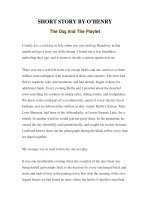
attached at opposite poles (Figure 9.3). Deviation
from twofold symmetry is observed at the interface of the dimer and in regions corresponding to
residues that are deduced to be in close contact
to the membrane-integral subunits (see section
on subunit–subunit interactions, below). In the
nucleotide-binding sites, only a pyrophosphate
molecule could be identified, while a density for
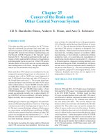
the adenine ring of ADP was missing (Figure 9.4).
Although the overall fold of the ATPase domain
is almost identical to that of HisP, with equivalent
catalytic (ArmI) and helical (ArmII) subdomains,
the structure of their dimers clearly differs. In the
HisP dimer, where the crystal structure was
P218
W265
G278
R228
S282
F241
S322
G346
G302
E119
A124
E308 (E.c.)
E306 (S.t.)
Figure 9.3. Ribbon representation of the MalK-Tl dimer. The ATPase core domains of each monomer are
colored yellow and blue, respectively. The C-terminal (transcript regulatory) domains are colored gray.
Labels indicate the numbers of helices and strands. The relative positions of residues discussed in the text are
indicated. Numbering of the residues is according to MalK-Ec except for E308/306, where the corresponding
numbering of MalK-St is also given (please note that residues M260P261 are deleted in MalK-St, resulting in a
total number of 369 compared to 371 residues in MalK-Ec). Color code: black, residues when mutated that
render the transporter insensitive to inducer exclusion; red, residues, when mutated that affect the repressing
activity of MalK; blue, mutation to lysine reduces ATPase activity; green, residue depicted for construction
of a truncated MalK variant by genetic engineering (Schmees and Schneider, 1998; see text for details).
Reproduced from Diederichs et al. (2000) with permission and modified.
IMPORT OF SOLUTES BY ABC TRANSPORTERS – THE MALTOSE SYSTEM
obtained in the presence of ATP, the monomers
associate via antiparallel beta sheets (Hung et al.,
1998) that, in the MalK-Tl structure, are located at
the top of the dimer (Figures 9.3 and 9.4). As a
consequence, in HisP the nucleotide-binding sites
are located opposite to each other at the outside
of the monomers, while in MalK-Tl both sites are
facing each other in the center part of the core
structure (Figure 9.4) (see Chapters 4 and 7 for a
detailed description of other crystal structures
of ABC proteins/domains).
Although the C-terminal regulatory domain
is clearly separated from the ATPase (core)
domain in MalK-Tl, mutational analysis of
MalK-St (Hunke et al., 2000a) (see below)
and a study using truncated MalK proteins and
chimeras suggested that both N- and C-terminal
parts of the protein are required for its structural integrity (Schmees and Schneider, 1998).
In the latter investigation, it was demonstrated
that when similar sized N- and C-terminal
half-molecules of MalK-St (split at L179) are
expressed they assemble into a transport complex in vivo which is still active. On the other
A-N-Term
M187
K106
Q82
V149
Lid
5
A85
L86
4
G145
Q140
V114
V117
G137
L123
Walker A Walker B
Signature motif
Switch
D-LooP
Figure 9.4. Core region of the MalK-Tl dimer.
The molecule is viewed along the interface
perpendicular to the pseudosymmetry axis.
The relative locations of conserved motifs are
indicated in the monomer colored yellow, while
single residues discussed in the text are indicated in
black in the monomer colored blue. The bound
pyrophosphate is shown in green. Residues written
in red in the lower helical (ArmII) domain are
thought to interact with the membrane-integral
subunits. Reproduced from Diederichs
et al. (2000) with permission and modified.
hand, when the site of splitting was shifted
towards the C-terminal domain, transport was
abolished. In particular, expression of fragments
that correspond exactly to one or both of the
ATPase and C-terminal domains of MalK-Tl
(split at P218, see Figure 9.3) did not result in
an active transporter, most probably due to misfolding of the peptides (Schmees and Schneider,
1998). This notion is supported by the finding
that transport function was retained in chimeras
composed of similar N- or C-terminal fragments
of MalK, with complementing fragments of
HisP (Schneider and Walter, 1991) or LacK, a
close homologue of the lactose ABC transporter
from A. radiobacter (Schmees and Schneider,
1998; Wilken et al., 1996). These studies also
indicated that a minimum portion necessary
transcription regulation by MalK would encompass residues Q263 to V369 (Schmees and
Schneider, 1998).
Functional amino acid residues
The malK gene has been the subject of extensive mutational analyses resulting in the identification of functionally important amino acid
residues and peptide fragments (Table 9.2).
From these studies a domain structure of the
protein was postulated, with an N-terminal
core (ABC) carrying the nucleotide-binding
sites and residues involved in the interaction
with the membrane components, together with
a C-terminal domain devoted to the transcript
regulatory activities of the protein (Kühnau et
al., 1991; Schmees and Schneider, 1998; Wilken,
1997). This view was largely confirmed by
the crystal structure of MalK-Tl. Nonetheless,
both domains (ABC and regulatory) are not
autonomous entities but talk to each other,
since mutations in both have been identified
that alter the activities of the other (Hunke
et al., 2000a; Kühnau et al., 1991; Schmees et al.,
1999b). The following section focuses on mutations affecting the transport activities of MalK
only. (For a description of mutations that eliminate the regulatory properties of MalK, see
Box 9.1 and Table 9.2.)
Mutations affecting ATPase activity
As shown in Figure 9.1, Table 9.2 mutations in
the ATPase domain, especially those affecting
the invariant lysine (K42) and aspartate (D158)
residues, respectively, in the nucleotide-binding
motifs A and B, usually abolish ATPase activity.
167
168
ABC PROTEINS: FROM BACTERIA TO MAN
However, depending on the chemical nature
of the substituting amino acid, such mutant
proteins may retain the capability to bind
ATP (Hunke et al., 2000a; Kühnau et al., 1991;
Panagiotidis et al. 1993, Schneider et al., 1994).
Replacement of cysteine-40 by serine, on the
other hand, is without functional consequences
(Hunke and Schneider, 1999). Other mutations
(P160L, D165N at the C-terminus of the B motif,
also called the D-loop, see Figure 9.1), although
remote from the ATP-binding site according to
the MalK-Tl structure (Figure 9.4), nonetheless
reduced the ATPase activity of the soluble
variants to less than 20% of the control (Hunke
et al., 2000a).
Amino acid substitutions in the ABC signature (‘LSGGQ’) motif have contrasting consequences for function. G137 cannot be replaced
by other residues without complete loss of
ATPase activity (Panagiotidis et al., 1993;
Schmees et al., 1999b). However, substituting
asparagine or lysine for glutamine-140 resulted
in an enzymatically active MalK variant when
analyzed separately but substantially reduced
MalE-maltose-dependent ATPase activity in the
assembled transport complex (Schmees et al.,
1999b). Thus, Q140 might be involved in the
activation of ATPase activity upon substrate
binding. From these genetic findings it was concluded that the ABC signature sequence could
sense an incoming signal through its C-terminal
half, while residues in the N-terminal part of the
motif may assist in the catalytic reaction. This
idea does not seem to be supported by the
MalK-Tl dimer structure, in which the signature
motif is located at the bottom of the helical
domain layer (Figure 9.4) and thus, is distant
both in cis and in trans from the nucleotidebinding sites. However, this may be different in
the assembled transport complex. In fact, the
first tertiary structure of a complete ABC transporter, which became available only recently,
lends support to this notion. In MsbA, a protein
mediating the export of the outer membrane
component lipid A in E. coli, the signature
sequence appears to be located rather closely to
the Walker B motif (Chang and Roth, 2001).
Moreover, by comparative analysis of the ATPbound form of HisP with the MgADP-bound
form of MJ0796, an ABC protein of the thermophilic archaeon Methanococcus jannaschii
(Yuan et al., 2001), suggested that the helical
domain may rotate outward from the
nucleotide-binding site upon hydrolysis, resulting in a substantial movement of the LSGGQ
motif. Although attractive, a note of caution
seems opportune as data from two different
proteins were compared. Furthermore, in
Rad50, an ABC-like protein that is not involved
in transport but is a soluble DNA repair
enzyme, the ABC signature motif contacts the
ATPase active site in the opposing monomer
(Hopfner et al., 2000). Whether the structure of
the Rad50 dimer can serve as a model for ABC
proteins devoted to transport processes is a
matter of current controversy (see also Chapter
4 for a detailed discussion). Again, one has to
keep in mind that the structure of the MalK
dimer in solution and of the other ABC transporter subunits for which structural data are
available might differ from that in the assembled transport complex. This aspect will be discussed further below.
The conserved sequence motif around
glutamine-82 (termed ‘lid’, see Figure 9.4)
was found in the MalK-Tl structure near the
nucleotide-binding site (Diederichs et al., 2000).
Substitution of lysine or glutamate for Q82 in
MalK-St reduced but did not abolish transport
activity in vivo (Walter et al., 1992b). Thus, the
absolute necessity of a glutamine residue at this
position can be excluded but the chemical
nature of the substitutes, K or E, does not rule
out a role in polarizing the water molecule that
attacks the ␥-phosphate of ATP during catalysis, as suggested from the HisP structure (Hung
et al., 1998). However, such a role is not supported by the MalK-Tl structure as the corresponding Q residue is too far away from the
pyrophosphate (Figure 9.4). Other candidates
for polarizing the water molecule (E64, E94),
as suggested by sequence comparison (Yoshida
and Amano, 1995), were eliminated by mutational analysis (Stein et al., 1997).
Another highly conserved residue from the
‘lid’ region, L86, when mutated to phenylalanine, was shown to cause the same phenotype
as the Q140K/N mutations described above.
Thus, the purified variant exhibits ATPase
activity comparable to wild type in the reconstituted transport complex, and ATP hydrolysis is abolished (Hunke et al., 2000a). These
results suggest that L86 may be involved in
activating the enzymatic activity of MalK upon
binding of substrated-loaded MalE to the complex and thus, be in close contact to MalF/
MalG. Consistent with this notion is the finding that in MsbA, the region encompassing the
corresponding residue (L428) is in direct contact with an intracellular domain that connects
the membrane-spanning helices to the ABC
domain (Chang and Roth, 2001).
IMPORT OF SOLUTES BY ABC TRANSPORTERS – THE MALTOSE SYSTEM
Residues within the so-called ‘switch’ region
of the ABC domain (see Figures 9.1 and 9.4),
located carboxy-terminal to the Walker B site,
are believed to propagate conformational
changes triggered by ATP hydrolysis, in analogy
to evidence provided by the crystal structure of
the E. coli recA protein (Story and Steitz, 1992;
Yoshida and Amano, 1995). In line with this
notion are results from mutational analysis of
the highly conserved histidine-192 in the switch
motif. Replacement by arginine was shown to
cause defective transport in vivo (Walter et al.,
1992b) and in vitro (Davidson and Sharma,
1997), and loss of ATPase activity of the purified
variant (Landmesser and Schneider, unpublished). (The previously reported retention of
ATPase activity by this mutant (Walter et al.,
1992b) could not subsequently be confirmed
when using an optimized purification protocol.)
How this residue might make contact with the
nucleotide-binding site is not obvious from the
MalK-Tl structure (Diederichs et al., 2000).
However, the authors proposed that other conformations of the protein may exist that, by analogy with the situation seen in the HisP structure,
could promote contact through an interspersed
water molecule.
So far, one residue located in the very
C-terminal, regulatory domain of MalK-St was
shown to affect the ATPase activity of the protein. E306 (E308 in E. coli MalK), when mutated
to lysine, resulted in the loss of transport activity in vivo and the purified MalK variant exhibited strongly reduced ATPase activity (Hunke
et al., 2000a). Although the crystal structure of
MalK-Tl does not provide any clue for a possible function of this residue, E306 is highly
conserved among members of the ‘MalK’ subfamily (Figure 9.1). Its function, like that of
the other conserved residues in the C-terminal
domain of these proteins, remains to be
elucidated.
Mutations affecting interactions with the
membrane components
Mutant analyses and biochemical evidence have
identified residues in MalK that are involved
in the functional and/or structural interaction
with the membrane integral subunits. Wilken
et al. (1996) isolated variants of the homologous
LacK protein (V114M, L123F, G145S) that partially or fully replace MalK in maltose transport.
Consequently, when introduced into MalK, the
same mutations reduced or abolished transport
activity (Scheffel, Brinkmann and Schneider,
unpublished). Mourez et al. (1997a) screened for
MalK mutants that could restore transport in
E. coli strains carrying mutations in the conserved
‘EAA’ loops of MalF and/or MalG (A85M,
V117M, V149M/I, M187I). With the exception of
A85 (part of the ‘lid’) and M187 (part of the
‘switch’), all are located in the largely ␣-helical
peptide connecting the Walker A and B sites
(Figure 9.4). In addition, limited proteolysis of
MalK in the presence and absence of MalFGcontaining membrane vesicles suggested that
K106 at the end of helix 3 is also in close contact
with the membrane integral subunits (Mourez
et al., 1998). (This theme will be continued in a
later section with evidence from crosslinking
experiments.)
The membrane-integral subunits
MalG and MalF
MalG (molecular mass 32 kDa), as shown by
extensive topological analysis using PhoA
fusions, very probably spans the membrane six
times, although two slightly differing models
have been proposed (Boyd et al., 1993; Dassa
and Muir, 1993). Thus, the protein represents a
typical hydrophobic domain of an ABC transporter. Linker insertion mutagenesis defined
regions in MalG that are crucial for transport, assembly and protein stability, respectively
(Dassa, 1993; Nelson and Traxler, 1998). Accordingly, most of transmembrane helix or segment
1 (TMS1) and parts of the first and second
periplasmic loop are tolerant to variations in
the primary structure and thus may be dispensable for function. Interestingly, mutation of
isoleucine-154 in the second periplasmic loop
to a serine renders the transport complex independent of the binding protein (see topology,
Figure 9.7). Binding protein-independent
mutants exhibit a much lower affinity for maltose (Km of 2 mM compared to 1 M for wild
type) and have lost the ability to transport maltodextrins (Treptow and Shuman, 1985). Thus,
residue 154 may be part of a substrate-binding
site (Covitz et al., 1994). Linker insertions close to
the conserved ‘EAA’ motif in the third cytoplasmic loop (here: EAAALDG), shown in Figure
9.7, are deficient in assembly into the transport
complex and thus abolish function in vivo. Moreover, single but radical mutations of the third
(A3D) and seventh (G7P) residue of the motif in
MalG eliminate transport and result in a dislocation of MalK from the membrane. In contrast,
169
170
ABC PROTEINS: FROM BACTERIA TO MAN
moderate changes in the chemical nature of the
side-chain of the same residues (A3S, G7A) or
replacement of the conserved glutamate at position 1 had no significant effect on function
(Mourez et al., 1997a). Mutations in the second
half of the first periplasmic loop and those in
TMSs 2, 4 and 5 were shown to affect protein stability (Nelson and Traxler, 1998). A region essential for substrate specificity was identified near
the C-terminus as insertion mutations resulted
in the loss of maltodextrin but not of maltose utilization (Dassa, 1993).
MalF (molecular mass 57 kDa) is somewhat unusual among ABC membrane-spanning
domains as it is predicted to contain eight transmembrane helices (see also HlyB, Chapter 11).
However, this topology seems to be confined
to the enterobacterial MalF proteins and a few
other examples (Ehrmann et al., 1998), as most
MalF proteins lack TM helices 1 and 2. In fact,
in E. coli MalF, the first membrane-spanning
helix is dispensable for function (Ehrmann
and Beckwith, 1991). In addition, the enterobacterial MalF proteins contain a large periplasmic peptide loop connecting the third and
fourth transmembrane helices, which is also not
conserved in evolution (see Figure 9.7). Nevertheless, mutations in this region are mostly not
tolerated with respect to transport function
because they affect MalK localization to the
membrane (Tapia et al., 1999). Interestingly
enough, overexpression of this periplasmic loop
caused the induction of a protein involved in
the extracytoplasmic stress response of E. coli
(Mourez et al., 1997b). Again, linker mutagenesis
identified regions in cytoplasmic loops 2 and 5,
in periplasmic loop 4 and in TM helix 8 as being
involved in the transport mechanism, while
mutations in cytoplasmic loop 3 and periplasmic loop 2 affected assembly (Tapia et al.,
1999). Strikingly, single mutations in the EAA
loop of MalF (here: EASAMDG) differ in their
phenotypic consequences from those affecting
the homologous positions in MalG. For example, in contrast to the results described above,
replacing the conserved glycine at position
7 by proline had no effect on transport in vivo
(Mourez et al., 1997a). As in the case of MalG,
substitution of different residues for glutamate1 also had no major effect on function. However,
when the glutamate residues in both EAA loops
were replaced by either lysine or leucine, transport was completely abolished (Mourez et al.,
1997a). These results clearly indicate an asymmetric but nonetheless crucial function of
the motif in both subunits, probably involving
contact with the MalK subunits (see above
and below).
Based on a detailed mutational analysis,
Ehrmann and collaborators assigned putative
functions to the TM helices 3–8 of MalF (Ehrle
et al., 1996; Steinke et al., 2001). Most mutations
in TM5 and those in TMs 3, 4 and 7 interfered
with MalF assembly. The defects of two of
the mutants in TM7 could be cured by secondsite mutations in TM helices 6 or 8 (Ehrle
et al., 1996), indicating close physical contact
between these helices. Mutations affecting substrate specificity, that is resulting in a loss of
maltodextrin utilization while maltose uptake is
retained, clustered in TM6 and TM8 and were
also found in TM helix 5 (L323Q). The L323Q
mutation is close to L334, which when mutated
to tryptophan, caused the transporter to accept
lactose as a substrate (Merino and Shuman,
1997). The very same mutation also renders
the system binding protein independent, when
combined with a second mutation in either
MalF or MalG (Covitz et al., 1994). Together,
these data support the notion that residues in
TM5 facing the periplasmic side of the membrane contribute to a substrate-binding site. TMs
6, 7 and 8, in which other mutations resulting
in a binding protein-independent transporter
were identified, are also likely to participate
in substrate binding. Based on the above data
and additional evidence from other systems as
well as on computational analysis of transmembrane domains of other ABC transporters,
Ehrmann et al. (1998) have proposed a hypothetical model for the arrangement of MalF and
MalG in the membrane (Figure 9.5). According
to this proposal, helices that form a channel for
substrate translocation include TMs 2, 3, 4 and
5 of MalG and TMs 4, 5, 6 and 7 of MalF. The
implications for a possible transport mechanism are discussed below.
THE MALFGK2 COMPLEX
Enzymatic properties
The maltose transport complex (MalFGK2)
can be purified from overproducing strains
either by conventional methods (Davidson and
Nikaido, 1991) or by affinity-tag technology
(Davidson and Sharma, 1997; Landmesser et al.,
2002; Reich-Slotky et al., 2000; Schmees et al.,
1999b) (see Box 9.3 for details). In detergent
solution, most preparations exhibit a low
basal ATPase activity (0.04 mol minϪ1 mgϪ1,
IMPORT OF SOLUTES BY ABC TRANSPORTERS – THE MALTOSE SYSTEM
Figure 9.5. Model of transmembrane domains of
MalF and MalG. A view of the transmembrane
helices from the extracellular (periplasmic) side of
the membrane is presented. Individual helices are
color coded as indicated by horizontal bars at the
top (MalG) and bottom (MalF). MalG TM1 is on
the upper left side, MalG TM6 on the upper right
side. MalF TM1 and 2 are in purple, MalF TM3 on
the lower right side, MalF TM8 on the lower left
side. Homologous TMs of both subunits are shown
in the same color. Reproduced from Ehrmann et al.
(1998), with permission.
Landmesser et al., 2002), which is enhanced
five- to sixfold in the presence of maltosebinding protein and maltose (0.2 mol minϪ1
mgϪ1, Chen et al., 2001; Landmesser et al., 2002).
To obtain rates of ATP hydrolysis that are coupled to ligand translocation the complex must
be incorporated into liposomes (see Boxes 9.3
and 9.4 for technical details). Under these conditions, Vmax values of MalE-maltose-dependent ATPase activity were obtained in the range
of 4–5 mol minϪ1 mgϪ1 with Michaelis constants of 0.1 to 0.2 mM (Chen et al., 2001;
Landmesser et al., 2002; Reich-Slotky et al.,
2000). The Km values are in good agreement
with those reported for the soluble MalK protein (see above). In proteoliposomes, ATP is
hydrolyzed cooperatively (Davidson et al.,
1996) and two intact copies of the MalK subunit
are required for function (Davidson and
Sharma, 1997). Maltose transport activity as a
function of ATP hydrolysis was also demonstrated with proteoliposomes, yielding widely
varying rates of uptake between 1.2 (Chen et al.,
2001; Davidson and Sharma, 1997) and
61 nmol minϪ1 mgϪ1 (Landmesser et al., 2002).
The preparation of an uncoupled MalFGK2
complex that exhibits high ATPase activity in
detergent solution, even in the absence of the
substrate-loaded binding protein was recently
reported. Addition of MalE/maltose resulted
in a marked stimulation of the catalytic activity.
When incorporated into liposomes, the complex returned to being dependent on the binding protein. Whether this unusual finding is
due to the location of the affinity tag that,
unlike in other preparations, is fused to the
C-terminal end of MalG, remains unclear
(Reich-Slotky et al., 2000).
In contrast to soluble MalK, the ATPase
activity of the transport complex is sensitive to
micromolar concentrations of vanadate (Hunke
et al., 1995). Inhibition is caused by the trapping
of ADP in the binding pocket after hydrolysis of
the ␥-phosphate of ATP (Sharma and Davidson,
2000). Under these conditions, that is, when the
transporter is locked in the transition state, a
tight association of the unloaded substratebinding protein with the transport complex is
observed (Chen et al., 2001).
Binding protein-independent transport complexes carrying certain mutations in MalF
and/or MalG exhibit a constitutive ATPase
activity (Covitz et al., 1994) and can be purified
by standard protocols (Davidson et al., 1992;
Sharma and Davidson, 2000). The mutations do
not significantly alter affinity, cooperativity,
vanadate sensitivity or substrate specificity of
the ATPase catalytic site (Davidson et al., 1996).
However, differences in fluorescence after binding a fluorophore to the MalK subunits suggested different conformations of wild type and
these mutant forms of MalFG in the transporter.
Moreover, the binding protein-independent
complexes containing these MalFG mutant proteins seem to resemble the transition (ADP.Pi)
state of the wild-type transporter (Mannering
et al., 2001).
THE HISQMP2 COMPLEX
The other well-characterized ABC importer,
the purified histidine transporter (HisQMP2),
exhibits essentially the same enzymatic properties as the maltose transporter (Ames et al.,
2001; Liu et al., 1997). In the reconstituted
171
172
ABC PROTEINS: FROM BACTERIA TO MAN
BOX 9.3. PRACTICAL ASPECTS. I: PURIFICATION OF THE S. TYPHIMURIUM
MALTOSE TRANSPORTER
Principle:
The first purification of the E. coli maltose transporter from an overproducing strain was achieved by Davidson and
Nikaido (1991) using conventional biochemical techniques. Today, purification at high protein yield of the maltose
transporter and of ABC transporters in general is largely facilitated by applying affinity-tag technology. To this end, one
of the subunits, usually the ABC protein, is synthesized as a fusion protein carrying a peptide or protein sequence at
either the N- or C-terminal end that is specifically recognized by a corresponding chromatography matrix. The most
popular approach is a fusion to six consecutive histidine residues that allows binding of the transport complex to
Ni-nitrilotriacetic acid, immoblized to agarose beads. After removal of unbound material, the complex is readily
eluted by imidazole. The following protocol combines the benefits of a newly constructed expression plasmid
(Landmesser et al., 2002) with a purification procedure basically devised by Davidson and Sharma (1997).
Procedure:
Cells of E. coli strain JM109 harboring plasmid pBB1 (his6-malK, malF, malG on expression vector pQE9 under the
control of the T5 promoter) are grown in rich medium to an OD650 ϭ 0.25. Expression of malK, malF, malG is induced by
the addition of 0.5 mM isopropyl -D-thiogalactopyranoside (IPTG) and growth continues to OD650 ϭ 4. Cells are
harvested by centrifugation for 10 min at 9000 ϫ g, resuspended in 150 ml of buffer 1 (50 mM Tris-HCl, pH 8, 5 mM
MgCl2, 20% glycerol, 0.1 mM phenylmethylsulfonylfluoride) and disruptured by one passage through a French press at
18 000 psi. Following a low speed spin for 15 min at 10 000 ϫ g, membrane vesicles are recovered by centrifugation for
1 h at 200 000 ϫ g, resuspended in buffer 1 and stored at Ϫ80°C until use. Solubilization of the transport complex is
achieved by adding n-dodecyl--D-maltoside (DM, final concentration: 1.1%) to membrane vesicles at 5 mg mlϪ1 in
buffer 1. After incubation for 1 h on ice under constant stirring, solubilized proteins are separated from the remaining
membranes by ultracentrifugation for 1 h at 200 000 ϫ g. Subsequently, the supernatant is mixed with Ni-NTA agarose
(1 ml of slurry per 9 ml of supernatant), equilibrated with buffer 1 containing 0.01% DM ( ϭ buffer 2) and incubated for
1 h on a shaking device in a cold room. The mixture is then poured into a disposable column and the matrix is washed
with 15 bed volumes of buffer 2, followed by 15 bed volumes of buffer 2 containing 20 mM imidazole. The transport
complex is finally eluted with 15 bed volumes of buffer 2 supplemented with 50 mM imidazole. Peak fractions are
combined, concentrated by ultrafiltration through Amicon filter YM30 and dialyzed against 500 volumes of buffer 2 to
remove imidazole. Finally, the protein is shock-frozen in liquid nitrogen and stored at Ϫ80°C. Typically, 4–5 mg of
highly purified complex are obtained from 1 liter of culture.
BOX 9.4. PRACTICAL ASPECTS. II: RECONSTITUTION OF SUBSTRATE-STIMULATED
ATPASE ACTIVITY AND ATP-INDUCED SUBSTRATE TRANSPORT IN
PROTEOLIPOSOMES
Principles:
The function of purified ABC importers, such as the MalFGK2 complex or the HisQMP2 complex, can be analyzed by
studying the binding protein/substrate-dependent ATPase activity and/or by monitoring ATP-dependent translocation
of radiolabeled substrate across a phospholipid bilayer. To this end, incorporation of the protein complexes into
liposomes is a prerequisite. The procedures used currently were introduced by Davidson and Nikaido (1991)
(see also Hall et al., 1998), based on the fundamental work by E. Racker and collegues (1979). To analyze MalE/maltosedependent ATPase activity, proteoliposomes containing the MalFGK2 complex are formed by detergent dilution in
the presence of maltose-binding protein and maltose. As the orientation of the complexes in the proteoliposomes cannot
be controlled, hydrolysis of added ATP is only due to the activity of those complexes that expose the MalK subunits to
the medium (see Figure 9.6A). Ames and collegues found that incubating proteoliposomes with high concentrations of
Mg2ϩ induces some leakage, thereby allowing substrate molecules and binding proteins to diffuse into the vesicles
(Liu et al., 1997). Thus, under these conditions, the ATPase activity of transport complexes of both orientations can be
monitored.
To measure the ATP-dependent uptake of radiolabeled maltose into the lumen of the proteoliposomes, the latter are
preloaded with ATP and the reaction is initiated by adding MalE and maltose to the medium. In this case, only the
IMPORT OF SOLUTES BY ABC TRANSPORTERS – THE MALTOSE SYSTEM
transport complexes that orient their MalK subunits to the interior of the vesicles add to the activity (Hall et al., 1998)
(Figure 9.6B). Alternatively, preformed proteoliposomes can be loaded with ATP by several cycles of freezing and
thawing, followed by passage through a filter to regenerate unilamellar vesicles (Chen et al., 2001; Liu and Ames, 1997).
Procedures:
Preparation of proteoliposomes for analyzing ATPase activity (according to Hall et al., 1998)
The reconstitution mixture (300 l) contains 50 g purified MalFGK2 complex, 120 g purified maltose-binding protein
(MalE) (prepared according to Dean et al., 1992), 60 mM maltose, 1% (w/v) octylglucoside and 2.5 mg liposomes. The
liposomes are preformed from soy bean or E. coli phospholipids by ultrasonication in 20 mM Tris-HCl, pH 8, 1 mM
dithiothreitol. After incubation on ice for 30 min under gentle stirring, 25 ml of 20 mM Tris-HCl, pH 8, 1 mM
dithiothreitol, are added and the mixture is centrifuged for 1 h at 200 000 ϫ g. The resulting proteoliposomes are
resuspended in 100 l of 20 mM Tris-HCl, pH 8, 1 mM MgCl2, 10 M maltose and stored on ice until use.
ATP hydrolysis assay (according to Nikaido et al., 1997)
100 l of proteoliposomes diluted in 20 mM Tris-HCl, pH 8, 1 mM MgCl2, 10 M maltose to a final concentration of
MalFGK2 complex of 40 g/ml are equilibrated at 37°C for 5 min and the reaction is initiated by the addition of 10 mM
MgCl2 and 2 mM ATP (final concentrations). Samples (25 l) are taken at 1 min intervals and placed into microtiter plate
wells containing 25 l of 12% SDS. The amount of Pi liberated is determined by a colorimetric assay (Chifflet et al., 1988)
using Na2HPO4 as a standard.To this end, 50 l of a solution containing 30 mg mlϪ1 ascorbic acid in 1 N HCl and 0.5%
ammonium molybdate are added to each well and the mixture is incubated for 5 min. The reaction is terminated by
adding 75 l of a solution containing 2% each of sodium citrate, sodium arsenate and acetic acid. After incubation at
room temperature for 20 min, absorption is measured in a microtiter plate reader at 750 nm.
Preparation of proteoliposomes for analyzing maltose transport (according to Hall et al., 1998)
Proteoliposomes are essentially prepared as described above with the following modifications: in the mixture,
maltose-binding protein and maltose are replaced by 5 mM ATP and the resulting proteoliposomes are resuspended
in 100 l 20 mM Tris-HCl, pH 8, 3 mM MgCl2.
Maltose transport assay (according to Hall et al., 1998)
Proteoliposomes (30–60 l) are diluted with 20 mM Tris-HCl, pH 8, 3 mM MgCl2, to a final volume of 135 l and the
reaction is initiated by adding 15 l of a solution containing maltose-binding protein (final concentration: 1 M) and
14
C-maltose (final concentration: 10 M; 0.86 Ci). Samples (25 l) are taken at 10 second intervals, diluted in 225 l
20 mM Tris-HCl, pH 8, 3 mM MgCl2 and filtered through a Millipore filter (0.22 m GSFT). The filters are quickly
washed with 5 ml ice-cold 50 ml LiCl, air-dried, and counted in a liquid scintillation counter.
system, ATP is hydrolyzed with a Vmax value of
2.1 mol minϪ1 mgϪ1 of protein in the presence
of liganded HisJ, while the maximal intrinsic
activity of the soluble protein is about 15-fold
lower. ATPase activity is vanadate-sensitive
and displays positive cooperativity. A transport rate for L-histidine of 8 nmol minϪ1 mgϪ1
was reported (Liu et al., 1997).
Mutations that render the histidine transporter independent of the binding protein were
mapped exclusively in the hisP gene (Speiser
and Ames, 1991). The isolated mutant transport complexes display a constitutive ATPase
activity, very similar to the values obtained
with the HisJ-stimulated wild-type complex,
although no cooperativity for ATP was observed
(Liu et al., 1999). However, and in marked
contrast to mutations that lead to binding
protein-independent maltose transport complexes (Covitz et al., 1994; Davidson et al., 1992),
ATP hydrolysis in the HisP constitutive
mutants is only poorly coupled to ligand transport unless HisJ is present (Liu et al., 1999).
Thus, depending on the subunit that is mutated,
the phenotype of constitutive ATPase activity
can reflect different steps in the transport process.
SUBUNIT–SUBUNIT INTERACTIONS
Suppressor analyses (Mourez et al., 1997a;
Wilken et al., 1996) and biochemical evidence
(Mourez et al., 1998) suggested that residues in
ArmII, the helical domain (mostly on ␣3 and
connecting loops, see Figure 9.4) of MalK, make
contact with the EAA loops of MalF and MalG,
respectively (see above). This notion was confirmed by site-directed chemical crosslinking in
membrane vesicles containing monocysteine
variants of the respective subunits (Hunke et al.,
173
ABC PROTEINS: FROM BACTERIA TO MAN
B
A
ATP
E
E
ADP ϩ Pi
E
K K
G F
F G
K K
E
ATP
E
E
ADP ϩ Pi
ATP
ATP
ATP
ATP
ADP ϩ Pi
14C-maltose
ϩ MalE
ϩ Maltose
Ϫ MalE
Ϫ Maltose
Time
Uptake of 14C-maltose
Maltose
ATP hydrolysis
174
ϩ ATP
Ϫ ATP
Time
Figure 9.6. Experimental protocol for assaying transporter activities in proteoliposomes. See Box 9.4 for
details. A, MalE/maltose-stimulated ATP hydrolysis catalyzed by the MalFGK2 complex is monitored by
assaying the release of inorganic phosphate. B, ATP-dependent transport activity of the MalFGK2 complex is
monitored by assaying the accumulation of radiolabeled maltose in the lumen of the proteoliposomes.
2000b) (Figure 9.7). According to this study,
MalK-K106, MalK-V117 and, to a lesser extent,
MalK-A85 contact the serine residue at position
3 in the EAA loop of MalF. In the conserved
EAA loop of MalG, alanine-3 and glycine-7 are
in close proximity to MalK-A85, while a looser
association was observed between G7 and
MalK-K106. Moreover, as revealed by crosslinking, both MalK monomers contact each
other via K106 (Figure 9.7A). These interactions
were altered in the presence of ATP (Figure
9.7B). In these conditions, MalK dimers also
formed intermolecular contacts at A85 that
simultaneously came within crosslinking distance of S3 in MalF. In addition, MalK-V114
also contacts MalF-S3. In MalG, a loose contact
of A3 to MalK-K106 was induced by the presence of ATP. These data not only confirmed the
notion that the MalK subunits interact asymmetrically with MalF and MalG (Mourez et al.,
1997a) but also provided additional evidence
for an ATP-induced conformational change of
MalK (Mourez et al., 1998; Schneider et al.,
1994). A similar conclusion was recently drawn
for the histidine transport complex from the
analysis of sulfhydryl modification by thiolspecific reagents, CD spectroscopy and intrinsic
fluorescence measurements (Kreimer et al.,
2000).
The observed crosslink between both MalK
monomers at alanine-85 is consistent with the
relative positions of these residues in the crystal structure of MalK-Tl dimer (Figure 9.4). In
contrast, the observed contact between the
MalK subunits at K106 seems less likely as the
residue is located in a loop connecting helices 2
and 3 that, in the MalK-Tl dimer, are positioned
at opposite ends. However, this might be taken
as evidence that the orientation of the MalK
monomers towards each other is different in
the assembled transport complex from that
seen in the crystal.
Together, both the genetic and biochemical
evidence in favor of helix 3 and connecting
loops of MalK to be crucial for interaction
with MalFG was beautifully confirmed by the
IMPORT OF SOLUTES BY ABC TRANSPORTERS – THE MALTOSE SYSTEM
Ϫ ATP
MalF
MalG
1
2
3
4
5
7
6
1
8
C
N
2
3
4
5
6
N
C
SAMDG
EA
AALDG
2
MalK
A
V11
4
V117
3
K10
6
A85
2
V11
4
V117
K10
6
A85
EA
3
MalK
ϩ ATP
MalG
MalF
1
2
3
4
5
7
6
1
8
C
N
2
33
4
5
6
N
C
SAMDG
EA
AALDG
V11
4
V117
6
2
K10
4
V117
V11
3
A85
2
MalK
K10
6
A85
EA
3
MalK
B
Figure 9.7. ATP-modulated subunit–subunit
interactions in the MalFGK2 transporter. Results
from site-directed crosslinking experiments as
described in Hunke et al. (2000b) are summarized.
Residues in MalK (A85, K106, V114, V117) and in
the ‘EAA’ loops of MalF (S3) and MalG (A3, G7),
respectively, that were substituted for by cysteines
and subjected to crosslinking are depicted in their
relative positions in the topological models of MalF
and MalG and in the secondary structural elements
of MalK, respectively. Thick lines between MalK
and MalF/MalG denote strong crosslinks induced
preferentially by Cu2ϩ-phenanthroline, thin lines
represent flexible crosslinks observed with
Cu2ϩ-phenanthroline and/or chemical linkers.
crystal structure of MsbA. Here, the corresponding region of the ABC domain was found to
make most intimate contact to an intracellular
domain (ICD1) connecting TM2 and TM3
(Chang and Roth, 2001).
NATURE AND ASSEMBLY OF THE
TRANSPORTER COMPLEX
Assembly of the MalFGK2 complex in vivo
apparently requires the initial formation of a
MalK dimer that subsequently interacts with
membrane-associated MalFG (Kennedy and
Traxler, 1999). MalF and MalG incorporate
spontaneously and independently into the
membrane. Upon interaction with MalG and
MalK, MalF apparently changes its conformation as suggested by limited proteolysis (Traxler
and Beckwith, 1992). The MalK dimer is formed
also in the absence of MalFG, both in vivo and
in vitro (Kennedy and Traxler, 1999). Furthermore, binding of purified MalK to MalFGcontaining membrane vesicles that were isolated
from cells lacking the malK gene is favored in the
presence of ATP (Mourez et al., 1998). The data of
this study also suggested that binding of MalK
occurs cooperatively and not linearly. Possibly,
the formation of the MalK dimer is induced by
ATP as in the case of the Rad50 dimer (Hopfner
et al., 2000), although no direct experimental
proof for this is available. However, as the experiments by Kennedy and Traxler (1999) were performed with intact cells and cell extracts that
usually contain millimolar concentrations of
ATP, the MalK proteins were likely to be in the
ATP-bound form. Also, based on the crosslinking data a role for alanine-85 in ATP-dependent
dimer formation would be attractive but needs
to be elucidated.
These data seem to contradict the reported
failure to detect dimers of purified MalK by
size exclusion chromatography (see above).
However, as already mentioned, it cannot be
excluded that the stability of the dimer is low
and thus, dissociation is favored under the
experimental conditions used.
In contrast to the above findings, functional
rebinding of MalK to MalFG-containing proteoliposomes that were previously depleted for
endogenous MalK by urea occurs independently of ATP (Landmesser et al., 2002) (see also
below). Thus, the conformational changes
observed with purified MalK upon binding
of ATP (Schneider et al., 1995b) seem not to
affect the site(s) of interaction with the membrane-integral subunits. Apparently, assembly
of a pair of ‘naive’ MalF and MalG subunits
with MalK (Mourez et al., 1998) has different
requirements than reassembly of a previously
dissociated intact complex.
The study by Mourez et al. (1998) also identified lysine-106 as being protected against proteolytic attack by MalFG-containing vesicles,
thereby adding to the notion that interaction
with the membrane components is mediated in
particular by helix 3 in the helical domain. Moreover, partial insertion of this peptide fragment
175
176
ABC PROTEINS: FROM BACTERIA TO MAN
into a pore formed by MalFG might explain the
tight association of the subunits, as indicated by
the high (molar) concentrations of chaotropic
reagents, such as urea, that are required for dissociation of MalK (Landmesser et al., 2002). This
may relate to previous findings, demonstrating
an accessibility of MalK to protease from the
periplasmic side of the membrane (Schneider et
al., 1995b). Similar experiments performed with
HisP (Baichwal et al., 1993), KpsT (Bliss and
Silver, 1997) and the isolated ABC domain of the
mammalian CFTR protein (Gruis and Price,
1997) also suggested a transmembrane orientation of ABC subunits/ domains. However, as
discussed by Blott et al. (1999), who failed to
detect accessibility of Mdr1 from the external
side of the membrane, the physiological significance of these findings is controversial. Also, the
crystal structure of the MsbA protein does not
support the above notion (Chang and Roth,
2001). Thus, to clarify unequivocally this matter,
we shall have to await other crystal structures to
be solved, especially that of an ABC importer.
In the histidine system, assembly of the subunits was studied after dissociation of HisP
from membrane vesicles containing the wildtype complex (P.-Q. Liu and Ames, 1998).
Treatment with 7.3 M urea resulted in only
about 40% dissociation of HisP from the membrane, while in the presence of relatively high
concentrations of Mg2ϩ ions and ATP (15 mM
each), 6.6 M urea were sufficient to obtain vesicles completely depleted of HisP. The authors
concluded that binding of ATP results in disengagement of the HisP subunits from HisQM
(P.-Q. Liu et al., 1999). Similar experiments with
the maltose transporter from S. typhimurium
showed no effect of ATP on dissociation of
MalK (Landmesser et al., 2002).
Analysis of the reassembly process of HisP
with HisQM was then found to occur independently of ATP, as in the case of MalK when
rebound to depleted MalFG-containing proteoliposomes (Landmesser et al., 2002). Moreover, and in contrast to the findings discussed
above for the maltose transporter, both copies
of HisP are apparently recruited separately per
HisQM.
Together, the available data suggest that
the maltose transporter assembles from a
membrane-associated MalFG subcomplex that
interacts with a MalK homodimer. The latter is
likely to be in an ATP-bound state. However,
with respect to the results reported for the histidine transporter, ABC importers belonging
to other subfamilies may require different
assembly pathways, owing to variations in
subunit structure, e.g. the lack of a C-terminal
extension.
CURRENT TRANSPORT
MODELS
Recent models describing putative individual
steps in the translocation of maltose through
MalFGK2 are essentially based on two lines of
experimental evidence:
• the ATPase activity of the purified transport
•
complex, incorporated into liposomes, is
substantially stimulated by liganded MalE
(Davidson and Nikaido, 1991);
binding protein-independent transport complexes exhibit a spontaneous ATPase activity
(Covitz et al., 1994; Davidson et al., 1992).
These findings suggested a series of signaling
events initiated by interaction of substrateloaded binding protein with the transport complex at the extracellular side of the membrane.
Subsequent conformational changes would
then result in coupling the hydrolysis of ATP to
the opening of a pore, which eventually leads
to translocation of the substrate molecule to the
cytoplasm (Davidson et al., 1992). Recent findings by Davidson and collaborators using
vanadate to lock the transporter in the transition state (Chen et al., 2001) changed this view
in that upon association of liganded MalE with
the membrane-bound complex, ATP hydrolysis and release of maltose from the binding
protein occur rather simultaneously. In the
following scenario (Figure 9.8) it is intended to
combine these and other data summarized
above into a tentative model that also takes into
account alternative views, especially that put
forward by Ames and co-workers for histidine
transport (Nikaido and Ames, 1999).
In the absence of substrate, the transport
complex is envisaged to reside in the ground
state with the MalK subunits partially inserted
into a pore formed by MalFG (1 in Figure 9.8).
Lysine-106 and helix 3 of MalK are postulated to be involved in this interaction (Hunke
et al., 2000b; Mourez et al., 1998; Wilken, 1997).
(In Figure 9.8, the orientation of the MalK
monomers towards each other is totally arbitrary, although it resembles the structure of the
HisP dimer in solution. However, with respect
to the variations in dimer crystal structures
IMPORT OF SOLUTES BY ABC TRANSPORTERS – THE MALTOSE SYSTEM
3
4
ADP
ADP
ATP
ADP
2 ADP
2 Pi
2 ATP
E
1
2
F
ATP
ATP K
ATP
ATP
K
G
2 ADP
2 ATP
Pi
4a
ADP
Pi
ADP
ATP
ADP
3a
MalK-K106
MalK-A85
Maltose
Figure 9.8. Tentative models of binding
protein-dependent transport. See text for details.
observed so far and with no experimental evidence that one of these holds true for the assembled complex, no preference is given to any of
the known tertiary structures.) Both MalK subunits are depicted in the ATP-bound form, as in
the E. coli cell the ATP concentration is in the
millimolar range and thus above the Kd (ATP)
determined for the transport complex. As a consequence, based on crosslinking experiments
(Hunke et al., 2000b), alanine-85 of both
monomers are in close contact with each other.
In the histidine transport model, the HisP
subunits are proposed to be rather deeply
embedded in the HisQM core but disengage
in the ATP-bound state (Nikaido and Ames,
1999). This notion is based on the observations
that (i) ATP facilitates dissociation of HisP by
urea (P.-Q. Liu and Ames, 1998) (already mentioned above) and (ii) mutant HisP subunits in
binding protein-independent transport complexes are disengaged in the absence of ATP
(P.-Q. Liu et al., 1999).
When maltose becomes available, substrateloaded MalE specifically interacts with extracellularly peptide loops of MalFG (Hor and
Shuman, 1993) (2 in Figure 9.8), thereby initiating conformational changes by which the
ATPase activity of MalK becomes activated.
Since fluorescence measurements indicated that
residues in the nucleotide-binding site are less
accessible to solvent in a vanadate-trapped
complex than in the ground state, activation
was suggested to occur by moving both catalytic sites closer together (Mannering et al.,
2001). Mutations in MalK that substantially
reduce ATPase activity in the complex but
allow ATP hydrolysis in the purified subunit
(L86F, Hunke et al., 2000a; Q140/N/K, Schmees
et al., 1999b) may thus interfere with a correct
orientation of both subunits towards each
other. The fact that hydrolysis of the soluble
variants remains unaffected adds to the notion
that the structure of the complex-associated
dimer may differ from that in solution. Whether
the tight association of the MalK subunits upon
binding of liganded MalE is best viewed
by assuming a localization of the nucleotidebinding sites at the dimer interface as in the
Rad50 dimer, which is also not excluded by the
MalK-Tl structure, is open for discussion and
will not be considered further. However, it is
intruiging that in the Rad50 dimer the ABC
signature motif of one subunit interacts with
the ribose and triphosphate moieties of the
nucleotide in the opposite subunit (Hopfner
et al., 2000). If so in MalK, this could provide
a possible explanation for the role of Q140
(helix 4, Figure 9.4) in the activation process
(Schmees et al., 1999b).
Concomitantly with ATP hydrolysis, MalE
is thought to release maltose by lowering its
affinity for the substrate through switching
into the open conformation (3 in Figure 9.8).
This idea is based on the finding that radiolabeled maltose was not associated with the
stable complex formed between MalE and
MalFGK2 upon vanadate trapping (Chen
et al., 2001). Moreover, the unliganded form
of MalE displays a five times lower affinity for
the membrane-bound complex than maltoseloaded MalE (Merino et al., 1995). As a consequence of initial binding of liganded MalE
and/or ATP hydrolysis, a translocation pathway
is opened to allow passage of the released ligand. According to a model by Ehrmann et al.
(1998) (see also Figure 9.5), substrate binding
initially occurs at hydrophilic residues in the
transmembrane helices 4 and 5 of MalG. However, further translocation is blocked by hydrophobic residues in helix 5. The conformational
changes induced by ATP hydrolysis at one site
177
178
ABC PROTEINS: FROM BACTERIA TO MAN
and possibly transmitted via changes in the
position of the helical domain relative to the EAA
loop in MalG (Hunke et al., 2000b) would
remove this hindrance. As a result, the substrate
is transferred to a binding site on transmembrane helix 4 of MalF. Release of maltose to the
cytoplasm would then occur via helix 6 of MalF,
provided hydrophobic residues of helix 5 are
moved out of the pathway by another conformational change. This might be accomplished
by ATP hydrolysis at the second copy of MalK
(4 in Figure 9.8). As a consequence of this step,
the MalK subunits also move apart in the ‘lid’
region, as suggested from the failure to form a
disulfide bond between alanine-85 in both
monomers in the absence of ATP (Hunke et al.,
2000b). The change in position of the residue
corresponding to glutamine-82 observed in
MJ0796 compared to the ATP-bound form of
HisP (Yuan et al., 2001) may also be taken as
evidence in favor of this notion. Moreover,
crosslinking studies indicated less intimate
contacts between K106 and residues in helix 3
to the EAA loops in MalFG in the ADP-bound
state (Hunke et al., 2000b). In a final step, the
free energy of ATP binding powers the return
of the complex to the ground state. Taking into
account the observed positive cooperativity of
ATP hydrolysis (Davidson et al., 1996), ATP
may first bind to one MalK subunit, thereby
increasing the affinity of the second ATPbinding site, which then would also bind ATP.
This scenario, illustrated in steps 3,4 in Figure
9.8, requires the hydrolysis of two molecules of
ATP per substrate molecule transported. Alternatively (lower part of Figure 9.8), hydrolysis
at one site might be sufficient to remove both
channel-blocking transmembrane helices from
the pathway. Hydrolysis at the other MalK
subunit (after dissociation of Pi and/or ADP
from the first site?) could then allow translocation of a second substrate molecule (3a/4a in
Figure 9.8). This would imply that either a second liganded binding protein enters the cycle or
that a bound receptor may sequester a second
substrate molecule. An apparent stoichiometry
of one to two molecules of ATP hydrolyzed per
molecule of substrate transported was found
in vivo (Mimmack et al., 1989), thus providing no
clue in favor of one of the above alternatives.
However, the histidine transport complex, for
which the latter model has been proposed, was
demonstrated to display equal affinity for both
forms of its binding protein, HisJ (Ames et al.,
1996), indicating that reloading of a bound receptor is in the range of possibility. In addition, this
model is essentially based on a study involving
a HisP variant that carries a mutation in the
highly conserved histidine residue in the
‘switch’ region (H211R) (see also Figure 9.1).
The mutant protein by itself is catalytically inactive in solution but apparently forms heterodimers with wild-type HisP that display
substantial ATPase activity. Moreover, when the
heterodimers were reassembled with HisQMcontaining membranes previously depleted of
endogenous HisP, about half of the ATPase
and transport activity of the wild type were
obtained. Thus, the authors concluded that in
the histidine transporter only one intact HisP
monomer is required for function (Nikaido and
Ames, 1999).
It should be noted, however, that in a similar
study no transport activity was observed with a
maltose transport complex of E. coli carrying
the same mutation (H192R) in one of the MalK
subunits (Davidson and Sharma, 1997). On
the other hand, partially active heterodimer formation of wild-type and mutant MalK variants
in solution were also observed (Landmesser
and Schneider, unpublished). The reason for
this discrepancy is currently unknown but is
probably due to different experimental protocols. It is obvious that a comparative study
with both transport complexes under identical
experimental conditions would help to unravel
this problem.
CONCLUSIONS AND
PERSPECTIVES
The huge body of experimental evidence that
has been accumulated on the transport systems
for maltose by numerous groups and for Lhistidine by Ames and collaborators, respectively, has contributed considerably to our
current understanding of the mechanism by
which ABC importers exert their functions. Both
systems are extensively characterized by various
means at the levels of intact cells, membrane
vesicles and, in recent years, proteoliposomes
containing the purified transport complexes.
Furthermore, the determination of the crystal
structures of their ABC subunits, MalK-Tl and
HisP-St, the discovery of a tight association
between the membrane-bound transporter and
the soluble substrate-binding protein at a particular stage of the translocation process, and the
identification of amino acid residues involved
in subunit–subunit interactions have provided
IMPORT OF SOLUTES BY ABC TRANSPORTERS – THE MALTOSE SYSTEM
important details on structural and functional
aspects of the system.
Nonetheless, most of the events during a
translocation cycle, including the energy
consuming step, still remain to be elucidated at
the molecular level. Moreover, the very same
data have created new questions, for example
which of the contrasting structures of the MalKTl and HisP-St dimers more likely reflects the situation in the assembled complex. A matter that
becomes even more complicated when other
currently available crystal structures of ABC proteins are also considered. Thus, attempts to crystallize an intact ABC importer and to solve its
structure are among the most obvious efforts
for the upcoming years. This holds true even
though the first tertiary structure of an ABC
transporter recently became available. However,
the MsbA protein, delivering a hydrophobic
substrate (lipid A) to the exterior of the E. coli
cell, is unlikely to be a close structural representative of transporters designed to translocate
hydrophilic compounds to the cytoplasm. Both
the maltose and histidine transporters of E. coli/
S. typhimurium, for which sufficient amounts of
highly purified preparations are at hand, are
among the best candidates to achieve this goal.
However, the use of transport complexes from
thermophilic microorganisms may prove to be
advantageous in this respect, as the successful
crystallizations of MalK-Tl, two ABC subunits
from M. jannaschii and the Rad50 protein from
P. furiosus have taught us. Unfortunately, and
regardless of recent progress, crystallization of
membrane proteins is still an empirical venture,
which makes estimates of when a first structure
will become available highly unpredictable.
Thus, to gain further insights into the architecture of an ABC importer in the absence of
a tertiary structure we shall in addition have
to rely on other approaches. These will include
two-dimensional crystallization and single
particle image analysis to obtain low-resolution
structures, which has successfully been used in
the case of the mammalian P-glycoprotein P
(Rosenberg et al., 2001) and the TAP1/TAP2
transporter (Velarde et al., 2001). Moreover, a
combination of well-established genetic and
biochemical means will provide further details
of protein–protein interactions in the assembled
complex, combined with the analysis of their
relevance to structural integrity and function.
Nevertheless, even with a tertiary structure
at our disposal, unraveling the dynamics of
the transport process, preferably on the level of
proteoliposomes, will require the increased
use of biophysical approaches, such as fluorescence energy transfer measurements. Again,
because of their modular organization, and
with mutant transport complexes at hand that
allow site-directed modifications of any subunit with fluorophores at will, both the maltose
and histidine transporters are most suited to
further serve as model systems for the investigation of ABC transporters in general.
At the time of proof-reading this manuscript
the first structure of an ABC importer, mediating the uptake of vitamin B12 in E. coli, was
published (Locher, K.P., Lee, A.T. and Rees, D.C.
(2002) The E. coli BtuCD structure: a framework
for ABC transporter architecture and mechanism. Science 296, 1091–1098). This report provides further support for the role of the EAA
loop in contacting the ABC domains, as well
as for the LSGGQ motif being part of the
nucleotide-binding site as in Rad50 (Hopfner
et al., 2000).
ACKNOWLEDGMENTS
I thank Wolfram Welte (University of Konstanz)
and Michael Ehrmann (University of Cardiff)
for providing computer files of Figures 9.3, 9.4
and 9.5. Work from the author’s laboratory was
supported by the Deutsche Forschungsgemeinschaft (SCHN274/6-4/7-2) and by the Fonds
der Chemischen Industrie.
REFERENCES
Ames, G.F.-L., Liu, C.E., Joshi, A.K. and
Nikaido, K. (1996) Liganded and unliganded receptors interact with equal affinity
with the membrane complex of periplasmic
permeases, a subfamily of traffic ATPases.
J. Biol. Chem. 271, 14264–14270.
Ames, G.F.-L., Nikaido, K., Wang, I.X., Liu, P.-Q.,
Liu, C.E. and Hu, C. (2001) Purification and
characterization of the membrane-bound
complex of an ABC transporter, the histidine
permease. J. Bioenerg. Biomembr. 33, 79–92.
Baichwal, V., Liu, D. and Ames, G.F.-L. (1993)
The ATP-binding component of a prokaryotic traffic ATPase is exposed to the periplasmic (external) surface. Proc. Natl Acad. Sci.
USA 90, 620–624.
Bliss, J.M. and Silver, R.P. (1997) Evidence that
KpsT, the ATP-binding component of an
ATP-binding cassette transporter, is exposed
179
180
ABC PROTEINS: FROM BACTERIA TO MAN
to the periplasm and associates with polymer during translocation of polysialic acid
capsule of Escherichia coli K1. J. Bacteriol.
179, 1400–1403.
Blott, E.J., Higgins, C.F. and Linton, K.J. (1999)
Cysteine-scanning mutagenesis provides no
evidence for the extracellular accessibility of
the nucleotide-binding domains of the multidrug resistance transporter P-glycoprotein.
EMBO J. 23, 6800–6808.
Boos, W. and Böhm, A. (2000) Learning new
tricks from an old dog. MalT of the Escherichia
coli maltose system is part of a complex regulatory network. Trends Genet. 16, 404–409.
Boos, W. and Shuman, H. (1998) Maltose/maltodextrin system of Escherichia coli: transport,
metabolism, and regulation. Microbiol. Mol.
Biol. Rev. 62, 204–229.
Borich, S.M., Murray, A. and Gormley, E.
(2000) Genomic arrangement of a putative
operon involved in maltose transport in the
Mycobacterium tuberculosis complex and
Mycobacterium leprae. Microbios 102, 7–15.
Boyd, D., Traxler, B. and Beckwith, J. (1993)
Analysis of the topology of a membrane
protein by using a minimum number of
alkaline phosphatase fusions. J. Bacteriol.
175, 553–556.
Brzostek, K., Heleszko, H. and Hrebenda, J.
(1993) Maltoporins and maltose-binding protein of Yersinia enterocolitica. J. Gen. Microbiol.
139, 195–201.
Chang, G. and Roth, C.B. (2001) Structure of
MsbA from E. coli: a homolog of the multidrug resistance ATP binding casette (ABC)
transporters. Science 293, 1793–1800.
Chen, J., Sharma, S., Quiocho, F.A. and
Davidson, A.L. (2001) Trapping the transition state of an ATP-binding cassette transporter: evidence for a concerted mechanism
of maltose transport. Proc. Natl Acad. Sci.
USA 98, 1525–1530.
Chifflet, S., Torriglia, A., Chiesa, R. and
Tolosa, S. (1988) A method for the determination of inorganic phosphate in the
presence of labile organic phosphate and
high concentrations of protein: application
to lens ATPases. Anal. Biochem. 168, 1–4.
Covitz, K.-M.Y., Panagiotidis, C.H., Hor, L.-I.,
Reyes, M., Treptow, N.A. and Shuman, H.A.
(1994) Mutations that alter the transmembrane signalling pathway in an ATP binding cassette (ABC) transporter. EMBO J. 13,
1752–1759.
Cummings, J.H. and Macfarlane, G.T. (1991)
The control and consequences of bacterial
fermentation in the human colon. J. Appl.
Bacteriol. 70, 443–459.
Danot, O. (2001) A complex signaling module
governs the activity of MalT, the prototype
of an emerging transactivator family. Proc.
Natl Acad. Sci. USA 98, 435–440.
Dassa, E. (1993) Structure–function relationships in MalG, an inner membrane protein
from the maltose transport system of
Escherichia coli. Mol. Microbiol. 7, 39–47.
Dassa, E. and Bouige, P. (2001) The ABC of
ABCs: a phylogenetic and functional classification of ABC systems in living organisms.
Res. Microbiol. 152, 211–229.
Dassa, E. and Muir, S. (1993) Membrane topology of MalG, an inner membrane protein
from the maltose transport system of
Escherichia coli. Mol. Microbiol. 7, 29–38.
Davidson, A.L. and Nikaido, H. (1991) Purification and characterization of the membraneassociated components of the maltose
transport system from Escherichia coli. J. Biol.
Chem. 266, 8946–8951.
Davidson, A.L. and Sharma, S. (1997) Mutation
of a single MalK subunit severely impairs
maltose transport activity in Escherichia coli.
J. Bacteriol. 179, 5458–5464.
Davidson, A.L., Shuman, H.A. and Nikaido, H.
(1992) Mechanism of maltose transport in
Escherichia coli: transmembrane signaling
by periplasmic binding proteins. Proc. Natl
Acad. Sci. USA 89, 2360–2364.
Davidson, A.L., Laghaeian, S.S. and
Mannering, D.E. (1996) The maltose transport system of Escherichia coli displays positive cooperativity in ATP hydrolysis. J. Biol.
Chem. 271, 4858–4863.
Dean, D.A., Reizer, J., Nikaido, H. and
Saier, M.H. (1990) Regulation of the maltose transport system of Escherichia coli by
the glucose-specific enzyme III of the
phosphoenolpyruvate-sugar phosphotransferase system. Characterization of inducer
exclusion-resistant mutants and reconstitution of inducer exclusion in proteoliposomes.
J. Biol. Chem. 265, 21005–21010.
Dean, D.A., Hor, L.I., Shuman, H.A. and
Nikaido, H. (1992) Interaction between
maltose-binding protein and the membraneassociated maltose transporter complex in
Escherichia coli. Mol. Microbiol. 6, 2033–2040.
Diederichs, K., Diez, J., Greller, G., Müller, C.,
Breed, J., Schnell, C., Vonrhein, C., Boos, W.
and Welte, W. (2000) Crystal structure
of MalK, the ATPase subunit of the
trehalose/maltose ABC transporter of
IMPORT OF SOLUTES BY ABC TRANSPORTERS – THE MALTOSE SYSTEM
the archaeon Thermococcus litoralis. EMBO J.
19, 5951–5961.
Diez, J., Diederichs, K., Greller, G., Horlacher,
R., Boos, W. and Welte, W. (2001) The
crystal structure of a liganded trehalose/
maltose-binding protein from the hyperthermophilic archaeon Thermococcus litoralis
at 1.85 Å. J. Mol. Biol. 305, 905–915.
DiRuggiero, J., Dunn, D., Maeder, D.L.,
Holley-Shanks, R., Chatard, J., Horlacher, R.,
Robb, F.T., Boos, W. and Weiss, R.B. (2000)
Evidence of recent lateral gene transfer
among hyperthermophilic archaea. Mol.
Microbiol. 38, 684–693.
Doige, C.A. and Ames, G.F.-L. (1993) ATPdependent transport systems in bacteria and
humans: relevance to cystic fibrosis and
multidrug resistance. Annu. Rev. Microbiol.
47, 291–319.
Dutzler, R., Wang, Y.-F., Rizkallah, P.J.,
Rosenbusch, J.P. and Schirmer, T. (1996)
Crystal structures of various maltooligosaccharides bound to maltoporin reveal a specific sugar translocation pathway. Structure
4, 127–134.
Ehrle, R., Pick, C., Ulrich, R., Hofmann, E. and
Ehrmann, M. (1996) Characterization of
transmembrane domains 6, 7, and 8 of MalF
by mutational analysis. J. Bacteriol. 178,
2255–2262.
Ehrmann, M. and Beckwith, J. (1991) Proper
insertion of a complex membrane protein in
the absence of its amino-terminal export signal. J. Biol. Chem. 266, 16530–16533.
Ehrmann, M., Ehrle, R., Hofmann, E., Boos, W.
and Schlösser, A. (1998) The ABC maltose
transporter. Mol. Microbiol. 29, 685–694.
Elferink, M.G.L., Albers, S.-V., Konings, W.N.
and Driessen, A.J.M. (2001) Sugar transport
in Sulfolobus solfataricus is mediated by two
families of binding protein-dependent ABC
transporters. Mol. Microbiol. 39, 1494–1503.
Evdokimov, A.G., Anderson, D.E., Routzahn,
K.M. and Waugh, D.S. (2001) Structural
basis for oligosaccharide recognition by
Pyrococcus furiosus maltodextrin-binding
protein. J. Mol. Biol. 305, 891–904.
Gardina, P.J., Bormans, A.F., Hawkins, M.A.,
Meeker, J.W. and Manson, M.D. (1997)
Maltose-binding protein interacts simultaneously and asymmetrically with both
subunits of the Tar chemoreceptor. Mol.
Microbiol. 23, 1181–1191.
Greller, G., Horlacher, R., DiRuggiero, J. and
Boos, W. (1999) Molecular and biochemical
analysis of MalK, the ATP-hydrolyzing
subunit of the trehalose/maltose transport
system of the hyperthermophilic archeon
Thermococcus litoralis. J. Biol. Chem. 274,
20259–20264.
Gruis, D.B. and Price, E.M. (1997) The
nucleotide binding folds of the cystic fibrosis transmembrane conductance regulator
are extracellularly accessible. Biochemistry
36, 7739–7745.
Hall, J.A., Davidson, A.L. and Nikaido, H.
(1998) Preparation and reconstitution of the
membrane-associated maltose transporter
complex of Escherichia coli. Methods
Enzymol. 292, 20–29.
Heidelberg, J.F., Eisen, J.A., Nelson, W.C.,
Clayton, R.A., Gwinn, M.L., Dodson, R.J.,
et al. (2000) DNA sequence of both chromosomes of the cholera pathogen Vibrio
cholerae. Nature 406, 477–483.
Hekstra, D. and Tommassen, J. (1993)
Functional exchangeability of the ABC proteins of the periplasmic binding proteindependent transport systems Ugp and Mal
of Escherichia coli. J. Bacteriol. 175, 6546–6552.
Higgins, C.F. (2001) ABC transporters: physiology, structure and mechanism – an overview. Res. Microbiol. 152, 205–210.
Höner zu Bentrup, K., Schmid, R. and
Schneider, E. (1994) Maltose transport in
Aeromonas hydrophila: purification, biochemical characterization and partial protein
sequence analysis of a periplasmic maltosebinding protein. Microbiology 140, 945–951.
Hopfner, K.-P., Karcher, A., Shin, D.S.,
Craig, L., Arthur, L.M., Carney, J.P. and
Tainer, J.A. (2000) Structural biology of Rad50
ATPase: ATP-driven conformational control
in DNA double-strand break repair and the
ABC-ATPase superfamily. Cell 101, 789–800.
Hor, L.-I. and Shuman, H.A. (1993) Genetic
analysis of periplasmic binding protein
dependent transport in Escherichia coli. Each
lobe of maltose-binding protein interacts with
a different subunit of the MalFGK2 membrane
transport complex. J. Mol. Biol. 233, 659–670.
Horlacher, R., Xavier, K.B., Santos, H.,
DiRuggiero, J., Kossmann, M. and Boos, W.
(1998) Archaeal binding protein-dependent
ABC transporter: molecular and biochemical analysis of the trehalose/maltose transport system of the hyperthermophilic
archaeon Thermococcus litoralis. J. Bacteriol.
180, 680–689.
Hülsmann, A., Lurz, R., Scheffel, F. and
Schneider, E. (2000) Maltose and maltodextrin transport in the thermoacidophilic
181