CHAPTER 25 – ABC TRANSPORTERS IN MITOCHONDRIA
Bạn đang xem bản rút gọn của tài liệu. Xem và tải ngay bản đầy đủ của tài liệu tại đây (376.22 KB, 17 trang )
515
25
CHAPTER
ABC TRANSPORTERS IN
MITOCHONDRIA
ROLAND LILL AND
GYULA KISPAL
INTRODUCTION
Mitochondria are essential organelles of most
eukaryotic cells including fungi, invertebrates,
vertebrates and plants. They perform various
processes such as oxidative phosphorylation,
the tricarboxylic acid cycle, fatty acid oxidation,
the biosynthesis of various amino acids, the
generation of iron-sulfur (Fe/S) clusters and
their insertion into apoproteins, as well as partial reactions of heme biosynthesis and the urea
cycle. According to the endosymbiont hypothesis, virtually all of these functions have been
inherited from the bacterial ancestor of the present-day mitochondrion, an ␣-proteobacterium.
Hence, both the components and mechanisms
of the shared processes are highly related in
mitochondria and bacteria.
In contrast to the aforementioned functions,
reactions including membrane transport of
proteins, peptides, sugars, metabolites, vitamins
and lipids into and out of the organelle differ
quite significantly from those operating in bacteria. For instance, the mitochondrial protein
import system involving the TOM and TIM
preprotein translocases does not exist in bacteria (Neupert, 1997; Pfanner and Geissler, 2001).
Likewise, only one of the bacterial protein
export systems has been maintained in mitochondria, namely the Oxa1/YidC complex
(Dalbey and Kuhn, 2000). Striking differences
between mitochondria and bacteria also exist
with respect to trafficking small molecules. To
ABC Proteins: From Bacteria to Man
ISBN 0-12-352551-9
facilitate this task, mitochondria contain more
than 30 so-called ‘carrier’ proteins, which transport a variety of compounds (e.g. nucleotides,
di- and tricarboxylates, vitamins and amino
acids) across the inner membrane (reviewed by
El Moualij et al., 1997; Nelson et al., 1998;
Palmieri et al., 2000).
No bacterial counterparts of these carrier proteins are known. Apparently, mitochondrial carrier proteins have replaced most of the versatile
membrane transport functions performed by
ATP-binding cassette (ABC) transporters of the
bacterial ancestors of mitochondria. In presentday bacteria such as Escherichia coli, more than
50 members of this large protein family are
found, and they are crucial for transport into
and out of the bacterial cytosol (Linton and
Higgins, 1998). In comparison, only a small
number of ABC transporters exist in mitochondria. Strikingly, both structural and functional
evidence suggests that these mitochondrial
transporters do not closely resemble any of the
bacterial counterparts, but rather represent proteins with a role specifically adapted for eukaryotic cells. Today, we can distinguish different
types of mitochondrial ABC transporters. Two
types belong to subclass B of the ABC transporter superfamily (MDR-like proteins) (Bauer
et al., 1999; Taglicht and Michaelis, 1998) and
are distinguished according to their degree of
homology to the three ABC transporters present
in the yeast Saccharomyces cerevisiae, namely the
Atm1p-like proteins and the Mdl1p/Mdl2p-like
Copyright 2003 Elsevier Science Ltd
All rights of reproduction in any form reserved
516
ABC PROTEINS: FROM BACTERIA TO MAN
proteins. An additional type of ABC transporter,
termed CcmAB, may exist in plant mitochondria, but to date only its membrane-spanning
domain (CcmB) has been identified.
This review will summarize our current
knowledge of mitochondrial ABC transporters.
We shall first address the properties and functions of the mitochondrial ABC transporters in
S. cerevisiae. Then, we shall introduce the ABC
transporters of mammalian cells and discuss
their (putative) functions in comparison to
those defined for the yeast proteins. Finally, we
shall briefly review recent insights into plant
mitochondrial ABC transporters and their
(putative) functions.
MITOCHONDRIAL ABC
TRANSPORTERS IN
S. CEREVISIAE
IDENTIFICATION OF THE FIRST
MITOCHONDRIAL ABC TRANSPORTER,
YEAST ATM1P
Based on the bacterial origin of mitochondria,
Leighton and Schatz (1995) predicted the
existence of ABC transporters in these
organelles. By using a polymerase chain reaction
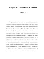
TABLE 25.1. MITOCHONDRIAL ABC TRANSPORTERS
Name of ABC
transporter
Chromosomal
localization
Yeast (Saccharomyces cerevisiae)
Atm1p
XIII
Amino acid
residues
Molecular
mass (kDa)
Homologous to
yeast protein
690
`
78
–
–
–
(Putative)
Function
Maturation of
cytosolic Fe/S
proteins, Iron
homeostasis
Peptide export
?
Mdl1p
Mdl2p
Man (Homo sapiens)
hABC7
XII
XVI
695
820
76
91
Xq13.1–q13.3
752
83
Atm1p (47%)
MTABC3
M-ABC1
2q36
7q35–q36
842
718
94
78
M-ABC2
1q42
738
79
Atm1p (38%)
Mdl2p (34%)
Mdl1p (32%)
Mdl1p (42%)
Mdl2p (38%)
Mouse (Mus musculus)
ABC-me
–
715
77
Mdl1p (39%)
Mdl2p (37%)
Heme transport ?
Plants
Sta1 (A. thaliana)
V
728
80
Atm1p (45%)
IV
IV
–
680
678
206
76
75
24
Atm1p (44%)
Atm1p (45%)
E. coli CcmB (27%)
Maturation of
cytosolic Fe/S
proteins
?
?
c-type cytochrome
biogenesis?
Sta2 (A. thaliana)
Sta3 (A. thaliana)
CcmB (Triticum aestivum)
(Membrane domain)
Maturation of
cytosolic Fe/S
proteins, Iron
homeostasis
Iron homeostasis
?
?
In all cases, a (putative) N-terminal mitochondrial presequence might be cleaved from the proteins, thus resulting in
slightly shorter mature forms. The highest sequence homology between the listed mammalian or plant proteins and
the Saccharomyces cerevisiae proteins is given as the fraction of identical amino acid residues in both proteins. For
references see text.
ABC TRANSPORTERS IN MITOCHONDRIA
cleavage site is not known but, based on the
consensus sequence recognized by matrix processing peptidase (MPP), it is predicted to
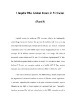
be after amino acid residues 25 or 41. Subcellular localization of Atm1p was demonstrated
by immunostaining of cell fractions and by
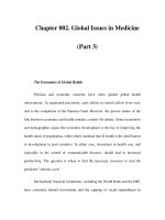
immunofluorescence. Atm1p is localized in
the mitochondrial inner membrane with the
nucleotide-binding domain facing the matrix
space (Figure 25.1). We presume, as will be
developed in later sections, that Atm1p is predicted to function as an exporter of compounds
from the matrix to the intermembrane space.
(PCR) approach, they identified genes for several
of the S. cerevisiae ABC transporters. The first
mitochondrial representative, termed Atm1p,
was identified by virtue of an N-terminal
sequence resembling a mitochondrial targeting
signal (presequence). In a parallel genetic screen
originally intended to isolate new components
of the biogenesis of c-type cytochromes (Kranz
et al., 1998), a temperature-sensitive mutant of
the yeast ATM1 gene (Kispal et al., 1997) was
found. This encodes a protein comprising 690
amino acid residues with six putative transmembrane segments and a C-terminal ATP-binding
domain (Table 25.1) exhibiting the characteristic
features of ABC transporter proteins. Atm1p
therefore belongs to the group of ‘half transporters’. It should be mentioned that no attempts
have been made so far to determine precisely
the structural mode of membrane integration of
Atm1p (or of the other mitochondrial ABC transporters). Different algorithms used to predict
transmembrane helices have identified five to
six hydrophobic sequences that fulfill the criteria
for membrane integration. Thus, by analogy
with classical ABC transporters (Higgins, 1992),
the Atm1p polypeptide chain may be expected
to span the membrane six times and the functional protein may be a homodimer consisting of
two molecules of Atm1p (Figure 25.1).
The function of the N-terminus of Atm1p as
a mitochondrial presequence was verified by
its ability to target attached proteins to mitochondria (Leighton and Schatz, 1995). The precise localization of the Atm1p presequence
DELETION OF THE YEAST ATM1 GENE
Cells deficient in the ATM1 gene (strain ⌬atm1)
display a strong growth defect on rich media
containing glucose (Kispal et al., 1997; Leighton
and Schatz, 1995) and do not grow on nonfermentable carbon sources such as glycerol.
The rate of growth of ⌬atm1 cells in the presence
of glucose is much slower than that of cells
harboring mitochondria defective in respiration.
Thus, Atm1p plays a role that goes beyond the
formation of respiratory competent mitochondria. Another phenotype resulting from the
deletion of ATM1 is a large reduction in the level
of holocytochromes (Kispal et al., 1997; Leighton
and Schatz, 1995). Immunostaining analysis
showed that this is not due to the defective
biosynthesis of the apoforms of the c-type
cytochromes in ⌬atm1 cells (Kispal et al., 1997).
Cytosol
MOM
Atm1p
Mdl2p
Mdl1p
IMS
MIM
N
N
ATP
N
ATP
ATP
ATP
Ma
N
N
N
ATP
ATP
Figure 25.1. Model for the membrane orientation of the yeast mitochondrial ABC transporters.
All three known yeast ABC transporters, Atm1p, Mdl1p and Mdl2p, share a similar membrane orientation
with the N-terminus (N) facing the matrix space, an N-terminal ATP-binding domain and a C-terminal
membrane-spanning domain with six putative transmembrane helices. The drawing represents
the predicted size of the loops between the membrane segments and indicates the formation of possible
homodimers. MOM, mitochondrial outer membrane; IMS, intermembrane space; MIM, mitochondrial
inner membrane; Ma, matrix.
517
518
ABC PROTEINS: FROM BACTERIA TO MAN
Moreover, heme biosynthesis occurs at wildtype rates in these cells (H. Lange, unpublished). Consequently, cells defective in Atm1p
seem to face a condition leading to the degradation of protein-bound heme, which can most
probably be explained by the oxidative stress
prevailing in ⌬atm1 cells (Kispal et al., 1997).
Reduced and oxidized glutathione, which are
the most important compounds required to balance the cellular redox level in yeast, are substantially increased in ⌬atm1 cells (Kispal et al.,
1997). The state of oxidative stress itself may be
a consequence of the dramatic increase in the
concentration of ‘free’ iron (i.e. non-heme and
non Fe/S iron), which appears to be an early
phenotype resulting from the loss of Atm1p
function (Kispal et al., 1999). Together with the
mitochondrial matrix protein Yfh1p (frataxin),
Atm1p was the first protein for which a function
in mitochondrial iron homeostasis could be
demonstrated (Babcock et al., 1997; Foury and
Cazzalini, 1997; Kispal et al., 1997).
In some genetic backgrounds, ⌬atm1 cells
lose mitochondrial DNA to yield so-called 0
cells (Leighton and Schatz, 1995; Senbongi
et al., 1999). This phenomenon is not an obligatory consequence of the inactivation of ATM1;
for example, deletion of the gene in strain W303
does not result in 0 cells (Kispal et al., 1997).
Thus, loss of mitochondrial DNA may well be
an indirect consequence of the oxidative damage resulting from iron overload and impairment of components of the machinery involved
in mitochondrial DNA maintenance (Kaufman
et al., 2000).
ROLE OF ATM1P IN THE MATURATION OF
CYTOSOLIC FE/S PROTEINS
Of all these pleiotropic phenotypes associated
with ⌬atm1 cells none provide any clues
towards the understanding of the function of
the ABC transporter. Initial insight into the
process in which Atm1p is involved came from
the observation that ⌬atm1 cells fail to grow
without added leucine (Kispal et al., 1999). In
yeast, leucine is synthesized from the common
leucine/valine precursor ␣-ketoisovalerate by
three specific steps catalyzed by the enzymes
␣-isopropyl malate synthase (Leu4p and Leu9p),
isopropyl malate isomerase (Leu1p) and isopropyl malate dehydrogenase (Leu2p) (see
reaction schemes in Hinnebusch, 1992; Jones
and Fink, 1982; Prohl et al., 2001). These enzymes
are compartmentalized, distributed between the
mitochondrial matrix (Leu4p and Leu9p) and
the cytosol (Leu4p, Leu1p and Leu2p) (Beltzer
et al., 1988; Casalone et al., 2000; Kohlhaw, 1988a,
1988b). Measurements of individual enzymatic
activities showed a quantitative deficiency of
isopropyl malate isomerase (Leu1p) in ⌬atm1
cells while the other enzymes were active at
wild-type levels (Kispal et al., 1999).
What is the reason for these observations?
Leu1p is a cytosolic protein that requires an
Fe/S cluster, generated in this mitochondrial matrix, for activity. Leu1p closely resembles aconitase of the mitochondrial matrix
(Kohlhaw, 1988b). However, in contrast to
Leu1p, mitochondrial aconitase, another Fe/S
protein, exhibits almost wild-type activity in
⌬atm1 cells, rendering a general defect in cellular Fe/S proteins unlikely (Kispal et al., 1997).
Rather, the specific defect in Leu1p indicated
that Atm1p may perform a function in the
maturation of extra-mitochondrial Fe/S proteins (Kispal et al., 1999).
To investigate the immediate effects of
Atm1p deficiency, as opposed to long-term consequences (see above), a yeast mutant in which
expression of the ATM1 gene was under the
control of a galactose-regulatable promoter
(Gal-ATM1 cells) was created (Kispal et al.,
1999). These cells can readily be depleted of
Atm1p when grown in the absence of galactose.
Nevertheless, in the presence of galactose they
do not exhibit a dramatic growth defect nor do
they display any of the pleiotropic phenotypes
reported above (e.g. cytochrome deficiency,
oxidative stress). Upon depletion of Atm1p, the
activity of Leu1p decreased at least 10-fold,
indicating that incorporation of the Fe/S cluster
into the cytosolic Leu1p apoprotein is an early
consequence of Atm1p deficiency. A direct function of Atm1p in the assembly of the Fe/S
cluster holoprotein, Leu1p, could be shown by
briefly radiolabeling wild-type cells with ferrous iron (55Fe), followed by immunoprecipitation of Leu1p from cell extracts using specific
antibodies (Kispal et al., 1999). The radioactive
iron associated with Leu1p served as a direct
measure of the formation of the Fe/S cluster in
Leu1p. Cells lacking Atm1p did not incorporate
any significant 55Fe radioactivity into Leu1p.
These results provided convincing evidence for
the involvement of Atm1p in the maturation of
a cytosolic Fe/S protein.
Recently, these results have been supported
and extended by the analysis of another cytosolic Fe/S protein, namely the essential protein
Rli1p, which harbors an Fe/S cluster domain at
ABC TRANSPORTERS IN MITOCHONDRIA
its N-terminus (G. Kispal, unpublished).
Assembly of the Fe/S cluster in Rli1p also
involves the function of Atm1p, suggesting a
general role for this ABC transporter in the biogenesis of extra-mitochondrial Fe/S proteins.
Atm1p function in cytosolic Fe/S protein maturation is highly specific because no defects were
observed in Fe/S proteins localized inside mitochondria upon depletion of Atm1p (Kispal
et al., 1999). For a better understanding of the
distinct function of Atm1p in the maturation of
cytosolic Fe/S proteins, it is necessary to provide a brief outline of the biogenesis of Fe/S
proteins in a eukaryotic cell. For a more comprehensive discussion of this recently discovered
process, the reader is referred to several detailed
reviews (Craig et al., 1999; Lill et al., 1999; Lill
and Kispal, 2000; Mühlenhoff and Lill, 2000).
beginning to understand Fe/S cluster biogenesis, and any putative mechanistic pathways are
based on rather limited experimental evidence.
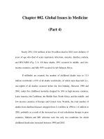
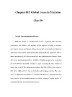
According to a present working model,
shown in Figure 25.2, iron, after its membrane
potential-dependent import into mitochondria
(Lange et al., 1999), binds to the two proteins,
Isu1p and Isu2p. The cysteine desulfurase Nfs1p
generates elemental sulfur (S0) from cysteine,
which is then used to form an ‘intermediate’
Apo
Holo
S Fe
Fe S
Extra-mitochondrial
Fe/S proteins
Erv1p
ISC export
machinery
BIOGENESIS OF EUKARYOTIC
FE/S PROTEINS
Assembly of mitochondrial Fe/S proteins
Many studies over the past four years have led
to the identification of some ten proteins of the
mitochondrial matrix, which play a role in the
formation of the Fe/S clusters and their incorporation into mitochondrial apoproteins (for
examples see Garland et al., 1999; Jensen and
Culotta, 2000; Kaut et al., 2000; Kim et al., 2001;
Kispal et al., 1999; Lange et al., 2000; Li et al.,
2001; Pelzer et al., 2000; Schilke et al., 1999;
Strain et al., 1998; Voisine et al., 2001). These
proteins are highly homologous to bacterial proteins encoded by the isc (iron sulfur cluster)
operons (Zheng et al., 1998), and were therefore
defined as compounds of the ‘ISC assembly
machinery’ (Lill and Kispal, 2000). Even though
virtually all of these proteins have been shown
to participate in the assembly of Fe/S clusters,
comparatively little is known about the precise
roles of individual proteins or the overall molecular mechanism of the pathway. Nevertheless,
a number of functional studies have been performed on the bacterial Isc proteins (for examples see Agar et al., 2000b, 2000c; Hoff et al.,
2000; Krebs et al., 2001; Ollagnier-de-Choudens
et al., 2001; Silberg et al., 2000; Yuvaniyama
et al., 2000; Zheng et al., 1993, 1994). Thus, the
following model combines knowledge gained
from studies on both mitochondrial and bacterial Isc proteins, assuming that the process
is highly similar in both environments. However, it should be emphasized that we are just
Mitochondrion
S Fe
Fe S
Isu1/2p
Ala
Cys
Arh1p
Yah1p
eϪ
ABC
transporter
Atm1p
?
ISC assembly
machinery
Nfs1p
Apo
Holo
S Fe
Fe S
Mitochondrial
Fe/S proteins
pmf
Iron
Cytosol
Figure 25.2. Working model for the function of
Atm1p in cytosolic Fe/S protein assembly in
eukaryotic cells. The assembly of Fe/S clusters, for
both mitochondrial and cytosolic Fe/S proteins, is
achieved by the ISC assembly machinery. First,
ferrous iron enters the mitochondrial matrix in a
membrane potential (pmf)-dependent step. Iron
binds to the Isu proteins which provide a scaffold
for the assembly of the Fe/S clusters. The cysteine
desulfurase, Nfs1p, generates elemental sulfur (S0)
from cysteine needed for Fe/S cluster formation on
the Isu proteins. The nascent Fe/S clusters are
released from the Isu proteins upon reduction by
the electron transfer chain shuttling electrons from
NAD(P)H to the ferredoxin reductase Arh1p and the
ferredoxin Yah1p. The Fe/S clusters are then
incorporated into the apoforms of mitochondrial
Fe/S proteins or exported to the cytosol, a step
most likely involving Atm1p. The exact nature of
the substrate of Atm1p is not known yet, but a
likely compound is a chelated Fe/S cluster. The
export process may be assisted by Erv1p, a
sulfhydryl oxidase in the intermembrane space. It
should be noted that many of the proposed steps of
this model need further experimental verification.
519
520
ABC PROTEINS: FROM BACTERIA TO MAN
[2Fe-2S] cluster on Isu1p/Isu2p. (Yuvaniyama
et al., 2000). This cluster may further be modified to generate a [4Fe-4S] cluster (Agar et al.,
2000a). The next steps of Fe/S cluster release
and incorporation into apoproteins have not
been defined experimentally, leaving us to speculate about the possible mechanism. In vitro, the
intermediate Fe/S cluster can be released from
the Isu proteins upon the addition of reducing
agents. Therefore, the ferredoxin reductase
Arh1p and the ferredoxin Yah1p may form an
electron transfer chain that provides the reducing electrons for the release of the Fe/S cluster
from the Isu proteins (Lange et al., 2000; Li
et al., 2001).
The fate of the released Fe/S cluster is
unknown. It may be transferred to and incorporated into the apoproteins spontaneously, or the
process may need the help of accessory proteins.
It is tempting to speculate that the insertion of
the Fe/S cluster into apoproteins is a proteinassisted reaction. Stabilization of the apoproteins before incorporation of the Fe/S cluster
could be an obvious task of the two mitochondrial heat shock proteins of the Hsp70/DnaK
and Hsp40/DnaJ classes, Ssq1p and Jac1p,
respectively (Kim et al., 2001; Lutz et al., 2001;
Schilke et al., 1999; Strain et al., 1998; Voisine
et al., 2001). However, evidence for an interaction between the chaperones and the apoproteins has not, so far, been reported. On the
contrary, the bacterial homologues of the two
heat shock proteins have been shown to bind to
the Isu proteins, leading to a stimulation of the
ATPase activity of the Hsp70 chaperone (Hoff
et al., 2000; Silberg et al., 2000). The mechanistic
significance of this interaction remains to be
discovered.
The Isa proteins have recently been shown
to be crucial for Fe/S cluster assembly (Jensen
and Culotta, 2000; Kaut et al., 2000; Pelzer
et al., 2000) and, according to in vitro data, they
may provide the necessary scaffold for the
assembly of these Fe/S clusters (Krebs et al.,
2001; Ollagnier-de-Choudens et al., 2001). Thus,
the Isa proteins may represent an alternative to
the Isu proteins in the assembly of the Fe/S
clusters. Finally, a requirement for frataxin
(yeast Yfh1p) for the normal activity of mitochondrial Fe/S proteins has been documented,
even though the effects of deleting the frataxin
gene were not dramatic (Foury, 1999; Rötig
et al., 1997). According to a recent study,
frataxin might play a role in the storage of iron
in mitochondria (Adamec et al., 2000). Thus,
the requirement for frataxin in Fe/S protein
maturation might well be an indirect consequence of the impaired delivery of iron to the
Isu and Isa proteins.
Maturation of extra-mitochondrial
Fe/S proteins
In addition to the assembly of mitochondrial
Fe/S proteins, the ISC assembly machinery also
plays a crucial role in the maturation of extramitochondrial Fe/S proteins. The currently
available data suggest that the Fe/S clusters
of cytosolic Fe/S proteins are assembled in the
mitochondrial matrix and, therefore, need to be
exported, in some form, from mitochondria
(summarized by Lill and Kispal, 2000). This
contention is based on the fact that depletion
of the mitochondrial Isc components abolishes
cytosolic Fe/S protein maturation. Nevertheless,
the molecular moiety leaving the organelle is not
known at present. Similarly, we are only just
beginning to understand the molecular mechanisms underlying the export process.
Since Atm1p is specifically required for the
assembly of cytosolic, but not mitochondrial
Fe/S proteins, it is thought to play a central role
in the release of a moiety synthesized by the ISC
assembly machinery from the organelles and
may be required for the assembly of cytosolic
Fe/S proteins. Only a few components of the
so-called ‘ISC export machinery’, other than
Atm1p, have been identified so far, namely
Erv1p and the two homologous proteins Bat1p
and Bat2p. Since these proteins appear to be
functionally related to Atm1p, the findings that
support their involvement in Fe/S protein maturation in the cytosol are briefly summarized as
follows.
Erv1p is a component of the intermembrane
space and is essential for yeast viability (Lange
et al., 2001; Lisowsky, 1992). Inactivation of
Erv1p leads to a dramatic reduction in the
assembly of cytosolic Fe/S proteins. Similar to
what is observed when Atm1p is depleted,
mitochondrial Fe/S protein assembly is not
affected in Erv1p-defective cells. Erv1p was
found to possess sulfhydryl oxidase activity
associated with the C-terminal domain of the
protein (Lee et al., 2000). Currently, the role of
this domain in Fe/S protein assembly in the
cytosol is unclear. Nevertheless, the localization
of Erv1p in the intermembrane space suggests
that it plays a role in the export pathway subsequent to that in which Atm1p is implicated.
Whether Erv1p transiently binds directly to the
ABC TRANSPORTERS IN MITOCHONDRIA
transported molecule, or introduces disulfide
bonds into a component of the pathway, remains
to be determined. Interestingly, the mammalian
homologue of Erv1p, termed ALR (‘augmenter
of liver regeneration’), can functionally replace
the yeast protein and thus the two proteins
appear to be orthologues. All of the components
of the ISC assembly machinery and Atm1p (see
below) are conserved in mammals, suggesting
that Fe/S cluster assembly follows similar pathways in virtually all eukaryotes.
The BAT1 gene was isolated as a high-copy
suppressor of a temperature-sensitive mutant of
ATM1 (Kispal et al., 1996). BAT1 and the highly
homologous gene BAT2 encode the mitochondrial and cytosolic forms of branched-chain
amino acid transaminases, respectively (Eden
et al., 1996; Kispal et al., 1996). The Bat proteins
catalyze the reversible inter-conversion of
branched-chain ␣-keto acids and amino acids
(i.e. leucine, isoleucine and valine). Additionally,
they perform a second function unrelated to
amino acid synthesis. This is evident from the
growth defect of ⌬bat1 ⌬bat2 cells, lacking both
BAT genes, on rich media containing glucose,
which occurs even after additional branchedchain amino acids are added to the medium
(Kispal et al., 1996). This observation may be
explained by the participation of the Bat
proteins in the maturation of cytosolic Fe/S proteins (Prohl et al., 2000; C. Prohl, unpublished).
The double mutant cells show a threefold reduction in the de novo synthesis of both Leu1p and
Rli1p Fe/S proteins in the cytosol. Thus, the Bat
proteins are not essential for maturation of
cytosolic Fe/S proteins, but apparently perform
an accessory function, increasing the efficiency
of the formation of holoprotein in an, as yet,
unknown way. Expression of either BAT gene is
sufficient for the normal formation of cytosolic
Fe/S proteins, indicating that the specific Bat
function can be performed either in mitochondria, or in the cytosol. Similar to Atm1p and
Erv1p, the Bat proteins are not required for the
biogenesis of Fe/S proteins within the mitochondria, suggesting that they participate in the
Atm1p-mediated export pathway. One possible
function may be the catalytic formation of a compound required for chelation of the Fe/S cluster
(or a related compound) during export from
the mitochondria.
In summary, there is ample evidence for the
involvement of Atm1p in the maturation of
cytosolic Fe/S proteins, yet the molecular
details underlying its precise function have not
been unraveled so far. Future progress in
understanding the roles of Atm1p, Erv1p and
the Bat proteins will require the identification of
the substrate for Atm1p and of any additional
components of the ISC export machinery.
MDL1P AND MDL2P, TWO HOMOLOGOUS
YEAST MITOCHONDRIAL ABC
TRANSPORTERS WITH DIFFERENT
FUNCTIONS
Recently, two additional ABC transporters,
termed Mdl1p and Mdl2p, have been identified
in yeast mitochondria, and were found to be
homologues of the human ABCB8 and ABCB10
genes (see below) (Young et al., 2001). Like
Atm1p, these proteins are half transporters
with an N-terminal membrane-spanning domain
(Figure 25.1) (Dean et al., 1994). The two proteins show rather high sequence homology (46%
identical amino acid residues) and have a
molecular mass of 76 kDa (Mdl1p) and 91 kDa
(Mdl2p), including a putative N-terminal extension serving as a mitochondrial presequence
(Table 25.1). In fact, only the N-terminus of
Mdl1p resembles a canonical mitochondrial targeting signal, whereas the N-terminal segment
of Mdl2p does not conform to the properties of
a presequence. According to biochemical fractionation experiments using specific antibodies,
both proteins are localized in the mitochondrial
inner membrane with the ABC domains facing
the matrix space (Young et al., 2001) (Figure
25.1). Thus, all three yeast mitochondrial ABC
transporters appear to exhibit the same membrane orientation and thus are presumed to
export substrates from the matrix towards the
cytosol.
Deletion of the MDL1 and MDL2 genes does
not cause major growth defects in S. cerevisiae
(Dean et al., 1994). However, whilst ⌬mdl1 cells
exhibit normal growth, growth of ⌬mdl2 cells is
retarded on glycerol-containing media. In part,
this may be explained by the finding that
⌬mdl2 cells tend to gradually lose mitochondrial DNA (J. Gerber, unpublished). Double
deletion of both MDL genes slightly exacerbates
the growth defect observed for ⌬mdl2 cells,
suggesting that the proteins may perform nonoverlapping functions. This is supported by
recent insights into the function of Mdl1p.
Both Mdl1p and Mdl2p are close homologues
of the yeast a-factor pheromone receptor Ste6p
and of another ABC protein, the mammalian
TAP transporter (ABCB2/ABCB3). This protein
mediates the transfer of antigenic peptides after
521
522
ABC PROTEINS: FROM BACTERIA TO MAN
25.3). When a double mutant ⌬mdl1 ⌬yme1 was
analyzed, a 75% reduction in peptide release
from the organelle was observed. The length of
the released peptides varied between 6 and 20
residues, strikingly similar in size to peptides
transported by the TAP transporter in the
ER (Elliott, 1997; Ritz and Seliger, 2001) (see
Chapter 26). The function of Mdl1p in peptide
export depended on a conserved motif in the
Walker A and B sites of the nucleotide-binding
domain and a loop characteristic for ATPases.
Peptide export through Mdl1p therefore seems
to require the hydrolysis of ATP (Young et al.,
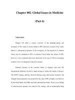
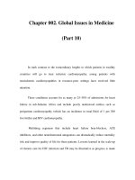
2001). For final exit from the organellar intermembrane space, as illustrated in Figure 25.3,
the peptides possibly pass the outer membrane
with the help of mitochondrial porin or the
TOM complex, both of which contain large
pores (Figure 25.3) (Künkele et al., 1998).
On the other hand, deletion of MDL2 does
not result in any alteration of peptide export
from the mitochondria, suggesting that only
Mdl1p mediates the release of peptides from
the mitochondrial matrix. These findings are
nicely corroborated by the observation that
Mdl1p and Mdl2p appear to be associated with
different high molecular mass complexes of
their generation by the cytosolic proteasome to
the class I major histocompatibility complex
(MHC class I) in the endoplasmic reticulum (ER)
(see Chapter 26). Hence, it was postulated that
the Mdl proteins may facilitate the export of
peptides from the matrix to the intermembrane
space. A direct test of this idea showed that
mitochondria derived from ⌬mdl1 mutant cells
displayed a 40% reduction in peptide release
(Young et al., 2001). In the assay system used,
the peptides were generated by the inner membrane protease Yta10p/Yta12p (also termed
Afg3p/Rca1p) from mitochondria-encoded
membrane proteins (Arlt et al., 1996; Rep et al.,
1996) (Figure 25.3). This member of the family of
ATP-dependent AAA proteases exposes its proteolytic domain in the matrix space and forms a
large hetero-oligomeric complex (for a recent
review on AAA proteases, see Langer, 2000).
The rather small decrease in peptide export
observed after deletion of MDL1 is explained by
the fact that another set of peptides generated by
the inner membrane protease Yme1p can still
leave the organelle in the absence of Mdl1p. This
second mitochondrial AAA protease forms a
homo-oligomer with its proteolytic domain in
the intermembrane space (Langer, 2000) (Figure
Peptides
TOM
complex
Porin
MOM
IMS
ATP
MIM
Yme1p
ATP
Yta10/12p
ATP
Ma
Mdl1p
Figure 25.3. Model for the function of Mdl1p in the export of peptides from the mitochondrial matrix.
Peptides generated by the inner membrane protease, Yta10p/Yta12p, are exported by the ABC transporter,
Mdl1p, in an ATP-dependent fashion. Another pool of proteolytic fragments is formed by the inner membrane
protease Yme1p in the intermembrane space. Most likely, the peptides leave the mitochondria via porin or
the TOM complex, both of which contain large pores. Currently, it is unknown how peptides generated by the
matrix protease Pim1p (not shown) are exported from the organelles. MOM, mitochondrial outer membrane;
IMS, intermembrane space; MIM, mitochondrial inner membrane; Ma, matrix.
ABC TRANSPORTERS IN MITOCHONDRIA
200 kDa and 300 kDa, respectively (Young et al.,
2001). This observation may argue for homodimer, rather than heterodimer, association of
the Mdl proteins.
Since deletion of MDL1 is not associated with
any detectable phenotype, the question arises as
to what the physiological significance of peptide
transport by Mdl1p might be. Moreover, mitochondria can further break down the longer
peptides to amino acids or di- and tripeptides,
which can be transported independently of
Mdl1p. Thus, the purpose of Mdl1p-mediated
peptide transport remains unclear, even though
the latter observation supports the finding that
Mdl1p is dispensable in yeast. In vertebrates,
proteins homologous to Mdl1p (see below)
might play an important role in the transport of
antigenic peptides derived from mitochondrial
proteins for presentation on the eukaryotic cell
surface. Functional complementation studies
with the mammalian homologues expressed in
the ⌬mdl1 background have not yet been conducted to test this attractive hypothesis.
MITOCHONDRIAL ABC
TRANSPORTERS IN
MAMMALS
The sequencing of the human genome has
provided us with a complete inventory of
ABC transporters in man (Klein et al., 1999;
). Amongst the 48
proteins with ABC domains, a few qualify as
potential mitochondrial components based on
the presence of a (putative) presequence at
their N-termini. Two of these proteins, termed
ABC7 (ABCB7, according to the nomenclature
of ) and MTABC3
(ABCB6) are homologous to the yeast Atm1p,
whereas another two proteins, namely M-ABC1
(ABCB8) and M-ABC2 (ABCB10), closely resemble yeast Mdl1p and Mdl2p. In mice, another
homologue of the latter subclass has been
identified and analyzed recently, the protein
ABC-me. The properties and functions of these
proteins are discussed in the following sections.
ABC7 AND MTABC3, FUNCTIONAL
ORTHOLOGUES OF YEAST ATM1P
The human ABC transporter ABC7 represents
the closest homologue of yeast Atm1p. The
cDNA corresponding to the gene has been
identified independently by several groups
(Allikmets et al., 1999; Csere et al., 1998; Mao
et al., 1998; Shimada et al., 1998). Sequencing of
the entire ABC7 gene revealed 16 introns and
the promoter structure (Bekri et al., 2000). At the
protein level ABC7 shares 47% amino acid
sequence identity with yeast Atm1p (Table 25.1).
Expression of the human gene in yeast has
demonstrated that the human protein is the
functional orthologue of Atm1p (Allikmets
et al., 1999; Csere et al., 1998). This gene is able
to revert growth of ⌬atm1 mutant cells to almost
wild-type rates and to restore normal cytochrome levels. Furthermore, mitochondria
harboring ABC7 instead of Atm1p do not
accumulate iron. All these observations strongly
indicate that upon expression in yeast, ABC7
can replace the primary function of Atm1p in
Fe/S cluster formation (Bekri et al., 2000). These
findings further suggest that ABC7 performs a
similar or identical function in the mammalian
cell as that carried out by Atm1p in yeast.
Mutations in the human ABC7 gene cause
X-linked sideroblastic anemia and cerebellar
ataxia (XLSA/A) (Allikmets et al., 1999; Bekri
et al., 2000). As a result of such mutations, mitochondria accumulate high concentrations of iron
and form so-called ring sideroblasts (i.e. ironloaded ring-shaped tubules which are concentrated around the nucleus). Thus, there exists a
striking similarity in phenotypes between yeast
and man upon impairment of Atm1p and ABC7
function, respectively. Biochemical studies indicate that yeast serves as an excellent model system to study the effects of the mutations in
ABC7. When expressed in yeast, mutant ABC7
proteins, or Atm1p bearing the corresponding
mutations, are functionally impaired (Allikmets
et al., 1999; Bekri et al., 2000). For instance, when
the ABC7-(E433K) mutants (mutation localized
towards the matrix following TM6), or the corresponding ATM1-(D398K) mutants, were
expressed in ⌬atm1 yeast cells, maturation of
cytosolic Fe/S proteins was twofold lower as
compared to wild-type cells (Bekri et al., 2000).
The surprisingly weak consequences of these
charge exchange mutations underlines the
importance of ABC7 function for a healthy cell.
In fact, only slight changes to ABC7 can dramatically affect cellular iron homeostasis and elicit
severe phenotypical consequences. These observations are consistent with the fact that, in yeast,
Fe/S cluster formation is an indispensable
process (Lill et al., 1999). Deletion of many genes
encoding components of the ISC assembly
523
524
ABC PROTEINS: FROM BACTERIA TO MAN
machinery is lethal, indicating a central role for
Fe/S proteins for life.
The human protein, termed MTABC3
(Taglicht and Michaelis, 1998), represents a
second functional orthologue of yeast Atm1p
(Mitsuhashi et al., 2000). The MTABC3 gene is
encoded by human chromosome 2 (Table 25.1)
and has been mapped to within the vicinity of
the locus for lethal neonatal metabolic syndrome, a disorder of mitochondrial function
associated with iron metabolism. Hence,
MTABC3 is a likely candidate gene for this disorder. The homology of MTABC3 and Atm1p is
less than that of ABC7 compared with Atm1p
(38% as compared to 47% identical amino
acid residues). Nevertheless, expression of
MTABC3 in ⌬atm1 yeast cells restores growth
to wild-type levels, reverts the increase in mitochondrial iron, and prevents the loss of mitochondrial DNA. Even though the role of
MTABC3 in the biogenesis of cytosolic Fe/S
proteins has yet to be analyzed, such a function
seems likely.
The relationship of the two human orthologues of yeast Atm1p is unclear. Based on their
common role in iron homeostasis it is conceivable that ABC7 and MTABC3 form a heterodimer in the human cell. An alternative
hypothesis predicts that both genes may be differentially expressed in human tissues. The
presence of two copies of Atm1p-like proteins
may offer the possibility to fine-tune the function of the ABC transporter, as found for numerous other mammalian proteins.
M-ABC1 AND M-ABC2, MAMMALIAN
HOMOLOGUES OF YEAST MDL1P AND
MDL2P
The human genome harbors four candidates
with homology to the yeast MDL genes. Only
two of the encoded proteins, termed M-ABC1
and M-ABC2 (ABCB8 and ABCB10 according to
nomenclature of ),
have been experimentally localized to mitochondria (Hogue et al., 1999; Zhang et al.,
2000a). Another gene product, ABCB9, has been
found in the lysosomal compartment (Zhang
et al., 2000b). Nevertheless, the protein is not a
close homologue of the vacuolar ABC transporters, Ycf1p of S. cerevisiae (Li et al., 1996) or
Hmt1p of Schizosaccharomyces pombe (Ortiz et al.,
1995). The fourth mammalian Mdl homologue,
ABCB5, has not yet been studied. The sequence
identity between the human M-ABC1/M-ABC2
and the yeast Mdl proteins varies between 32%
and 42% (Table 25.1). Based on sequence comparisons, M-ABC1 may be the counterpart of
Mdl2p, while M-ABC2 is more closely related to
Mdl1p. However, the differences in homology
may be too small to infer a close functional relationship.
Currently, it is not known whether M-ABC1
and M-ABC2 form homo- or heterodimers in the
mitochondrial inner membrane. Similarly, the
membrane orientation of these proteins is
not yet clear, even though it is likely that it is
the same as for Mdl1p and Mdl2p, with the
nucleotide-binding domain facing the matrix
space (however, see Zhang et al., 2000a). No
experimental evidence has been obtained for
any function of M-ABC1 and M-ABC2 in the
transport of peptides out of the mitochondrial
matrix, even though such a role, similar to that
of Mdl1p, seems probable (see above).
ABC-ME, A MURINE MITOCHONDRIAL
ABC TRANSPORTER WITH A FUNCTION IN
HEME METABOLISM
Only one mitochondrial ABC transporter has
been identified to date in mice, the protein
ABC-me (mitochondrial erythroid), and its cellular role has been investigated in some detail
(Shirihai et al., 2000). The ABC-me gene has
been isolated as one factor that is induced upon
expression of the erythropoietic transcription
factor GATA-1. ABC-me is highly expressed in
erythroid tissues of embryos and adults. In
murine erythroleukemia (MEL) cells, overexpression of ABC-me strongly increased the heme
concentration. Conversely, ABC-me mRNA
levels are decreased by physiological concentrations of heme. Together, these findings are consistent with a role for ABC-me in the trafficking
of intermediates of heme biosynthesis. The
heme biosynthetic steps are partitioned between
the mitochondrial matrix and the cytosol, with
the first reaction and the last three steps taking
place in the matrix. The ABC domains of
ABC-me face the mitochondrial matrix and this
has been taken to indicate that the protein
should function as an exporter (Shirihai et al.,
2000). ABC-me could be involved in translocating either ␦-aminolevulinate or heme from the
mitochondrial matrix to the cytosol. The rather
specific expression of ABC-me in erythroid cells
may be necessary to satisfy the extraordinarily
high needs for transporting heme biosynthetic
metabolites across the mitochondrial inner
ABC TRANSPORTERS IN MITOCHONDRIA
membrane. At present, it is unclear which, if
any, human ABC protein may represent the
counterpart of murine ABC-me, since there is no
known human Mdl-like protein with specific
expression in erythroid tissues. Further, all
known human candidates exhibit a similar
degree of sequence similarity to ABC-me.
MITOCHONDRIAL ABC
TRANSPORTERS IN
PLANTS
The recent sequencing of the genome of the
weed Arabidopsis thaliana has allowed access
to the inventory of plant ABC transporters
(Sanchez-Fernandez et al., 2001). The plant
genome contains more than 100 distinct members of this protein family. From the homology
with yeast ABC transporters, several counterparts to Atm1p and Mdl1p/Mdl2p can be identified in Arabidopsis. While two of the three
homologues of Atm1p have been characterized
as mitochondrial proteins (Kushnir et al., 2001),
the subcellular localization of the two Mdl1plike proteins is not clear. The latter show high
sequence similarity to both yeast mitochondrial
Mdl1p/Mdl2p and to the mammalian TAP1/
TAP2 (ABCB2/ABCB3) transporters of the ER
(see Chapter 26). Thus, in the absence of experimental evidence, it is uncertain whether these
members of the ABC transporter family resemble mitochondrial or microsomal constituents.
In addition to these plant ABC proteins, another
potential ABC transporter, termed CcmAB, may
exist. CcmB, the membrane-spanning part of
this ABC protein, is encoded by the mitochondrial genome of various plants. Bacterial homologues of the plant CcmB protein play a role in
c-type cytochrome biogenesis. The Atm1p-like
and the CcmAB-like ABC proteins of plant mitochondria will be discussed in more detail in the
following sections.
THE STA (ATM) PROTEINS, HOMOLOGUES
OF YEAST ATM1P
A. thaliana contains three genes, termed STA1,
STA2 and STA3 (also known as ATM3, ATM2
and ATM1, respectively), the products of which
share about 45% amino acid identity with the
yeast ATM1 gene product (Kushnir et al., 2001;
Sanchez-Fernandez et al., 2001) (Table 25.1).
The sequence identity between the three plant
proteins varies from 71% (Sta1/Sta2) to 83%
(Sta2/Sta3). Although all three Sta proteins
contain a putative mitochondrial presequence
at their N-termini, mitochondrial localization
has only been experimentally determined for
Sta1 and Sta2 (Kushnir et al., 2001).
Inactivation of the STA1 gene results in
chlorosis and dwarfism of mutant plants
(Kushnir et al., 2001). The most severe phenotype was seen when plants were grown on synthetic media. Nevertheless, mutant plants are
photoautotrophic and fertile. Plant leaves in
the mutants exhibit a number of abnormalities
such as enlarged cells with more air space in
between them. The STA1 mutant can be partially complemented by ectopic expression of
STA2, resulting in plants which grow to almost
wild-type size and show no signs of chlorosis.
Even though the two proteins apparently have
overlapping functions, their roles are not
entirely redundant. A plausible reason for these
observations may be differential and tissuespecific expression of the STA genes.
A function for Sta1 in Fe/S protein maturation
in the cytosol could be inferred from complementation studies in yeast (Kushnir et al., 2001).
In these studies, expression of STA1 in ⌬atm1
mutant yeast cells fully complemented the
defects of Atm1p deficiency. The Sta1 protein
supported cytosolic Fe/S protein maturation
with an efficiency comparable to yeast Atm1p.
However, despite the apparent functional similarities, biochemical analyses of Sta1-deficient
plants revealed striking differences compared to
yeast ⌬atm1 cells. In contrast to the 25- to
30-fold increase in iron concentration in yeast
mitochondria derived from ⌬atm1 cells, plant
mitochondria displayed only a small increase in
free iron concentration. Furthermore, plant cells
showed no obvious signs of oxidative stress.
The differences between yeast and plants may
be explained by the multiplicity of the ATM1like gene in plants. The functional redundancy
of the Sta proteins may lead to comparatively
weak phenotypic consequences of the inactivation of a single STA gene. These results support
the view that the phenotypes observed in yeast
are indirect (secondary) consequences of the
defect in Atm1p.
The function of Sta1 in cytosolic Fe/S protein
maturation, together with the presence of
numerous plant genes encoding components of
the ISC assembly machinery (Kushnir et al.,
2001), indicates that the process of Fe/S protein
biogenesis in plants resembles that of the model
525
526
ABC PROTEINS: FROM BACTERIA TO MAN
organism yeast. Thus, the function of the three
Sta proteins in this biosynthetic process may be
to transport a component required for Fe/S
protein assembly outside the mitochondria.
CCMB, A NOVEL COMPONENT OF AN
ABC TRANSPORTER OF LAND PLANTS?
The sequencing of various plant mitochondrial
genomes has led to the suggestion that an ABC
transporter, with homology to bacterial proteins implicated in c-type cytochrome biogenesis, might be present in these organelles. This
expectation is supported by the recent identification of the independently encoded membranespanning protein component, CcmB, of a
putative ABC transporter (Faivre-Nitschke
et al., 2001). In wheat, CcmB consists of 206
amino acid residues (Table 25.1). The plant
CcmB protein has a large number of hydrophobic amino acid residues and shares significant
sequence identity with CcmB proteins of various bacteria (24–29%). Both plant and bacterial
CcmB proteins have hydrophobicity profiles
characteristic of membrane proteins with six
predicted transmembrane helices. Typical of
many genes encoded by the plant mitochondrial genome, the CcmB transcript is highly
edited (42 C to U editing positions), affecting
32 out of the 206 amino acid residues.
CcmB has been identified as a constituent of
the mitochondrial inner membrane by employing an antibody raised against CcmB (FaivreNitschke et al., 2001). This detects a 28 kDa
protein, compared to the calculated molecular
mass of 24 kDa (Table 25.1), that is enriched
in the mitochondrial membrane fraction. Association of CcmB with its putative ABC domain,
CcmA, has not been established so far. This is
mainly due to the fact that, in land plants, the
expected CcmA protein is not encoded by the
mitochondrial genome, but rather is thought to
derive from a nuclear gene.
The function of CcmB in land plants has not
been addressed experimentally so far. The
homology to bacterial CcmB, however, has led
to the suggestion that the plant protein, like the
bacterial counterparts, performs a function in
c-type cytochrome biogenesis (Faivre-Nitschke
et al., 2001). For a better understanding of the
potential function of CcmAB, a brief sketch of
c-type cytochrome biogenesis follows. For
further details, the reader is referred to recent
review articles by Kranz et al. (1998), Page
et al. (1998), and Thony-Meyer (2000). c-Type
cytochromes carry a heme moiety covalently
attached to two conserved cysteine residues via a
thioether bond. The best-known examples are
the cytochromes c and c1, which are located in the
mitochondrial intermembrane space, or the bacterial periplasm, where they participate in electron transfer during oxidative phosphorylation.
During evolution, three systems have evolved
for the biogenesis of these heme proteins (Kranz
et al., 1998). System I is the most complex and is
found in ␣- and ␥-proteobacteria and in mitochondria of land plants. System II is used by
Gram-positive bacteria, cyanobacteria and
chloroplasts, and system III is present in fungal,
vertebrate and invertebrate mitochondria. The
components of the individual systems differ in
both structure and number. The simplest pathway (system III) uses just one protein for biogenesis, the cytochrome heme lyases that attach
heme to the apocytochromes. Biogenesis in system I, on the other hand, involves some ten proteins which share no obvious homology with
cytochrome heme lyases. Studies in E. coli and
Rhodobacter capsulatus have provided us with a
rudimentary view of the individual steps of biogenesis in system I (reviewed by Thony-Meyer,
2000). In brief, apocytochrome c is translocated
into the periplasm by the canonical Sec translocase. In an unknown way, heme is transferred
from the cytosol to a periplasmic heme chaperone, CcmE, where it becomes covalently bound
in a transient fashion (Schulz et al., 1998). Earlier
genetic studies of the bacterial CcmA, CcmB
and CcmC (another integral membrane protein)
showed their involvement in cytochrome biogenesis, and it was, therefore, suggested that a
complex comprising these three proteins may
form an ABC transporter necessary to export
heme from its site of synthesis in the cytosol to
the periplasm (Goldman and Kranz, 2001;
Goldman et al., 1998). However, this view was
rendered rather unlikely by the findings in
E. coli that heme can be transferred to the periplasmic heme chaperone CcmE in the absence of
CcmA and CcmB, but not without CcmC
(Schulz et al., 1999). Furthermore, CcmC and a
periplasmic protein, CcmE, were found to interact tightly with each other and with heme (Ren
and Thony-Meyer, 2001). Based on these most
recent results, it may be expected that CcmC
facilitates transport of heme from the bacterial
cytosol to CcmE in the periplasm. The function
of CcmA and CcmB is not yet clear, but it has
recently been proposed that they export a compound required to maintain the reduced states
of apocytochrome cysteines, the vinyl groups of
ABC TRANSPORTERS IN MITOCHONDRIA
protoheme or heme iron (Faivre-Nitschke et al.,
2001). Biosynthesis of the holoform of cytochrome c is then completed by the covalent
attachment of heme to apocytochrome c, a reaction most probably catalyzed by CcmF. Prerequisites for this reaction are a number of redox steps
that lead to the reduction of both heme and the
disulfide bridges of the apoprotein. In plant
mitochondria, further studies are needed to verify the existence of the ABC transporter CcmAB
and to examine its potential function in c-type
cytochrome biogenesis.
FUTURE DIRECTIONS
This review on mitochondrial ABC transporters
clearly shows that we are only beginning to
understand the biological roles of these interesting proteins. While it is possible that all of the
yeast and human mitochondrial ABC transporters have been identified (Bauer et al., 1999;
Decottignies and Goffeau, 1997; Klein et al.,
1999; Taglicht and Michaelis, 1998), the biochemical characterization of these components
lags behind. Future studies on mitochondrial
ABC transporters will include the purification
and functional reconstitution of these proteins,
the identification of substrates and the elucidation of the molecular mechanisms underlying
transport. Further challenges include the discovery of new diseases associated with mutations in these proteins, and understanding the
structural/functional relationships between
these important proteins. Clearly, the most
interesting years of research on mitochondrial
ABC transporters lie ahead of us.
ACKNOWLEDGMENTS
Our work was supported generously by grants
from the Sonderforschungsbereich 286,
Deutsche Forschungsgemeinschaft, Deutsches
Humangenomprojekt,
Volkswagen-Stiftung,
Fonds der Chemischen Industrie and the
Hungarian Funds OKTA.
REFERENCES
Adamec, J., Rusnak, F., Owen, W.G., Naylor, S.,
Benson, L.M., Gacy, A.M. and Isaya, G.
(2000) Iron-dependent self-assembly of
recombinant yeast frataxin: implications for
Friedreich ataxia. Am. J. Hum. Genet. 67,
549–562.
Agar, J.N., Krebs, C., Frazzon, J., Huynh, B.H.,
Dean, D.R. and Johnson, M.K. (2000a) IscU
as a scaffold for iron-sulfur cluster biosynthesis: sequential assembly of [2Fe-2S] and
[4Fe-4S] clusters in IscU. Biochemistry 39,
7856–7862.
Agar, J.N., Yuvaniyama, P., Jack, R.F., Cash, V.L.,
Smith, A.D., Dean, D.R. and Johnson, M.K.
(2000b) Modular organization and identification of a mononuclear iron-binding site
within the NifU protein. J. Biol. Inorg.
Chem. 5, 167–177.
Agar, J.N., Zheng, L., Cash, V.L., Dean, D.R.
and Johnson, M.K. (2000c) Role of the IscU
protein in iron-sulfur cluster biosynthesis:
IscS-mediated assembly of a [Fe2S2] cluster
in IscU. J. Am. Chem. Soc. 122, 2136–2137.
Allikmets, R., Raskind, W.H., Hutchinson, A.,
Schueck, N.D., Dean, M. and Koeller, D.M.
(1999) Mutation of a putative mitochondrial
iron transporter gene (ABC7) in X-linked
sideroblastic anemia and ataxia (XLSA/A).
Hum. Mol. Gen. 8, 743–749.
Arlt, H., Tauer, R., Feldmann, H., Neupert, W.
and Langer, T. (1996) The YTA10-12 complex, an AAA protease with chaperone-like
activity in the inner membrane of mitochondria. Cell 85, 875–885.
Babcock, M., De Silva, D., Oaks, R., DavisKaplan, S., Jiralerspong, S., Montermini, L.,
Pandolfo, M. and Kaplan, J. (1997)
Regulation of mitochondrial iron accumulation by Yfh1p, a putative homolog of
frataxin. Science 276, 1709–1712.
Bauer, B.E., Wolfger, H. and Kuchler, K. (1999)
Inventory and function of yeast ABC proteins: about sex, stress, pleiotropic drug and
heavy metal resistance. Biochim. Biophys.
Acta 1461, 217–236.
Bekri, S., Kispal, G., Lange, H., Fitzsimons, E.,
Tolmie, J., Lill, R. and Bishop, D.F. (2000)
Human ABC7 transporter: Gene structure
and mutation causing X-linked sideroblastic
anemia with ataxia (XLSA/A) with disruption of cytosolic iron-sulfur protein maturation. Blood 96, 3256–3264.
Beltzer, J.P., Morris, S.R. and Kohlhaw, G.B.
(1988) Yeast LEU4 encodes mitochondrial
and nonmitochondrial forms of alphaisopropylmalate synthase. J. Biol. Chem.
263, 368–374.
Casalone, E., Barberio, C., Cavalieri, D. and
Polsinelli, M. (2000) Identification by
functional analysis of the gene encoding
527
528
ABC PROTEINS: FROM BACTERIA TO MAN
alpha-isopropylmalate synthase II (LEU9) in
Saccharomyces cerevisiae. Yeast 16, 539–545.
Craig, E.A., Voisine, C. and Schilke, B. (1999)
Mitochondrial iron metabolism in the yeast
Saccharomyces cerevisiae. Biol. Chem. 380,
1167–1173.
Csere, P., Lill, R. and Kispal, G. (1998)
Identification of a human mitochondrial
ABC transporter, the functional orthologue
of yeast Atm1p. FEBS Lett. 441, 266–270.
Dalbey, R.E. and Kuhn, A. (2000) Evolutionarily related insertion pathways of bacterial, mitochondrial, and thylakoid. Annu.
Rev. Cell Dev. Biol. 16, 51–87.
Dean, M., Allikmets, R., Gerrard, B., Stewart, C.,
Kistler, A., Shafer, B., Michaelis, S. and
Strathern, J. (1994) Mapping and sequencing of two yeast genes belonging to the
ATP-binding cassette superfamily. Yeast 10,
377–383.
Decottignies, A. and Goffeau, A. (1997) Complete inventory of the yeast ABC proteins.
Nat. Genet. 15, 137–145.
Eden, A., Simchen, G. and Benvenisty, N.
(1996) Two yeast homologs of ECA39, a target for c-Myc regulation, code for cytosolic
and mitochondrial branched-chain amino
acid aminotransferases. J. Biol. Chem. 271,
20242–20245.
El Moualij, B., Duyckaerts, C., LamotteBrasseur, J. and Sluse, F.E. (1997) Phylogenetic classification of the mitochondrial
carrier family of Saccharomyces cerevisiae.
Yeast 13, 573–581.
Elliott, T. (1997) Transporter associated with
antigen processing. Adv. Immunol. 65,
47–109.
Faivre-Nitschke, S.E., Nazoa, P., Gualberto, J.M.,
Grienenberger, J.M. and Bonnard, G. (2001)
Wheat mitochondria ccmB encodes the
membrane domain of a putative ABC transporter involved in cytochrome c biogenesis.
Biochim. Biophys. Acta 1519, 199–208.
Foury, F. (1999) Low iron concentration and
aconitase deficiency in a yeast frataxin homologue deficient strain. FEBS Lett. 456, 281–284.
Foury, F. and Cazzalini, O. (1997) Deletion of
the yeast homologue of the human gene
associated with Friedreich’s ataxia elicits
iron accumulation in mitochondria. FEBS
Lett. 411, 373–377.
Garland, S.A., Hoff, K., Vickery, L.E. and
Culotta, V.C. (1999) Saccharomyces cerevisiae
ISU1 and ISU2: members of a wellconserved gene family for iron-sulfur cluster assembly. J. Mol. Biol. 294, 897–907.
Goldman, B.S. and Kranz, R.G. (2001) ABC
transporters associated with cytochrome
c biogenesis. Res. Microbiol. 152, 323–329.
Goldman, B.S., Beck, D.L., Monika, E.M. and
Kranz, R.G. (1998) Transmembrane heme
delivery systems. Proc. Natl Acad. Sci. USA
95, 5003–5008.
Higgins, C.F. (1992) ABC transporters: from
microorganisms to man. Annu. Rev. Cell
Biol. 8, 67–113.
Hinnebusch, A.G. (1992) General and pathwayspecific regulatory mechanisms controlling
the synthesis of amino acid biosynthetic
enzymes in Saccharomyces cerevisiae. In: The
Molecular and Cellular Biology of the Yeast
Saccharomyces: Vol. II: Gene Expression (ed.
E.W. Jones, J.R. Pringle and J.R. Broach),
pp. 319–414. Cold Spring Harbor: CSH
Laboratory Press.
Hoff, K.G., Silberg, J.J. and Vickery, L.E.
(2000) Interaction of the iron-sulfur cluster
assembly protein IscU with the Hsc66/
Hsc20 molecular chaperone system of
Escherichia coli. Proc. Natl Acad. Sci. USA 97,
7790–7795.
Hogue, D.L., Liu, L. and Ling, V. (1999)
Identification and characterization of a
mammalian mitochondrial ATP-binding
cassette membrane protein. J. Mol. Biol. 285,
379–389.
Jensen, L.T. and Culotta, V.C. (2000) Role of
Saccharomyces cerevisiae ISA1 and ISA2 in
iron homeostasis. Mol. Cell. Biol. 20,
3918–3927.
Jones, E.W. and Fink, G.R. (1982) Regulation
of amino acid and nucleotide biosynthesis
in yeast. In: The Molecular Biology of the
Yeast Saccharomyces. Metabolism and Gene
Expression (ed. J.N. Strathern, E.W. Jones
and J.R. Broach), pp. 181–299. Cold Spring
Harbor: CSH Laboratory Press.
Kaufman, B.A., Newman, S.M., Hallberg, R.L.,
Slaughter, C.A., Perlman, P.S. and
Butow, R.A. (2000) In organello formaldehyde crosslinking of proteins to mtDNA:
identification of bifunctional proteins. Proc.
Natl Acad. Sci. USA 97, 7772–7777.
Kaut, A., Lange, H., Diekert, K., Kispal, G.
and Lill, R. (2000) Isa1p is a component of
the mitochondrial machinery for maturation
of cellular iron-sulfur proteins and requires
conserved cysteine residues for function.
J. Biol. Chem. 275, 15955–15961.
Kim, R., Saxena, S., Gordon, D.M., Pain, D.
and Dancis, A. (2001) J-domain protein,
Jac1p, of yeast mitochondria required for
ABC TRANSPORTERS IN MITOCHONDRIA
iron homeostasis and activity of Fe-S cluster
proteins. J. Biol. Chem. 276, 17524–17532.
Kispal, G., Steiner, H., Court, D.A., Rolinski, B.
and Lill, R. (1996) Mitochondrial and
cytosolic branched-chain amino acid transaminases from yeast, homologs of the myc
oncogene-regulated Eca39 protein. J. Biol.
Chem. 271, 24458–24464.
Kispal, G., Csere, P., Guiard, B. and Lill, R.
(1997) The ABC transporter Atm1p is
required for mitochondrial iron homeostasis. FEBS Lett. 418, 346–350.
Kispal, G., Csere, P., Prohl, C. and Lill, R. (1999)
The mitochondrial proteins Atm1p and
Nfs1p are required for biogenesis of cytosolic
Fe/S proteins. EMBO J. 18, 3981–3989.
Klein, I., Sarkadi, B. and Varadi, A. (1999) An
inventory of the human ABC proteins.
Biochem. Biophys. Res. Commun. 1461,
237–262.
Kohlhaw, G.B. (1988a) Beta-isopropylmalate
dehydrogenase from yeast. Methods
Enzymol. 166, 429–435.
Kohlhaw, G.B. (1988b) Isopropylmalate dehydratase from yeast. Methods Enzymol. 166,
423–429.
Kranz, R., Lill, R., Goldman, B., Bonnard, G.
and Merchant, S. (1998) Molecular mechanisms of cytochrome c biogenesis: three distinct systems. Mol. Microbiol. 29, 383–396.
Krebs, C., Agar, J.N., Smith, A.D., Frazzon, J.,
Dean, D.R., Huynh, B.H. and Johnson, M.K.
(2001) IscA, an alternate scaffold for Fe-S
cluster biosynthesis. Biochemistry 40,
14069–14080.
Künkele, K.P., Juin, P., Pompa, C., Nargang, F.E.,
Henry, J.P., Neupert, W., Lill, R. and
Thieffry, M. (1998) The isolated complex of
the translocase of the outer membrane of
mitochondria. Characterization of the cationselective and voltage-gated preproteinconducting pore. J. Biol. Chem. 273,
31032–31039.
Kushnir, S., Babiychuck, E., Storozhenko, S.,
Davey, M.W., Papenbrock, J., De Rycke, R.,
et al. (2001) A mutation of the mitochondrial
ABC transporter Sta1 leads to dwarfism and
chlorosis in the Arabidopsis mutant starik.
Plant Cell 13, 89–100.
Lange, H., Kispal, G. and Lill, R. (1999)
Mechanism of iron transport to the site of
heme synthesis inside yeast mitochondria.
J. Biol. Chem. 274, 18989–18996.
Lange, H., Kispal, G., Kaut, A. and Lill, R.
(2000) A mitochondrial ferredoxin is
essential for biogenesis of intra- and
extra-mitochondrial Fe/S proteins. Proc.
Natl Acad. Sci. USA. 97, 1050–1055.
Lange, H., Lisowsky, T., Gerber, J.,
Mühlenhoff, U., Kispal, G. and Lill, R.
(2001) An essential function of the mitochondrial sulfhydryl oxidase Erv1p/ALR in the
maturation of cytosolic Fe/S proteins. EMBO
Rep. 2, 715–720.
Langer, T. (2000) AAA proteases: cellular
machines for degrading membrane proteins.
Trends Biochem. Sci. 25, 247–251.
Lee, J.E., Hofhaus, G. and Lisowsky, T. (2000)
Erv1p from Saccharomyces cerevisiae is a FADlinked sulfhydryl oxidase. FEBS Lett. 477,
62–66.
Leighton, J. and Schatz, G. (1995) An ABC
transporter in the mitochondrial innermembrane is required for normal growth of
yeast. EMBO J. 14, 188–195.
Li, J., Saxena, S., Pain, D. and Dancis, A. (2001)
Adrenodoxin reductase homolog (Arh1p) of
yeast mitochondria required for iron homeostasis. J. Biol. Chem. 276, 1503–1509.
Li, Z.S., Szczypka, M., Lu, Y.P., Thiele, D.J. and
Rea, P.A. (1996) The yeast cadmium factor
protein (YCF1) is a vacuolar glutathione
S-conjugate pump. J. Biol. Chem. 271,
6509–6517.
Lill, R. and Kispal, G. (2000) Maturation of
cellular Fe/S proteins: The essential function
of mitochondria. Trends Biochem. Sci. 25,
352–356.
Lill, R., Diekert, K., Kaut, A., Lange, H.,
Pelzer, W., Prohl, C. and Kispal, G. (1999)
The essential role of mitochondria in the
biogenesis of cellular iron-sulfur proteins.
Biol. Chem. 380, 1157–1166.
Linton, K.J. and Higgins, C.F. (1998) The
Escherichia coli ATP-binding cassette (ABC)
proteins. Mol. Microbiol. 28, 5–13.
Lisowsky, T. (1992) Dual function of a new
nuclear gene for oxidative phosphorylation
and vegetative growth in yeast. Mol. Gen.
Genet. 232, 58–64.
Lutz, T., Westermann, B., Neupert, W. and
Herrmann, J.M. (2001) The mitochondrial
proteins Ssq1 and Jac1 are required for the
assembly of iron sulfur clusters in mitochondria. J. Mol. Biol. 307, 815–825.
Mao, M., Fu, G., Wu, J.S., Zhang, Q.H., Zhou, J.,
Kan, L.X., et al. (1998) Identification of genes
expressed in human CD34(ϩ) hematopoietic stem/progenitor cells by expressed
sequence tags and efficient full-length
cDNA cloning. Proc. Natl Acad. Sci. USA 95,
8175–8180.
529
530
ABC PROTEINS: FROM BACTERIA TO MAN
Mitsuhashi, N., Miki, T., Senbongi, H.,
Yokoi, N., Yano, H., Miyazaki, M., et al.
(2000) MTABC3, a novel mitochondrial
ATP-binding cassette protein involved in
iron homeostasis. J. Biol. Chem. 275, 17536–
17540.
Mühlenhoff, U. and Lill, R. (2000) Biogenesis
of iron-sulfur proteins in eukaryotes: a novel
task of mitochondria that is inherited from
bacteria. Biochim. Biophys. Acta 1459,
370–382.
Nelson, D.R., Felix, C.M. and Swanson, J.M.
(1998) Highly conserved charge-pair networks in the mitochondrial carrier family.
J. Mol. Biol. 277, 285–308.
Neupert, W. (1997) Protein import into mitochondria. Ann. Rev. Biochem. 66, 861–915.
Ollagnier-de-Choudens, S., Mattioli, T.,
Takahashi, Y. and Fontecave, M. (2001)
Iron-sulfur cluster assembly: characterization of IscA and evidence for a specific and
functional complex with ferredoxin. J. Biol.
Chem. 276, 22604–22607.
Ortiz, D.F., Ruscitti, T., McCue, K.F. and
Ow, D.W. (1995) Transport of metal-binding
peptides by HMT1, a fission yeast ABC-type
vacuolar membrane protein. J. Biol. Chem.
270, 4721–4728.
Page, M.D., Sambongi, Y. and Ferguson, S.J.
(1998) Contrasting routes of c-type cytochrome assembly in mitochondria, chloroplasts and bacteria. Trends Biochem. Sci. 23,
103–108.
Palmieri, L., Runswick, M.J., Fiermonte, G.,
Walker, J.E. and Palmieri, F. (2000) Yeast
mitochondrial carriers: bacterial expression,
biochemical identification and metabolic significance. J. Bioenerg. Biomemb. 32, 67–77.
Pelzer, W., Mühlenhoff, U., Diekert, K.,
Siegmund, K., Kispal, G. and Lill, R. (2000)
Mitochondrial Isa2p plays a crucial role in
the maturation of cellular iron-sulfur proteins. FEBS Lett. 476, 134–139.
Pfanner, N. and Geissler, A. (2001) Versatility
of the mitochondrial protein import machinery. Nature Rev. Mol. Cell Biol. 2, 339–349.
Prohl, C., Kispal, G. and Lill, R. (2000)
Branched-chain amino acid transaminases
of yeast Saccharomyces cerevisiae. Methods
Enzymol. 324, 365–375.
Prohl, C., Pelzer, W., Diekert, K., Kmita, H.,
Bedekovics, T., Kispal, G. and Lill, R. (2001)
The yeast mitochondrial carrier Leu5p and its
human homologue Graves’ disease protein
are required for accumulation of coenzyme A
in the matrix. Mol. Cell Biol. 21, 1089–1097.
Ren, Q. and Thony-Meyer, L. (2001) Physical
interaction of CcmC with heme and the
heme chaperone CcmE during cytochrome
c maturation. J. Biol. Chem. 276, 32591–32596.
Rep, M., van Dijl, J.M., Suda, K., Schatz, G.,
Grivell, L.A. and Suzuki, C.K. (1996)
Promotion of mitochondrial membrane
complex assembly by a proteolytically inactive yeast Lon. Science 274, 103–106.
Ritz, U. and Seliger, B. (2001) The transporter
associated with antigen processing (TAP):
structural integrity, expression, function, and
its clinical relevance. Mol. Med. 7, 149–158.
Rötig, A., de Lonlay, P., Chretien, D., Foury, F.,
Koenig, M., Sidi, D., Munnich, A. and
Rustin, P. (1997) Aconitase and mitochondrial iron-sulphur protein deficiency in
Friedreich ataxia. Nat. Genet. 17, 215–217.
Sanchez-Fernandez,
R.,
Davies,
T.G.,
Coleman, J.O. and Rea, P.A. (2001) The
Arabidopsis thaliana ABC protein superfamily, a complete inventory. J. Biol. Chem. 276,
30231–30244.
Schilke, B., Voisine, C., Beinert, H. and Craig, E.
(1999) Evidence for a conserved system for
iron metabolism in the mitochondria of
Saccharomyces cerevisiae. Proc. Natl Acad. Sci.
USA 96, 10206–10211.
Schulz, H., Hennecke, H. and Thony-Meyer, L.
(1998) Prototype of a heme chaperone essential for cytochrome c maturation. Science
281, 1197–1200.
Schulz, H., Fabianek, R.A., Pellicioli, E.C.,
Hennecke, H. and Thony-Meyer, L. (1999)
Heme transfer to the heme chaperone CcmE
during cytochrome c maturation requires
the CcmC protein, which may function independently of the ABC-transporter CcmAB.
Proc. Natl Acad. Sci. USA 96, 6462–6467.
Senbongi, H., Ling, F. and Shibata, T. (1999)
A mutation in a mitochondrial ABC transporter results in mitochondrial dysfunction
through oxidative damage of mitochondrial
DNA. Mol. Gen. Genet. 262, 426–436.
Shimada, Y., Okuno, S., Kawai, A.,
Shinomiya, H., Saito, A., Suzuki, M., et al.
(1998) Cloning and chromosomal mapping
of a novel ABC transporter gene (hABC7), a
candidate for X-linked sideroblastic anemia
with spinocerebellar ataxia. J. Hum. Genet.
43, 115–122.
Shirihai, O.S., Gregory, T., Yu, C., Orkin, S.H.
and Weiss, M.J. (2000) ABC-me: a novel
mitochondrial transporter induced by
GATA-1 during erythroid differentiation.
EMBO J. 19, 2492–2502.
ABC TRANSPORTERS IN MITOCHONDRIA
Silberg, J.J., Hoff, K.G., Tapley, T.L. and
Vickery, L.E. (2000) The Fe/S assembly
protein IscU behaves as a substrate for the
molecular chaperone Hsc66 from Escherichia
coli. J. Biol. Chem. 276, 1696–1700.
Strain, J., Lorenz, C.R., Bode, J., Garland, S.,
Smolen, G.A., Ta, D.T., Vickery, L.E. and
Culotta, V.C. (1998) Suppressors of superoxide dismutase (SOD1) deficiency in
Saccharomyces cerevisiae. Identification of
proteins predicted to mediate iron-sulfur
cluster assembly. J. Biol. Chem. 273,
31138–31144.
Taglicht, D. and Michaelis, S. (1998)
Saccharomyces cerevisiae ABC proteins and
their relevance to human health and disease.
Methods Enzymol. 292, 130–162.
Thony-Meyer, L. (2000) Haem-polypeptide
interactions during cytochrome c maturation. Biochim. Biophys. Acta 1459, 316–324.
Voisine, C., Cheng, Y.C., Ohlson, M., Schilke,
B., Hoff, K., Beinert, H., Marszalek, J. and
Craig, E.A. (2001) Jac1, a mitochondrial
J-type chaperone, is involved in the biogenesis of Fe/S clusters in Saccharomyces cerevisiae.
Proc. Natl Acad. Sci. USA 98, 1483–1488.
Young, L., Leonhard, K., Tatsuta, T.,
Trowsdale, J. and Langer, T. (2001) Role of
the ABC transporter Mdl1 in peptide export
from mitochondria. Science 291, 2135–2138.
Yuvaniyama, P., Agar, J.N., Cash, V.L.,
Johnson, M.K. and Dean, D.R. (2000) NifSdirected assembly of a transient [2Fe-2S]
cluster within the NifU protein. Proc. Natl
Acad. Sci. USA 97, 599–604.
Zhang, F., Hogue, D.L., Liu, L., Fisher, C.L.,
Hui, D., Childs, S. and Ling, V. (2000a)
M-ABC2, a new human mitochondrial
ATP-binding cassette membrane protein.
FEBS Lett. 478, 89–94.
Zhang, F., Zhang, W., Liu, L., Fisher, C.L.,
Hui, D., Childs, S., Dorovini-Zis, K. and
Ling, V. (2000b) Characterization of ABCB9,
an ATP binding cassette protein associated
with lysosomes. J. Biol. Chem. 275,
23287–23294.
Zheng, L., White, R.H., Cash, V.L., Jack, R.F.
and Dean, D.R. (1993) Cysteine desulfurase
acitivity indicates a role for NifS in metallocluster biosynthesis. Proc. Natl Acad. Sci.
USA 90, 2754–2758.
Zheng, L., White, R.H., Cash, V.L. and
Dean, D.R. (1994) Mechanism for the desulfurization of L-cysteine catalyzed by the nifS
gene product. Biochemistry 33, 4714–4720.
Zheng, L., Cash, V.L., Flint, D.H. and Dean,
D.R. (1998) Assembly of iron-sulfur clusters.
Identification of an iscSUA-hscBA-fdx gene
cluster from Azotobacter vinelandii. J. Biol.
Chem. 273, 13264–13272.
531