Acupuncture in manual therapy 4 the shoulder
Bạn đang xem bản rút gọn của tài liệu. Xem và tải ngay bản đầy đủ của tài liệu tại đây (1.63 MB, 17 trang )
4
The shoulder
Jennie Longbottom
CHAPTER CONTENTS
Background . . . . . . . . . . . . . . . . . . . . . . . . . . . 57
Mechanisms of myofascial pain . . . . . . . . . . . 59
Rotator cuff disease . . . . . . . . . . . . . . . . . . . . 59
Muscles involved . . . . . . . . . . . . . . . . . . . . . . . 60
The supraspinatus muscle . . . . . . . . . . . . . . . . . . 60
The infraspinatus muscle . . . . . . . . . . . . . . . . . . . 61
The subscapularis muscle . . . . . . . . . . . . . . . . . . 62
What if inflammation is present? . . . . . . . . . . . 63
Return of normal shoulder movement . . . . . . . 65
Muscle imbalance re-education . . . . . . . . . . . . . . 65
Re-establishment of movement synchrony . . . . . . 66
The unresolving shoulder . . . . . . . . . . . . . . . . 66
Chronic shoulder pain and stiffness . . . . . . . . 67
References . . . . . . . . . . . . . . . . . . . . . . . . . . . 72
Background
Musculoskeletal shoulder pain is a frequent presentation within physiotherapy, often with a multifactorial
aetiology. It is a commonly treated problem in primary
care: between seven and twenty five per 1000 adults
consult general practitioners for shoulder problems
(Lamberts et al 1991); and one in every three people
experience shoulder pain at some stage of their lives.
Of these, 54% of sufferers report ongoing symptoms
at 3 years (Lewis & Tennent 2007). The most frequent diagnosis is that of rotator cuff disease (RCD)
© 2010 Elsevier Ltd.
DOI: 10.1016/B978-0-443-06782-2.00004-9
(van der Windt 1995); however, there is extremely
poor correlation between magnetic resonance imaging, X-ray, ultrasound findings, and symptoms (Lewis
& Tennent 2007). In addition, histological research
does not provide strong evidence for an inflammatory tendon component associated with this condition; rather, the evidence points to the potential role
of oxidative stress and the biochemical mediation of
symptoms. Cytokines, vascular endothelial growth
factor, interleukin-1beta (IL-1), tumour necrosis factor alpha (TNF-), and the neuropeptide substance
P have all been cited as potential factors involved
in tendon pathology and pain (Lewis & Tennent
2007). For those whose recovery is not self-limiting,
slower or incomplete, a multitude of structures can
contribute to the pain mechanism that will form the
foundation of the treatment hypothesis.
Donatelli (1997) refers to the shoulder as complex, which is composed of a number of joint structures and articulations that maintain the humerus
in the joint space. Integrated and harmonious links
between all structures are required for full mobility
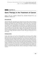
and function (Dempster 1965). The synchronized
movement of four joints must occur for elevation
to take place and for function to be achieved
Glenohumeral;
Scapulothoracic;
Sternoclavicular; and
Acromioclavicular (Fig. 4.1).
l
l
l
l
It is necessary for the manual therapist to have a
comprehensive understanding of functional biomechanics, movement phases, muscle imbalance, and injury
chapter 4
The shoulder
Acromioclavicular joint
Clavicle
Subacromial space
Sternoclavicular joint
Coracoid
process
Head of humerus
Ribs
Humerus
Glenohumeral joint
Scapulothoracic joint
Figure 4.1 Shoulder complex.
l
pathology, including trauma, microtrauma, or disease
processes that may interfere with any of the movement
mechanisms giving rise to pain and dysfunction:
‘Acupuncture may be more or less effective for different
pain types; therefore diagnosis of the predominant pain
mechanisms should always underpin treatment decisions
and prognosis.’ (Lundeberg & Ekholm 2001).
It is essential that relevant pain presentation
mechanisms are addressed with the help of manual
therapy, electrotherapy, and acupuncture intervention; once pain is under control, functional rehabilitation is facilitated (Lewis 2007). We cannot
expect patients to enter into a therapeutic alliance
without understanding how and why we are trying
to achieve pain modulation; similarly, we must ask
whether it is correct to treat the pain presentation
if we do not understand the mechanisms ourselves.
Assessment of these mechanisms is crucial for the
development of the hypothesis that will dictate
58
whether the manual or acupuncture intervention is
to be effective (Lundeberg & Ekholm 2001).
Consider some of the structures involved in
shoulder dysfunction:
Anatomical abnormalities such as congenital
acromial osteophyte variations;
Poor scapula control;
Shoulder instability whether through
hypermobility, trauma, or RCD; and
Poor glenohumeral, scapulothoracic, or shoulder
girdle mechanisms.
l
l
l
l
The shoulder is an inherently mobile complex,
with varying joint surfaces allowing the freedom of
movement, and vast mobility occurs at the expense
of stability (Donatelli 1997). Because there are over
20 muscles acting upon the joint to provide stability, the possibility of pain provoked from myofascial
structures should never be overlooked. Indeed, it is
recommended that this may well be the first line
Jennie Longbottom
of investigation since restoration of full movement
and full stability cannot occur if the muscle component is the pain-provoking structure (Ceccherelli
et al 2001). Restoration of full muscle balance cannot occur with the presence of a dysfunctional
motor end-plate, which prevents full muscle length.
A shortened, abnormal muscle length will result in
pain provoked by loading of the muscle, a characteristic presentation of myofascial pain involvement
and resulting muscle weakness.
Mechanisms of myofascial
pain
Mechanisms of myofascial pain occur as a result
of nociceptor stimulation in peripheral tissues via
mechanical structures associated with conditions
such as:
l
l
l
l
Impingement;
Entrapment;
Bony abnormalities; and
Mechanical pressure.
The alleviation of nociceptive or myofascial pain
must be directed towards the tissues causing this
pain. The source of dysfunctional tissues involved
can only be revealed by careful assessment and
elimination; similarly, the mechanism of acupuncture can only be effective if treatment targets the
structures involved. The presence of active myofascial pain can result in:
Increased acetylcholine at the motor end plate;
Shortened muscle fibres, ischaemic and/or
mechanical pressure on associated blood
vessels; or
Increased production of cytokines and substance
P within the area.
l
l
l
If any of the above is the cause, then the aim of
acupuncture intervention must be:
To deactivate the myofascial trigger point
(MTrPt);
To restore muscle length and relaxation;
To restore blood flow; and
To assist in the removal of neuropeptideaggravating chemicals.
l
l
l
l
Patients will clearly report a myofascial component to their pain if they describe:
chapter 4
Pain eased on off-loading;
Pain eased by touch, heat or ice, indicating an
ischaemic component;
Pain referred along a given muscle referral
pattern; and/or
Reproduction of pain on palpation of tender spot
or taut band.
l
l
l
l
If any of the above is involved in the pain presentation, then a full myofascial assessment with a subsequent TrPt deactivation of the myofascial component
is the first requirement for the needle application
whether in the rotator cuff and/or cervical muscles.
Rotator cuff disease
Rotator cuff disease (RCD) represents the most
common cause of modern shoulder pain and disability. Much of the clinical literature on RCD focuses
on subacromial impingement and supraspinatus
tendinopathy, although other patterns of lesions are
also recognized. Both extrinsic and intrinsic factors
to the cuff tendon are thought to be involved in the
pathogenesis, leading on to a spectrum of conditions ranging from subacromial bursitis to mechanical failure of the cuff tendon itself (Barying et al
2007). Careful history and examination followed
by pertinent investigation are essential to establish
the correct diagnosis. The main aim of treatment is
to improve symptoms and restore the function of
the affected shoulder.
There is no definitive evidence for the efficacy of
physical therapy interventions in the management of
RCD (Al-Shenqiti & Oldham 2005). Myofascial pain
syndromes are common conditions that result from
active TrPts (Sola et al. 1955). Myofascial pain has
two important components: motor dysfunction of
the muscle, and sensory abnormality characterized
by either local or referred pain (Whyte-Ferguson &
Gerwin 2005). There are a number of clinical diagnostic characteristics that may be presented during assessment that can be used to confirm and/or
exclude the presence of MTrPts. The reliability of
TrPt identification has been the subject of much
criticism (Bohr 1996), but the reliability of physical signs is essential to obtaining meaningful clinical
information (Al-Shenqiti & Oldham 2005; Nice et al
1992). These indicators include: spot tenderness,
pain recognition, and referred pain pattern.
Pain aggravated on muscle loading;
l
59
chapter 4
The shoulder
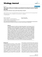
Patients demonstrating diagnostic rotator cuff tears
on magnetic resonance imaging (MRI) investigation
may respond favourably to the deactivation of TrPts,
but it is essential to understand both the anatomical presentation of pain and the muscles commonly
involved (Fig. 4.2). It is equally important to adopt
rigor and standardization of assessment in order to
eliminate the contributing myofascial pain component
of rotor cuff pain presentation. The TrPts must be
deactivated prior to shoulder stability exercise, postural and ergonomic retraining, and any future muscle
imbalance and scapula retraining. The most common
TrPts are found in the infraspinatus muscle, whilst
the subscapularis is least affected muscle in RCD
(Al Shenqiti & Oldham 2005).
Suprascapular nerve
Muscles involved
The supraspinatus muscle
A major function of the supraspinatus (Figs. 4.3
and 4.4) is to maintain balance amongst the other
rotator cuff muscles and therefore offer stability to
the joint. A common clinical symptom is ‘a catch’
of severe pain whilst the movement of elevation is
attempted, with a positive Neer or Hawkins sign,
or both. Pain is referred to the mid-deltoid region,
extending to the arm and forearm if severe, especially at the lateral epicondyle of the elbow. It may
often be mistaken for subdeltoid bursitis or later
1
4
2
Suprascapular nerve
Axillary nerve
3
Subscapular nerve
Muscle
Origin
Insertion
Action
1 Supraspinatus
Supraspinous fossa
of the scapula
Greater tuberosity
of the humerus
Abduction
2 Infraspinatus
Infraspinous fossa
of the scapula
Greater tuberosity
of the humerus
External rotation
3 Teres minor
Lateral border of
the scapula
Greater tuberosity
of the humerus
Abduction
4 Subscapularis
Subscapular fossa
of the scapula
Lesser tuberosity
of the humerus
Internal rotation
Figure 4.2 l The muscles of the rotator cuff.
60
Innervation
Suprascapular nerve (C4–C6)
Suprascapular nerve (C4–C6)
Axillary nerve (C5,C6)
Subscapular nerve (C5–C6)
Jennie Longbottom
chapter 4
A
B
Figure 4.3 l Supraspinatus pain referral pattern.
epicondylitis (Simons et al 1999), but in reality, the
supraspinatus muscle is in direct contact with the
bursa and, hence, we are presented with nociceptive sensitization. It is necessary to undertake TrPt
release and manage the patient with appropriate
stretching and muscle re-education. This muscle
should not be stretched if related RCD processes
are present (Fig. 4.5).
Medial to lateral needling
across supraspinatus fossa
Lateral to medial needling for
musculo-tendinous junction
The infraspinatus muscle
Infraspinatus injury is a common presentation characterized by deep, intense pain at the anterior edge
of the shoulder within the bicipital groove, radiating
down the radial aspect of arm and forearm, and it
Figure 4.4 l Direction of trigger point needling for
supraspinatus muscle.
61
chapter 4
The shoulder
Stretch excercise 1: Supraspinatus
Stretch excercise 2: Supraspinatus
Figure 4.5 l Stretching exercises for supraspinatus muscle.
is identified as a major source of arm pain (Figs. 4.6
and 4.7) (Travell 1952). The pain is associated with
abduction and medial rotation, and is most commonly a result of the acute overload associated with
whiplash injury. If joint restriction accompanies the
trigger point, then mobilization of the acromioclavicular and sternoclavicular articulations may be
required. If there is suspicion of rotator cuff damage, the infraspinatus should not be stretched, but
sustained myofascial contract–relax should be used
(Fig. 4.8).
Isolated posterior pain is usually not involved in
a single muscle pain presentation. However, if the
patient complains of dysaesthesia in the fourth and
fifth fingers, this may well be attributed to a single muscle element (Escobar & Ballesteros 1998).
This is usually the result of overload stresses, and
repetition of upward reaching and extension of the
shoulder, commonly associated with window cleaning. Its action is often coupled with the infraspinatus, and it is necessary to deactivate both muscles
before any muscle imbalance retraining.
62
The subscapularis muscle
Subscapularis trigger point pain referral presents
with posterior scapula and shoulder pain in the
form of a ‘watchstrap band’ of pain on the affected
arm (Fig. 4.9) (Zohn 1988). The subscapularis
medially rotates and adducts the arm and patients
initially have pain on medial rotation and abduction; for example, when throwing a ball or playing
golf. It can also manifest in patients following hemiplegia. Gradually abduction is restricted to below
45° and is often diagnosed as frozen shoulder. The
subscapularis is often overlooked in shoulder dysfunction (Donatelli 1997; Simons et al 1999). It
has a large and relatively inaccessible muscle mass
that serves to sensitize the other rotator cuff muscles, which often develop latent TrPts. This leads to
loss of rotation and pain patterns that may mimic
joint range of movement loss, especially in lateral
rotation. Management aims to identify the factors
involved, whilst pain management remains a priority because pain leads to inhibition of rotator cuff
Jennie Longbottom
chapter 4
A
B
Figure 4.6 l Infraspinatus muscle pain referral pattern.
and shoulder weakness (Donatelli 1997; Itoi et al
2007). The goals of the rehabilitation process
should include:
l
l
l
l
l
Reduction of TrPt dysfunction;
Return of normal shoulder movement;
Muscle imbalance re-education;
Re-establishment of movement synchrony; and
Progressive return to function.
What if inflammation is
present?
Figure 4.7 l Direction of needling for infraspinatus
muscle.
Although the evidence for the presentation of
inflammatory processes in RCD is poor, there are
some indications that these processes are present
63
chapter 4
The shoulder
Figure 4.8 l Stretching for Infraspinatus muscle.
Figure 4.9 l Subscapularis pain referral pattern.
in cases of acute injury. Acupuncture is thought
to have a modulating effect on both the systemic
and peripheral mechanisms implicated in neurogenic inflammation (Ceccherelli et al 2002). After
64
stimulation with acupuncture, calcitonin generelated peptide (CGRP), substance P, and betaendorphin are all released (Raud & Lundeberg 1991).
Substance P initiates mast cells and macrophages
Jennie Longbottom
to secrete inflammatory mediators; CGRP stimulates vasodilatation and thus induces peripheral
events, improving tissue function and pain relief.
If the acupuncture is too intense and too frequent,
it can result in overstimulation of substance P and
CGRP, causing a proinflammatory effect. Wellperformed acupuncture (obtaining de Qi) that
is low dose and frequently applied (two or three
times per week for 10 to 20 minutes) using points
distal to the injury site, at the segmental dorsal
horn or on the contralateral side (Bradnam 2002)
at the start of the injury process, could provoke a
sustained low-dose release of CGRP with resulting anti-inflammatory effects (Sandberg et al 2004)
and without activation of proinflammatory agents
(Raud & Lundeberg 1991). This offers a case for
promoting early acupuncture intervention at the
acute stage of the inflammatory process. How
often have we turned to acupuncture after three
or more treatments when pain modulation has not
been met? If inflammation and pain are preventing
manual intervention and active return to function,
then acupuncture should be considered within the
first few treatments to promote cortisol release,
increase blood flow, and facilitate manual intervention and rehabilitation (Tables 4.1 and 4.2). Distal
points, He-Sea points, and Qi Cleft points should
all be considered for the activation of Qi and blood
flow and for the promotion of homeostasis and
healing. Qi Cleft points are referred to in traditional Chinese medicine (TCM) for the treatment
of acute conditions where inflammatory agents are
causing pain, swelling, and limited movement. It is
common to choose Qi Cleft points that correspond
to the injury site and affected meridians.
Return of normal shoulder
movement
Normal movement may be restored by a variety of
therapeutic means, including: proprioceptive training; stretching; and a range of movement (ROM)
home exercise programme.
Muscle imbalance re-education
There are no significant differences between
patients who are given customized exercises and
chapter 4
Table 4.1 Suggested points for increased blood flow
Points Traditional Chinese
medicine
Western
SI3
Alleviates pain in arm
and face
Clears heat
Upper quadrant pain
LI4/5
Alleviates pain
Expels pathogens
Alleviates pain and
swelling in upper extremity
LI11
Arm pain
Stimulates Qi flow in LI
meridian
Increases blood flow in the
meridian
GB20
Removes pain and heat in
the area of neck and arm
Increases blood flow to
head and neck
LIV3
Alleviates pain and
induces relaxation
GV14
Moves Qi and alleviates
stiffness
Increases blood flow to
head and neck
BL40
He-Sea point of meridian
Increases blood flow in
meridian
BL60
Removes heat and
activates the channel
BL62
Activates channel and
alleviates pain
ST44
Alleviates pain and
swelling
ST36
Tonifies Qi
Nourishes blood
Alleviates pain and
swelling in lower extremity
those who are given standard exercises on measures of pain, intensity, functional status, shoulder
ROM, and strength (Wang 2004). The best exercise protocol for RCD or subacromial impingement syndrome (SIS) has not yet been established,
although the benefit of subjecting patients to a
reinforcement programme for the glenohumeral
and scapulothoracic muscles to improve joint stability, reduce pain, and regain strength is generally accepted. Rehabilitative programmes based on
either non-specific or specific exercises seem to
give favourable results but further research is necessary in order to verify which protocol is the most
effective. Stretching is often proposed to be associated with re-enforcement exercises to lengthen
shortened muscular and ligamentous structures,
and manual therapy has been demonstrated to be
a valid instrument for reducing in the impingement
syndrome. At the moment, muscular reinforcement
65
chapter 4
The shoulder
Table 4.2 Suggested points for enhancing acute
symptom resolution
Points
Area supplied
Suggested conditions
LU6
PC4
HT6
Palmer aspect of
wrist and forearm
Acute swelling and
inflammation to contralateral
wrist and forearm
Tendinosis of wrist flexors
Repetitive strain injury
Distal points for shoulder/
elbow injury
LI7
SJ7
SI6
Postero-ulnar aspect
of wrist and forearm
Acute swelling and injury to
contralateral wrist.
Extensor tendinosis
Repetitive strain injury
Distal points for shoulder/
elbow injury
ST34
GB36
SP8
LIV6
KID5
GB35
Acute knee injury,
swelling and stiffness
Sports injuries
All soft tissue injuries
Acute flare up
of inflammatory
processes
Contralateral knee if area
within point location swollen
May be used as distal points
if outside the area of swelling
BL63
BL59
KID8
KID9
Acute ankle or lower
limb injury
Shin splints
Contralateral ankle if area
within point location swollen
May be used as distal points
if outside the area of swelling
hip and knee pain
using distal, He Sea, or Qi Cleft points may well
provide the modulating effect to facilitate cortisol
release and blood flow, thus enhancing rehabilitation. However, if the pain nature is caused by myofascial structures, a variety of other factors must be
explored.
The unresolving shoulder
Patients are often referred to physiotherapy with
the catch all diagnosis of frozen shoulder (FS)
(Neviaser 1945), which is loosely defined as a
painful, stiff shoulder, varying in duration from
several weeks to several months. Pain, along with
diminished function, usually motivates the patient
to seek help (Cailliet 1981; DePalma 1983). It is
essential to eliminate any cervical or thoracic spine
involvement along with acromioclavicular, sternoclavicular, and scapulothoracic dysfunction, or first
rib involvement. Although there is little agreement
on treatment protocols, the goals for rehabilitation
remain clear, namely, pain relief and restoration of
function. Pain tends to be more long standing, radiating beyond the shoulder joint and involving sleep
disruption; therefore, the aim of acupuncture intervention should be directed towards activation of
descending inhibitory mechanisms involving:
l
l
is the recommended approach for an impingement
syndrome and instability problems because of the
dependence of the scapulohumeral girdle on the
surrounding muscle (Casonato 2003).
Re-establishment of movement
synchrony
Re-establishment of movement synchrony is necessary to restore the patient to previous performance and functional levels. In the case of the
athlete, the development of a throwing or activity
programme that pertains to the individual sport
is necessary, and with this, a progressive return to
function simulating sport activity in the resisted
exercise programme. If a build-up of inflammatory neuropeptides aggravating the peripheral
pain mechanisms is the cause, then acupuncture
66
l
l
Pain modulation;
Sleep enhancement;
Well being; and
Functional restoration.
Within TCM, FS is referred to as Jianning and
belongs to the yin group of disease patterns known
as Bi syndrome (Sun & Vangermeersch 1955), or
painful obstructive syndrome (Maciocia 1994). It is
mainly confined to superficial meridian or channel
blockage, stagnation or obstruction caused by an
attack of pathogenic factors such as cold (Han Bi),
dampness (Shi Bi), or wind (Feng Bi) or a combination of all three. External pathogens will only
invade the channel when defensive Qi (Wei Qi)
or internal organ Qi and/or blood is weak, and
cannot counteract the stronger pathogen factor.
Within the flow of Qi dynamics, joints are important areas of convergence of Qi and blood. Through
the joints, yin and yang Qi meet (Maciocia 1994),
Qi and blood enter and exit, and pathogenic factors
converge after penetrating the channels causing
Jennie Longbottom
an obstruction to the flow, resulting in stagnation.
The concept of Bi encompasses superficial disease
processes in connective tissue structures paralleled
in Western anatomical theory, such as tendons,
ligaments, muscles, and joints. Stagnation causes
pain and obstruction results in loss of normal joint
range.
Within the diagnosis of FS, all three pathogens
may be responsible, but cold and damp predominate.
Cold freezes and contracts, leading to the intense,
stabbing pain consistent with the first stages of FS.
Damp will produce the numbness, loss of movement, and deep ache characteristic of the second and
third stages of FS. The Large Intestine and Stomach
meridians are both superficial to and cross the shoulder joint, offering vulnerable areas to the invasion of
cold and damp (Needles 1982). Emotional trauma,
such as anger, grief, or shock, is classed as pathogenic
agents and may influence Qi and blood flow; Cyriax
(1978) refers to the shoulder as the most emotional
joint of the body.
The Large Intestine meridian is thought to be
important for shoulder function because of its close
proximity to the joint. Because Bi syndrome corresponds to a yin disease and the philosophy of TCM
is to maintain a balance between yin and yang,
stimulation of yang energy is desirable to address
this yin excess. In classical acupuncture, stimulation of a distal yang point on the channel will open
the channel (Maciocia 1994), eliminate stagnation,
and promote Qi and blood flow and help to expel
pathogenic factors. One channel can affect another
related channel on the same polarity with opposite potential (e.g. Large Intestine and Stomach on
the Yang Ming Stomach meridian intersects with
the Large Intestine meridian crossing the shoulder
and is known as Yang Ming in ancient Chinese literature). In order to facilitate descending inhibitory
processes in pain modulation and stimulate Qi flow
for restoration of function, traditional local and distal points may be used to facilitate these two objectives (Table 4.3).
Pain modulation may be enhanced by the use of
transcutaneous electrical nerve stimulation (TENS)
at home, or in the case of more prolonged dysfunction, electroacupuncture. Using a frequency
of 2 to 4 Hz at distal points may enhance opioid
and endorphin production, whilst a frequency of
80 to 100 Hz at local points may enhance production of leu-enkephalins and meta-enkephalins
for segmental pain gate modulation (Han &
Terenius 1982).
chapter 4
Table 4.3 Traditional local & distal points
Local points
Function (segmental dorsal horn
inhibition)
LI15/14
Stimulate Qi within the shoulder joint
TE14
Improve blood flow
GB21
Stiffness of shoulder
Extra points
JianQian (M-UE-48)
Stiffness of shoulder
Distal points
Function (descending inhibitory
(bilateral application) control)
LI4
Pain above the sternum
TE5
Pain in shoulder
ST38
Activates the Large Intestine and
Stomach channels to move Qi
GB34
Action on soft tissue structures
He-Sea point
Extra points
Yintang (M-HN-3)
Sleep enhancement
Amnian (N-HN-54)
Activates melatonin within pineal
gland
Chronic shoulder pain and
stiffness
There is no clear evidence to support one or a combination of treatments for the patient with FS;
reports of success in the literature are equally outnumbered by research to the contrary (Hunt 2005).
Frozen shoulder affects 2 to 5% of the general population (Kordell 2002). The exact mechanism of
the onset is unknown, but changes to the capsule
are thought to be similar to that of Dupuytrens contracture (Bunker et al 2000). The diagnosis is based
on detailed history and assessment with decreased
ROM (up to 50%) with:
Stiff end feel;
Negative instability tests; and
Normal X-ray to rule out bony injury or
calcification of the rotator cuff tendons
(Lundeberg 1969).
l
l
l
As stated, the primary aim of treatment should be
pain relief. It is likely to increase patient compliance
with his rehabilitation programme, and affect any painrelated muscle inhibition and abnormal biomechanics.
67
chapter 4
The shoulder
Case Study 1
Dan Franklin
A 39-year-old male lawyer presented with a 5-week
history of right shoulder pain; he had woken with the
pain one morning, but had not been able to attribute it to
any incident or activity. The subject rested his shoulder,
and when the pain did not abate after 3 weeks, sought
advice from his general practitioner, who prescribed
ibuprofen; there were no further investigations. The
medication helped somewhat, and three days before
presentation to physiotherapy, the subject decided
to test his shoulder with a social game of tennis; it
soon became obvious that he could not continue, and
therefore he rested again and made a physiotherapy
appointment for further input. The subject described
sharp and localized right shoulder pain over the lateral
aspect of the deltoid that occurred in conjunction with
arm movements, especially abduction or fast movements
in any direction. The subject was not able to lie on his
right side, but did not report any sleep disturbances;
there were no neural signs and there was no concurrent
neck pain. Previous medical history revealed that he had
twice dislocated his right shoulder while playing rugby;
the last episode had occurred over 15 years previously
and he had experienced no further problems until this
recent episode of pain.
Examination findings
On examination, the subject was found to have an
increased middle and upper thoracic kyphosis, and
a protracted cervical spine. Both scapulae were also
protracted, the right more so than the left, and his right
humeral head was observed to be sitting anteriorly
in the glenoid relative to the left side. Cervical spine
movements were slightly reduced in all directions from
what the present author would expect in a subject of
this age group, and his cervical paraspinal muscles were
a little tender on palpation, but neither reproduced his
shoulder pain. The subject’s thoracic spine was stiff
in extension, and posteroanterior mobilizations of the
spinous processes and costovertebral joints at thoracic
levels 1 to 4 (T1 to T4) and ribs 2 to 4 on the right
revealed hypomobility and reproduced local pain. The
subject’s right shoulder demonstrated flexion to 170°,
with slight pain at the end of ROM. Abduction revealed
a painful arc between 80° and 120° before resistance
and the return of pain at 170°. Poor scapulohumeral
rhythm was present in flexion and more obviously
in abduction. This included a reduced glenohumeral
contribution to flexion and abduction in mid-ranges,
and a compensatory increase in scapular elevation and
upward rotation. The hand-behind-back movement,
a combination of shoulder extension, adduction, and
internal rotation, was painful and restricted. Resisted
external rotation on the right was weak compared with
the left, but range was full and pain-free bilaterally.
Resisted isometric flexion, abduction, adduction,
68
extension, and internal rotation with the right shoulder in
neutral were of full strength and pain-free.
The subject underwent three tests indicative of
impingement, as described by Brukner and Khan (2002):
Neer test, the Hawkins-Kennedy test, and the ‘empty
can test’ (resisted abduction in 90° abduction, with 30°
horizontal flexion. Speed’s (biceps) test and O’Brien’s
superior labrum anteroposterior lesion test were both
negative. An apprehension test was painful, but not
positive. A diagnosis of SIS was made on the basis of the
above examination. MRI provides an accurate anatomical
image of the subacromial space and is the current gold
standard in the diagnosis of SIS (Silva et al 2008). Actual
shoulder diseases can be differentiated aetiopathologically
according to a primary and secondary impingement
syndrome. Narrowing of the subacromial space, which
is caused by an osseous shape variant, leads to primary
impingement. Secondary impingement develops when the
subacromial space is reduced by swollen tissue below the
osseous shoulder roof (Adamietz et al 2008). Factors that
needed to be addressed by the treatment included:
l Improvement of the glenoid alignment of the humeral
head;
l Strengthening of and coordination work for the rotator
cuff, especially the external shoulder rotators;
l Mobilization to restore extension range throughout
the upper thoracic spine and lower cervical spine;
l Improvement of right-sided scapulohumeral rhythm;
l Achieving pain relief as quickly as possible to ease
discomfort; and
l Reduction of antalgic biomechanics and promotion of
compliance with further treatment.
A visual analogue scale (VAS) for pain was completed
at the time of the initial assessment, and this, along
with flexion and abduction ROM measures, was used
throughout treatment to assess progress.
Treatment
The primary treatment goal for the first session was pain
relief. It was also felt that pain relief would be likely to
increase the subject’s compliance with his rehabilitation
programme, and affect any pain-related muscle inhibition
and abnormal biomechanics. The first treatment choice
to achieve this aim was acupuncture, given its accepted
analgesic effects. Treatment consisted of:
l Grade II anterior–posterior mobilization of the
glenohumeral joint;
l Grade III posterior–anterior mobilization of the T1 to
T4 spinal segments, right costovertebral joints, and
ribs 2 to 4;
l Soft-tissue massage to the upper trapezius, posterior
shoulder muscles, and pectoralis muscles of the right
side;
(Continued)
Jennie Longbottom
chapter 4
Case Study 1 (Continued)
Gentle horizontal or cross-flexion stretches for the
posterior of the right shoulder; and
l Taping to encourage better alignment of the right
humeral head in the glenoid fossa.
Three days later, the subject reported aggravation
of his symptoms, possibly as a result of the initial
examination and treatment. Distal acupuncture points
were chosen during this second session, because of
their strong analgesic potential. Manual techniques
had potentially aggravated the subject’s condition
previously and local acupuncture would also have the
potential to aggravate the injury (Lundeberg & Ekholm
2001). Because the subject demonstrated an acute to
subacute presentation, it was decided to needle the
contralateral shoulder, thereby triggering the pain-gate
mechanism at the correct spinal segment without risking
an inflammatory response in the affected shoulder. For
the local shoulder points, Large Intestine 15 (LI15) and
Triple Energizer 14 (TE14) were chosen because these
points are in the same dermatome as the shoulder and
are known to be effective in the treatment of shoulder
pain (Hecker et al 2001; Kleinhenz et al 1999; White &
Ernst 1999). Large Intestine 4 (LI4) was used bilaterally
because it is also a well-recognized point for shoulder
dysfunction (Hopwood et al 1997; He et al 2005; Hecker
et al 2001; Kleinhenz et al 1999), and is acknowledged to
be one of the strongest points in the body for analgesia
since it is a strong instigator of opioid release and
descending inhibition (Table 4.4) (Carlsson 2002; He et al
2005; Hecker et al 2001; Hopwood et al 1997; Kleinhenz
et al 1999).
The subject had improved objectively by the time
of the third treatment in terms of VAS score and ROM,
although he still felt subjectively worse than prior to the
first treatment. Two treatments per week were booked
since this may be more effective than less frequent
sessions (White & Ernst 1999), and because there
had been an objective improvement but no subjective
recovery, it was decided to change the distal point from
LI4 to Stomach 38 (ST38), one which is more specific
to shoulder injury (Hecker et al 2001; Hopwood et al
1997). Having increased the subject’s pain with the first
treatment using manual therapy a concern remained
about the potential irritability of the condition, and
therefore the present author was not prepared to risk
needling locally, preferring to continue with contralateral
needling of the shoulder and arm instead.
l
Fourth session
The subject felt much improved by the fourth session,
but he still had pain on sudden movements and any
abduction with an internal rotation component. With
his pain now significantly reduced, a change was made
to the treatment, which now included ipsilateral local
needling at LI15 and TE14, as well as LI11. Additional
manual therapy was used during this session.
Table 4.4 Case study 1: treatment choice justification
Day VAS
ROM pretreatment
Treatment
ROM post
treatment
1
37/100 Flexion
Mobilization T/S
170° R2, P1 GHJ, massage,
Abduction
taping
80° P1
170° P2
Flexion
170°
Abduction:
70-120° P1
170° P2
2
65/100 Flexion
60° P2
Abduction
60° P2
LI15, TE14,
LI11C
LI4B
Mobilization
GHJ
Pendular
exercises
Flexion
130° P1
Abduction
70° P1
3
65/100 Flexion
175°
Abduction
175°
LI15, TE14,
LI11C,
ST38B
Scapula stability
Retraction
exercises
Flexion
130°
Abduction
70°
6
43/100 Flexion
175°
Abduction
175°
LI15, TE14,
LI11C
St 38B
Scapula stability
LI15 TE14, LI11R
Flexion
170°
Abduction
170°
9
27/100 Flexion
175°
Abduction
175°
T/S, STM post
shoulder
Neer test
positive
Rotational
exercises
Flexion
175°
Abduction
175°
Notes: ROM, range of motion; C, contralateral; B, bilateral; R, right;
VAS, visual analogue scale; R2, end of ROM caused by resistance
rather than pain; P1, the point in a ROM where pain is felt for the
first time, but does not cause cessation of movement; P2, end of
ROM because of resistance (pain also present at this point, but
not restrictive of movement); mobilization T/S, posterior/anterior
mobilization centrally and unilaterally (right) of thoracic spine
segments T1–T4; mobilization GHJ, anteroposterior mobilization of
the glenohumeral joint; STM, soft-tissue massage.
Discussion
While it was disappointing that the first manual therapy
treatment appeared to aggravate the subject’s condition,
his improvement following the commencement
of acupuncture was encouraging. Unfortunately,
(Continued)
69
chapter 4
The shoulder
Case Study 1 (Continued)
acupuncture was not used during the initial treatment
session because he disclosed that he had not eaten
all day, and it is accepted that acupuncture can have
an effect on blood glucose levels (Carlsson 2002;
Chen et al 1994). Once he had experienced the acute
exacerbation of his condition after the first treatment
session, descending inhibition of pain might have been
enhanced by including Liver 3 (LIV3) with LI4 (the four
gates), which are known for their very powerful central
effects (Carlsson 2002). Small Intestine 3 (SI3), which
aids the release of cortisol, could also have been chosen
to reduce inflammation (Roth et al 1997; Toyama et al
1982). One point that will be included in this subject’s
future treatments is Gall Bladder (GB21) because it has
been incorporated in successful studies of acupuncture
in shoulder pain (He et al 2005).
Case Study 2
Kevin Hunt
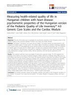
A 40-year-old female shop assistant presented with a
3-month history of pain in her right shoulder that had
become worse in 3 weeks prior to her assessment.
The pain pattern was distributed over the anterior and
posterior aspects of the shoulder, radiating to the
deltoid insertion in a band around the deltoid muscle
(Fig. 4.10).
The subject’s VAS was 40/100 at best and 90/100 at
worst with movement (A). Pain along the lateral border
of the scapula (B) was 90/100. Pain along the anterior
chest in line with the axilla (C) was rated 90/100 and
the patient was very anxious about whether this might
be associated with a more serious pathology. There
had been a previous injury to her right shoulder 2 years
before that had required 6 months of physiotherapy
for subacromial dysfunction. The subject had been
prescribed co-codamol (30/500 mg q.d.s) and X-ray
showed no bony changes. The treatment plan is shown
in Table 4.5.
Clinical reasoning
The deactivation of the subscapularis trigger point
and the consistent pain pattern from an active trigger
point at B resulted in a dramatic increase in ROM
(flexion increased from 84° to 140°; abduction from
82° to 140°). MRI findings to subscapularis tendons
in FS show that there are synovitis-like abnormalities
relating to the superior border (Mengiardi et al 2004;
Pearsall et al 2000). The improvement in pain and
ROM after deactivation of subscapularis trigger point
is consistent with those following surgical release
(Pearsall et al 2000). The subject reported improved
sleep, reduced anxiety levels, and resolution of pain
B. Subsequent treatments involved acupuncture to
improve the cumulative pain management. Acupuncture
stimulation releases endorphins and enkephalins such
as adrenocorticotrophic hormone into the blood stream,
providing further systemic pain inhibition as well as the
potential for sympathetic nervous system inhibition (Ma
2004). Other hormones and neurotransmitters, such as
serotonin, catecholamines, inorganic chemicals, and
70
A
C
B
C
Figure 4.10 l Case Study 2 pain presentation.
amino acids (e.g. glutamate and aminobutyric acid),
have been proposed as mediators of certain analgesic
effects of acupuncture, and research is ongoing into
their contributing effect. Recent functional MRI (fMRI)
trials have demonstrated an effect on limbic and
paralimbic structures involved in the modulation of pain
that is strongest when de Qi is elicited by peripheral
acupuncture stimulation (Brooks & Tracey 2005; Hui
1995; Tracey 2007).
As the treatment progressed, local tender and joint
acupuncture points were added especially Lung 1 (LU1);
however, this also corresponds to the TrPt presentation
of the pectoralis major muscle and a greater release of
pain and ROM might have been achieved by adding the
pectoralis TrPt, if positive (Fig. 4.11).
Conclusion
The subject reported an improvement of 70% in her
condition, ceased taking medication; slept through the
night again, and was able to perform normal activities of
daily living. The pain reduction achieved in the present
(Continued)
Jennie Longbottom
chapter 4
Case Study 2 (Continued)
Table 4.5 Treatment summary of patient with secondary frozen shoulder
Day
VAS
ROM pre-treatment
Treatment
ROM post treatment
0
A 90/100
B 90/100
C 90/100
Flexion: 84°
Abduction: 82°
Subscapularis Trigger point
deactivation
Flexion: 140°
Abduction: 104°
5
A 70/100
B 0/100
C 70/100
Flexion: 125°
Abduction: 100°
LI4 B
LI11, 14,15 R
LI4 B
Flexion: 125°
Abduction: 100°
13
A 80/100
B 0/100
C 70/100
Flexion: 120°
Abduction: 90°
SI9, 11. 12R
GB21 R
Flexion: 120°
Abduction: 90°
18
A 40/100
B 0/100
C 70/100
Flexion: 140°
Abduction: 110°
LI4 B
LU1 R
SI9, 11, 12 R
GB21 R
Flexion: 120°
Abduction: 90°
23
A 40/100
B 0/100
C 70/100
Flexion: 150°
Abduction: 110°
LI4 B
SI9, 11, 12 R
GB21 R
Flexion: 150°
Abduction: 110°
Notes: C, contralateral; B, bilateral; R, right; A, B, C: see Fig. 4.10.
Figure 4.11 l Pain presentation in the pectoralis major muscle.
small case report was consistent with that found in other
studies using acupuncture for pain modulation and
as a precursor to active rehabilitation (Lin et al 1994;
Tukmachi 1999), and as a postoperative pain modulator
following acromioplasty (Gilbertson et al 2003). More
frequent treatment involving an increased use of distal
and bilateral points could have enhanced the effect
reported in the present study (Guerra et al 2003).
71
chapter 4
The shoulder
References
Adamietz, B., Sauer, R., Keilholz, L.,
2008. Radiotherapy for shoulder
impingement. Strahlenther Onkol.
184 (5), 245–250.
Al-Shenqiti, A.M., Oldham, J.A., 2005.
Test–retest reliability of myofascial
trigger point detection in patients
with rotator cuff tendonitis. Clin.
Rehabil. 19 (5), 482–487.
Barying, T., Emery, R., Reilly, P., 2007.
Management of rotator cuff disease:
specific treatment for specific
disorders. Best practice and
research. Clin. Rheumatol. 21 (2),
279–294.
Bohr, T., 1996. Problems with
myofascial pain syndrome and
fibromyalgia. Neurology 46,
593–597.
Bradnam, L., 2002. Western
acupuncture point selection: a
scientific clinical reasoning model.
J. Acupunct. Assoc. Chartered
Psychother. 1, 21–29.
Brooks, J., Tracey, I., 2005. From
nociception to pain perception:
imaging the spinal and supraspinal
pathways. J. Anat. 207 (1), 19–33.
Brukner, P., Khan, K., 2002. Clinical
Sports Medicine, 2nd edn. McGrawHill, New York.
Bunker, T.D., Reilly, K.S., Hambleden,
D.L., 2000. Express of growth
factors, cytokines and matrix
metalloproteinases in frozen
shoulder. J. Bone Joint Surg. 82,
768–773.
Cailliet, R., 1981. Shoulder Pain, 2nd
edn. FA Davis, Philadelphia.
Carlsson, C., 2002. Acupuncture
mechanisms for clinically relevant
long-term effects- reconsideration
and a hypothesis. Acupunct. Med.
20 (2–3), 82–99.
Casonato, O., 2003. The role of
therapeutic exercise in the
conflicting and unstable shoulder.
Phys. Ther. Rev. 8 (10833196),
69–84.
Ceccherelli, F., Bordin, M., Gagliardi,
G., et al., 2001. Comparison
between superficial and deep
acupuncture in the treatment of
the shoulder’s myofascial pain: a
randomised and controlled study.
Acupunct. Electrother. Res. 26 (4),
229–238.
Ceccherelli, F., Gagliardi, G.,
Ruzzanti, L., et al., 2002.
Acupuncture modulation of
capsaicin-induced inflammation:
effect of intraperitoneal and local
administration of naloxone in rats.
72
A blinded controlled study. J. Altern.
Complement. Med. 8 (3), 341–349.
Chen, D., Gong, D., Zhai, Y., 1994.
Clinical and experimental studies
in treating Diabetes Mellitus with
acupuncture. J. Tradit. Chin. Med.
14 (3), 163–166.
Cyriax, J., 1978. Textbook of
Orthopaedic Medicine, 7th edn.
Bailliere Tindall, London.
DePalma, A., 1983. Surgery of
the Shoulder. JB Lippincott,
Philadelphia.
Dempster, W., 1965. Mechanism of
shoulder movement. Arch. Phys.
Med. Rehabil. 46A (49), 49–70.
Donatelli, R., 1997. Physical Therapy
of the Shoulder, 3rd edn. Churchill
Livingstone, Edinburgh.
Escobar, P., Ballesteros, J., 1998.
Teres minor: source of symptoms
resembling ulnar neuropathy or C8
radiculopathy. Am. J. Phys. Med.
Rehabil. 67 (3), 120–122.
Gilbertson, B., Wenner, K., Russell,
L.C., 2003. Acupuncture and
arthroscopic acromioplasty. J.
Orthop. Res. 21, 752–758.
Guerra, J., Bassas, E., Andres, M., et al.,
2003. Acupuncture for soft tissue
shoulder disorders: a series of 201
cases, including commentary by
White AR. Acupunct Med. 21 (1-2),
18–22.
Han, J., Terenius, L., 1982. The
neurochemical basis of acupuncture
analgesia. Annu. Revis. Pharmacol.
Toxicol. 22, 91–104.
He, D., Hostermark, A.T., Veirsted, K.B.,
et al., 2005. Effects of intensive
acupuncture on pain related social
and psychological variables for
women with chronic neck and
shoulder pain-an RCT with six
months and three year follow up.
Acupunct Med. 23 (2), 52–61.
Hecker, H.U., Steveling, A., Peuker, E.,
et al., 2001. Color Atlas of
Acupuncture: Body Points, Ear
Points, Trigger Points, 2nd edn.
Thieme Publishing, Stuttgart.
Hopwood, V., Lovesey, M., Makone, S.,
1997. Acupuncture and Related
Techniques in Physiotherapy.
Churchill Livingstone, Edinburgh.
Hui, H., 1995. A review of treatment
for diabetes by acupuncture during
the post forty years. J. Tradit. Chin.
Med. 15 (2), 145–154.
Hunt, K., 2005. Acupuncture in a
female patient with secondary
frozen shoulder. J. Acupunct. Assoc.
Chartered Psychother. (Jan.), 50–55.
Itoi, E., Managawa, H., Sato, T., et al.,
2007. Isokinetic strength after tears
of the suraspinatus tendon. J. Bone
Joint Surg. 79B (1), 77–82.
Kleinhenz, J., Streitberger, K., Windeler,
J., et al., 1999. Randomised clinical
trial comparing the effects of
acupuncture and a newly designed
placebo needle in rotator cuff
tendinitis. Pain 83 (2), 235–241.
Kordell, T., 2002. Frozen shoulder and
diabetes. Diabetes Forecast 55 (8),
60–64.
Lewis. J., 2007. Rotator cuff pathology.
Personal Communication, AACP
Conference.
Lewis, J., Tennent, T., 2007. How
effective are our diagnostic tests
for rotator cuff pathology?. In:
MacAuley, D., Best, T.M. (Eds.)
Evidence-Based Sports Medicine,
2nd edn. Blackwell, Oxford.
Lin, M.L., Huang, C.T., Lin, J.G.,
et al., 1994. A comparison
between the pain relief effect of
electroacupuncture, regional nerve
block and electroacupuncture
plus regional nerve block in frozen
shoulder. Acta Anaesthesiol. Sin.
32 (4), 237–242.
Lundeberg, B.J., 1969. The
frozen shoulder. Clinical and
radiographical observations. The
effect of manipulation under
general anaesthetic. Structure
and glycosaminoglycan content
of the joint capsule. Local bone
metabolism. Acta Ophthalmol.
Scand. Suppl. 19, S1–S59.
Lundeberg, T., Ekholm, J., 2001.
Pain—from periphery to brain.
J. Acupunct. Assoc. Chartered
Psychother. (Feb.), 13–19.
Maciocia, G., 1994. Painful obstruction
syndrome. In: The Practice of
Traditional Chinese Medicine:
The Treatment of Diseases with
Acupuncture and Chinese Herbs.
Churchill Livingstone, Edinburgh.
Mengiardi, B., Pfirmann, C.W., Gerber, C.,
et al., 2004. Frozen shoulder: MRI
arthrographic findings. Radiology
233 (2), 486–492.
Needles, J., 1982. Bi syndrome. J. Chin.
Med. 10 (1), 1–4.
Neviaser, J., 1945. Adhesive
capsulitis of the shoulder; study of
pathological findings in periarthritis
of the shoulder. J. Bone Joint Surg.
27, 211.
Nice, D., Riddle, D., Lamb, R.,
et al., 1992. Inter-tester reliability
of judgements of the presence of
Jennie Longbottom
trigger points in patients. Arch. Phys.
Med. Rehabil. 73, 893–898.
Pearsall, A.W., Holovacs, T.F., Speed,
K.P., 2000. The intra-articular
component of the Subscapularis
tendon: anatomic and histological
correlation in reference to surgical
release in patients with frozenshoulder syndrome. Arthroscopy 16,
236–242.
Raud, J., Lundeberg, T., 1991. Potent
anti-inflammatory action of
calcitonin gene-related peptide.
Biochem. Biophys. Res. Commun.
180, 1419–1435.
Roth, L., Maret-Maric, A., Adler, R.,
et al., 1997. Acupuncture points
have subjective (needling sensation)
and objective (serum cortisol
increase) specificity. Acupunct. Med.
15 (1), 2–5.
Sandberg, M., Lindberg, L., Gerdle,
B., 2004. Effects of acupuncture
on skin and muscle blood flow in
healthy subjects. Eur. J. Pain. 8 (2),
163–171.
Silva, L., Andréu, J., Muñoz, P.,
et al., 2008. Accuracy of physical
examination in subacromial
impingement syndrome.
Rheumatology 47 (5), 679–683.
Simons, D.G., Travell, J., Simons,
L.S., 1999. Myofascial Pain and
Dysfunction: The Trigger Point
Manual, 2nd edn. Lippincott
Williams & Wilkins, Baltimore.
Sola, A., Rodenberger, M., Gettys, B.,
1955. Incidence of hypersensitive
areas in posterior shoulder muscles.
Am. J. Phys. Med. 34, 585–590.
Sun, P., Vangermeersch, L., 1955.
Classification of Bi syndrome.
J. Chin. Med. 47, 8–14.
Toyama, P., Popell, C., Evans, J.,
et al., 1982. Beta endorphin and
cortisol measurements following
acupuncture and moxibustion.
J. Holistic Med. 4 (1), 58–67.
Tracey, I., 2007. Objectifying pain:
lessons from imaging somatic and
visceral pain in humans using FMRI.
Cephalalgia 24 (9), 722.
chapter 4
Travell, J., 1952. Ethyl chloride spray
for painful muscle spasm. Arch.
Phys. Med. Rehabil. 33, 291–298.
Tukmachi, E.S., 1999. Frozen shoulder:
a comparison of Western and
traditional Chinese approaches and
a clinical study of its acupuncture
treatment. Acupunct. Med. 9 (1),
9–21.
Wang, S.S., 2004. Comparison of
customised versus standard exercises
in rehabilitation of shoulder
disorders. Isokinet. Exerc. Sci. 12
(2), 135–141.
White, A.R., Ernst, E., 1999. A
systematic review of randomised
controlled acupuncture trails for
neck pain. Rheumatology 38 (2),
143–147.
Whyte-Ferguson, L., Gerwin, R., 2005.
Clinical Mastery in the Treatment of
Myofascial Pain. Lippincott Williams
and Wilkins, Baltimore.
Zohn, D., 1988. Musculoskeletal Pain:
Diagnosis and Physical Treatment,
2nd edn. Little, Brown, Boston.
73