Acute care handbook for physical therapists (fourth edition) chapter 4 pulmonary system
Bạn đang xem bản rút gọn của tài liệu. Xem và tải ngay bản đầy đủ của tài liệu tại đây (2.79 MB, 31 trang )
C H A PT E R
4
Pulmonary System
Paul E.H. Ricard
CHAPTER OUTLINE
CHAPTER OBJECTIVES
Body Structure and Function
Structure
Function
Evaluation
Patient History
Physical Examination
Inspection
Diagnostic Testing
Health Conditions
Obstructive Pulmonary
Conditions
Restrictive Pulmonary Conditions
Restrictive Extrapulmonary
Conditions
Chest Wall Restrictions
Management
Pharmacologic Agents
Thoracic Procedures
Physical Therapy Intervention
The objectives of this chapter are the following:
1. Provide a brief review of the structure and function of the pulmonary system
2. Give an overview of pulmonary evaluation, including physical examination and diagnostic testing
3. Describe pulmonary diseases and disorders, including clinical findings, medical-surgical management, and
physical therapy intervention
PREFERRED PRACTICE PATTERNS
The most relevant practice patterns for the diagnoses discussed in this chapter, based on the
American Physical Therapy Association’s Guide to Physical Therapist Practice, second edition,
are as follows:
• Impaired Aerobic Capacity/Endurance Associated with Deconditioning: 6B
• Impaired Ventilation, Respiration/Gas Exchange, and Aerobic Capacity/Endurance
Associated with Airway Clearance Dysfunction: 6C
• Impaired Ventilation and Respiration/Gas Exchange Associated with Ventilatory Pump
Dysfunction or Failure: 6E
• Impaired Ventilation and Respiration/Gas Exchange Associated with Respiratory
Failure: 6F
• Impaired Ventilation, Respiration/Gas Exchange, and Aerobic Capacity/Endurance
Associated with Respiratory Failure in the Neonate: 6G
Please refer to Appendix A for a complete list of the preferred practice patterns, as individual
patient conditions are highly variable and other practice patterns may be applicable.
To safely and effectively provide exercise, bronchopulmonary hygiene program(s), or both to
patients with pulmonary system dysfunction, physical therapists require an understanding of
the pulmonary system and of the principles of ventilation and gas exchange. Ventilation is
defined as gas (oxygen [O2] and carbon dioxide [CO2]) transport into and out of lungs, and
respiration is defined as gas exchange across the alveolar-capillary and capillary-tissue interfaces.
The term pulmonary primarily refers to the lungs, their airways, and their vascular system.1
Body Structure and Function
Structure
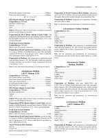
The primary organs and muscles of the pulmonary system are outlined in Tables 4-1 and
4-2, respectively. A schematic of the pulmonary system within the thorax is presented in
Figure 4-1.
Function
To accomplish ventilation and respiration, the pulmonary system is regulated by many neural,
chemical, and nonchemical mechanisms, which are discussed in the sections that follow.
Neural Control
Ventilation is regulated by two separate neural mechanisms: one controls automatic ventilation, and the other controls voluntary ventilation. The medullary respiratory center in the
53
54
CHAPTER 4 Pulmonary System
TABLE 4-1 Structure and Function of Primary Organs of the Pulmonary System
Structure
Description
Function
Nose
Paired mucosal-lined nasal cavities supported by bone
and cartilage
Passageway that connects nasal and oral cavities to
larynx, and oral cavity to esophagus
Subdivisions naso-, oro-, and laryngopharynx
Passageway that connects pharynx to trachea
Opening (glottis) covered by vocal folds or by the
epiglottis during swallowing
Flexible tube composed of C-shaped cartilaginous
rings connected posteriorly to the trachealis muscle
Divides into the left and right main stem bronchi at
the carina
Right and left main stem bronchi subdivide within
each lung into secondary bronchi, tertiary bronchi,
and bronchioles, which contain smooth muscle
Paired organs located within pleural cavities of the
thorax
The right lung has three lobes, and the left lung has
two lobes
Microscopic sacs at end of bronchial tree immediately
adjacent to pulmonary capillaries
Functional unit of the lung
Double-layered, continuous serous membrane lining
the inside of the thoracic cavity
Divided into parietal (outer) pleura and visceral (inner)
pleura
Conduit that filters, warms, and humidifies air entering
lungs
Conduit for air and food
Facilitates exposure of immune system to inhaled
antigens
Prevents food from entering the lower pulmonary tract
Voice production
Pharynx
Larynx
Trachea
Bronchial tree
Lungs
Alveoli
Pleurae
Cleans, warms, and moistens incoming air
Warms and moistens incoming air from trachea to alveoli
Smooth muscle constriction alters airflow
Contains air passageways distal to main stem bronchi,
alveoli, and respiratory membranes
Primary gas exchange site
Surfactant lines the alveoli to decrease surface tension and
prevent complete closure during exhalation
Produces lubricating fluid that allows smooth gliding of
lungs within the thorax
Potential space between parietal and visceral pleura
Data from Marieb E: Human anatomy and physiology, ed 3, Redwood City, Calif, 1995, Benjamin-Cummings; Moldover JR, Stein J, Krug PG: Cardiopulmonary
physiology. In Gonzalez EG, Myers SJ, Edelstein JE et al: Downey & Darling’s physiological basis of rehabilitation medicine, ed 3, Philadelphia, 2001,
Butterworth-Heinemann.
TABLE 4-2 Primary and Accessory Ventilatory Muscles with Associated Innervation
Primary inspiratory muscles
Accessory inspiratory muscles
Primary expiratory muscles
Accessory expiratory muscles
Pulmonary Muscles
Innervation
Diaphragm
External intercostals
Trapezius
Sternocleidomastoid
Scalenes
Pectorals
Serratus anterior
Latissimus dorsi
Rectus abdominis
External obliques
Internal obliques
Internal intercostals
Latissimus dorsi
Phrenic nerve (C3-C5)
Spinal segments T1-T9
Cervical nerve (C1-C4), spinal part of cranial nerve XI
Spinal part of cranial nerve XI
Cervical/brachial plexus branches (C3-C8, T1)
Medial/lateral pectoral nerve (C5-C8, T1)
Long thoracic nerve (C5-C7)
Thoracodorsal nerve (C5-C8)
Spinal segments T5-T12
Spinal segments T7-T12
Spinal segments T8-T12
Spinal segments T1-T9
Thoracodorsal nerve (C5-C8)
Data from Kendall FP, McCreary EK, editors: Muscles: testing and function, ed 3, Baltimore, 1983, Lippincott, Williams, and Wilkins; Rothstein JM, Roy SH, Wolf
SL: The rehabilitation specialist’s handbook, ed 2, Philadelphia, 1998, FA Davis; DeTurk WE, Cahalin LP: Cardiovascular and pulmonary physical therapy: an
evidence-based approach, New York, 2004, McGraw-Hill Medical Publishing Division.
brain stem, which is responsible for the rhythmicity of
breathing, controls automatic ventilation. The pneumotaxic
center, located in the pons, controls ventilation rate and
depth. The cerebral cortex, which sends impulses directly to
the motor neurons of ventilatory muscles, mediates voluntary
ventilation.3
Chemical Control
Arterial levels of CO2 (Pco2), hydrogen ions (H+), and O2 (Po2)
can modify the rate and depth of respiration. To maintain
homeostasis in the body, specialized chemoreceptors on the
carotid arteries and aortic arch (carotid and aortic bodies, respectively) respond to either a rise in Pco2 and H+ or a fall in Po2.
CHAPTER 4 Pulmonary System
55
A
B
C
FIGURE 4-1
A, Right lung positioned in the thorax. Bony landmarks
assist in identifying normal right lung configuration.
B, Anterior view of the lungs in the thorax in conjunction
with bony landmarks. Left upper lobe is divided into apical
and left lingula, which match the general position of the
right upper and middle lobes. C, Posterior view of the lungs
in conjunction with bony landmarks. (From Ellis E, Alison
J, editors: Key issues in cardiorespiratory physiotherapy,
Oxford, 1992, Butterworth-Heinemann, p 12.)
56
CHAPTER 4 Pulmonary System
Stimulation of these chemoreceptors results in transmission of
impulses to the respiratory centers to increase or decrease the
rate or depth, or both, of respiration. For example, an increase
in Pco2 would increase the ventilation rate to help increase the
amount of CO2 exhaled and ultimately lower the Pco2 levels in
arterial blood. The respiratory center found in the medulla
primarily responds to a rise in Pco2 and H+.4,5
Nonchemical Influences
Coughing, bronchoconstriction, and mucus secretion occur in
the lungs as protective reflexes to irritants such as smoke or
dust. Emotions, stressors, pain, and visceral reflexes from lung
tissue and other organ systems also can influence ventilation rate
and depth.
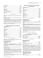
Mechanics of Ventilation
Ventilation occurs as a result of changes in the potential space
(volume) and subsequent pressures within the thoracic cavity
created by the muscles of ventilation. The largest primary
muscle of inhalation, the diaphragm, compresses the contents
of the abdominal cavity as it contracts and descends, increasing
the volume of the thoracic cavity.
CLINICAL TIP
The compression of the abdominal contents can be observed
with the protrusion of the abdomen. Clinicians use the term
“belly breathing” to facilitate diaphragmatic breathing.
The contraction of the intercostal muscles results in two
motions simultaneously: bucket and pump handle. The combined motions further increase the volume of the thorax. The
overall increase in the volume of the thoracic cavity creates a
negative intrathoracic pressure compared with outside the body.
As a result, air is pulled into the body and lungs via the pulmonary tree, stretching the lung parenchyma, to equalize the
pressures within the thorax with those outside the body.
Accessory muscles of inspiration, noted in Table 4-2, are
generally not active during quiet breathing. Although not the
primary actions of the individual muscles, their contractions can
increase the depth and rate of ventilation during progressive
activity by increasing the expansion of the thorax. Increased
expansion results in greater negative pressures being generated
and subsequent larger volumes of air entering the lungs.
CLINICAL TIP
In healthy lungs, depth of ventilation generally occurs before
increases in rate.
Although inhalation is an active process, exhalation is a
generally passive process. The muscles relax, causing a decrease
in the thoracic volume while the lungs deflate to their natural
resting state. The combined effects of these actions result in an
increase of intrathoracic pressure and flow of air out of the lungs.
Contraction of the primary and accessory muscles of exhalation,
found in Table 4-2, results in an increase in intrathoracic
pressure and a faster rate of decrease in thoracic size, which
forces air out of the lungs. These motions are outlined schematically in Figure 4-2.6,7
In persons with primary or secondary chronic pulmonary
health conditions, changes in tissue and mechanical properties
in the pulmonary system can result in accessory muscle use
being observed earlier in activity or may even be present at rest.
Determination of the impairment(s) resulting in the observed
activity limitation can help a clinician focus a plan of care. In
addition, clinicians should consider the reversibility, or the
degree to which the impairment can be improved, when determining a patient’s prognosis for improvement with physical
therapy. If reversing a patient’s ventilatory impairments is
unlikely, facilitation of accessory muscle use can be promoted
during functional activities and strengthening of these accessory
muscles (e.g., use of a four-wheeled rolling walker with a seat
and accompanying arm exercises).
CLINICAL TIP
Patients with advanced pulmonary conditions may automatically assume positions to optimize accessory muscle use, such
as forward leaning on their forearms (i.e., tripod posturing).
Gas Exchange. Once air has reached the alveolar spaces,
respiration or gas exchange can occur at the alveolar-capillary
membrane. Diffusion of gases through the membrane is affected
by the following:
• A concentration gradient in which gases will diffuse from
areas of high concentration to areas of low concentration:
Alveolar O2 = 100 mm Hg → Capillary O2 = 40 mm Hg
• Surface area, or the total amount of alveolar-capillary interface available for gas exchange (e.g., the breakdown of alveolar membranes that occurs in emphysema will reduce the
amount of surface area available for gas exchange)
• The thickness of the barrier (membrane) between the two
areas involved (e.g., retained secretions in the alveolar spaces
will impede gas exchange through the membrane)
Ventilation and Perfusion Ratio. Gas exchange is optimized when the ratio of air flow (ventilation V) to blood flow
(perfusion Q ) approaches a 1 : 1 relationship. However, the
actual V/Q ratio is 0.8 because alveolar ventilation is approximately equal to 4 L per minute and pulmonary blood flow is
approximately equal to 5 L per minute.2,8,9
Gravity, body position, and cardiopulmonary dysfunction
can influence this ratio. Ventilation is optimized in areas of least
resistance. For example, when a person is in a sitting position,
the upper lobes initially receive more ventilation than the lower
lobes; however, the lower lobes have the largest net change in
ventilation.
Perfusion is greatest in gravity-dependent areas. For example,
when a person is in a sitting position, perfusion is the greatest
at the base of the lungs; when a person is in a left side-lying
position, the left lung receives the most blood.
A V/Q mismatch (inequality in the relationship between
ventilation and perfusion) can occur in certain situations. Two
CHAPTER 4 Pulmonary System
57
FIGURE 4-2
Respiratory mechanics (bucket and pump handle motions). (From Snell RS, editor: Clinical anatomy by regions,
ed 9, Baltimore, 2012, Lippincott, Williams & Wilkins.)
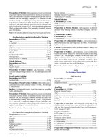
terms associated with V/Q mismatch are dead space and shunt.
Dead space occurs when ventilation is in excess of perfusion, as
with a pulmonary embolus. A shunt occurs when perfusion is
in excess of ventilation, as in alveolar collapse from secretion
retention. These conditions are shown in Figure 4-3.
Gas Transport. O2 is transported away from the lungs to
the tissues in two forms: dissolved in plasma (Po2) or chemically
bound to hemoglobin on a red blood cell (oxyhemoglobin). As
a by-product of cellular metabolism, CO2 is transported away
from the tissues to the lungs in three forms: dissolved in plasma
(Pco2), chemically bound to hemoglobin (carboxyhemoglobin),
and as bicarbonate.
Approximately 97% of O2 transported from the lungs is
carried in chemical combination with hemoglobin. The majority of CO2 transport, 93%, occurs in the combined forms of
carbaminohemoglobin and bicarbonate. A smaller percentage,
3% of O2 and 7% of CO2, is transported in dissolved forms.10
Dissolved O2 and CO2 exert a partial pressure within the plasma
and can be measured by sampling arterial, venous, or mixed
venous blood.11 See the Arterial Blood Gas section for further
description of this process.
Evaluation
Pulmonary evaluation is composed of patient history, physical
examination, and interpretation of diagnostic test results.
Patient History
In addition to the general chart review presented in Chapter 2,
other relevant information regarding pulmonary dysfunction
that should be ascertained from the chart review or patient
interview is listed as follows11-13:
58
CHAPTER 4 Pulmonary System
Bronchiole
Alveoli
Capillary
A
B
C
FIGURE 4-3
Ventilation and perfusion mismatch. A, Normal alveolar ventilation. B, Capillary shunt. C, Alveolar dead
space.
• History of smoking, including packs per day or pack years
(packs per day × number of years smoked) and the amount
of time that smoking has been discontinued (if applicable)
• Presence, history, and amount of O2 therapy at rest, with
activity and at night
• Exposure to environmental or occupational toxins (e.g.,
asbestos)
• History of pneumonia, thoracic procedures, or surgery
• History of assisted ventilation or intubation with mechanical
ventilation
• History or current reports of dyspnea either at rest or with
exertion. Dyspnea is the subjective complaint of difficulty
with respiration, also known as shortness of breath. A visual
analog scale or ratio scale (Modified Borg scale) can be used
to obtain a measurement of dyspnea. The American Thoracic
Society Dyspnea Scale can be found in Table 4-3. Note: The
abbreviation DOE represents “dyspnea on exertion”
• Level of activity before admittance
• History of baseline sputum production, including color (e.g.,
yellow, green), consistency (e.g., thick, thin), and amount.
Familiar or broad terms can be applied as units of measure
for sputum (e.g., quarter-sized, tablespoon, or copious)
• Sleeping position and number of pillows used
CLINICAL TIP
Dyspnea also may be measured by counting the number of
words a person can speak per breath. For example, a patient
with one- to two-word dyspnea is noticeably more dyspneic
than a person who can speak a full sentence per breath. Measurement of dyspnea can be used in goal writing (e.g., “Patient
will ascend/descend 10 stairs with one rail with reported
dyspnea < 2/10.”).
Physical Examination
The physical examination of the pulmonary system consists of
inspection, auscultation, palpation, mediate percussion, and
cough examination. Suggested guidelines for physical therapy
intervention(s) that are based on examination findings and diagnostic test results are found at the end of this chapter.
TABLE 4-3 American Thoracic Society Dyspnea Scale
Grade
Degree
0
None
1
Slight
2
Moderate
3
Severe
4
Very severe
Not troubled with breathlessness
except with strenuous exercise
Troubled by shortness of breath
when hurrying on the level or
walking up a slight hill
Walks slower than people of the
same age on the level because
of breathlessness, or has to
stop for breath when walking
at own pace on the level
Stops for breath after walking
about 100 yards or after a few
minutes on the level
Too breathless to leave the house
or breathless when dressing or
undressing
From Brooks SM: Surveillance for respiratory hazards, ATS News 8:12-16,
1982.
Inspection
A wealth of information can be gathered by simple observation
of the patient at rest and with activity. Physical observation
should proceed in a systematic fashion and include the
following:
• General appearance and level of alertness
• Ease of phonation
• Skin color
• Posture and chest shape
• Ventilatory or breathing pattern
• Presence of digital clubbing
• Presence of supplemental O2 and other medical equipment
(refer to Chapter 18)
• Presence and location of surgical incisions
Observation of Breathing Patterns
Breathing patterns vary among individuals and may be influenced by pain, emotion, body temperature, sleep, body position,
activity level, and the presence of pulmonary, cardiac, metabolic, or nervous system disease (Table 4-4). The optimal time,
clinically, to examine a patient’s breathing pattern is when he
CHAPTER 4 Pulmonary System
59
TABLE 4-4 Description of Breathing Patterns and Their Associated Conditions
Breathing Pattern
Description
Associated Conditions
Apnea
Lack of airflow to the lungs for >15 seconds
Biot’s respirations
Constant increased rate and depth of respiration
followed by periods of apnea of varying lengths
Ventilation rate <12 breaths per minute
Airway obstruction, cardiopulmonary arrest, alterations
of the respiratory center, narcotic overdose
Elevated intracranial pressure, meningitis
Bradypnea
Cheyne-Stokes
respirations
Hyperpnea
Hyperventilation
Hypoventilation
Kussmaul respirations
Orthopnea
Paradoxic ventilation
Sighing respirations
Tachypnea
Hoover’s sign*
Increasing depth of ventilation followed by a period
of apnea
Increased depth of ventilation
Increased rate and depth of ventilation resulting in
decreased Pco2
Decreased rate and depth of ventilation resulting in
increased Pco2
Increased regular rate and depth of ventilation
Dyspnea that occurs in a flat supine position. Relief
occurs with more upright sitting or standing
Inward abdominal or chest wall movement with
inspiration and outward movement with
expiration
The presence of a sigh >2-3 times per minute
Ventilation rate >20 breaths per minute
The inward motion of the lower rib cage during
inhalation
Use of sedatives, narcotics, or alcohol; neurologic or
metabolic disorders; excessive fatigue
Elevated intracranial pressure, CHF, narcotic overdose
Activity, pulmonary infections, CHF
Anxiety, nervousness, metabolic acidosis
Sedation or somnolence, neurologic depression of
respiratory centers, overmedication, metabolic
alkalosis
Diabetic ketoacidosis, renal failure
Chronic lung disease, CHF
Diaphragm paralysis, ventilation muscle fatigue, chest
wall trauma
Angina, anxiety, dyspnea
Acute respiratory distress, fever, pain, emotions, anemia
Flattened diaphragm often related to decompensated or
irreversible hyperinflation of the lungs
Data from Kersten LD: Comprehensive respiratory nursing: a decision-making approach, Philadelphia, 1989, Saunders; DesJardins T, Burton GG: Clinical manifestations and assessment of respiratory disease, ed 3, St Louis, 1995, Mosby;
*Hoover’s sign has been reported to have a sensitivity of 58% and specificity of 86% for detection of airway obstruction. Hoover’s sign is associated with a patient’s
body mass index, severity of dyspnea, and frequency of exacerbations and is seen in up to 70% of patients with severe obstruction.†
†Data from Johnson CR, Krishnaswamy N, Krishnaswamy G: The Hoover’s sign of pulmonary disease: molecular basis and clinical relevance, Clin Mol Allergy
6:8, 2008.
CHF, Congestive heart failure; Pco2, partial pressure of carbon dioxide.
or she is unaware of the inspection because knowledge of the
physical examination can influence the patient’s respiratory
pattern.
Observation of breathing pattern should include an assessment of rate (12 to 20 breaths per minute is normal), depth,
ratio of inspiration to expiration (one to two is normal), sequence
of chest wall movement during inspiration and expiration,
comfort, presence accessory muscle use, and symmetry.
CLINICAL TIP
If possible, examine a patient’s breathing pattern when he or
she is unaware of the inspection because knowledge of the
physical examination can influence the patient’s respiratory
pattern. Objective observations of ventilation rate may not
always be consistent with a patient’s subjective complaints of
dyspnea. For example, a patient may complain of shortness of
breath but have a ventilation rate within normal limits. Therefore the patient’s subjective complaints, rather than the objective observations, may be a more accurate measure of treatment
intensity.
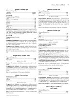
Auscultation
Auscultation is the process of listening to the sounds of air
passing through the tracheobronchial tree and alveolar spaces.
The sounds of airflow normally dissipate from proximal to distal
airways, making the sounds less audible in the periphery than
the central airways. Alterations in airflow and ventilation effort
result in distinctive sounds within the thoracic cavity that may
indicate pulmonary disease or dysfunction.
Auscultation proceeds in a systematic, side-to-side, and
cephalocaudal fashion. Breath sounds on the left and right sides
are compared in the anterior, lateral, and posterior segments of
the chest wall, as shown in Figure 4-4. The diaphragm (flat side)
of the stethoscope should be used for auscultation. The patient
should be seated or lying comfortably in a position that allows
access to all lung fields. Full inspirations and expirations are
performed by the patient through the mouth, as the clinician
listens to the entire cycle of respiration before moving the
stethoscope to another lung segment.
All of the following ensure accurate auscultation:
• Make sure stethoscope earpieces are pointing up and inward
(toward your patient) before placing in the ears.
60
CHAPTER 4 Pulmonary System
A
B
C
FIGURE 4-4
Landmarks for lung auscultation on (A) anterior, (B) posterior, and (C) lateral aspects of the chest wall.
(Courtesy Peter P. Wu.)
• Long stethoscope tubing may dampen sound transmission.
Length of tubing should be approximately 30 cm (12 in) to
55 cm (21 to 22 in).12
• Always check proper function of the stethoscope before auscultating by listening to finger tapping on the diaphragm
while the earpieces are in place.
• Apply the stethoscope diaphragm firmly against the skin so
that it lays flat.
• Observe chest wall expansion and breathing pattern while
auscultating to help confirm palpatory findings of breathing
pattern (e.g., sequence and symmetry). For example,
decreased chest wall motion palpated earlier in the left lower
lung field may present with decreased breath sounds in that
same area.
Breath sounds may be normal or abnormal (adventitious or
added) breath sounds; all breath sounds should be documented
according to the location and the phase of respiration (i.e.,
inspiration, expiration, or both) and in comparison with the
opposite lung. Several strategies can be used to reduce the
chance of false-positive adventitious breath sound findings,
including the following:
• Ensure full, deep inspirations (decreased effort can be misinterpreted as decreased breath sounds).
• Be aware of the stethoscope tubing’s touching other objects
(especially ventilator tubing) or chest hair.
• Periodically lift the stethoscope off the chest wall to help
differentiate extraneous sounds (e.g., chest or nasogastric
tubes, patient snoring) that may appear to originate from the
thorax.
To maximize patient comfort, allow periodic rest periods
between deep breaths to prevent hyperventilation and
dizziness.
Normal Breath Sounds. Clinically, tracheal or bronchial
and vesicular breath sounds generally are documented as
“normal” or “clear” breath sounds; however, the use of tracheal
or vesicular breath sounds is more accurate.
Tracheal, Bronchial, or Bronchovesicular Sounds. Normal tracheal or bronchial breath sounds are loud tubular sounds heard
over the proximal airways, such as the trachea and main stem
bronchi. A pause is heard between inspiration and expiration;
the expiratory phase is longer than the inspiratory phase.
Normal bronchovesicular sounds are similar to bronchial breath
sounds; however, no pause occurs between inspiration and
expiration.11,12
Vesicular Sounds. Vesicular sounds are soft rustling sounds
heard over the more distal airways and lung parenchyma. Inspiration is longer and more pronounced than expiration because
a decrease in airway lumen during expiration limits transmission of airflow sounds.11,12
Note: In most reference books, a distinction between normal
bronchial and bronchovesicular sounds is made to help with
standardization of terminology. Often, however, this distinction
is not used in the clinical setting.
CLINICAL TIP
The abbreviation CTA stands for “clear to auscultation.”
Abnormal Breath Sounds. Breath sounds are abnormal if
they are heard outside their usual location in the chest or if they
are qualitatively different from normal breath sounds.14 Despite
efforts to make the terminology of breath sounds more
CHAPTER 4 Pulmonary System
TABLE 4-5 Possible Sources of Abnormal
Breath Sounds
Sound
Possible Etiology
Bronchial (abnormal if heard
in areas where vesicular
sounds should be present)
Decreased or diminished (less
audible)
Absent
Fluid or secretion consolidation
(airlessness) that could
occur with pneumonia
Hypoventilation, severe
congestion, or emphysema
Pneumothorax or lung collapse
consistent, terminology may still vary from clinician to clinician
and facility to facility. Always clarify the intended meaning of
the breath sound description if your findings differ significantly
from what has been documented or reported. Abnormal breath
sounds with possible sources are outlined in Table 4-5.
Adventitious Breath Sounds. Adventitious breath sounds
occur from alterations or turbulence in airflow through the
tracheobronchial tree and lung parenchyma. These sounds can
be divided into continuous (wheezes and rhonchi) or discontinuous (crackles) sounds.12,14
The American Thoracic Society and American College of
Chest Physicians have discouraged use of the term rhonchi, recommending instead that the term wheezes be used for all continuous adventitious breath sounds.15 Many academic institutions
and hospitals continue to teach and practice use of the term
rhonchi; therefore it is mentioned in this section.
Continuous Sounds
Wheeze. Wheezes occur most commonly with airway obstruc-
tion from bronchoconstriction or retained secretions and commonly are heard on expiration. Wheezes also may be present
during inspiration if the obstruction is significant enough.
Wheezes can be high pitched (usually from bronchospasm or
constriction, as in asthma) or low pitched (usually from secretions, as in pneumonia).
STRIDOR. Stridor is an extremely high-pitched wheeze that
occurs with significant upper airway obstruction and is present
during inspiration and expiration. The presence of stridor indicates a medical emergency. Stridor is also audible without a
stethoscope.
CLINICAL TIP
Acute onset of stridor during an intervention session warrants
immediate notification of the nursing and medical staff.
Rhonchi. Low-pitched or “snoring” sounds that are continu-
ous characterize rhonchi. These sounds generally are associated
with large airway obstruction, typically from secretions lining
the airways.
Discontinuous Sounds
Crackles. Crackles are bubbling or popping sounds that represent the presence of fluid or secretions, or the sudden opening
of closed airways. Crackles that result from fluid (pulmonary
edema) or secretions (pneumonia) are described as “wet” or
61
“coarse,” whereas crackles that occur from the sudden opening
of closed airways (atelectasis) are referred to as “dry” or “fine.”
CLINICAL TIP
Wet crackles also can be referred to as rales, but the American
Thoracic Society–American College of Chest Physicians has
moved to eliminate this terminology for purposes of
standardization.15
Extrapulmonary Sounds. These sounds are generated from
dysfunction outside of the lung tissue. The most common sound
is the pleural friction rub. This sound is heard as a loud grating
sound, generally throughout both phases of respiration, and
almost always is associated with pleuritis (inflamed pleurae
rubbing on one another).12,14 The presence of a chest tube
inserted into the pleural space also may cause a sound similar
to a pleural rub.
CLINICAL TIP
Asking the patient to hold his or her breath can help differentiate a true pleural friction rub from a sound artifact or a pericardial friction rub.
Voice Sounds. Normal phonation is audible during auscultation, with the intensity and clarity of speech also dissipating
from proximal to distal airways. Voice sounds that are more or
less pronounced in distal lung regions, where vesicular breath
sounds should occur, may indicate areas of consolidation or
hyperinflation, respectively. The same areas of auscultation
should be used when assessing voice sounds. The following
three types of voice sound tests can be used to help confirm
breath sound findings:
1. Whispered pectoriloquy. The patient whispers “one, two,
three.” The test is positive for consolidation if phrases are
clearly audible in distal lung fields. This test is positive
for hyperinflation if the phrases are less audible in distal
lung fields.
2. Bronchophony. The patient repeats the phrase “ninety-nine.”
The results are similar to whispered pectoriloquy.
3. Egophony. The patient repeats the letter e. If the auscultation
in the distal lung fields sound like a, then fluid in the air
spaces or lung parenchyma is suspected.
Palpation
The third component of the physical examination is palpation
of the chest wall, which is performed in a cephalocaudal direction. Figure 4-5 demonstrates hand placement for chest wall
palpation of the upper, middle, and lower lung fields. Palpation
is performed to examine the following:
• Presence of fremitus (a vibration caused by the presence of
secretions or voice production, which is felt through the
chest wall) during respirations11
62
CHAPTER 4 Pulmonary System
A
B
C
FIGURE 4-5
Palpation of (A) upper, (B) middle, and (C) lower chest wall motion.
(Courtesy Peter P. Wu.)
• Presence, location, and reproducibility of pain, tenderness,
or both
• Skin temperature
• Presence of bony abnormalities, rib fractures, or both
• Chest expansion and symmetry
• Presence of subcutaneous emphysema (palpated as bubbles
popping under the skin from the presence of air in the subcutaneous tissue). This finding is abnormal and represents
air that has escaped or is escaping from the lungs. Subcutaneous emphysema can occur from a pneumothorax (PTX), a
complication from central line placement, or after thoracic
surgery1
CLINICAL TIP
To decrease patient fatigue while palpating each of the chest
wall segments for motion, all of the items listed above can be
examined simultaneously.
Chest Wall and Abdominal Excursion. Direct measurement of chest wall expansion can be used for objective data
FIGURE 4-6
Demonstration of mediate percussion technique. (From Hillegass EA, Sadowsky HS: Essentials of cardiopulmonary physical therapy, ed 2, Philadelphia, 2001, Saunders.)
collection, intervention, or goal setting. Begin by placing a tape
measure snugly around the circumference of the patient’s chest
wall at three levels:
1. Angle of Louis
2. Xyphoid process
3. Umbilicus
Measure the change in circumference in each of these areas
with normal breathing and then deep breathing. The resulting
values can be used to describe breathing patterns or identify
ventilation impairments. Changes in these values after an intervention may indicate improvements in breathing patterns and
can be used to evaluate treatment efficacy. Normal changes in
breathing patterns exist in supine, sitting, and standing.
CLINICAL TIP
By placing your thumb tips together on the spinous processes
or xyphoid process, you can estimate the distance of separation
between your thumb tips to qualitatively measure chest wall
motion.
Mediate Percussion. Mediate percussion can evaluate
tissue densities within the thoracic cage and indirectly measure
diaphragmatic excursion during respirations. Mediate percussion also can be used to confirm other findings in the physical
examination. The procedure is shown in Figure 4-6 and is performed by placing the palmar surface of the index finger, middle
finger, or both from one hand flatly against the surface of the
chest wall within the intercostal spaces. The tip(s) of the other
index finger, middle finger, or both then strike(s) the distal third
of the fingers resting against the chest wall. The clinician proceeds from side to side in a cephalocaudal fashion, within the
intercostal spaces, for anterior and posterior aspects of the chest
CHAPTER 4 Pulmonary System
CLINICAL TIP
Do not confuse this examination technique with the intervention technique of percussion, which is used to help mobilize
bronchopulmonary secretions in patients.
Cough Examination. An essential component of bronchopulmonary hygiene is cough effectiveness. The cough mechanism can be divided into four phases: (1) full inspiration, (2)
closure of the glottis with an increase of intrathoracic pressure,
(3) abdominal contraction, and (4) rapid expulsion of air. The
inability to perform one or more portions of the cough mechanism can lead to pulmonary secretion clearance. Cough examination includes the following components11,12:
• Effectiveness (ability to clear secretions)
• Control (ability to start and stop coughs)
• Quality (wet, dry, bronchospastic)
• Frequency (how often during the day and night cough
occurs)
• Sputum production (color, quantity, odor, and consistency)
The effectiveness of a patient’s cough can be examined
directly by simply asking the patient to cough or indirectly by
observing the above components when the patient coughs
spontaneously.
Hemoptysis. Hemoptysis, the expectoration of blood
during coughing, may occur for many reasons. Hemoptysis is
usually benign postoperatively if it is not sustained with successive coughs. The therapist should note whether the blood is
dark red or brownish in color (old blood) or bright red (new or
frank blood). The presence of new blood in sputum should be
documented and the nurse or physician notified.
Patients with cystic fibrosis may have periodic episodes of
hemoptysis with streaking or larger quantities of new blood.
During these episodes airway clearance techniques (ACT) may
need to be modified. Current recommendations for patients who
have scant hemoptysis (<5 ml) are to continue with all ACT,
and those with massive hemoptysis should discontinue all ACT.
For persons with mild to moderate hemoptysis (≥5 ml), no clear
recommendations exist for continuing or discontinuing ACT.
However, expert consensus is that autogenic drainage or active
cycle of breathing techniques are least likely to exacerbate
hemoptysis while maintaining the needs of assisted sputum
clearance.16
Diagnostic Testing
Oximetry
Pulse oximetry is a noninvasive method of determining arterial
oxyhemoglobin saturation (Sao2) through the measurement of
the saturation of peripheral oxygen (Spo2). It also indirectly
examines the partial pressure of O2. Finger or ear sensors generally are applied to a patient on a continuous or intermittent
basis. O2 saturation readings can be affected by poor circulation
(cool digits), movement of sensor cord, cleanliness of the sensors,
nail polish, intense light, increased levels of carboxyhemoglobin
(Hbco2), jaundice, skin pigmentation, shock states, cardiac dysrhythmias (e.g., atrial fibrillation), and severe hypoxia.17,18
CLINICAL TIP
To ensure accurate O2 saturation readings, (1) check for proper
waveform or pulsations, which indicate proper signal reception,
and (2) compare pulse readings on an O2 saturation monitor
with the patient’s peripheral pulses or electrocardiograph readings (if available).
Oxyhemoglobin saturation is an indication of pulmonary
reserve and is dependent on the Po2 level in the blood. Figure
4-7 demonstrates the direct relationship of oxyhemoglobin
saturation and partial pressures of O2. As shown on the steep
portion of the curve, small changes in Po2 levels below
100
SaO2 (O2 saturation %)
wall. Mediate percussion is a difficult skill and is performed
most proficiently by experienced clinicians; mediate percussion
also can be performed over the abdominal cavity to assess tissue
densities, which is described further in Chapter 8.
Sounds produced from mediate percussion can be characterized as one of the following:
• Resonant (over normal lung tissue)
• Hyperresonant (over emphysematous lungs or PTX)
• Tympanic (over gas bubbles in abdomen)
• Dull (from increased tissue density or lungs with
decreased air)
• Flat (extreme dullness over very dense tissues, such as the
thigh muscles)12
To evaluate diaphragmatic excursion with mediate percussion, the clinician first delineates the resting position of the
diaphragm by percussing down the posterior aspect of one side
of the chest wall until a change from resonant to dull (flat)
sounds occurs. The clinician then asks the patient to inspire
deeply and repeats the process, noting the difference in landmarks when sound changes occur. The difference is the amount
of diaphragmatic excursion. The other also is examined, and a
comparison then can be made of the hemidiaphragms.
63
80
60
40
20
0
0
20
40
60
80
100 120
PaO2 (O2 partial pressure)
FIGURE 4-7
The oxyhemoglobin dissociation curve. (Courtesy Marybeth Cuaycong.)
64
CHAPTER 4 Pulmonary System
TABLE 4-6 Relationship Between Oxygen Saturation,
the Partial Pressure of Oxygen, and the
Signs and Symptoms of Hypoxemia
Oxyhemoglobin
Saturation
(SaO2) (%)
Oxygen Partial
Pressure (PaO2)
(mm Hg)
Signs and
Symptoms of
Hypoxemia
97-99
95
90-100
80
90
60
None
Tachypnea
Tachycardia
As above
Restlessness
Malaise
Impaired judgment
Incoordination
Vertigo
Nausea
As above
Labored respiration
Cardiac dysrhythmia
Confusion
As above
As above
85
50
80
75
45
40
TABLE 4-7 Causes of Acid-Base Imbalances
Acidosis
Alkalosis
From Frownfelter DL, Dean E: Principles and practice of cardiopulmonary
physical therapy, ed 4, St Louis, 2006, Mosby.
60 mm Hg result in large changes in oxygen saturation, which
is considered moderately hypoxic.11 The relationship between
oxygen saturation and Po2 levels is further summarized in Table
4-6. The affinity or binding of O2 to hemoglobin is affected by
changes in pH, Pco2, temperature, and 2,3-diphosphoglycerate
(a by-product of red blood cell metabolism) levels. Note that
pulse oximetry can measure only changes in oxygenation (Po2)
indirectly and cannot measure changes in ventilation (Pco2).
Changes in ventilation must be measured by arterial blood gas
(ABG) analysis.19
Blood Gas Analysis
Arterial Blood Gases. ABG analysis examines acid-base
balance (pH), ventilation (CO2 levels), and oxygenation (O2
levels) and guides medical or therapy interventions, such as
mechanical ventilation settings or breathing assist techniques.11
For proper cellular metabolism to occur, acid-base balance must
be maintained. Disturbances in acid-base balance can be caused
by pulmonary or metabolic dysfunction (Table 4-7). Normally,
the pulmonary and metabolic systems work in synergy to help
maintain acid-base balance. Clinical presentation of carbon
dioxide retention, which can occur in patients with lung disease,
is outlined in Box 4-1.
The ability to interpret ABGs provides the physical therapist
with valuable information regarding the current medical status
of the patient, the appropriateness for bronchopulmonary
hygiene or exercise treatments, and the outcomes of medical and
physical therapy intervention.
ABG measurements usually are performed on a routine basis,
which is specified according to need in the critical care setting.
For the critically ill patient, ABG sampling may occur every 1
to 3 hours. In contrast, ABGs may be sampled one or two times
Respiratory
Metabolic
Chronic obstructive
pulmonary disease
Sedation
Head trauma
Drug overdose
Pneumothorax
Central nervous system
disorders
Pulmonary edema
Sleep apnea
Chest wall trauma
Pulmonary embolism
Pregnancy
Anxiety/fear
Hypoxia
Pain
Fever
Sepsis
Congestive heart
failure
Pulmonary edema
Asthma
Acute respiratory
distress syndrome
Lactic acidosis
Ketoacidosis:
Diabetes
Starvation
Alcoholism
Diarrhea
Parenteral nutrition
Vomiting
Nasogastric suction
Diuretics
Steroids
Hypokalemia
Excessive ingestion of
antacids
Administration of
HCO3
Banked blood
transfusions
Cushing’s syndrome
From George-Gay B, Chernecky CC, editors: Clinical medical-surgical nursing:
a decision-making reference, Philadelphia, 2002, WB Saunders.
BOX 4-1 Clinical Presentation of Carbon Dioxide
Retention and Narcosis
•
•
•
•
•
•
•
•
•
•
Altered mental status
Lethargy
Drowsiness
Coma
Headache
Tachycardia
Hypertension
Diaphoresis
Tremor
Redness of skin, sclera, or conjunctiva
From Kersten LD: Comprehensive respiratory nursing: a decision-making
approach, Philadelphia, 1989, Saunders, p 351.
a day in a patient whose pulmonary or metabolic status has
stabilized. Unless specified, arterial blood is sampled from an
indwelling arterial line. Other sites of sampling include arterial
puncture, venous blood from a peripheral venous puncture or
catheter, and mixed venous blood from a pulmonary artery
catheter. Chapter 18 describes vascular monitoring lines in more
detail.
Terminology. The following terms are frequently used in
ABG analysis:
• Pao2 (Po2): Partial pressure of dissolved O2 in plasma
• Paco2 (Pco2): Partial pressure of dissolved CO2 in plasma
• pH: Degree of acidity or alkalinity in blood
• HCO3: Level of bicarbonate in the blood
CHAPTER 4 Pulmonary System
• Percentage of Sao2 (O2 saturation): A percentage of the
amount of hemoglobin sites filled (saturated) with O2 molecules (Pao2 and Sao2 are intimately related but are not
synonymous)
Normal Values. The normal ranges for ABGs are as follows20:
Greater than 80 mm Hg
35 to 45 mm Hg
7.35 to 7.45
22 to 26 mEq/liter
Pao2
Paco2
pH
HCO3
ABGs generally are reported in the following format: pH/Paco2/Pao2/
HCO3 (e.g., 7.38/42/90/26).
Interpretation. Interpretation of ABGs includes the ability
to determine any deviation from normal values and hypothesize
a cause (or causes) for the acid-base disturbance in relation to
the patient’s clinical history. Acid-base balance—or pH—is the
most important ABG value for the patient to have within
normal limits (Figure 4-8). It is important to relate ABG values
with medical history and clinical course. ABG values and vital
signs generally are documented on a daily flow sheet, an invaluable source of information. Because changes in ABG are not
immediately available in most circumstances, the value of this
test is to observe changes over time. Single ABG readings
should be correlated with previous ABG readings, medical
status, supplemental O2 or ventilator changes, and medical procedures. Be sure to note if an ABG sample is drawn from mixed
venous blood, as the normal O2 value is lower. Po2 of mixed
venous blood is 35 to 40 mm Hg.
Acid-base disturbances that occur clinically can arise from
pulmonary and metabolic disorders; therefore interpretation of
the ABG results may not prove to be as straightforward as
shown in Figure 4-8. Therefore the clinician must use this
information as part of a complete examination process to gain
full understanding of the patient’s current medical status.
Venous Blood Gas Analysis. Although not as common as
ABGs, venous or mixed venous blood gases (VBGs) also can
provide important information to the clinician. VBGs CO2
(Svco2) and O2 (Svo2) values represent the body’s metabolic
workload and efficiency for any given state. Large increases in
Svco2 values can represent inefficient/deconditioned peripheral
muscles or overall deconditioning associated with acute/chronic
illness.
Svco2 and cardiac output (estimated) values can be observed
in patients with central catheters and may be continuously
monitored in those receiving tailored therapy for advanced heart
failure. Direct monitoring of Svco2 values and cardiac output
during an exercise session can drive your treatment and
recommendations.
Evaluate pH & Blood Gases
pH < 7.40
pH > 7.40
Acidosis
Alkalosis
Decreased HCO3–
Increased PaCO2
Decreased PaCO2
Increased HCO3–
Metabolic
Acidosis
Respiratory
Acidosis
Respiratory
Alkalosis
Metabolic
Alkalosis
Decreased
PaCO2
Attempting to
Compensate
Normal
PaCO2
Normal
HCO3–
No Compensation
65
Increased
HCO3–
Decreased
HCO3–
Attempting to
Compensate
Attempting to
Compensate
Normal
HCO3–
Normal
PaCO2
No Compensation
FIGURE 4-8
Methods to analyze arterial blood gases. (From Cahalin LP: Pulmonary evaluation. In DeTurk WE, Cahalin
LP, editors: Cardiovascular and pulmonary physical therapy, ed 2, New York, 2011, McGraw Hill, p 265.)
Increased
PaCO2
Attempting to
Compensate
66
CHAPTER 4 Pulmonary System
Chest X-Rays
Radiographic information of the thoracic cavity in combination
with a clinical history provides critical assistance in the differential diagnosis of pulmonary conditions. Diagnosis cannot be
made by CXR alone; the therapist should use CXR reports as
a guide for decision making and not as an absolute parameter
for bronchopulmonary hygiene evaluation and treatment.
CLINICAL TIP
CXRs sometimes lag behind significant clinical presentation
(e.g., symptoms of pulmonary infection may resolve clinically,
whereas CXR findings remain positive for infection). CXR also
can be a helpful tool pre-and post-physical therapy sessions for
bronchopulmonary hygiene to determine the effectiveness of
the treatment. This is more common in the ICU setting or in
hospital units where patients receive daily CXR.
Indications for CXRs are as follows21,22:
• Assist in the clinical diagnosis and monitor the progression
or regression of the following:
• Airspace consolidation (pulmonary edema, pneumonia,
adult respiratory distress syndrome [ARDS], pulmonary
hemorrhage, and infarctions)
• Large intrapulmonary air spaces and presence of mediastinal or subcutaneous air, as well as PTX
• Lobar atelectasis
• Other pulmonary lesions, such as lung nodules and
abscesses
• Rib fractures
• Determine proper placement of endotracheal tubes, central
lines, chest tubes, or nasogastric tubes
• Evaluate structural features, such as cardiac or mediastinal
size and diaphragmatic shape and position
CXRs are classified according to the direction of radiographic
beam projection. The first word describes where the beam enters
the body, and the second word describes the exit. Common types
of CXRs include the following:
• Posterior-anterior (P-A): Taken while the patient is
upright sitting or standing
• Anterior-posterior (A-P): Taken while the patient is
upright sitting or standing, semireclined, or supine
• Lateral: Taken while the patient is upright sitting or
standing, or decubitus (lying on the side)
Upright positions are preferred to allow full expansion of
lungs without hindrance of the abdominal viscera and to visualize gravity-dependent fluid collections. Lateral films aid in
three-dimensional, segmental localization of lesions and fluid
collections not visible in P-A or A-P views.
The appearance of various chest structures on CXR depends
on the density of the structure. For example, bone appears white
on CXR because of absorption of the x-ray beams, whereas air
appears black. Moderately dense structures such as the heart,
aorta, and pulmonary vessels appear gray, as do fluids such as
pulmonary edema and blood.2 Figure 4-9 outlines the anatomic
structures used for chest x-ray (CXR) interpretation.
A
B
FIGURE 4-9
A, Normal chest radiograph (posteroanterior view). B, Same radiograph as
in A with normal anatomic structures labeled or numbered. (1, Trachea;
2, right main stem bronchus; 3, left main stem bronchus; 4, left pulmonary
artery; 5, pulmonary vein to the right upper lobe; 6, right interlobar artery;
7, vein to right middle and lower lobes; 8, aortic knob; 9, superior
vena cava; 10, ascending aorta.) (From Fraser RS, Müller NL, Colman N,
Paré MD: Diagnosis of diseases of the chest, ed 4, Philadelphia, 1999,
Saunders.)
A systematic approach to a basic CXR interpretation is
important. First, assess the densities of the various structures to
identify air, bone, tissue, and fluid. Next, determine if the findings are normal or abnormal and if they are consistent on both
sides of the lungs. Common CXR findings with various pulmonary diagnoses are discussed in the Health Conditions section
of this chapter.
Sputum Analysis
Analysis of sputum includes culture and Gram stain to isolate
and identify organisms that may be present in the lower respiratory tract. Refer to Chapter 13 for more details on culture and
Gram stain. After the organisms are identified, appropriate
CHAPTER 4 Pulmonary System
antibiotic therapy can be instituted. Sputum specimens are collected when the patient’s temperature rises or the color or consistency of sputum changes. They also can be used to evaluate
the efficacy of antibiotic therapy. Sputum analysis can be inaccurate if a sterile technique is not maintained during sputum
collection or if the specimen is contaminated with too much
saliva, as noted microscopically by the presence of many squamous epithelial cells. Therapists involved in bronchopulmonary
hygiene and collecting sputum samples should have sterile
sputum collection containers and equipment on hand before
beginning the treatment session to ensure successful sputum
collection.
CLINICAL TIP
Patients who present with a sputum analysis negative for active
infection may still have retained secretions that could hinder gas
exchange and tolerance to activity. Therefore therapists must
evaluate clinically the need for secretion clearance techniques.
Flexible Bronchoscopy
A flexible, fiberoptic tube is used as a diagnostic and interventional tool to visualize directly and aspirate (suction) the bronchopulmonary tree. If a patient is mechanically ventilated, the
bronchoscope is inserted through the endotracheal or tracheal
tube. Refer to Chapter 18 for more information on mechanical
ventilation and endotracheal and tracheal tubes. If the patient
is spontaneously breathing, a local anesthetic is applied and
light sedation via intravenous access is given before the bronchoscope is inserted through one of the patient’s nares.
CLINICAL TIP
Bronchoscopy also can be performed with a rigid bronchoscope. This is primarily an operative procedure.22-24
Box 4-2 summarizes the diagnostic and therapeutic indications
of bronchoscopy.
Ventilation-Perfusion Scan
The V/Q scan is used to rule out the presence of pulmonary
embolism (PE) and other acute abnormalities of oxygenation
and gas exchange and as preoperative and postoperative evaluation of lung transplantation.
During a ventilation scan, inert radioactive gases or aerosols
are inhaled, and three subsequent projections (i.e., after first
breath, at equilibrium, and during washout) of airflow are
recorded.
During a perfusion lung scan, a radioisotope is injected
intravenously into a peripheral vessel, and six projections are
taken (i.e., anterior, posterior, both laterals, and both posterior
obliques). The scan is sensitive to diminished or absent blood
flow, and lesions of 2 cm or greater are detected.
Perfusion defects can occur with pulmonary embolus,
asthma, emphysema, and virtually all alveolar filling, destructive or space-occupying lesions in lung, and hypoventilation. A
67
BOX 4-2 Diagnostic and Therapeutic Indications
for Flexible Bronchoscopy
Diagnostic Indications
Therapeutic Indications
Evaluation of neoplasms (benign
or malignant) in air spaces and
mediastinum, tissue biopsy
Evaluation of the patient before
and after lung transplantation
Endotracheal intubation
Infection, unexplained chronic
cough, or hemoptysis
Tracheobronchial stricture and
stenosis
Hoarseness or vocal cord paralysis
Fistula or unexplained pleural
effusion
Localized wheezing or stridor
Chest trauma or persistent
pneumothorax
Postoperative assessment of
tracheal, tracheobronchial,
bronchial, or stump anastomosis
Removal of retained
secretions, foreign bodies,
and/or obstructive
endotracheal tissue
Intubation or stent
placement
Bronchoalveolar lavage
Aspiration of cysts or
drainage of abscesses
Pneumothorax or lobar
collapse
Thoracic trauma
Airway maintenance
(tamponade for bleeding)
Data from Hetzed MR: Minimally invasive techniques in thoracic medicine and
surgery, London, 1995, Chapman & Hall; Rippe JM, Irwin RS, Fink MP et al:
Procedures and techniques in intensive care medicine, Boston, 1994, Little,
Brown; Malarkey LM, McMorrow ME: Nurse’s manual of laboratory tests and
diagnostic procedures, ed 2, Philadelphia, 2000, Saunders.
CXR a few hours after the perfusion scan helps the differential
diagnosis.
Ventilation scans are performed first, followed by perfusion
scan. The two scans are then compared to determine extent of
V/Q matching. As described earlier, in the Ventilation and
Perfusion Ratio section, average reference V/Q ratio is approximately equal to 0.8.23,25
Computed Tomographic Pulmonary Angiography
Computed tomographic pulmonary angiography (CT-PA) is a
minimally invasive test that allows direct visualization of the
pulmonary artery and subsequently facilitates rapid detection
of a thrombus. CT-PA is most useful for detecting a clot in the
main or segmental vasculature. In recent years, CT-PA has
become the preferred method to diagnose acute PE, rather than
V/Q scanning.26,27 Benefits of CT-PA include its wide availability for testing, high sensitivity, and rapid reporting. The
test is also useful in determining other pulmonary abnormalities
that may be contributing to a patient’s symptoms. The American and European Thoracic Societies have incorporated CT-PA
into their algorithms for diagnosing PE.28,29 Prospective Investigation of Pulmonary Embolism Diagnosis (PIOPED II) investigators also recommend CT-PA as a first-line imaging test to
diagnose PE.30
Pulmonary Function Tests
Pulmonary function tests (PFTs) consist of measuring a patient’s
lung volumes and capacities, in addition to inspiratory and
expiratory flow rates. Lung capacities are composed of two or
more lung volumes. Quantification of these parameters helps to
68
CHAPTER 4 Pulmonary System
distinguish obstructive from restrictive respiratory patterns, in
addition to determining how the respiratory system contributes
to physical activity limitations. The respiratory system’s volumes
and capacities are shown in Figure 4-10. Alterations in volumes
and capacities occur with obstructive and restrictive diseases;
these changes are shown in Figure 4-11. Volume, flow, and gas
dilution spirometers and body plethysmography are the measurement tools used for PFTs. A flow-volume loop also is
included as part of the patient’s PFTs and is shown in Figure
4-12. A comprehensive assessment of PFT results includes comparisons with normal values and prior test results. PFT results
may be skewed according to a patient’s effort. Table 4-8 outlines
the measurements performed during PFTs. FEV1, FVC, and the
FEV1/FVC ratio are the most commonly interpreted PFT values.
These measures represent the degree of airway patency during
expiration, which affects airflow in and out of the lung.
The normal range of values for PFTs is variable and is based
on a person’s age, gender, height, ethnic origin, and weight
(body surface area). Normal predicted values can be extrapolated
from a nomogram or calculated from regression (prediction)
equations obtained from statistical analysis.
CLINICAL TIP
Predicted normal values for a person’s given age, gender, and
height are provided in the PFT report for reference to the person’s actual PFT result.
For example, based on a nomogram, the following predicted
values for forced vital capacity (FVC) and forced expiratory
volume in 1 second (FEV1) would be approximately the
following31:
• FVC = 4.1 L, FEV1 = 3 L for a man who is 55 years old and
66 inches tall
• FVC = 2.95 L, FEV1 = 2.2 L for a woman who is 55 years
old and 62 inches tall
Because results can vary from person to person, compare a
person’s PFT results from his or her previous tests. Indications
for PFTs are as follows31-33:
• Detection and quantification of respiratory disease
• Evaluation of pulmonary involvement in systemic diseases
• Assessment of disease progression
• Evaluation of impairment, activity limitation, or disability
• Assessment for bronchodilator therapy or surgical intervention, or both, along with subsequent response to the respective intervention
• Preoperative evaluation (high-risk patient identification)
Health Conditions
Respiratory disorders can be classified as obstructive or restrictive. A patient may present with single or multiple obstructive
and restrictive processes, or with a combination of both as a
result of environmental, traumatic, orthopedic, neuromuscular,
nutritional, or drug-induced factors. These disorders may be
infectious, neoplastic, or vascular or involve the connective
tissue of the thorax.11
Common terminology often used to describe respiratory dysfunction is listed below:
• Air trapping: Retention of gas in the lung as a result of partial
or complete airway obstruction
FIGURE 4-10
Lung volumes. (From Yentis SM, Hirsch NP, Smith GB, editors: Anaesthesia and intensive care a-z: an encyclopedia of principles and practice, ed 2, Oxford, 2000, Butterworth-Heinemann, p 340.)
CHAPTER 4 Pulmonary System
69
A
B
FIGURE 4-11
A, How obstructive lung disorders alter lung volumes and capacities. B, How restrictive lung disorders alter
lung volumes and capacities. ERV, Expiratory reserve volume; FRC, functional residual capacity; IC, inspiratory
capacity; IRV, inspiratory reserve volume; RV, residual volume; TLC, total lung capacity; VC, vital capacity;
VT, tidal volume. (From Des Jardins T, Burton GC, editors: Clinical manifestations and assessment of respiratory disease, ed 3, St Louis, 1995, Mosby, pp 40, 49.)
• Bronchospasm: Smooth muscle contraction of the bronchi and
bronchiole walls resulting in a narrowing of the airway
lumen
• Consolidation: Transudate, exudate, or tissue replacing alveolar air
• Hyperinflation: Overinflation of the lungs at resting volume
as a result of air trapping
• Hypoxemia: A low level of oxygen in the blood, usually a Pao2
less than 60 to 80 mm Hg
• Hypoxia: A low level of oxygen in the tissues available for
cell metabolism
• Respiratory distress: The acute or insidious onset of dyspnea,
respiratory muscle fatigue, abnormal respiratory pattern and
rate, anxiety, and cyanosis related to inadequate gas exchange;
70
CHAPTER 4 Pulmonary System
A
B
C
D
FIGURE 4-12
Characteristic flow-volume loops: (A) normal, (B) obstructive lung disease, (C) restrictive lung disease, (D)
tracheal/laryngeal obstruction. RV, Residual volume; TLC, total lung capacity. (From Yentis SM, Hirsch NP,
Smith GB, editors: Anaesthesia and intensive care a-z: an encyclopedia of principles and practice, ed 2, Oxford,
2000, Butterworth-Heinemann.)
TABLE 4-8 Description and Clinical Significance of Pulmonary Function Tests
Test
Lung Volume Tests
Tidal volume (VT)
Description
Significance
The volume of air inhaled or exhaled during a
single breath in a resting state
Decreased tidal volume could be indicative of
atelectasis, fatigue, restrictive lung disorders,
and tumors.
Decreased IRV could be indicative of obstructive
pulmonary disease.
ERV is necessary to calculate residual volume and
FRC. Decreased values could be indicative of
ascites, pleural effusion, or pneumothorax.
RV helps to differentiate between obstructive and
restrictive disorders.
An increased RV indicates an obstructive disorder,
and a decreased RV indicates a restrictive
disorder.
TLC helps to differentiate between obstructive and
restrictive disorders.
An increased TLC indicates an obstructive
disorder; a decreased TLC indicates a restrictive
disorder.
A decreased VC can result from a decrease in lung
tissue distensibility or depression of the
respiratory centers in the brain.
Inspiratory reserve volume
(IRV)
Expiratory reserve volume
(ERV)
The maximum amount of air that can be
inspired following a normal inspiration
The maximum amount of air that can be
exhaled after a normal exhalation
Residual volume (RV)
The volume of air remaining in the lungs at the
end of maximal expiration that cannot be
forcibly expelled
Total lung capacity (TLC)
The volume of air contained in the lung at the
end of maximal inspiration (TLC = VT +
IRV + ERV + RV)
Vital capacity (VC)
The maximum amount of air that can be
expired slowly and completely following a
maximal inspiration (VC = VT + IRV +
ERV)
The volume of air remaining in the lungs at the
end of a normal expiration
Calculated from body plethysmography (FRC =
ERV + RV)
Functional residual
capacity (FRC)
FRC values help differentiate between obstructive
and restrictive respiratory patterns.
An increased FRC indicates an obstructive
respiratory pattern, and a decreased FRC
indicates a restrictive respiratory pattern.
CHAPTER 4 Pulmonary System
71
TABLE 4-8 Description and Clinical Significance of Pulmonary Function Tests—cont’d
Test
Description
Significance
Inspiratory capacity (IC)
Residual volume to total
lung capacity ratio
(RV : TLC × 100)
The largest volume of air that can be inspired
in one breath from the resting expiratory
level (IC = VT + IRV)
The percentage of air that cannot be expired in
relation to the total amount of air that can
be brought into the lungs
Changes in IC usually parallel changes in VC.
Decreased values could be indicative of restrictive
disorders.
Values >35% are indicative of obstructive
disorders.
Ventilation Tests
Minute volume (VE) or
minute ventilation
The total volume of air inspired or expired in 1
minute (VE = VT × respiratory rate)
VE is most commonly used in exercise or stress
testing.
VE can increase with hypoxia, hypercapnia,
acidosis, and exercise.
VD provides information about available surface
area for gas exchange.
Increased dead space = decreased gas exchange
VA measures the amount of oxygen available to
tissue, but it should be confirmed by arterial
blood gas measurements.
Respiratory dead space
(VD)
Alveolar ventilation (VA)
Pulmonary Spirometry Tests
Forced vital capacity
(FVC)
Forced expiratory volume
timed (FEVt)
The volume of air in the lungs that is ventilated
but not perfused in conducting airways and
nonfunctioning alveoli
The volume of air that participates in gas
exchange
Estimated by subtracting dead space from tidal
volume (VA = VT – VD)
The volume of air that can be expired forcefully
and rapidly after a maximal inspiration
The volume of air expired over a time interval
during the performance of an FVC maneuver
The interval is usually 1 second (FEV1)
After 3 seconds, FEV should equal FVC
FEV% (usually FEV1/FVC
× 100)
The percent of FVC that can be expired over a
given time interval, usually 1 second
Forced expiratory flow
25%-75% (FEF25%-75%)
The average flow of air during the middle 50%
of an FEV maneuver
Used in comparison with VC
Represents peripheral airway resistance
The maximum flow rate attainable at any time
during an FEV
The largest volume of air that can be breathed
per minute by maximal voluntary effort
Test lasts 10 or 15 seconds and is multiplied by
6 to 4, respectively, to determine the
amount of air that can be breathed in a
minute (liters/min)
A graphic analysis of the maximum forced
expiratory flow volume followed by a
maximum inspiratory flow volume
Peak expiratory flow rate
(PEFR)
Maximum voluntary
ventilation (MVV)
Flow-volume loop (F-V
loop)
Gas Exchange
Diffusing capacity of
carbon monoxide
(DLCO)
A known mixture of carbon monoxide and
helium gas inhaled and then exhaled after
10 seconds, and the amount of gases are
remeasured
FVC is normally equal to VC, but FVC can be
decreased in obstructive disorders.
A decrease in FEV1 can indicate either obstructive
or restrictive airway disease.
With obstructive disease, a decreased FEV1 results
from increased resistance to exhalation.
With restrictive disease, a subsequent decrease in
FEV1 results from a decreased ability to
initially inhale an adequate volume of air.
FEV% is a better discriminator of obstructive and
restrictive disorders than FEVt.
An increase in FEV1/FVC indicates a restrictive
disorder, and a decrease in FEV1/FVC indicates
an obstructive disorder.
A decrease in (FEF25%-75%) generally indicates
obstruction in the medium-sized airways.
PEFR can assist with diagnosing obstructive
disorders such as asthma.
MVV measures status of respiratory muscles, the
resistance offered by airways and tissues, and
the compliance of the lung and thorax.
The distinctive curves of the F-V loop are created
according to the presence or absence of disease.
Restrictive disease demonstrates an equal reduction
in flow and volume, resulting in a vertical oval
loop. Obstructive disease demonstrates a greater
reduction in flow compared with volume,
resulting in a horizontal tear-shaped loop.
DLCO assesses the amount of functioning
pulmonary capillary bed in contact with
functioning alveoli (gas exchange area).
Adapted from Thompson JM, McFarland GK, Hirsch JE, et al, editors: Clinical nursing practice, ed 5, St. Louis, 2002, Mosby; and data from Malarkey LM,
Morrow ME, editors: Nurse’s manual of laboratory tests and diagnostic procedures, ed 2, Philadelphia, 2000, Saunders, pp 293-297.
72
CHAPTER 4 Pulmonary System
the clinical presentation that usually precedes respiratory
failure
• Respiratory failure: The inability of the pulmonary system to
maintain an adequate exchange of oxygen and carbon dioxide
(see Chapter 18)
Obstructive Pulmonary Conditions
Obstructive lung diseases or conditions may be described by
onset (acute or chronic), severity (mild, moderate, or severe),
and location (upper or lower airway). Obstructive pulmonary
patterns are characterized by decreased airflow out of the lungs
as a result of narrowing of the airway lumen. This causes
increased dead space and decreased surface area for gas exchange.
Chronic obstructive pulmonary disease (COPD) describes
airflow limitation that is not fully reversible. The Global
Initiative for Obstructive Lung Disease (GOLD) states that
the airflow limitation in COPD is usually progressive and associated with an abnormal inflammatory response to noxious particles or gases.34 The diagnosis of COPD is confirmed with
spirometric testing. Patients with COPD typically have a combination of chronic bronchitis, emphysema, and small airway
obstruction.35 Table 4-9 outlines obstructive disorders, their
general physical and diagnostic findings, and their general
clinical management.
Asthma
Asthma is an immunologic response that can result from allergens (e.g., dust, pollen, smoke, pollutants), food additives, bacterial infection, gastroesophageal reflux, stress, cold air, and
exercise.8 The asthmatic exacerbation may be immediate or
delayed, resulting in air entrapment and alveolar hyperinflation
during the episode with symptoms disappearing between
attacks. The primary characteristics of an asthma exacerbation
are as follows:
• Bronchial smooth muscle constriction
• Mucus production (without infection) resulting from the
increased presence of leukocytes, such as eosinophils
• Bronchial mucosa inflammation and thickening resulting
from cellular and fluid infiltration36
Admission to a hospital occurs if signs and symptoms of an
asthma exacerbation do not improve after several hours of
medical therapy, especially if FEV1 is less than 50% of normal.37
Status asthmaticus is a severe, life-threatening airway obstruction with the potential for cardiopulmonary complications, such
as arrhythmia, heart failure, and cardiac arrest. Status asthmaticus is not responsive to basic medical therapies and is characterized by severe hypoxemia and hypercarbia that require assisted
or mechanical ventilation.38
Chronic Bronchitis
Chronic bronchitis is the presence of cough and pulmonary
secretion expectoration for at least 3 months, 2 years in a
row.20,39 Chronic bronchitis usually is linked to cigarette
smoking or, less likely, to air pollution or infection. It begins
with the following8:
• Narrowing of large, then small, airways because of inflammation of bronchial mucosa
• Bronchial mucous gland hyperplasia and bronchial smooth
muscle cell hypertrophy
• Decreased mucociliary function
These changes result in air trapping, hyperinflated alveoli, bronchospasm, and excess secretion retention.
The definition of an acute exacerbation of chronic bronchitis
is vague.40 The patient often describes (1) worsened dyspnea at
rest or with activity, with a notable inability to ambulate, eat,
or sleep; (2) fatigue; and (3) abnormal sputum production or
inability to clear sputum. On clinical examination, the patient
may have hypoxemia, hypercarbia, pneumonia, cor pulmonale,
or worsening of comorbidities. Hospital admission is determined by the degree of respiratory failure, hemodynamic stability, the number of recent physician visits, home oxygen use, and
doses of pulmonary medications.40
Emphysema
Emphysema may be genetic (α1-antitrypsin protein deficiency),
in which the lack of proteolytic inhibitors allows the alveolar
interstitium to be destroyed, or it may be caused by cigarette
smoking, air pollutants, or infection. Three types of emphysema
occur: centrilobular (centriacinar), panlobular (panacinar), and
paraseptal. Centrilobular emphysema affects the respiratory
bronchioles and the proximal acinus, mostly within the upper
lobes. Panlobular emphysema affects the respiratory bronchioles, alveolar ducts and sacs, and alveoli. Paraseptal emphysema
affects the distal acinus and can be associated with bullae formation and pneumothorax.41
Emphysema leads to progressive destruction of alveolar walls
and adjacent capillaries secondary to the following8:
• Decreased pulmonary elasticity
• Premature airway collapse
• Bullae formation (a bulla is a pocket of air surrounded by
walls of compressed lung parenchyma)
These changes result in decreased lung elasticity, air trapping, and hyperinflation.42 Reasons for hospital admission are
similar to those of a patient with chronic bronchitis, except cor
pulmonale does not develop until the late stages of emphysema.
A spontaneous PTX is a sequela of emphysema in which a bleb
(a pocket of air between the two layers of visceral pleura) ruptures to connect with the pleural space.
Cystic Fibrosis
Cystic fibrosis (CF) is a lethal, autosomal-recessive trait
(chromosome 7) that affects exocrine glands of the entire
body, particularly of the respiratory, gastrointestinal, and
reproductive systems. Soon after birth, an initial pulmonary
infection occurs that leads to the following changes throughout
life8:
• Bronchial and bronchiolar walls become inflamed.
• Bronchial gland and goblet cells hypertrophy to create tenacious pulmonary secretions.
• Mucociliary clearance is decreased.
These changes result in bronchospasm, atelectasis, V/Q mismatch, increased airway resistance, hypoxemia, and recurrent
pulmonary infections.42 Hospitalization may be indicated if
there is increased sputum production or cough for longer than
Tachypnea
Fatigue
Anxiety
Pursed lip breathing
Active expiration
Cyanosis, if severe
Accessory muscle use
“Blue bloater” with stocky build
and dependent edema
Tachypnea with prolonged
expiratory phase
Pursed lip breathing
Accessory muscle use, often with
fixed upper extremities
Elevated shoulders
Barrel chest
Fatigue
Anxiety
“Pink puffer” with cachexia
Otherwise, see Chronic
bronchitis, above
Tachypnea
Fatigue
Accessory muscle use
Barrel chest
Cachexia
Clubbing
See Cystic fibrosis, above
Asthma
(exacerbation)
Chronic
bronchitis
Cystic fibrosis
Bronchiectasis
See Chronic
bronchitis, above
See Chronic
bronchitis, above
See Chronic
bronchitis, above
Tachycardia with weak
pulse on
inspiration
Increased A-P chest
diameter
Decreased tactile and
vocal fremitus
Hyperresonant
percussion
Pulsus paradoxus
(systolic blood
pressure decreases
on inspiration), if
severe
Tachycardia
Hypertension
Decreased tactile and
vocal fremitus
Hyperresonant
percussion
Increased A-P chest
diameter
Palpation
See Cystic fibrosis,
above
Crackles
Diminished breath
sounds
Rhonchi
Very diminished
breath sounds
Wheeze
Crackles
Rhonchi
Diminished breath
sounds
Crackles
Polyphonic
wheezing on
expiration
>inspiration
Diminished breath
sounds
Auscultation
Purulent, odorous
sputum
± Hemoptysis
Cough likely tight,
either controlled
or spasmodic
Usually very viscous,
greenish sputum
± blood streaks
Usually absent and
nonproductive
Spasmodic cough
Sputum ranges from
clear to purulent
Often most
productive in the
morning
Tight, usually
nonproductive,
then slightly
productive of
benign sputum
Cough
Patchy infiltrates
± Atelectasis
+ Honeycombing, if
advanced
Increased vascular
markings
Crowded bronchial
markings
Translucent lung fields
Flattened diaphragms
Bullae.
± Small heart with
decreased vascular
markings
Translucent lung fields
Flattened diaphragms
Fibrosis
Atelectasis
Enlarged right ventricle
Linear opacities
Translucent lung fields
Flattened diaphragms
± Cardiomegaly with
increased
bronchovascular
markings
During exacerbation:
translucent lung
fields, flattened
diaphragms,
increased A-P
diameter of chest,
more horizontal ribs
Chest x-ray normal
between asthma
exacerbations
Chest X-Ray
Antibiotics
Bronchodilators
Mucolytics
Supplemental O2
Bronchopulmonary hygiene
Nutritional support
Psychosocial support
Lung transplantation
Antibiotics
Bronchodilators
Corticosteroids
Supplemental O2
IV fluid administration
Nutritional support
Bronchopulmonary hygiene
± Pain control for pleuritic pain
Lung transplantation
Smoking cessation
Bronchodilator
Steroids
Expectorants
Antibiotics if infection exists
Diuretics if cor pulmonale
present
Supplemental O2
Bronchopulmonary hygiene
Assisted or mechanical
ventilation, if severe
Bronchodilators
Supplemental O2
Nutritional support
Removal of causative agent
Bronchodilators
Corticosteroids
Supplemental O2
IV fluid administration
Management
CHAPTER 4 Pulmonary System
±, With or without; A-P, anterior-posterior.
Emphysema
Observation
Disorder
TABLE 4-9 Characteristics and General Management of Obstructive Disorders
73
74
CHAPTER 4 Pulmonary System
2 weeks; worsened dyspnea or pulmonary function; weight loss;
or the development of hemoptysis, PTX, or cor pulmonale.43
CLINICAL TIP
Periodic admissions for infections are referred to as “cleanouts.”
A progressive exercise program in conjunction with bronchopulmonary hygiene during a cleanout has been shown to significantly improve secretion expectoration and increase muscle
strength and aerobic capacity, lasting up to 1 month after
discharge.44,45
Bronchiectasis
Bronchiectasis is an obstructive, restrictive disorder characterized by the following8:
• Destruction of the elastic and muscular bronchiole walls
• Destruction of the mucociliary escalator (in which normal
epithelium is replaced by nonciliated mucus-producing
cells)
• Bronchial dilatation
• Bronchial artery enlargement
Bronchiectasis is defined as the permanent dilatation of
airways that have a normal diameter of greater than 2 mm.46
Bronchiectasis results in fibrosis and ulceration of bronchioles,
chronically retained pulmonary secretions, atelectasis, and
infection. The etiology of bronchiectasis includes previous bacterial respiratory infection, CF, tuberculosis, and immobile cilia
syndromes.46 In order of frequency, bronchiectatic changes occur
in the left lower lobe, right middle lobe, lingula, entire left
lung, right lower lobe, and entire right lung.46 Hospitalization
usually occurs when complications of bronchiectasis arise,
including hemoptysis, pneumonia, PTX, empyema, or cor
pulmonale.
Restrictive Pulmonary Conditions
Restrictive lung diseases or conditions may be described by
onset (acute or chronic) or location (pulmonary or extrapulmonary). Restrictive patterns are characterized by low lung volumes
that result from decreased lung compliance and distensibility
and increased lung recoil. The result is increased work of breathing. Table 4-10 outlines restrictive disorders, their general
physical and diagnostic findings, and their general clinical
management.
Atelectasis
Atelectasis involves the partial or total collapse of alveoli, lung
segment(s), or lobe(s). It most commonly results from hypoventilation or ineffective pulmonary secretion clearance. The following conditions also may contribute to atelectasis:
• Inactivity
• Upper abdominal or thoracic incisional pain
• Compression of lung parenchyma
• Diaphragmatic restriction from weakness or paralysis
• Postobstructive pneumonia
• Presence of a foreign body
The result is hypoxemia from V/Q mismatch, transpulmonary shunting, and pulmonary vasoconstriction of variable
severity depending on the amount of atelectasis.8 General risks
for the development of atelectasis include cigarette smoking or
pulmonary disease, obesity, and increased age. Perioperative or
postoperative risk factors include altered surfactant function
from anesthesia, emergent or extended operative time, altered
consciousness or prolonged narcotic use, hypotension, and
sepsis.
Pneumonia
Pneumonia is the multistaged inflammatory reaction of the
distal airways from the inhalation of bacteria, viruses, microorganisms, foreign substances, gastric contents, dusts, or chemicals, or as a complication of radiation therapy.8 Pneumonia often
is described as community or hospital (nosocomial) acquired.
Hospital-acquired pneumonia is defined as pneumonia occurring after 48 hours within a hospital stay and is associated with
ventilator use, contaminated equipment, or poor hand
washing.47,48 The consequences of pneumonia are V/Q mismatch and hypoxemia. The phases of pneumonia are the
following46:
1. Alveolar edema with exudate formation (0 to 3 days)
2. Alveolar infiltration with bacterial colonization, red and
white blood cells, and macrophages (2 to 4 days)
3. Alveolar infiltration and consolidation with dead bacteria,
white blood cells, and fibrin (4 to 8 days)
4. Resolution with expectoration or enzymatic digestion of
infiltrative cells (after 8 days)
5. Pneumonia may be located in single or multiple lobes either
unilaterally or bilaterally. The complete clearance of pneumonia can take up to 6 weeks.47 Resolution of pneumonia is
slower with increased age, previous pneumonia, positive
smoking history, poor nutritional status, or coexisting
illness.
CLINICAL TIP
Viral pneumonias may not produce the same quantity of secretions as bacterial pneumonias. Necessity and efficacy of bronchopulmonary clearance techniques should be considered
before providing these interventions to patients with viral
pneumonias.
Pulmonary Edema
The etiology of pulmonary edema can be categorized as either
cardiogenic or noncardiogenic. Cardiogenic pulmonary edema
is an imbalance of hydrostatic and oncotic pressures within the
pulmonary vasculature that results from backflow of blood from
the heart.8 This backflow increases the movement of fluid from
the pulmonary capillaries to the alveolar spaces. Initially, the
fluid fills the interstitium and then progresses to the alveolar
spaces, bronchioles, and, ultimately, the bronchi. A simultaneous decrease in the lymphatic drainage of the lung may occur,
exacerbating the problem. Cardiogenic pulmonary edema can
occur rapidly (flash pulmonary edema) or insidiously in association with left ventricular hypertrophy, mitral regurgitation, or
aortic stenosis. Cardiogenic pulmonary edema results in atelectasis, V/Q mismatch, and hypoxemia.8
± Tachycardia
Decreased tactile
fremitus and vocal
resonance
± Tachypnea
± Fever
± Shallow respirations
See Atelectasis
Fatigue
± Accessory muscle use
Tachypnea
Orthopnea
Anxiety
Accessory muscle use
Labored breathing and
altered mental
status at onset
Tachypnea
Increased PA pressure
Rapid onset of
tachypnea
± Chest pain
Anxiety
Dysrhythmia
Lightheadedness
Tachypnea
Chest wall ecchymosis
Cyanosis, if severe
Atelectasis
Pneumonia
Pulmonary edema
Adult respiratory
distress
syndrome
(ARDS)
Pulmonary
embolism (PE)
Lung contusion
Wet crackles
Diminished or absent
breath sounds at
involved site
Diminished or absent
breath sounds distal
to PE
Wheeze
Crackles
Diminished breath
sounds
Crackles
Wheeze
Rhonchi (rare)
Symmetric wet crackles,
especially at bases
± Wheeze
Crackles
Rhonchi
Bronchial breath sounds
over area of
consolidation
Crackles at involved site
Diminished breath
sounds
If lobar collapse exists,
absent or bronchial
breath sounds
Auscultation
Weak cough if pain
present, dry or wet
Sputum may be clear,
white, or bloodtinged
Generally without
sputum, although
sputum may be
present if infection
exists or from the
presence of an
endotracheal tube
Usually absent
Sputum may be thin,
frothy, clear, white,
or pink
Initially dry to more
productive
Sputum may be yellow,
tan, green, or rusty
Dry or wet
Sputum ranges in color,
depending on reason
for atelectasis
Cough
Nondiagnostic for PE
May show density at
infarct site with
lucency distal to the
infarct
Decreased lung volume
Dilated PA with
increased vascular
markings
± Atelectasis
Patchy, irregular
opacities localized to
a segment or lobe
± Consolidation
Increased hilar vascular
markings
Kerley’s B lines (short,
horizontal lines at
lung field periphery)
± Pleural effusion
Left ventricular
hypertrophy
Cardiac silhouette
Fluffy opacities
Pulmonary edema with
diffuse bilateral
patchy opacities
“Ground glass”
appearance
Linear opacity of
involved area
If lobar collapse exists,
white triangular
density
Fissure and
diaphragmatic
displacement
Well-defined density at
the involved lobe(s)
± Air bronchogram
± Pleural effusion
Chest X-Ray
Pain management
Supplemental O2
Mechanical ventilation
IV fluid administration
Anticoagulation
Hemodynamic
stabilization
Supplemental O2 or
mechanical ventilation
Inferior vena cava filter
placement
Thrombolysis
Embolectomy
Mechanical ventilation
Hemodynamic monitoring
IV fluid administration
Prone positioning
Nitrous oxide therapy
Antibiotics
Supplemental O2
IV fluid administration
Functional mobilization
Bronchopulmonary
hygiene
Diuretics
Other medications,
dependent on etiology
Supplemental O2
Hemodynamic monitoring
Incentive spirometry
Supplemental O2
Functional mobilization
Bronchopulmonary
hygiene
Management
CHAPTER 4 Pulmonary System
Data from Thompson JM, McFarland GK, Hirsch JE et al, editors: Clinical nursing practice, St Louis, 1993, Mosby; Malarkey LM, McMorrow ME, editors: Nurse’s manual of laboratory tests and diagnostic procedures,
ed 2, Philadelphia, 2000, Saunders.
±, With or without; PA, pulmonary artery.
Hypotension
Tachycardia
Crepitus resulting from
rib fracture
Hypotension
Tachycardia
Decreased chest wall
expansion at
involved site
Hypotension
Tachycardia or
bradycardia
Decreased bilateral
chest wall expansion
Dull percussion
Increased tactile and
vocal fremitus
See Atelectasis
Decreased chest wall
expansion at
involved site
Dull percussion
Palpation
Observation
Disorder
TABLE 4-10 Characteristics and General Management of Restrictive Disorders
75
76
CHAPTER 4 Pulmonary System
Noncardiogenic pulmonary edema can result from alterations in capillary permeability (as in adult respiratory distress
syndrome [ARDS] or pneumonia), intrapleural pressure from
airway obstruction(s), or lymph vessel obstruction. The results
are similar to those of cardiogenic pulmonary edema.
CLINICAL TIP
Beware of a flat position in bed or other positions that worsen
dyspnea during physical therapy intervention in patients with
pulmonary edema.
Adult Respiratory Distress Syndrome
ARDS is an acute inflammation of the lung generally associated
with aspiration, drug toxicity, inhalation injury, pulmonary
trauma, shock, systemic infections, and multisystem organ
failure.49 It is considered a critical illness and has a lengthy
recovery and a high mortality rate. Characteristics of ARDS
include the following:
• An exudative phase (hours to days), characterized by increased
capillary permeability, interstitial and alveolar edema, hemorrhage, and alveolar consolidation with leukocytes and
macrophages
• A proliferative stage (days to weeks) characterized by hyaline
formation on alveolar walls and intraalveolar fibrosis resulting in atelectasis, V/Q mismatch, severe hypoxemia, and
pulmonary hypertension
Latent pulmonary sequelae of ARDS are variable and range
from no impairments to mild exertional dyspnea to mixed
obstructive-restrictive abnormalities.50
CLINICAL TIP
Prone positioning can be used in the ICU setting as a treatment
strategy in patients with ARDS. Prone positioning facilitates
improved aeration to dorsal lung segments, improved V/Q
matching, and improved secretion drainage.51,52 Prone positioning should be performed only by experienced clinicians and
with proper equipment (specialty frames or beds).
Pulmonary Embolism
PE is the partial or full occlusion of the pulmonary vasculature
by one large or multiple small emboli from one or more of the
following possible sources: thromboembolism originating from
the lower extremity (more than 90% of the time),53 air entering
the venous system through catheterization or needle placement,
fat droplets from traumatic origin, or tumor fragments.
CLINICAL TIP
PT intervention should be discontinued if the signs and symptoms of PE arise during treatment (see Table 4-10). Seat or lay
the patient down, and call for help immediately.
A PE results in the following54:
• Decreased blood flow to the lungs distal to the occlusion
• Atelectasis and focal edema
• Bronchospasm from the release of humeral agents
• Possible parenchymal infarction
Emboli size and location determine the extent of V/Q mismatch, pulmonary shunt, and thus the degree of hypoxemia and
hemodynamic instability.53 The onset of a PE is usually acute
and may be a life-threatening emergency, especially if a larger
artery is obstructed.
CLINICAL TIP
If you are evaluating the patient for the first time since a PE,
make sure the patient has received a therapeutic level of anticoagulation medicine or that other medical treatment has been
completed. Refer to Chapter 7 for more information on
anticoagulation.
Interstitial Lung Disease
Interstitial lung disease (ILD) is a general term for the destruction of the respiratory membranes in multiple lung regions.
This destruction occurs after an inflammatory phase, in which
the alveoli become infiltrated with macrophages and mononuclear cells, followed by a fibrosis phase, in which the alveoli
become scarred with collagen.46 Fibrotic changes may extend
proximally toward the bronchioles. More than 100 suspected
predisposing factors exist for ILD, such as infectious agents,
environmental and occupational inhalants, and drugs; however,
no definite etiology is known.8,55 Clinically, the patient presents
with exertional dyspnea and bilateral diffuse chest radiograph
changes and without pulmonary infection or neoplasm.56 ILD
has a variety of clinical features and patterns beyond the scope
of this text; however, the general sequela of ILD is a restrictive
pattern with V/Q mismatch.
Lung Contusion
Lung contusion is the result of a sudden compression and
decompression of lung tissue against the chest wall from a direct
blunt (e.g., fall) or blast (e.g., air explosion) trauma. The compressive force causes shearing of the alveolar-capillary membrane and results in microhemorrhage, whereas the decompressive
force causes a rebound stretching of the parenchyma.57 A diffuse
accumulation of blood and fluid in the alveoli and interstitium
causes alveolar shunting, decreased lung compliance, and
increased pulmonary vascular resistance.58 The resultant degree
of hypoxemia is dependent on the size of contused tissue. Lung
contusion usually is located below rib fracture(s) and is associated with PTX and flail chest.
Restrictive Extrapulmonary Conditions
Disorders or trauma occurring outside of the visceral pleura also
may affect pulmonary function. Table 4-11 outlines restrictive
extrapleural disorders, their general physical findings, and their
general medical management.
Tachypnea
± Discomfort from
pleuritis
Decreased chest
expansion on
involved side
See Pleural effusion,
above
See Pneumothorax,
above
Pleural effusion
Pneumothorax
(PTX)
Hemothorax
±, With or without.
Observation
Disorder
See Pleural effusion,
above
See Pneumothorax,
above
Diminished breath
sounds near
involved site
Absent if tension PTX
Normal to decreased
breath sounds or
bronchial breath
sounds at the level
of the effusion
± Tachycardia
Decreased tactile
fremitus
Dull percussion
See Pleural effusion,
above
Auscultation
Palpation
TABLE 4-11 Characteristics and General Management of Extrapleural Disorders
Usually absent, unless
associated with
significant lung
contusion in
which hemoptysis
may occur
Usually absent
Usually absent
Cough
Translucent area usually at
apex of lung
± Associated depressed
diaphragm, atelectasis,
lung collapse, mediastinal
shift, if severe
Visceral pleura can be seen as
thin white line
See Pleural effusion, above
Homogenous density in
dependent lung
Fluid obscures diaphragm
and fills costophrenic
angle
Fluid shifts with change in
patient position
Mediastinal shift to opposite
side, if severe
Chest X-Ray
Supplemental O2
Chest tube placement
Pain management if pleuritic
pain present
Monitor and treat for shock
Blood transfusion, as needed
If effusion is small and
respiratory status is stable,
monitor only
Supplemental O2
Chest tube placement for
moderate or large effusion
Thoracocentesis if persistent
Pleurodesis
Diuretics
Workup to determine cause if
unknown
Pain management if pleuritic
pain present
If PTX is small and respiratory
status is stable, monitor only
If PTX is moderate-sized or
large, chest tube placement
Supplemental O2
Pain management if pleuritic
pain present
Management
CHAPTER 4 Pulmonary System
77