Nguyen Lan HieuManagement of VSD
Bạn đang xem bản rút gọn của tài liệu. Xem và tải ngay bản đầy đủ của tài liệu tại đây (14.96 MB, 62 trang )
Management of VSD
(Ventricular septal defect)
Overview
The most common lesion seen in congenital heart
disease (about 2.5 per 1000 live births).
Ventricular defects may be located anywhere in
the ventricular septum, single or multiple, of
variable size and shape, with associated defects.
Ventricular defects are classified by their location
in the septum -> 4 types.
Anatomy
Anatomy
1. Membranous defects:
Originally considered the most common type, are
now just more common in complicated lesions than
muscular defects.
A small translucent structure located immediately
superior to the division of the septal band and
adjacent to the commissure between the anterior and
septal leaflets of the tricuspid valve. It lies directly
under the aortic valve on the left side and overlaps a
small segment of the right atrium.
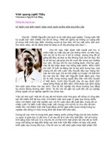
Associated aortic valvar abnormalities, ventricular
Aortic rim
Perimembranous VSD
Anatomy
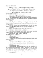
2. Muscular defects:
May be located anywhere in the apical,
mid, anterior or posterior muscular
septum and are often multiple.
With echocardiography, now more
frequently identified & more common
than membranous defects when
uncomplicated.
Anatomy
3. Infundibular (subpulmonary) defects:
Located under the pulmonary valve when
viewed from the right ventricle &
immediately beneath the aortic valve
viewed from the left ventricle.
Associated prolapse of the adjacent right
coronary aortic valve cusp…
Anatomy
4. Endocardial cushion type of defects:
Located beneath the tricuspid valve,
extending to the tricuspid valve ring.
Occupy the area when an
atrioventricularis communis opening
would be found.