Biochemical pathways an atlas of biochemistry and molecular biology second edition 2
Bạn đang xem bản rút gọn của tài liệu. Xem và tải ngay bản đầy đủ của tài liệu tại đây (18.52 MB, 211 trang )
189
3 Metabolism
3.12.1
Cloroplasts
(similar: cyanobacteriae)
PLASTOCYANINE (OR
CYTOCHROME C6)
PLASTOCYANINE (OR
CYTOCHROME C6)
Oxygen evolving
complex
cyt
c550
Thylacoid
lumen
(pH ca. 5)
CP47
D1
D2 PROTEIN
TyrZ
TyrD
CP43
Heme
f
Reaction
center P680
Psa A
Q cycle
Chl
D1
P
D1
P
D2
Chl
D2
2Fe2S2
(Rieske)
Heme
bp
Pheo
D1
Pheo
D2
JP
Heme
bn
Cyt
659
Heme
cn
Heme
bn
Heme
cn
Psa B
PROTEIN
or
Chlacc
ChlA0
PQA1
PQA1
Fx
Arrangement
simplified
Stroma
(pH ca. 8)
Exciton
Reaction
center P700
Heme
f
Analogous electron transfer
at this side
Mn4Ca
FA
Psa D F
B
Psa E
Arrangement
simplified
Psa C
ATRAZINE
or DCMU
Purple bacteria
(e.g. Rhodobacter
sphaeroides)
Excitons
LH1
L PROTEIN
M PROTEIN
LH1
B Chl
BA
PA
LH2
Heme
f
Heme
f
B Chl
BB
PB
2Fe2S2
(Rieske)
18 B Chl
B850
9 BChl B800
Arrangement
simplified
Preferred electron transfer mode
Q cycle
Reaction
center P865
B Pheo
HA
UQA
–
2UQ
Heme
bp
B Pheo
HB
Heme
cn
UQB
Heme
cn
Analogous electron transfer
at this side
LH2
{2H}
To Calvin cycle
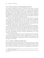
Figure 3.12-1. Photosynthetic Systems in Green Plants and Cyanobacteria (Top); in Purple Bacteria (Below)
The structures (in particular of the light-harvesting complexes) have been simplified.
Cyclic electron flow
PS II
Cyt b6f
Noncyclic electron flow
PS I
PS II
Cyt b6f
PS I
Figure 3.12-2. Electron Flow in Green Plants and in Cyanobacteria
Cyclic electron flow
REACTION
CENTER
Cyt bc1
Noncyclic electron flow
REACTION
CENTER
NADH
DEHYDROGENASE
Figure 3.12-3. Electron Flow in Purple Bacteria
violaxanthin can be interconverted into zeaxanthin and back, depending on the light intensity (Fig. 3.5.3-2). While the former compound
does not accept energy from excited states of chlorophylls, the latter is
open for this energy transfer and dissipates the energy into heat via a
short- living excited state. This results in a protective role by eliminating the dangerous triplet state of chlorophyll (3Chl*) at high light intensity, which could give rise to singlet oxygen (3.2.5.8). Cyanobacteria
and red algae additionally use phycobilisomes as the major light- harvesting complexes. These are large rod-shaped, membrane-attached
antenna complexes, which contain phycocyanobilin, phycoerythrobilin
and other pigments (3.3.3). While chlorophylls a and b absorb in the
blue and red regions, these pigments fill the ‘green gap’ (Fig. 3.12-4).
3 Metabolism
3H
190
Absorption
3.12.1
+
(Chi,p)
(Pheo)
FERREDOXIN
NADP+
REDUCTASE
Wavelength (nm)
pi
Figure 3.12-4. Absorption Spectra of Light Absorbing Chromophores
(Line colors: green-plants, blue-cyanobacteria).
ADP
FAD
NADP
ATP
+
NADPH + H
+
3H
+
To Calvin cycle
The structure of PS II has been resolved in high resolution in
cyanobacteria, however, the photosystem in higher plants appears to
be closely related.
Photosystem I (PSI) can be considered to be a light-driven plastocyanin-ferredoxin oxidoreductase. The main proteins PsaA and PsaB carry
the components of the electron transfer chain in pseudo-symmetric C2
fashion. They consist of a pair of chlorophylls a (eC-A1 and eC-B1,
likely representing the primary donor P700), associated with two more
pairs of chlorophylls a (eC-A2, eC-B2, eC-A3 and eC-B3, also named
chlacc and A0 pairs), two phylloquinones (QK-A and QK-B, also named
A1 pair) and a central [4Fe-4S] cluster FX. A pair of [4Fe-4S] clusters FA
and FB is bound to the protein PsaC. Additionally, docking sites exist for
ferredoxin (or flavodoxin) at the stromal surface and for plastocyanin
(or cytochrome c6 in cyanobacteria) at the luminal surface. The basic
structures of the plant and the cyanobacterial PSI are closely related,
however the plant system is monomeric and the bacterial one is mostly
trimeric. As a core antenna in green plants, 79 chlorophylls are tightly
coordinated by PsaA/PsaB for fast energy transfer, surrounded by more
chlorophylls, b-carotenes and xanthophylls (3.5.3.2). Again, cyanobacteria have phycobilisomes attached to the PS core.
The composition of the antenna complexes is listed in Table 3.12-1,
the absorption spectra are given in Figure 3.12-4.
Cytochrome b6f Complex: This complex provides the electronic
connection between PSII and PSI. In connection with the quinone
pool, it provides proton translocation from the stromal to the thylacoid
(luminal) side. In plants and cyanobacteria, it is a symmetrical dimer
of a Rieske [2Fe-2S] protein, a cytochrome f (containing heme f),
a cytochrome b6 polypeptide (containing hemes bp, bn cn), subunit IV
and some minor proteins.
Structure of the photosystem in purple bacteria: There is only one
photosystem, which resembles the photosytem II of plants and cyanobacteria and shows a twofold symmetry as well. The reaction center is
a cluster of four bacteriochlorophylls (two of them closely associated =
P 865). Two bacteriophaeophytins take the place of the phaeophytins and
two ubiquinones take the place of the plastoquinones. Most purple bacteria have two antenna complexes containing bacteriochlorophylls and
carotenoids. LH1 as core complex forms a tight ring around the reaction
center, while several LH2 rings are arranged around this core. The cytochrome bc1 complex is closely related to the complex III of the mitochondrial chain (3.11.1.3). It lacks the heme cn present in the b6 f complex.
Light absorption step: An absorbed light quantum excites an electron
in one of the LHC molecules, which transfers its energy (‘exciton’) by
resonance interaction via other LHC molecules quickly (ca.10−13 sec,
> 90 % efficiency) to the reaction center. In photosytem II of plants
and cyanobacteria, or in the only reaction center of purple bacteria, it
excites a pigment in the cluster of four closely associated chlorophylls
(P680 Æ P680* or P865 Æ P865*, respectively).This pigment, in turn,
donates an electron extremely quickly to a primary acceptor (pheophytine,
Figure 3.12-5. Time Course of Electron Transfer in Purple Bacteria
Table 3.12-1. Cofactors of the Light Harvesting Complexes (LHC)
Purple bacteria
Plants
(1 reaction center)
32 bacteriochlorophylls,
16 carotenes
Cyanobacteria (Blue
Algae)
(2 photosystems)
Ph. Sys. I: ca. 200 Chl., a > b
50 carotenoids
phycocyanobilin
phycoerythrobilin
Ph. Sys. II: ca. 250 Chl., a > b
110 carotenoids
phycoviolobilin
phycouvobilin
3.3.4 or bacteriochlorphyll, 3.3.4), causing the reaction to become increasingly irreversible. Via the quinones PQA or UQA, the electron finally
reaches phylloquinone B (PQB, 3.2.7.2) or ubiquinone B (UQB, 3.2.7.2)
respectively, where two electrons and two protons (from the cytoplasm)
accumulate, forming a hydroquinone (quinol, Fig. 3.12-5). In photosystem II only the D1- side is operative, while in photosystem I of plants and
of cyanobacteria both branches may contribute to electron transfer.
Regeneration of the reaction center: In plants and cyanobacteria,
P680+ replaces the lost electron by abstraction of another electron
from the Mn4Ca-protein complex (oxygen evolving complex, OEC)
via a tyrosine residue, Tyrz. After four repetitions, OEC4+ reacts with
water and is reduced again.
OEC4+ + 2 H2O = OEC0 + 4 H+ + O2
In purple bacteria, in the case of ‘cyclic electron flow’, the lost electron of P865+ (the special pair) is returned from the cytochrome bc1
complex via diffusing cytochrome c2. No extra reducing power for
other purposes becomes available in this way. In case of ‘noncyclic electron flow’ in these bacteria, an oxidation reaction (of H2S, S,
H2S2O3, succinate etc.) takes place:
H2S = Ssolid + 2 H+ + 2 e−
or succinate = fumarate + 2 H+ + 2 e−
(in periplasm)
(in cytoplasm).
The liberated electrons enter the reaction center via a bound cytochrome complex (e.g. in Rhodopseudomonas viridis, 0.27 μsec) or via
soluble cytochrome c2 (e.g., in Rh. sphaeroides, μsec to msec) and
reduce the special pair again.
Cytochrome b6f and bc1 complexes: The hydroquinone (quinol) formed
in the primary photosynthetic reaction transfers its hydrogen via the ‘quinone pool’ to the cytochrome complexes b6f (in plants) or bc1 (in bacteria),
where protons are released to the thylakoid space or to the periplasm,
191
3 Metabolism
3.12.1
Extracellular
space
Purple bacteria
(e.g. Rhodobacter
sphaeroides)
Chloroplasts
(green plants)
Cytoplasm
Extracellular space
Photoactivation
PHOTOSYSTEM I
SB = Schiff base
Cytoplasm
Figure 3.12-7. Photosynthesis and Reaction Mechanism in
Halophilic Archeaea
Arrows:
cyclic electron
flow
Photoactivation
PHOTOSYSTEM II
Arrows: noncyclic electron
flow
Figure 3.12-6. Standard Redox Potentials in Photosynthesis
(Purple Bacteria/Plants and Cyanobacteria)
In vivo, the actual potentials can differ due to protein binding,
variant concentration ratios, etc.
respectively. These complexes closely resemble the mitochondrial ubiquinol-cytochrome c reductase (complex III). Correspondingly, a ‘Q
cycle’ operates for transfer of additional protons to the thylakoid space or
to the periplasm, respectively. For details, see 3.11.4.3. The corresponding electrons are finally transferred to photosystem I (in plants via plastocyanine, in cyanobacteria via cytochrome c6) or returned to the reaction
center (in purple bacteria: cyclic electron flow via cytochrome c2).
NAD+ or NADP+ reduction: In plants and cyanobacteria, illumination
excites the primary donor P700 in photosystem I to release an electron
to the primary acceptor chlorophyll A0 (the role of the chlorophyllacc is
unclear). Then it is transferred to phylloquinone A1 and further on to the
iron-sulfur cluster Fx. This electron transfer proceeds either through the
cofactor sequence bound to the protein PsaA or to the ones bound to
PsaB. From Fx, the electron reaches the iron-sulfur clusters FA and FB,
which are bound to the peptide PsaC. These clusters release 2 electrons
to the two [4Fe-4S] clusters in ferredoxin (or to the FMN in flavodoxin).
These are then conferred either to the NADP+ reductase (noncyclic
electron flow), or alternatively back to the cytochrome b6f-complex
for additional proton transfer (cyclic electron flow). This allows a fine
adaptation to the requirements of the cell, since NADPH reduction
equivalents or ATP energy can be supplied in variable ratios. The graph
of the reduction potential of the steps passed through resembles a ‘Z’
(Fig. 3.12-6, for details of the redox potentials see 3.11.4).
As described above, purple bacteria cannot follow this mechanism.
They have to obtain reducing power from the environment to be able
to reduce NADP+ (noncyclic = reverse electron flow, since the electrons have to flow ‘uphill’ of the redox potential).
Halophilic archaea (Fig. 3.12-7): The photosystem of these archaea is
unrelated to photosynthesis in higher plants. It uses bacteriorhodopsin,
a small retinal protein (26 kDa) with 7 transmembrane passes, which
pumps protons upon absorption of photons through the membrane,
quantum yield y = 0.65.
It is mediated by light- induced trans-cis isomerization of the retinyliden chromophore and involves the following steps:
•
•
•
•
•
•
Isomerization of retinal from the all-trans to the 13 cis-configuration [BR568 to J state (0.5 psec) and on to K and L states]
Transfer of a proton from the protonated Schiff base (SBH) to the
carboxylate of Asp85 (L to MI states), followed by its release to the
extracellular medium
Modification of chromophore/protein structure. This changes the
accessibility from the extracellular side to accessibility from the
cytoplasmatic side (MI to MII states)
Transfer of a proton from Asp 96 to the Schiff base (M to N state,
several msec)
Thermal cis-trans reisomerization (N to O state, several msec)
Restoration of the initial state (O to BR568 state).
Isomerization of retinal (11-cis ´ all-trans) also plays a role in the
visual process of vertebrates (7.4.6).
Literature:
Barros, T. et al. EMBO J. 2009;28:298–306.
Cramer, W.A., Zhang, H. Biochim. biophys. Acta 2006;1757:339–345.
3.12.1...2
3 Metabolism
192
Fromme, P. Photosynthetic Protein Complexes. A Structural
Approach. Wiley-VCh Verlag, 2008 (Very detailed survey).
Guerkova-Kuras, M. et al. Proc.Nat. Acad.Sci. (USA)
2001;98:4437–4442.
Haupts, U. et al. Biochemistry 1997;36:2–7.
Holzwarth, A.R. et al. Proc. Natl. Acad. Sci. (USA) 2006;103:
6895–6900.
Jansson, S. Biochim. Biophys. Acta 1994;1184:1–19.
Loll, B. et al. Nature 2005;438:1040–1044.
Stroebel, D. et al. Nature 2003;426:413–418.
and concomitant cleavage into two 3-phosphoglycerate molecules.
This is followed by phosphorylation and reduction reactions. Then
an aldol condensation and a series of transfer reactions takes place,
mostly using reactions closely related to the pentose phosphate cycle
(3.1.6.1). As a result, the carboxylation of 6 C5 molecules yields 1 C6
molecule (glucose-P or fructose-P) and the reconstitution of the original 6 C5 molecules:
3.12.2 Dark Reactions
As described above, the light reactions provide both the energy carrier
ATP and the reductant NADPH. For the consecutive synthesis of biological material (initially carbohydrates), CO2 and water are also required.
The produced hexose is converted in chloroplasts into starch (3.1.2.2)
or in the cytosol into sucrose (3.1.4.1).
The enzyme ribulose bisphosphate carboxylase/oxygenase (Rubisco)
catalyzes the key reaction of the Calvin cycle
Calvin cycle (Fig. 3.12-8): CO2 fixation takes place in a cyclic process within the stroma by carboxylation of ribulose 1,5-diphosphate
Ribulose bisphosphate + CO2 + H2O = 2 3-phospho-D-glycerate
DG¢0 = −35,1 kJ/mol.
6 C5 + 6 CO2 Æ 6 C5 + 1 C6
according to the overall reaction of the Calvin cycle
6 CO2 + 12 H2O + 18 ATP4− + 12 NADPH = C6H12O6 + 18 ADP3− + 18 Pi2−
+12 NADP+ + 6 H+.
STARCH (3.1.3.2)
Enzyme activity regulation
Light reaction (3.1.2.1)
3.1.9.2
SUCROSE (3.1.4.1)
+
NADPH + H
+
(NADH + H )
Calvin
Cycle
ATP
H2O
CO2
Photorespiration
ATP
(Recycling of
2-PHOSPHOGLYCOLATE)
Figure 3.12-8. CO2 Fixation by the Calvin Cycle and its Regulation
Numbers in circles indicate
the number of molecules
reacting in order to produce
1 molecule of glucose 6-P.
193
3 Metabolism
3.12.2, 3.13
The enzyme is apparently the most abundant enzyme in the biosphere. It consists of eight large and eight small subunits (51 … 58
and 12 … 18 kDa). It has a low catalytic efficiency (kcat = 3.3 sec−1
per large subunit). Although the carboxylase reaction is usually preferred, it also performs an oxygenase side reaction (Fig. 3.12-9, see
also ‘photorespiration’ below).
Regulation of the Calvin cycle: The cycle has to operate only if sufficient NADPH and ATP from the light reaction are available in order
to prevent useless degradation reactions. This is performed by lightinduced activation of rubisco, fructose bisphosphatase (FBPase) and
sedoheptulose bisphosphatase (SBPase).
•
•
•
•
The pH in the stroma increases during the light reaction (3.12.1),
since protons are pumped out. It approaches the pH optimum of
rubisco, FBPase and SBPase.
Reduced ferredoxin, the reaction product of photosystem I, reduces
thioredoxin, which in turn activates FBPase and SBPase by reduction of enzyme -SS- bridges (Fig. 3.12-5). Simultaneously, phosphofructokinase (3.1.1.2) is deactivated by this reduction and thus
decreases the competing glycolysis reaction (3.1.1.1).
Mg++, which flows into the stroma during illumination, activates
rubisco, FBPase and SBPase.
NADPH, which is produced by the light reaction, activates FBPase
and SBPase.
During dark, these reactions are switched off. The energy supply of
photosynthesizing cells is then provided the same way as in non-photosynthesizing cells by glycolysis (3.1.1.1), pentose phosphate cycle
(3.1.6.1) and oxidative phosphorylation (3.11).
Photorespiration and C4 cycle: The rubisco side reaction with O2
yields at first 3-phosphoglycerate and 2-phosphoglycolate, which later
on is partially oxidized, resulting in CO2 liberation (photorespiration,
Figure 3.12-8, see also 3.1.9.2). This counteracts photosynthesis and
requires additional energy input for recycling. The rate of this reaction
increases relatively to the rate with CO2 at higher temperatures and
at low CO2 concentration at the site of synthesis (e.g., on hot, bright
days), and limits the growth rate of plants.
A number of plants (C4 plants, mostly tropical ones) have developed a mechanism for increasing the CO2 concentration in the fluid
phase of chloroplasts from ca. 5 μmol/l to ca. 70 μmol/l (Fig. 3.12-10).
So-called mesophyll cells surround the bundle-sheath cells, which
contain the Calvin cycle enzymes. The mesophyll cells, which lack
MESOPHYLL CELL
Cytoplasm
Chloroplast
Figure 3.12-9. Carboxylase (Top) and Oxygenase
(Below) Reaction Mechanisms of Rubisco
rubisco, perform a CO2 fixation by the highly exergonic (and thus
practically irreversible) reaction (3.1.3.4):
Phosphoenolpyruvate + HCO3− Æ oxaloacetate + Pi
and transfer this bound CO2 through a number of further reactions to
the chloroplasts of bundle-sheath cells, where it is released to be used
in the Calvin cycle. Several reaction types exist (Fig. 3.12-10). These
reactions require five energy-rich P bonds/ CO2 (instead of three in the
Calvin cycle). Therefore, this mechanism is of advantage only in hot,
sunny climates.
Literature:
Furbank, R.T., Taylor, W.C. The Plant Cell 1995;7:797–807.
Gutteridge, S., Gatenby, A. The Plant Cell 1995;7:809–819.
Heldt, H.W., Flügge, U.I. in Tobin, A.K. Plant Organelles.
Cambridge University Press, 1992.
Heldt, H.W. Plant Brochemistry and Molecular Biology. Oxford
University Press, (1998).
Portis, A.R. Ann. Rev. Plant Physiol. Plant Mol. Biol. 1992;43:415–437.
3.13 Plant Secondary Metabolism
Antje Chang
Plant metabolism can be divided into primary and secondary metabolism. The term primary metabolism encompasses all processes and
compounds that are essential for the fundamental functions of life,
BUNDLE-SHEATH CELL
Cytoplasm
Chloroplast
Figure 3.12-10. CO2 Pumping by the C4 Cycle (NADP+-Malate Enzyme Type, e.g., in Maize and Sugar Cane)
3.13, 3.13.1
3 Metabolism
SHIKIMIC ACID (3.2.7.1)
like growth, development, and reproduction. In contrast, secondary
metabolism which is characterized by its immense chemical diversity,
is required for the survival of the individual in its respective environment. Therefore, these natural products, traditionally referred to as
secondary metabolites have an ecological function for the organism in
its interaction with its biotic and abiotic environment. Their role had
been overlooked for a long time, but is widely accepted now.
The functions, which in general can be regarded as the plant’s
chemical interaction, are studied in the field of so-called chemical
ecology, considering the following aspects:
Chemical defense (constitutive or induced defense against pathogens and herbivores). Plants have developed different strategies
for the defense against herbivores and pathogens:
- The bioactive compounds are synthesized constitutively and accumulated in specialized cells (e.g., hair) or in subcellular compartments (e.g., vacuole), and are released by plant tissue destruction.
- Non-toxic precursors (e.g., glycosylated precursor of toxic aglycons) are stored apart from the corresponding specific enzyme,
e.g., a glycosidase. After destruction of the cell compartments the
enzymatic reaction is initiated and the toxic aglycone is released.
- The formation of defensive compounds, e.g., phytoalexins and
proteinase inhibitors, may be induced by signal substances
(elicitors) as a response to the attack by pathogens (e.g., by phytoalexins) and herbivores (e.g., by proteinase inhibitors).
•
Attraction of pollinators and seed distributors (flower pigments,
volatile compounds).
•
Adaptation to the environment (e.g., UV protection).
JUGLONE
INDOLE ACETIC ACID
INDOLE ALKALOIDS
PHYLLOQUINONE
L-AROGENATE
TOCOPHEROL
L-PHENYLALANINE
L-TYROSINE
PLASTOQUINONE
ALKALOIDS
CINNAMIC ACID DERIVATIVES
COUMARINE
FLAVONOIDS
UBIQUINONE
CINNAMOYL ALCOHOL
LIGNIN
part of the shikimic acid pathway
subsequent reactions
Figure 3.13-1. Products Produced by the Shikimate Pathway
ACETYL-CoA
Secondary metabolism is not only found in plants, but also in bacteria (e.g., antibiotics 3.10.9), fungi and marine sessile organisms. This
chapter will focus on the plant secondary compounds, since 80 % of the
secondary metabolites are produced by higher plants. Many of these
reactions originate from pathways of the primary metabolism, therefore
only the differing parts are described here and references are given for
the common reactions. The biosynthetic origins of the secondary metabolites are also often used as base for their classification (Table 3.13-1).
CH3
MALONYL-CoA
S
HO
CoA
5
hexaketide intermediate
CH3
Terpenoids/isoprenoids:
hemiterpenes, monoterpenes, sesquiterpenes,
diterpenes, triterpenes, tetraterpenes, polyterpenes
Pseudo-alkaloids:
terpenoid alkaloids, piperidine alkaloids
Alkaloids:
Nicotiana alkaloids, pyrrolizidine alkaloids,
tropane alkaloids, benzylisoquinoline alkaloids,
indole alkaloids, purine alkaloids
shikimic acid, phenylalanine,
polyketide
O
O
derived from
O
S-Enz
O
O
6-(2,4-DIHYDROXY-6-METHYLPHENYL)PYRAN-2-ONE
OH
O
O
decarboxylative condensation reaction
6 CO2 + 6 CoA-SH
Table 3.13-1. Major Groups of Plant Secondary Metabolites
Phenolic compounds:
polyphenols, phenols, phenylpropane
derivatives, flavonoids, stilbenes
S
CoA
O
O
Classes of secondary metabolites
L-TRYPTOPHAN
CHORISMATE
OH
cyclization
TYPE III POLYKETIDE SYNTHASE
•
194
C5-unit (‘activated isoprene’)
O
terpenes, polyketides, acetate
O
CH3
3-METHYLNAPHTHALENE-1,8-DIOL
CH3
amino acids
OH
3.13.1 Phenolics
Unlike animals, plants, fungi, and bacteria are able to perform the
de novo biosynthesis of aromatic metabolites. In higher plants most
of the phenolics are formed by the shikimate pathway with aromatic
amino acids as intermediates (3.2.7.1). Another major pathway leading to aromatic natural products is the polyketide pathway, which proceeds via linear coupling of acetate units. Flavonoids are an example
of mixed biosynthesis of aromatic metabolites (3.13.1.3).
H2O
OH
oxidation
similar: ERYTHROMYCIN, TETRACYCLINE,
GRISEOFULVINE biosynthesis etc. (3.10.9)
{2H}
PLUMBAGIN
O
CH3
3.13.1.1 Biosynthesis
Shikimate pathway: The biosynthesis of the three aromatic amino acids
L-phenyalanine, L-tyrosine, and L-tryptophan by the shikimate pathway
is described in detail in 3.2.7.1 and Figure 3.2.7-1. The pathway is localized in plastids of plants and in the cytoplasm of bacteria and fungi.
Originating from D-erythrose 4-phosphate and phosphoenolpyruvate,
OH
O
Figure 3.13-2. Polyketide Pathway (biosynthesis of plumbagin,
putative reaction in Plumbago indica)
195
3 Metabolism
3.13.1
the pathway includes shikimate, chorismate and prephenate as intermediates. Contrary to the general pathway, part of the sequence is reversed
in higher plants: prephenate is first transaminated to arogenate, the
dehydratase/decarboxylase or dehydrogenase/decarboxylase reactions
take place afterwards (arogenate pathway). These aromatic amino acids
are precursors of numerous aromatic compounds in bacteria, fungi, and
plants. A survey of these interrelationships is given in Figure 3.13-1.
Polyketide pathway (Fig. 3.13-2): Polyketides are natural products
found mainly in bacteria and fungi, but also in plants and animals. They
are synthesized by linear condensation reactions of acetate units, deriving
from malonyl-CoA via decarboxylation. This is a process similar to fatty
acid biosynthesis (3.4.1.1). The polyketide synthases are multi-enzyme
complexes that produce a wide range of structural diverse secondary
metabolites, also depending on the kind of starter molecule. In plants, the
polyketide pathway is involved in mixed biosyntheses, like in the biosynthesis of flavonoids (3.13.1.3) and stilbenes (3.13.1.4), where a phenylpropane is the starter molecule. Several type III polyketide synthases are
known in plants, such as chalcone synthase or stilbene synthase. Related
reactions are found in the biosynthesis of, e.g., erythromycin (3.10.9.3),
tetracycline (3.10.9.4) and other antibiotics.
3.13.1.2 Phenylpropane Derivatives (Fig. 3.13-3)
Phenylpropanes encompass a broad range of plant secondary metabolites. They are mainly synthesized from phenylalanine. Phenylalanine
ammonia lyase (PAL) is a key enzyme between the primary and secondary metabolism, producing trans-cinnamate by release of ammonia. The activity of PAL is influenced by light and temperature and is
regulated by feedback inhibition.
trans-Cinnamate is a central intermediate for a wide range of
derivatives (Table 3.13-2, Fig. 3.13-4). They are synthesized mainly
by hydroxylation and methylation reactions catalyzed by specific
enzymes. Examples are phenylpropanoids, i.e., eugenol, anethol, and
estragol, which are major constituents of essential oils. The corresponding alcohols (4-coumarol, sinapol, coniferol, ferulol) are formed
by reduction of carboxylic groups and represent the monomeric components of lignin (monolignol).
STILBENES
TRANS-CINNAMATE
2-MONOOXYGENASE
FLAVONOIDS + POLYMERS
Figure 3.13-3. Phenylpropanoid Compounds in Plants
3.13.1
3 Metabolism
The polymerization reaction leading to lignin in the cell walls of
the plants is catalyzed by lignin peroxidase (Fig. 3.13-3). The extracellular process is initiated by the formation of a radical, presumably
by H2O2 (3.2.5.8) and progresses via chain reaction mechanisms. The
result is a closely meshed, irregular network. Its overall composition
depends on the ratio of the originating alcohols and the reaction conditions and varies among different species. Lignin is the second most
frequent compound in the biosphere (after cellulose, the annual synthesis rate is ca. 2 * 1010 t). It brings about the pressure resistance of
plant cell walls (3.1.6.3). Only a few organisms, mostly fungi, can
degrade lignin. Suberin has a similar structure with alcoholic groups
esterified by (mostly) long-chain fatty acids. It occurs in cork, the
endodermal cells of roots and other parts of plants.
The pathway to coumarin starts with hydroxylation of transcinnamate, resulting in trans-2-coumarate (Fig. 3.13-3). The product
accumulates in the vacuole of the mesophyll cells in the form of glucosylated cis- and trans-isomers. When the plants are wounded, a
specific glucosidase in the cytoplasm catalyzes the hydrolysis of the
cis-isomer, producing coumarin by lactonization.
Coumarin is a toxin found in many plants, e.g. in woodruff (Galium
odoratum) or tonga bean (Dipteryx odorata, common name: cumaru).
Coumarin derivatives have been used in the perfume industry. They are
important in pharmacology due to their anticoagulant effect and likewise
as rat poison, causing internal hemorrhage and death (e.g., Warfarin®).
O
OH
Figure 3.13-4. Trans-Cinnamate
Derivatives
R3
4-COUMARYL-CoA
R1
R2
OH
MALONYL-CoA
196
O
OH
CoA
3
O
OH
HO
R1
R2
R3
3-Coumarate
OH
H
H
4-Coumarate
H
OH
H
Caffeate
OH
OH
H
Ferulate
OCH3
OH
H
Sinapate
OCH3
OH
OCH3
O
CHALCONE SYNTHASE
NARINGENIN-CHALCONE
Table 3.13-2. Some Trans-Cinnamate Derivatives
ACo – S
S
RESVERATROL SYNTHASE
4 CoA-SH
+ 3 CO2
4 CoA-SH
+ 4 CO2
RESVERATROL (3,4',5-TRIHYDROXYSTILBENE)
OH
OH
CHALCONEFLAVANONEISOMERASE
OH O
HO
OH
NARINGENIN (a FLAVANONE)
OH
B
O
HO
A
C
GENISTEIN (an ISOFLAVONE)
OH
O
HO
OH
O
2 {H}
O2
2-OXOGLUTARATE
O
CO2
SUCCINATE
OH
APIGENIN (a FLAVONE)
OH
O
HO
2 {H}
NARINGENIN
3-DIOXYGENASE
OH
O
CATECHIN (a FLAVAN-3-OL)
OH
DIHYDROKAEMPFEROL (a FLAVANOL)
OH
OH
O2
O
HO
O
HO
2-OXOGLUTARATE
DIHYDROKAEMPFEROL
DIOXYGENASE CO2
H 2O
SUCCINATE
OH
OH
O
OH
via LEUCOPELARGONIDIN
2 {H}
OH
{H2O}
(a FLAVAN-3,4-DIOL)
OH
KAEMPFEROL (a FLAVONOL)
OH
OH
OH
O
O
HO
O
HO
PELARGONIDIN (an ANTHOCYANIDIN)
OH
O+
HO
OH
OH
OH
OH
Figure 3.13-5. Biosynthesis of Flavonoids and Stilbenes
OH
197
3 Metabolism
3.13.1
3.13.1.3 Flavonoids
The flavonoids are a large group of plant secondary metabolites. They
display a great variety in structure and function and are widely distributed in the plant kingdom.
The biosynthesis (Fig. 3.13-5) combines the products of the shikimate pathway and of the polyketide pathway (3.13.1.1). 4-CoumaroylCoA ligase activates 4-coumarate to its CoA derivative. Thereafter,
chalcone synthase catalyzes the addition of three malonyl-CoA units
(originating from the polyketide pathway) and removal of 3 CO2 to
naringenine chalcone, forming the flavan backbone that is characteristic of all flavonoids. These compounds can be assigned to several subgroups depending on the substitution pattern, as listed in Table 3.13-3.
Some flavonoid structures are shown in Figure 3.13-6.
Table 3.13-3. Subgroups of Flavonoids
Flavonoid
subgroup
Examples
Source
Flavanone
hesperetin, naringenin, eriodictyol
grapefruit, orange
Flavone
luteolin, apigenin, tangeritin
pepper, celery
Flavonol
quercetin, rutin, kaempferol, myricetin
onion, endive
Flavanol
catechin, gallocatechin, epicatechin,
theaflavin
red grape, apple, green
tea
Flavanonol
taxiflorin, dihydrokaempferol
gingko
Isoflavone
genistein, daidzein, licoricidin
soybean
Anthocyanidin
cyanidin, delphinidin, malvidin,
pelargonidin, peonidin, petunidin
cherry, blueberry, red
grape
Flavonoids accumulate in cell vacuoles, mostly in their glycosylated
form. Many color pigments in flowers and fruits serve as attractants of
pollinators and animals for seed distribution. Anthocyanins, the glycosides of anthocyanidines, are water-soluble vacuolar pigments. Their
colors depend on the substitution patterns of the B-ring, the pH-value
in the vacuole, the binding of metal ions etc.
Flavonoids in the epidermis serve as UV-protection for the inner
cell layers, e.g,. the mesophyll cells. These compounds play an important role in the interaction of rhizobia and plants. They act as plant
signals activating the expression of nodulation genes, thus initiating
the formation of N2-fixing root nodules. Some flavonoid metabolites
are produced by plants as phytoalexins (stress compounds) or antibiotics or exert antioxidant activity.
3.13.1.4 Stilbenes
The biosynthesis of stilbenes (Fig. 3.13-5) is similar to that of the
flavonoids. Three malonyl-CoA units (produced via the polyketide
pathway) react with 4-coumaroyl-CoA (3.13.1.3). In this manner,
4 CO2 are removed by decarboxylation and a diphenylethylene structure
is formed. The resveratrol synthesis is shown as an example. This compound is a phytoalexin, which is produced by plants under the attack
of bacteria and fungi. It has anti-cancer and anti-inflammatory activity.
3.13.1.5 Tannins (Fig. 3.13-7)
Tannins are plant polyphenols, widely occurring in gymnosperms and
angiosperms. They can be classified chemically into two main groups,
hydrolyzable (gallotaninns) and non-hydrolyzable (condensed) tannins, formed from flavonoid units (3.13.1.3). The gallotannins are
glycosylated derivatives of gallic acid, which is derived from shikimic
acid (3.2.7.1). The hydroxyl groups of a hexose (usually D-glucose) in
the center of hydrolyzable tannins is esterified with numerous gallic
acid molecules. The condensed tannins (proanthocyanidins) are oligoor polymers of flavonoids units.
Tannins are mainly localized in the vacuoles or in specialized cells
of the tree bark, wood, fruit, leaves, roots and plant galls for protection
GALLIC ACID
(3,4,5-TRIHYDROXYBENZOATE)
GALLOTANNIN
(HYDROLYZABLE TANNINS)
R
OH
O
O
O
O
HO
OH
O
R
O
OH
O
R
O
HO
PELARGONIDIN
CYANIDIN
HO
OH
O
O
OH
OH
OH
B
HO
HO
O+
A
O+
=R
HO
C
OH
OH
OH
CONDENSED TANNINS
OH
OH
OH
O
HO
DELPHINIDIN
OH
PAEONIDIN
OH
OCH3
OH
OH
OH
OH
OH
HO
O
HO
+
HO
O+
OH
OH
O
OH
OH
OH
OH
OH
OH
OCH3
O
HO
MALVIDIN
PETUNIDIN
OH
OH
OCH3
OH
OH
OH
OH
OH
HO
HO
O+
O+
OH
OH
OH
OCH3
O
HO
OH
OH
Figure 3.13-6. Some Flavonoids
OH
OH
OH
Figure 3.13-7. Gallic Acid and Tannins
R
3.13.1...2
against herbivores and pathogens. When the plant is wounded, the
tannins are released and their phenolic groups bind to amino groups
of plant proteins, converting the proteins into an indigestible form.
This drastically reduces the food quality of the plant for herbivores. In
addition, tannins have a bitter and astringent taste.
3.13.2 Terpenoids
The ubiquitously occurring terpenoids are the largest group of natural products, showing a wide structural diversity in carbon skeletons
and functional groups, particularly within the plant kingdom. A part
of these compounds is essential for plant development and hence is
assigned to the primary metabolism, e.g., hormones, members of the
electron transport system or pigments for light absorption. Most of
the terpenoids, however, have an important function in the secondary
metabolism, e.g., components of the essential oils, steroids, waxes,
resins and natural rubber. A major number serve as defensive compounds against herbivores and pathogens, or as in the case of colors
and scents, as attractants for pollinators. Due to the biological activity
many of them have pharmacological significance.
3 Metabolism
3.13.2.3 Sesquiterpenes (Fig. 3.13-8)
Sesquiterpenes (C15) represent the largest group within the terpenoids.
Several hundred sesquiterpene backbone structures have been identified and thousands of naturally occurring derivatives have been isolated. They are found in all tracheophyta, in mosses, fungi, brown and
red algae, and in insects. The precursor is farnesyl-PP, synthesized by
the transfer of two 3-isopentenyl-PP to a dimethylallyl-PP starter unit
(head to tail addition, Fig. 3.5.1-1). Sesquiterpenes can be classified
into acyclic, mono-, bi-, and tricyclic compounds.
•
•
•
3.13.2.1 Biosynthesis
All terpenoids are derived from the C5-units 3-isopentenyl-PP (IPP)
and dimethylallyl-PP (DMAPP), and are classified into hemiterpenes
(C5), monoterpenes (C10), sesquiterpenes (C15), diterpenes (C20), triterpenes (C30), tetraterpenes (C40) and polyterpenes, according to the
number of linked C5-units. The biosynthesis of the precursors IPP
and DMAPP of all terpenoids proceed via two different pathways, a
mevalonate-dependent and a mevalonate-independent pathway:
•
•
Mevalonate pathway: It is localized typically in the cytosol and
is identical to the first part of cholesterol synthesis (Fig. 3.5.1-1)
via the intermediate mevalonate up to IPP, which is catalyzed to
DMAPP by isopentenyl-diphosphate isomerase.
Rohmer pathway (non-mevalonate pathway or DOXP/MEP pathway, Fig. 3.5.3-1): It is localized in plastids and the precursors of
it can be obtained from pyruvate and D-glyceraldehyde 3-P via
producing the intermediates 1-deoxy-D-xylulose 5-P, 2-C-methylD-erythritol 4-P and further phosphorylated intermediates leading
to IPP and DMAPP.
3.13.2.2 Hemiterpenes and Monoterpenes (Fig. 3.13-8)
The C5-structure isoprene is a representative example of hemiterpenes. The synthesis takes place in the chloroplasts and is induced by
light and high temperature. Isoprene is released by the cleavage of the
diphosphate unit from dimethylallyl-PP (Fig. 3.5.1-1). It can contribute to the emission of organic aerosols together with other terpenoids
in forest atmosphere, especially in coniferous forests.
Monoterpenes (C10, Fig. 3.13-8) derive from geranyl-PP, which is
formed by adding IPP to a DMAPP starter unit (head-to-tail addition, for
mechanism see Fig. 3.5.1-2). In some cases, neryl-PP (i.e. the native substrate for the monoterpene synthase in tomato, Solanum lycopersicum)
or linalyl-PP are the starting compounds. Most of them are volatile and
typical scent and aroma compounds from higher plants. They are often
found as components of the essential oils, together with the sesquiterpenes. Monoterpenes occur as acyclic, mono- or bicyclic molecules.
•
•
•
Acyclic monoterpenes: Geraniol is the main constituent of rose
oil and citronella oil. The essential oils of various Citrus species
contain citronellol.
Examples of monocyclic monoterpenes are menthol and limonene
from Mentha species and thymol from thyme (Thymus vulgaris). All
of them are synthesized by cyclization of geranyl-PP, a typical enzyme
being limone synthase, synthesizing limonene. The resulting structures
are further diversified by additional rearrangements and oxidations.
Bicyclic monoterpenes are formed by two sequential cyclization
reactions of geranyl-PP. Examples are pinene (in pine resin), camphene and thujene (a neurotoxic compound in absinth). Structures
containing ketone, alcohol, and ether groups are, e.g., camphor,
borneol and eucalyptol.
198
Acyclic sesquiterpenes are not common. Farnesol, an alcohol derivative of farnesyl diphosphate is present in essential oils of, e.g., rose
flower, sandalwood and lemon grass.
Monocyclic sesquiterpenes are based on several structural skeletons, e.g., bisabolane, germacrene, elemane and humulane. The
cyclization reactions are catalyzed by specific cyclases. Bisabolol,
an alcohol derivative has an anti-inflammatory effect and occurs
in the essential oil of chamomile (Matricaria chamomilla) and in
bergamot oil (Citrus bergamia).
Bicyclic sesquiterpenes are, e.g., cadinenes and caryophyllenes. The latter are constitutents of many essential oils, e.g.,
clove (Syzygium aromaticum), hemp (Cannabis sativa), rosemary (Rosmarinus officinalis) and cinnamon (Cinnamomum
verum). The phytoalexin capsidiol (Fig. 3.5.3-2) derives from
the germacrene structure and can be found in pepper (Capsicum
anuum) and tobacco (Nicotiana tabacum) in response to fungal
infection. Azulenes (e.g., guaiazulene of chamomile (Matricaria
chamomilla), Fig. 3.5.3-2) contain a condensed aromatic 5- and
7-ring system.
3.13.2.4 Diterpenes (Fig. 3.13-8)
Diterpenes (C20) consist of four C5-units and derive from geranylgeranyl-PP. They occur in plants and fungi. Most of them are primary
metabolites, such as the phytohormone gibberilic acid or phytol (Fig.
3.5.3-2), which is esterified to chlorophyll, both promoting growth
and elongation during germination (Fig. 3.5.3-2). Paclitaxel (formerly
named taxol), isolated from the bark from the pacific yew tree (Taxus
brevifolia), has an anti-cancer effect and is used as a mitotic microtubule inhibitor in cancer therapy.
3.13.2.5 Triterpenes (Fig. 3.13-9)
Triterpenes (C30) are derivatives of the acyclic squalene. This compound is synthesized through a head-to-head condensation of two
farnesyl-PP molecules catalyzed by squalene synthase. In plants,
squalene is converted to the tetracyclic cycloartenol by cycloartenol
synthase. Cycloartenol is a precursor of plant steroids (phytosterols,
Fig. 3.5.2-1), e.g., sitosterol, stigmasterol and campesterol (occurring in, e.g., soybean oil or rapeseed oil). Squalene can also be converted into a/b-amyrin by 2,3-oxidosqualene a- or b-amyrin cyclase.
Amyrin is a precursor of pentacyclic triterpenes (see below). In animals, squalene is the precursor of cholesterol (3.5.1.1).
Cardiac glycosides occur only in glycosylated form in nature. Their
aglycones can be classified as
•
•
cardenolides (Fig. 3.5.2-1, exclusively synthesized in plants) and
bufadienolides (formed in plants and in toads of the genus Bufo).
The characteristic features are additional 5-membered or 6-membered lactone rings, respectively. The glycosides of Digitalis lanata
and Digitalis purpurea (digoxin and digitoxin) and other plants are
important for pharmacological purposes, being the active components of drugs for treatment of heart insufficiency. Their effect is
based on the inhibition of the Na+/K+ ATPase (TC 3.A.3.1.1, see sections 6.1.4 and 7.2.1), which is also responsible for their toxicity in
higher doses.
Ecdysone (3.5.2.3 and Fig. 3.5.2-1) and 20-hydroxyecdysone are
the major steroidal hormones of molting insects, which synthesize
them from cholesterol or from plant sterols. Ecdysone analogues and
some derivatives (e.g., abutasterone) were also isolated from the fern
199
3 Metabolism
3.13.2
GERANYL-PP
CH3
FARNESYL-PP
CH3
CH3
H3C
PP
CH3
CH3
H3C
PP
SESQUITERPENES (C15)
MONOTERPENES (C10)
CITRONELLOL (acyclic) MENTHOL (monocyclic) a-PINENE (bicyclic)
CH3
CH3
H3C
FARNESOL (acyclic)
CH3
BISABOLOL (monocyclic)
CH3
OH
OH
H3C
H3C
CH3
H3C
GERANIOL
CH3
CH3
LIMONENE
CH3
OH
CH3
OH
CAMPHOR (bicyclic)
H3C
CH3 OH
δ-CADINENE (bicyclic)
CH3
OH
CARYOPHYLLENE (bicyclic)
CH3
H3C
CH3
CH3
H3C
CH3
O
H3C
CH3
THYMOL
CH3
H
H
CH3
CH3
CH3
all-trans GERANYLGERANYL-PP
CH3
OH
H3C
H2C
CH3
CH2
H3C
CH3
H3C
CH3
H3C
3
PP
CH3
DITERPENES (C20)
PACLITAXEL (TAXOL)
O
HO
GIBBERILLIC ACID GA3
O
O
NH
O
OH
H
O
HO
CH2
O
O
CH3
CH3
O
OH
O
HO
H
CH3
HO
OH
H3C
O
Figure 3.13-8. Examples for Monoterpenes, Sesquiterpenes and Diterpenes
O
O
O
O
CH3
3.13.2
3 Metabolism
200
SQUALENE
H3C
FARNESYL DIPHOSPHATE
CH3
CH3
PP
CH3
PP
NADPH + H+
CH3
NADP+
CH3
CH3
CH3
H3C
H3C
CH3
SQUALENE SYNTHASE
CH3
NADPH + H+
SQUALENE MONOOXYGENASE
O2
H2O
NADP+
SQUALENE-2,3-EPOXIDE
H3C
CH3
CH3
CHOLESTEROL
CH3
CH3
CH3
CH3
CH3
O
PREGNENOLONE
CARDENOLIDES (see Fig. 3.5.2-1)
2,3-OXIDOSQUALENECYCLOARTENOLCYCLASE
2,3-OXIDOSQUALENE-β-AMYRIN CYCLASE
2,3-OXIDOSQUALENE-α-AMYRIN CYCLASE
H3C
CYCLOARTENOL
CH3
CH3
H3C
H3C
H3C
CH3
H
CH3
CH3
α-AMYRIN H3C
β-AMYRIN
H
CH3
CH3
H
CH3
CH3
CH3
CH3
CH3
HO
H
H3C CH3
HO
HO
CH3
H3C
CH3
H
CH3
H3C
CH3
CH3
H
CH3
CH3
BETULINIC ACID
α-BOSWELLIC ACID
H
e.g. SITOSTEROL,
STIGMASTEROL
CH3
CH3
H3C
H2C
STEROIDS, see Fig. 3.5.2-1
CH3
CH3
H
CH3
COOH
H
CH3
CH3
HO
O
HO
H3C
CH3
CH3
OH
STEROIDAL ALKALOIDS
TOMATIDINE
STEROIDAL SAPONINS
TRITERPENOID SAPONINS
(25S)-5β-SPIROSTANOL
OLEANOLIC ACID
CH3
H3C
H3C
CH3
CH3
CH3
H
H
HO
H
CH3
O
CH3
N
H
H
H3C
CH3
OH
CH3
O
H
H
H
H
R
O
H
H
H
R
R = GLYCOSIDIC GROUPS
Figure 3.13-9. Triterpenes
O
H3C
H
CH3
CH3
H
CH3
O
201
3 Metabolism
Polypodium vulgare. These phytoecdysteroids act as insect feeding
deterrents, disturb the precise synchronization of insect development
and lead to the appearance of malformed animals.
Steroidal alkaloids are a group of nitrogen-containing steroids. Most
of them are synthesized in higher plants. The insertion of nitrogen
into the terpene structure results in an alkaline character and thus they
share common properties with alkaloids. They belong to the so-called
pseudoalkaloids (3.13.3.2). The nitrogen does not derive from amino
acids but is inserted as NH3 at a late stage of the biosynthesis. Like
most of the alkaloids, the steroidal alkaloids are toxic to herbivores.
The poisonous glycoalkaloids solasodine and solanine, isolated from
potato (Solanum tuberosum) and tomatine from tomato (Solanum
lycopersicum) are responsible for the toxicity of the green parts of the
potato tuber and of unripe tomato fruits.
Non-glycosylated pentacyclic triterpenes are not a widespread
group. Amyrin can be found in the latex of poinsettia (‘Christmas
Star’, Euphorbia pulcherrima). Betulinic acid occurs in betulin (the
pigment from the bark of white birch, Betula pendula). It shows antiinflammatory and anti-tumor activities. Boswellic acid from frankincense (the resin of Boswellia sacra) is studied for anti-inflammatory
applications.
Saponins (glycosylated pentacyclic triterpenes), are a group of plant
secondary metabolites, which are localized in roots, rhizomes and
seeds. Due to their amphiphilic character they can act as detergents,
destroying cell membranes by surface action. Thus these agents protect
against pathogens, fungi, and herbivores. Many of them are of pharmaceutical interest. According to the ring system of the aglycones, they
are classified into steroidal saponins (with a spirostane structure, e.g.,
spirostanol) and triterpenoid saponins (e.g., oleanolic acid or solanine).
3.13.2.6 Tetraterpenes (Fig. 3.5.3-2)
A head-to-head condensation of two geranylgeranyl-PP molecules
results in the C40 skeleton of phytoene, the precursor of all tetraterpenes. The carotinoids are the major group of the tetraterpenes, encompassing carotins and xanthophylls as their oxidation products. These
compounds and their derivatives play important roles in the primary
metabolism of plants (e.g., as pigments in the light-harvesting complexes of the photosynthesis system), as well as in other kingdoms of
life. More details are given in chapters 3.5.3.2 … 4.
Under the aspect of secondary metabolism in plants, the lipophylic
carotinoids are responsible for many colors in fruits and flowers varying from yellow (i.e., violaxanthine from pansy, Viola tricolor) to red
(i.e., lycopene from tomato, Solanum lycopersicum). They accumulate in plastids.
3.13.2.7 Oligo- and Polyterpenes
Several plants are able to form polymers of the IPP and DMAPP
derived isoprene, the polyterpenes. Natural rubber from the latex
of the rubber tree (Hevea brasiliensis) consists of 500 to 5000 linearly bound C5-units forming an all-cis-polyisoprene (Fig. 3.5.3-2).
The biosynthesis is localized in the laticifers. Starting from geranyl
diphosphate, isoprene units are added successively by rubber cis-polyprenylcistransferase. Guttapercha is composed of isoprene units forming a trans-polyisoprene in latex. It is isolated from dried leaves of
Palaquium gutta, a tropical tree, a native of Southeast Asia.
3.13.3 Nitrogen-Containing Secondary Metabolites
The large group of nitrogen-containing secondary compounds encompasses the glucosinolates, the cyanogenic glycosides and the alkaloids. Most compounds in this group derive from amino acids. Also
non-proteinogenic amino acids belong to this group.
3.13.3.1 Cyanogenic Glycosides and Glucosinolates
(Fig. 3.13-10)
The two classes of secondary metabolites share common properties.
Their biosynthetic pathways are evolutionarily related. Both occur in
3.13.2...3
a non-toxic glycosylated form. These water-soluble compounds are
stored in the vacuoles of specialized cells. In case of plant damage,
they release toxic compounds by the action of an enzyme localized in
a different cell compartment.
Cyanogenic glycosides are widely distributed in the plant kingdom
and can be encountered in gymnosperms and angiosperms, e.g., in
seeds of bitter almonds (Prunus dulcis), in the tuberous root of cassava (Manihot esculenta, a tropical native of South America) and in
sorghum (Sorghum bicolor). Dhurrin, a cyanogenic glycoside in sorghum is localized in the vacuole of the epidermis cells. When the plant
tissue is destroyed, cytosolic b-glucosidase cleaves the glucoside
bond, releasing 4-hydroxy-(S)-mandelonitrile, which, in turn, is split
by cytosolic mandelonitrile lyase into 4-hydoxybenzaldehyde and the
toxic hydrogen cyanide.
Glucosinolates are nitrogen- and sulfur-containing compounds, likewise derived from amino acids and a-D-glucose. They occur in almost
all plants of the Brassicaceae and related families and have deterrent
effect against herbivores, due to their bitter and sharp taste, which
is characteristic for, e.g., horseradish, cauliflower, cabbage, mustard,
and broccoli. Sinigrin, a glucosinate from horseradish derives from
L-methionine and a-D-glucose and is accumulated in the vacuole.
When the plant is damaged by a herbivore, cytosolic myrosinase (thioglucosidase) cleaves the glucoside and degrades sinigrin to allyl isothiocyanate, which is a effective deterrent.
Non-proteinogenic amino acids are metabolites produced by plants
serving as an efficient defense against herbivores. They are toxic substances due to their structural similarity to proteinogenic amino acids.
As an example, canavanine, isolated from several Fabaceae (leguminous plants) is structurally related to L-arginine (Fig. 3.13-11). After
consuming these compounds, the herbivores mistakenly insert those
amino acids into their own proteins, causing inactivation.
L-CANAVANINE
NH2
O
O
N
H2N
OH
NH2
L-ARGININE
NH
H2N
O
N
H
OH
NH2
Figure 3.13-11. Canavanine and L-Arginine
3.13.3.2 Alkaloids
Alkaloids are a large class of naturally “alkali-like” secondary
metabolites containing heterocylic nitrogen. They can be found in
ca. 20 % of the vascular plants, mainly in angiosperms. Many of them
are poisonous and function as a defense against herbivores and pathogens (e.g., colchicine). Thus, the highest concentration can often be
detected in those tissues that are most important for the reproduction
and survival of the plant or with the highest probability of attack,
e.g., seeds, flowers, young and also growing peripheral tissues. The
structures of ca. 12,000 plant alkaloids have been elucidated, and
many of them show biological activity, mostly with toxic effects.
They are used as stimulants (e.g., caffeine, nicotine) and drugs (e.g.,
morphine, quinine).
Their structural diversity is classified according to their occurrence
in certain plant lineages (e.g., Nicotiana alkaloids from tobacco), their
amino acid origin (Table 3.13-4) or to their structural skeleton:
3.13.3
•
•
•
3 Metabolism
‘True’ alkaloids contain a heterocyclic nitrogen atom, originating
from an amino acid.
Proto-alkaloids are also amino acid-derived, but their nitrogen is
outside of the ring system (including peptides and polyamines).
In pseudoalkaloids the nitrogen is bound in a heterocycle, but
does not originate from amino acids. An example is coniin, a
piperidine derivate. It is a neurotoxin, found in Conium maculatum, which paralyzes and disrupts the peripheral nervous system.
Paclitaxel, another example is a diterpenoid-derived pseudoalkaloid (3.13.2.4)
Table 3.13-4. Major Types of Alkaloids and Their Amino Acid Precursor
Alkaloid class
Biosynthetic precursor
examples
Pyridine (Nicotiana) alkaloids
aspartate
nicotine
Quinolizidine (Lupin) alkaloids
lysine
lupinine
Purine alkaloids
glycine
caffeine
Pyrrolizidine alkaloids
ornithine
senecionine
Indole alkaloids
tryptophan
strychnine
Ergoline alkaloids
tryptophan
D-lysergic
Isoquinoline, benzylisoquinoline
alkaloids
tyrosine
codein, morphine
Tropane alkaloids
ornithine
atropine
Quinolizidine alkaloids (Fig. 3.13-13): Quinolizidine alkaloids are
also termed ‘lupin alkaloids’ due to their major occurrence in the
genus Lupinus (Fabaceae). The basic structure, quinolizidine, is a
bicyclic 6-membered ring system, sharing a nitrogen atom. The biosynthesis is located in the chloroplasts. It starts from lysine, which is
transformed to cadaverine by lysine decarboxylase. Thereafter, one
or more cadaverine molecules are integrated, yielding bi-, tri- or tetracyclic structures. The exact mechanism is not fully understood yet.
Lupinine, a bicyclic compound is isolated from yellow lupin
(Lupinus luteus). Cytisine, a tricylic alkaloid from the golden chain
(Laburnum anagyroides) is highly toxic and can be found in all parts
of the plant. Sparteine (Lupinus scoparius) and lupanine from the white
lupin (Lupinus albus) are typical compounds with a tetracylic structure.
Purine alkaloids (Fig 3.13-14): The precursor of purine alkaloids is
xanthine, a degradation product of the purine pathway (Fig. 3.6.1-4).
Caffeine, theobromine, and theophylline are methylated derivatives of
xanthine and have stimulating effects. They occur in seeds of coffee
(Coffea arabica) and cacao (Theobroma cacao) and in the leaves of
the tea plant (Camellia sinensis).
acid
diethylamide (LSD)
Nicotiana/tobacco alkaloids (Fig. 3.13-12): Nicotine, nornicotine,
anabasine, and anatabine can be found in the nightshade family
(Solanaceae), mainly in the Nicotiana species, with nicotine being the
major metabolite. Their biosynthesis is exclusively localized in
the roots. The alkaloids are subsequently transported into the shoot via the
xylem. Nicotine is strongly toxic to the nervous system and functions
as a defense compound, especially as a natural insecticide. The backbone structure of nicotine is composed of two heterocyclic rings: the
pyridine ring originates from nicotinic acid (Fig. 3.7.9-1), while the
N-methylpyridine ring is synthesized from ornithine via putrescine.
The heterocycles of anabasine and anatabine originate from nicotinic
acid and lysine, respectively.
Pyrrolizidine alkaloids: Pyrrolizidine alkaloids are a group of more
than 400 structures. They are esters of the ‘necine base’ and one or
more ‘necic acids’. The ‘necic acids’ derive from branched-chain
aliphatic amino acids isoleucine or valine (3.2.6). The ‘necine base’
1-hydroxymethylpyrrolizidine is biosynthesized from putrescine and
spermidine (3.2.9.3) via homospermidine.
Esterification results in a backbone structure (senecionine N-oxide,
in the case of Senecio plants, Fig. 3.13-15), which is later structurally
diversified by one- or two step transformations, e.g., dehydrogenations,
position-specific hydroxylations, epoxidations, and O-acetylations.
Pyrrolizidine alkaloids occur in distantly related plant families of angiosperms, e.g., in the genera Senecio and Eupatorium
(Asteraceae), Heliotropium and Cynoglossum (Boraginaceae) as well
as in certain orchids such as Phalaenopsis. In Senecio species the biosynthesis of the alkaloids is localized in the roots and the synthesized
polar N-oxides are subsequently transported into the vacuoles of the
aerial parts via the phloem. If an animal is feeding on these plants,
the N-oxides are spontaneously transformed into the pro-toxic free
DHURRIN
OH
CH2
HO
O
HO
4-HYDROXY-(S)-MANDELONITRILE
O
N
OH
H2O
4-HYDROXYBENZALDEHYDE
N
HO
β-GLUCOSIDASE
O
HCN
GLUCOSE
OH
SINIGRIN
O
O
N
H2C
S OH
O
OH
ALLYL ISOTHIOCYANATE
H2O
SO42– GLUCOSE
O
N
H2C
OH
S
H
HYDROXYNITRILE LYASE
OH
OH
202
MYROSINASE
OH
OH
Figure 3.13-10 Examples of Cyanogenic Glycosides and Glucosinolates
C
S
203
3 Metabolism
3.13.3
base in the intestine. After resorption, the free base is bioactivated by
cytochrome P450 enzymes into reactive pyrrolic intermediates (Fig.
3.13-16). These compounds react with proteins and nucleic acids and
thus exert severe cell toxicity. They are strong feeding deterrents for
livestock, wildlife, and insects.
Monoterpene indole alkaloids (Fig. 3.13-17): Monoterpene indole
alkaloids encompass a group of more than 2500 compounds that were
ORNITHINE
H2N
HO
isolated mainly from the plant families Rubiaceae, Loganiaceae, and
Apocynaceae. The alkaloids are synthesized from geranyl-PP (obtained
via the Rohmer/non-mevalonate pathway, Fig. 3.5.3-1), which is converted into secologanin, a monoterpene (3.13.2.2). This compound
undergoes an addition reaction with tryptamine (3.2.7.3) catalyzed by
strictosidine synthase (Fig. 3.13-18). The resulting strictosidine is the
central intermediate for all monoterpene indole alkaloids, e.g., yohimbine, catharanthine, strychnine, quinine and bisindole alkaloids. A
number of these multi-step pathways have been described.
Many of these compounds show strong biological activity. An example is strychnine and its derivative brucin from the seeds of Strychnos
nux-vomica. Both cause strong muscular convulsions, which could
NH2
O
LUPININE
LUPANINE
CH2OH
ORNITHINE DECARBOXYLASE
H
O
CO2
N
N
N
H2N
PUTRESCINE
NH2
CYTISINE
SPARTEINE
S-ADENOSYL-LMETHIONINE
H
PUTRESCINE N-METHYLTRANSFERASE
O
NH
N
N
S-ADENOSYL-LHOMOCYSTEINE
N
H
H2N
N-PUTRESCINE
H
N CH3
Figure 3.13-13. Examples of Quinolizidine Alkaloids
O2
H2O
N-METHYLPUTRESCINE OXIDASE
H2O2
NH3
N-METHYLAMINOBUTANAL
H
N CH3
O
H2O
Biosynthesis
see Figure 3.7.9-1
(S)-ANATABINE
spontaneous
N
H
N
?
N-METHYL-Δ1-PYRROLINIUM cation
NICOTINIC ACID
O
N+
ANABASINE
OH
N
CH3
N
H
Δ1-PIPERIDINE
NICOTINE SYNTHASE
N
CO2
(S)-NICOTINE
H
N
N
N
CO2
CH3
(S)-NORNICOTINE
N
H
NICOTINE N-DEMETHYLASE
CH3
Figure 3.13-12. Biosynthesis of Nicotine
N
3.13.3
3 Metabolism
204
ARGININE
SPERMIDINE
PUTRESCINE
see Figure 3.2.9-2
NH2
H2N
1,3-DIAMINOPROPANE
H2N
NH2
N
N
CH3 O
H3C
HOMOSPERMIDINE
H
N
OH
NH2
NH2
CH3
O
CH3
O
O
ISOLEUCINE
THEOBROMINE
CAFFEINE
N
NH2
HOMOSPERMIDINE SYNTHASE
H2N
H3C
H
N
H2N
N
HN
O
N
NECINE BASE
(HYDROXYMETHYLPYRROLIZIDINE)
N
N
H3C
H3C
NECIC ACID
OH
HO
H
O
H3C
THEOPHYLLINE
N
CH OH
OH 3
O
O
H
N
H3C
N
O
N
SENECIONINE N-OXIDE
HO CH3
O
H3C
CH3 O
O O
H
N
H3C
Figure 3.13-14. Examples of Purine Alkaloids
N+
O–
EXAMPLES OF PYRROLIZIDINE ALKALOIDS
SENECIONINE-type
HO
H3C
O
TRIANGULARINE-type
HO
CH3
O
CH3
H3C
CH3 O
O H
O
N
H 3C
O H
N
MONOCROTALINE-type
OH
O
O
O
PHALAENOPSINE-type
LYCOPSAMINE-type
H3C
CH3
O
OHH
N
H3C
O
CH3
OH
O
OH
OH
H3C
H
O
O
CH3
O
N
Figure 3.13-15. Synthesis and Structures of Pyrrolizidine Alkaloids
O
CH3 CH3
O
O H
N
205
3 Metabolism
3.13.3
SENECIONINE-N-OXIDE
SENECIONINE
"non-toxic N-oxide"
"pro-toxic free base"
CH3
O
HO
H3C
CH3
O
O
O
CH3
O
HO
In the gut:
spontaneous
reduction
toxic pyrrolic intermediate
H3C
CH3
O
CH3
O
H3C
CH3
O CYTOCHROME P450 enzymes
O
H
HO
After resorption
bioactivation by
O
O
O
H
H2O
NADP+
N+
O2
NADPH + H+
N
SENECIONINE N-OXYGENASE
N
O–
Figure 3.13-16. Bioactivation of Pyrrolizidine Alkaloids
Ergoline alkaloids (Fig. 3.13-19): Like the monoterpene indole alkaloids (see above), ergoline alkaloids are tryptophan-derived secondary
metabolites. They can be divided into three compound classes:
lead to death by exhaustion. Extracts from other Strychnos species
contain the bisindole alkaloids toxiferin and tubocurarin, which are
the components of curare, an arrow poison from South America.
These alkaloids inhibit the neuromuscular transmission resulting in
paralysis of the peripheral nerves, causing respiratory paralysis and
death.
Indole alkaloids are used as anti-cancer, anti-malarial and antiarrhythmic agents. The pharmacological use of, e.g., vinblastine
and vincristine as anti-cancer drugs is due to their inhibition of
microtubule formation during mitosis, disruption of mitotic spindle
assembly and arrest of tumor cells in the M phase of the cell cycle
(4.3.5). Ajmaline is used in the treatment of cardiac arrhythmia. It
is produced in Rauwolfia serpentina cell cultures involving many
enzymatic steps.
AJMALICINE
•
Lysergic acid amides (e.g., ergometrine).
Lysergic acid peptide derivatives (e.g., ergotamine and ergotoxine). This group contains a complex cyclolactam-tripeptide structure generated from the three amino acids a-hydroxyalanine,
proline, and phenylalanine.
Clavine alkaloids, derivatives of 6,8-dimethylergolines. They are
biologically inactive.
Lysergic acid derivatives show strong biological activity: Ergometrine
causes rhythmical contractions of the uterus (German name
AJMALINE
N
N
H
•
•
CH2
CH3
HO
H
QUININE
H
N
CH3
H
H
O
H
N H H
CH3
CH3
H3C–O
N
O
STRYCHNINE
YOHIMBINE
CATHARANTHINE
N
N
H
N
H3 C
H
N
H
N
OCH3
H
OH
H3C–O
H
N H
N
H
O
H
O
CH3
O
OCH3
O
VINBLASTINE
OH
TOXIFERIN
TUBOCURARIN
N
HO
CH3
OH
CH3
H
N
H
H3C–O
H3C
H
H
H3C
N+
O
O
CH3
N
CH3
O
O
N
O
H3C
H3C
N+
H
O
H
N+
N
N H
H3C
H
H
H
H3C
O HO
O
CH3
N+
OH
OH
H3C
O
Figure 3.13-17. Indole Alkaloids
O
H
CH3
3.13.3
3 Metabolism
TRYPTOPHAN
HO
206
TRYPTAMINE
O
TRYPTOPHAN DECARBOXYLASE
NH2
STRICTOSIDINE
NH2
N
H
N
H
CO2
NH
H
N
STRICTOSIDINE SYNTHASE H
OH
O
HO
OH
O
O
SECOLOGANIN
H
O
CH2
O
Non-mevalonate (DOXP/MEP) pathway
Terpene biosynthesis (3.5.3.)
O
O
H3C
H2O
OH
CH2
OH
O
CH3
O
O HO
OH
OH
O
Figure 3.13-18. Biosynthesis of Strictosidine
‘Mutterkorn’ for the alkaloid group), ergotamine and ergotoxine have
styptic effects. The peptide alkaloids also show sympatholytic effects
and inhibit the action of epinephrine, norepinephrine and serotonin.
The synthetic derivative lysergic acid diethylamide (LSD) produces
hallucinogenic effects.
Ergotamine and ergotoxine alkaloids were first isolated from the
fungus Claviceps purpurea. This fungus infects different genera of
grains and grasses and Convolvulaceae, forming a violet-black dormant form (sclerotium), which is resistant against low temperature
ERGOLINE (basic structure)
D-LYSERGIC ACID-L-PROPANOLAMIDE
CH3
NH
and drought. The sclerotium contains up to 1–2 % alkaloids. During
the Middle Ages infections of the grain with the fungi frequently
caused food poisoning (ergotism).
Benzylisoquinoline alkaloids (Fig. 3.13-20): These compounds
occur mainly in the plant families Papaveraceae, Ranunculaceae,
Berberidaceae, and Menispermaceae. Presently more than 2500 structures are elucidated. The most prominent natural products, which are
mainly isolated from the latex, are codeine, morphine, and papaverine from opium poppy (Papaver somniferum), chelidonine from
Chelidonium majus and berberine from Berberis vulgaris.
The compounds can be classified into the morphine-type, the benzylisoquinoline-type, the benzophenanthridine-type and the protoberberine-type alkaloids.
OH
HN
CH3
O
N
MORPHINE (MORPHINE-type)
H
CHELIDONINE (BENZOPHENANTHRIDINE-type)
O
HO
N
H
O
O
N
H
ERGOTAMINE
D-LYSERGIC ACID DIETHYLAMIDE (LSD)
(synthetic)
CH3
N
CH3
H
H3C
N
H
N
H
H
HO
O
CH3
O
N
O
CH3
H
O
N
PAPAVERINE (BENZYLISOQUINOLINE-type)
N
H3C
O
H
HO O
NH
H3C
N
H
O
H
O
H3C
H3C
NH
N
O
BERBERINE (PROTOBERBERINE-type)
O
O
N+
O
H
O
O
O
NH
Figure 3.13-19. Ergoline Alkaloids
CH3
CH3
Figure 3.13-20. Benzylisoquinoline Alkaloids
CH3
CH3
207
3 Metabolism
3.13.3
DOPAMINE
S-ADENOSYL-LMETHIONINE
(S)-NORCOCLAURINE
HO
HO
NH2
HO
HC
S-ADENOSYL-L- 3
HOMOCYSTEINE
H2O
NH
4-HYDROXYPHENYLACETALDEHYDE
H
H
O
NH
HO
HO
(S)-NORCOCLAURINE
SYNTHASE
(S)-COCLAURINE
O
NORCOCLAURINE 6-OMETHYLTRANSFERASE
HO
HO
S-ADENOSYL-L-METHIONINE
-
COCLAURINE N
METHYLTRANSFERASE
HO
S-ADENOSYL-L-HOMOCYSTEINE
S-ADENOSYL-LMETHIONINE
O
S-ADENOSYL-LHOMOCYSTEINE
H3C
HO
H3C
O
BERBERINE etc.
H3C
N
HO
3'-HYDROXY-N-METHYL-(S)COCLAURINE 4'-OMETHYLTRANSFERASE
SYNTHASE
1,2-DEHYDRORETICULINE
SYNTHASE
H3C
NADPH + H+ H C O
3
O2
NADPH + H
NADP
H3C
N+
METHYLCOCLAURINE
3'-MONOOXYGENASE
HO
1,2-DEHYDRORETICULINE
REDUCTASE (NADPH)
O
N
HO
CH3
H
HO
H3C
SALUTARIDINE
NADP+
O
2 H2O
H3C
NADPH + H+
O2
O
HO
CH3
SALUTARIDINE
SYNTHASE
H
H3C
O
SALUTARIDINE
REDUCTASE
(NADPH)
NADP+
SALUTARIDINOL-7-O-ACETATE
CoA-SH
O
H3C
H3C
O
N
HO
spontaneous
CH3
N
H
H 3C
H3C
O
O
O
NEOPINONE
CoA-S-Ac
H3C
N
H3C
H
CH3
SALUTARIDINOL
O
HO
SALUTARIDINOL 7-OACETYLTRANSFERASE
H
O
THEBAINE 6-ODEMETHYLASE
CH3
H
O
HO
H
ORIPAVINE
O
HO
H3C
CODEINE 3-ODEMETHYLASE
O
H
N
O
CH3
CH3
H
H3C
O
ORIPAVINE 6-ODEMETHYLASE
CODEINONE
MORPHINONE
O
HO
H3C
O
H
H
N
O
CH3
O
CH3
O
CODEINONE REDUCTASE
(NADPH)
MORPHINE 6-DEHYDROGENASE
NADP+
MORPHINE
O
CODEINE 3-ODEMETHYLASE
O
CH3
H
HO
N
NADPH + H+
CODEINE
H3C
H
H
NADPH + H+
NADP+
N
O
spontaneous
H
N
HO
O
O2
2-OXOGLUTARATE
CO2 FORMALDEHYDE
SUCCINATE
Figure 3.13-21. Morphine Biosynthesis
HO
H
H
N
CH3
O
NADPH + H
THEBAINE
N
O
+
O
CH3
H
HO
+
N
HO
CH3
(R)-RETICULINE
+
HO
N
H
HO
1,2-DEHYDRORETICULINE
H3C O
NADP+
H2O
O
HO
CH3
H
HO
(S)-N-METHYLCOCLAURINE
(S)-3'-HYDROXY-N-METHYLCOCLAURINE
(S)-RETICULINE
CH3
CH3
3.13.3
3 Metabolism
ATROPINE
ORNITHINE
N
H2N
OH
H3 C
208
NH2
HO
O
O
O
PyrP
ORNITHINE DECARBOXYLASE
HYOSCYAMINE
CO2
OH
N
H3 C
O
H2N
PUTRESCINE
NH2
S-ADENOSYL-L-METHIONINE
O
PUTRESCINE N-METHYLTRANSFERASE
SCOPOLAMINE
S-ADENOSYL-L-HOMOCYSTEINE
H3C
N
O
OH
N -METHYLPUTRESCINE
H
N CH3
H2N
O2
H2O
N-METHYL PUTRESCINE OXIDASE
O
O
COCAINE
H 3C
N
NH3
H2O2 + H+
O
CH3
OO
spontaneous
H2O
4-(1-METHYL-2-PYRROLIDINYL)-3-OXOBUTANOATE
H3C
O
O
N
Figure 3.13-22. Tropane Alkaloids
HO
O
TROPINONE
N
H3C
O
TROPINONE REDUCTASE I
TROPINONE REDUCTASE II
NADPH + H+
TROPINE
NADPH + H
+
+
NADP
N
PSEUDOTROPINE
NADP+ N
H3C
H3C
OH
OH
TROPINONE ACYLTRANSFERASE
LITTORINE MUTASE
CH3
HYOSCAMINE
N
CALYSTEGINE A3 (and further
CALYSTEGINES)
HN
OH
HO
OH
OH
O
O
HYOSCAMINE 6-β HYDROXYLASE
HYOSCAMINE DIOXYGENASE
H3C
O
N
SCOPOLAMINE
OH
O
O
Figure 3.13-23. Biosynthesis of Tropane Alkaloids
209
3 Metabolism
Morphine is the major alkaloid from opium poppy, one of the oldest
medicinal plants and is a highly potent narcotic and analgesic opiate
drug. It acts directly on the peripheral and central nervous system to
decrease pain or to cause respiratory depression.
Morphine biosynthesis (Fig. 3.13-21): Almost all enzymes of this pathway have been described and the pathway is well understood. The first
step in the biosynthesis is the condensation reaction of the tyrosine derivatives dopamine (3.2.7.3) and 4-hydroxyphenylacetaldehyde. The product norcoclaurine is O-methylated at position 6 yielding (S)-coclaurine.
Then a N-methylation and a 3¢-hydroxylation lead to (S)-3¢-hydroxy-Nmethylcoclaurine. The last step to (S)-reticuline is a 4¢-O-methylation.
This compound is also the branching point leading to various other benzylisoquinolines, e.g., berberine, palmatine and sanguinarine.
The specific morphine pathway starts with the two-step epimerization of (S)-reticuline. The subsequent synthesis to salutaridine and
salutaridinol takes place through an intramolecular carbon-carbon
phenol coupling. Afterwards salutaridinol is acetylated. Depending
on the pH, the product salutaridinol 7-O-acetate can spontaneously
cyclize to thebaine, a pentacyclic morphinan alkaloid, which is a
precursor for synthetic morphine derivatives, e.g., diacetylmorphine
(heroin). The final steps in the morphine biosynthesis consist of two
demethylations yielding codeinone and reduction to codeine, which
finally is demethylized to morphine.
Tropane alkaloids (Fig. 3.13-22): The occurrence of the tropane alkaloids is restricted to some genera of the Solanaceae and
Erythroxylaceae. The compounds are found in, e.g., deadly nightshade (Atropa belladonna), and coca plant (Erythroxylum coca).
The initial part of the biosynthesis (Fig. 3.13-12) is shared with
the formation of the Nicotiana alkaloids (see above). The backbone structure is tropane, a nitrogen-containing bicyclic ring system. It derives from ornithine/arginine via putrescine forming a
3.13.3
N-methyl-D1-pyrrolinium cation, which is further metabolized to tropinone. The subsequent reductions performed by tropinone reductase I
and tropinone reductase II lead to tropine and pseudotropine, respectively. The former is catalyzed to L-hyoscyamine or DL-hyoscyamine
(atropine) and scopolamine, whereas pseudotropine is the precursor
for the biosynthesis of calystegines.
Atropine inhibits competitively the muscarinic actions of acetylcholine, e.g., it causes the relaxation of the circular eye muscle resulting in the dilation of the pupil. Besides hyoscyamine and scopolamine,
cocaine shows high biological activity. Cocaine can be isolated from
the leaves of the coca plant Erythroxylum coca. It is a powerful
addictive stimulant of the nervous system.
Literature:
Ahimsa-Müller, M.A. J. Nat. Prod. 2007;70:1955–1960.
Bohlmann, J., Gershenzon, J. Proc. Natl Acad. Sci. USA
2009;106:10402–10403.
Facchini, P.J., De Luca,V. Plant J. 2008;54:763–784.
Flores-Sanchez, I.J., Verporrte, R. Plant Physiol. Biochem.
2009;47:167–175.
Hartmann, T., Ober, D. Top. Curr. Chem. 2000;209:207–244.
Hunter, W.N. J. Biol. Chem. 2007;282:21573–21577.
Munk, K. Taschenlehrbuch Biologie Botanik. Stuttgart: Thieme,
2008.
O’Connor, S.E., Maresh, J.J. Nat. Prod. Rep. 2006;23:532–547.
Richter, G. Biochemie der Pflanzen. Stuttgart: Thieme, 2008.
Stöckigt, J., Panjikar, S. Nat. Prod. Rep. 2007;24:1382–1400.
Träger, B. Phytochemistry 2006;67:327–337.
Weiler, E., Nover, L. Allgemeine und molekulare Botanik. Stuttgart:
Thieme, 2008.
4.1.1
210
4 Protein Biosynthesis, Modifications and Degradation
4.1 Protein Synthesis in Bacteria
Martina Jahn and Dieter Jahn
Table 4.1.1-1. Components of the Bacterial RNA Polymerase Holoenzyme
Subunit Copies in
Gene
holoenzyme
Mol. mass
kDa
Function
a
2
rpoA
36.5
enzyme assembly, regulatory factor
binding, C-terminal aCTD domain binds
upstream of promoter elements
b
1
rpoB
150
catalytic subunit binds NTP, forms
phosphodiester bonds
b¢
1
rpoC
155
binds nonspecifically DNA, contains
2 Zn++
s
1
rpoD
70
recognizes the promoter and initiates
synthesis
w
1
rpoZ
10
Protective chaperon function, binds to
b¢ subunit
4.1.1 Bacterial Transcription
Transcription is the selected transfer of genetic information stored in
the DNA into single-stranded RNA. The base sequence of the produced RNA is identical with that of one strand of the DNA duplex, but
with the exception of the use of uracil instead of thymine nucleotides
(Fig. 4.1.1-1).
Table 4.1.1-2. The Sigma Subunits of E. coli RNA Polymerase
Figure 4.1.1-1. Principle of Transcription
In bacteria, this leads to the synthesis of three classes of RNAs:
•
•
•
messenger RNA (mRNA), encoding proteins
combined ribosomal/transfer RNA (rRNA/tRNA) transcripts from
which tRNAs and rRNAs are obtained by cleavage (Fig. 4.1.1-3)
additional RNA transcription units:
– small regulatory and antisense RNAs (micro RNAs)
– 6S RNA as a RNA polymerase regulator
– 4.5S RNA (E. coli) as part of the signal recognition particle for
protein export
– Catalytic RNA, ribozymes (RNA part of RNAse P)
A usual transcriptional unit for a bacterial protein encoding gene (Fig.
4.1.2-2) consists of several typical regions including:
•
•
•
•
a promoter region, which binds the RNA polymerase, positive and
negative transcriptional regulatory proteins and RNAs
an upstream non-coding region between the transcriptional and
translational start site harboring the information for the ribosome
binding site
the actual coding region (which frequently contains several contiguously arranged structural genes), beginning at the translational
start site (position +1). This region is also involved in the binding
regulatory proteins
a termination sequence.
4.1.1.1 Bacterial RNA Polymerase, Promoters and Initiation of
Transcription (Fig. 4.1.1-2)
All classes of bacterial RNAs are synthesized by one type of DNAdirected RNA-polymerase. The core RNA polymerase consists of
five subunits (a2bb¢w) and requires an additional s-(sigma) factor
for activity (Tables 4.1.1-1 and 4.1.1-2). Unlike DNA polymerases,
no primer is needed for initiation. The core RNA polymerase is DNA
binding per se, however, without any DNA sequence specificity. The
sigma factor confers specificity to the regions of transcriptional initiation, called promoter regions. Depending on the environmental
conditions, the different sigma factors direct the enzyme to different
promoters for the transcription of appropriate genes. During heat
shock, genes for chaperones and proteases are induced by the heatshock specific sigma factor s32. However, the majority of genes is
under control of the vegetative sigma factor s70. Here additional
regulatory mechanisms are required. They are mediated by additional
transcription factors with specific binding sites in the promoter
Sigma
factor
Gene
Environmental stimuli
Molecules per
cell, exponential
growth
s70
rpoD
vegetative growth
700
s54
rpoN
reduced nitrogen assimilation
110
s 38
rpoS
stationary phase
s32
rpoH
heat-shock
s28
rpoF
flagellin and chemotaxis genes
s24
rpoE
extreme/extracellular heat shock adaptation
s19
fecJ
iron depletion
1
10
370
10
1
region. Additionally, small regulatory RNAs (micro RNAs) might
participate in gene regulation. Some details about gene regulations
are given below.
RNA polymerase initiates transcription at the transcriptional
start site upstream of the translational start codon. Therefore, the
resulting transcript contains a 5¢ untranslated region, which often
contains a ribosome binding site. Subsequently, transcription proceeds through the coding region (elongation) and continues until
a site for termination is reached. The regulation of bacterial transcription is discussed in detail below. The mechanism of the actual
bond-forming reaction is analogous to that of DNA replication
(Fig. 3.8.1-2) except that it involves ribonucleotides instead of
deoxyribonucleotides.
Initiation: Bacterial s70 dependent promoters usually consist of
2 modules with the following consensus sequences (the subscripts
indicate the frequency of occurrence):
–35 sequence
(in highly effective promoters)
T0.69T0.79G0.61A0.56C0.54A0.54
–10 sequence (Pribnow-box)
(general)
T0.77A0.76T0.60A0.61A0.56T0.82
The RNA polymerase holoenzyme binds unspecifically to the DNA
and slides along the DNA until it recognizes the DNA promoter region
by its s subunit. There it binds very tightly with a KM of up to 10–14
mol/1. Two turns of the DNA from the –12 base pair to the +4 base
pair are unwound and both DNA strands are separated to form the
initiation complex (Fig. 4.1.1-2). The initial two ribonucleoside triphosphates are joined, the first being most commonly ATP, less
frequently GTP. Then the s subunit dissociates from the complex.
Elongation: RNA is synthesized at the template in the 5¢Æ3¢ direction, while the transcription bubble moves along the DNA at a rate
of 50 to 100 nucleotides/sec. Usually 12 nucleotides of the newly
formed RNA form a hybrid with the template DNA before both
strands become separated. The movement of the transcription bubble
Biochemical Pathways: An Atlas of Biochemistry and Molecular Biology, Second Edition. Edited by Gerhard Michal and Dietmar Schomburg.
© 2012 John Wiley & Sons, Inc. Published 2012 John Wiley & Sons, Inc.
211
4 Protein Biosynthesis, Modifications and Degradation
4.1.1
DEOXYRIBONUCLEIC ACID
INITIATION COMPLEX
(4.2)
ELONGATION COMPLEX
TERMINATION
COMPLEX
Assembly
Assembly
Figure 4.1.1-2. Bacterial Transcription
(Fig. 4.1.1-5)
(4.2)
Figure 4.1.1-4. Processing of Untranslated RNAs
within the elongation complex is not uniform, since purified RNA
polymerase has been observed to pause in vitro for seconds or even
minutes. Duration and frequency of these pauses can be decreased
by binding of antitermination factors to RNA polymerase (e.g., NusA
and G together with phage lambda N protein). Transcription may also
be resumed by action of GreA, which removes a short piece from the
3¢ end of the synthesized RNA.
4.1.1.2 RNA Processing
RNA coding for proteins (mRNA): As mentioned before, bacterial
genes are frequently organized in polycistronic operons, coding for
a series of proteins, which are often involved in the same biochemical pathway. Each operon is transcribed as a whole, thus yielding a
polycistronic mRNA chain. The consecutive translation of mRNA
into proteins, however, occurs separately for each protein. Usually,
no posttranslational processing of the mRNAs takes place in bacteria. In a few cases, group II introns (4.2.1.3) have been found in
bacteria. They have inherent ribozyme activity, which is involved in
their splicing.
Untranslated RNAs: E. coli cells possess seven separated operons
containing the genes for rRNAs and tRNAs. The 30S primary transcripts are about 5500 nucleotides long. They usually comprise one
copy each of the 16S rRNA, 23S rRNA and 5S rRNA and several
tRNA genes. The individual sequences are cut from the primary transcript while transcription is still going on (Fig. 4.1.1-3).
Termination: There are two classes of termination sites:
•
•
r-independent DNA sites do not require any additional factor.
They consist of a palindromic, G∫C-rich sequence followed downstream by an A=T-rich region. It is thought that the RNA oligo-U
transcribed from the A-rich sequence destabilizes the DNA/RNA
hybrid. Simultaneously, the RNA forms a G∫C-rich stem-loop
structure, which retracts RNA from the transcription bubble and
causes the structure to disassemble.
No common DNA or RNA motifs were found for the much less abundant r-dependent termination sites, which require factor r for termination. The hexameric factor r presumably moves along nascent
RNA in 5¢Æ3¢ direction until it catches up with the RNA polymerase
stalled at a pausing site and releases the RNA from the enzyme.
Figure 4.1.1-3. Cleavage of the 30S Primary Transcript
4.1.1...2
4 Protein Biosynthesis, Modifications and Degradation
Ribonucleases (RNases) III, P, F cut at definite positions, producing
pre-16S, 23S and 5S rRNAs, as well as pre-tRNAs (Fig. 4.1.1-4). Likely,
stem-loops in the RNA act as recognition sequences for RNase III.
Both the 5¢ and the 3¢ ends of the pre-rRNAs are trimmed by action of
the RNases M16, D, M23 and M5 to their final lengths. Then methylation takes place, yielding N6, N6-dimethyl adenine and other methylated bases (Fig. 4.1.1-5).
Figure 4.1.1-5. Examples for Modification of tRNA
The self assembly of the large and the small ribosome subunits
from rRNAs and ribosomal proteins is a sequential process. Some
proteins bind at distinct sites of the rRNAs, causing conformational
changes and thus creating proper binding sites (scaffolds) for other
ribosomal proteins.
The pre-tRNAs contain additional nucleotide sequences at both
ends (Fig. 4.1.1-4), which have to be removed. The 19 nucleotide
long 5¢ extension is trimmed by RNase P. This enzyme contains a
RNA component (M1 RNA) which actually catalyzes the cleavage.
Thus, RNase P is a ribozyme. The 3¢ extension of tRNA is removed
by RNases E or F and D. Finally, the bases of the tRNAs are modified similarly to the eukaryotic tRNAs (4.2.1.8), but smaller in
number.
Accuracy of Transcription: The error rate of transcription is about
1 per 104 nucleotides, thus considerably higher than in DNA replication (3.8.1.4). This rate is a compromise between speed and accuracy
and is apparently tolerable for the following reasons:
•
•
•
•
The products of transcription are not transferred to the progeny
Transcription of an encoding gene takes place repeatedly
The genetic code contains synonyms for amino acids (2.7)
Many amino acid substitutions do not affect protein activity.
4.1.1.3 Inhibitors of Transcription
The antibiotic rifampicin inactivates only RNA polymerase of bacteria by blocking the initiation step, while streptolydigin blocks the
elongation step. Actinomycin D binds to DNA and inhibits both bacterial and eukaryotic transcription.
212
4.1.2.1 Regulation of Transcription Initiation
Core RNA polymerase, albeit nonspecifically DNA binding, is not
able to recognize the promoter region by itself. Binding of a sigma
subunit to the core enzyme confers the ability of promoter recognition. Even at this early stage, different sigma subunits provide the cell
with the possibility to respond to important environmental stimuli,
including depletion of the carbon, nitrogen and iron sources or to heat
stress. The seven sigma factors of E. coli are listed in Table 4.1.1-2.
Streptomyces avermitilis has genes for 60 sigma factors.
An outdated concept stated that the s70 recognizes the consensus
promoters −35 (TTGACA) −10 (TATAAT) for the so called ‘housekeeping’, constitutively expressed genes in E. coli. However, transcriptional profiling using the DNA array technology revealed, that
all genes are regulated by the growth rate, including the housekeeping
genes. Moreover, many tightly controlled genes are also s70-dependent. Furthermore, transcriptional initiation by the sigma subunit
binding RNA polymerase is controlled by many additional transcription factors. At certain promoters more than ten regulatory proteins
and RNAs are involved in transcriptional control. This way various
different, even oppositional stimuli are integrated into a useful gene
regulatory response. In most cases gene regulation is not an on/off
switch, but rather a continuous ‘more or less’ procedure.
RNA polymerase binding to promoters can be influenced by regulatory proteins in two different ways:
•
•
Negative control: A repressor protein binds to or near the promoter
and prevents RNA polymerase binding or activity.
Positive control: An activator protein binds to or near the promoter
and assists in RNA polymerase binding or transcription initiation.
The same regulator can act as a repressor of a certain gene and as an
activator of another gene. The regulatory proteins show an extremely
strong binding to their binding site (KM typically 10−13 mol/1 with
high selectivity). Often DNA binding is mediated by a helix-turnhelix motif.
4.1.2.2 Examples for Gene Regulation in Bacteria
DNA binding regulatory proteins can respond to environmental
stimuli (by stimulons, Table 4.1.2-1). In such a case binding of a signal molecule activates or inactivates the regulator. Thus, a repressor
effects either repression or de-repression, an activator either activation or deactivation of gene transcription.
Many extracellular stimuli are mediated by so-called two-component regulatory systems. As an example, the redox regulating ArcAB
system is shown (Fig. 4.1.2-1). A membrane bound receptor kinase
ArcB senses the environmental signal (here a more negative redox
state) and autophosphorylates itself at a histidine residue. This phosphate group gets transferred to an aspartate residue of the response
regulator protein ArcA. This transfer induces a conformational change
in ArcA, so that it can bind to specific sequences in the upstream regulator sequence (URS) of all the operons belonging to the same regulon
and thus modulate their transcription.
4.1.2 Regulation of Bacterial Gene Expression
Bacteria continuously adapt to their environment for successful
survival and growth. Consequently, they possess multiple receptors for environmental stimuli and connected regulatory circuits
for the metabolic and gene expression control. Metabolic control
is the quickest response of a bacterial cell to its environment.
It acts on the existing components of the metabolism, the enzymes,
their complexes and control compounds. Gene expression control
ensures the formation of the proteins and RNAs needed under
certain environmental conditions. It takes place at the level of
transcription, translation, protein modification and degradation.
It requires some time and is employed for middle-term and longterm adaptations.
Figure 4.1.2-1. Regulation by the 2-Component System ArcAB
213
4 Protein Biosynthesis, Modifications and Degradation
4.1.2
Table 4.1.2-1. Examples of Stimulons (E. coli)
Regulon
Responding to
Mechanism
Crp
carbon starvation
cAMP level (see below)
Heat shock
elevated temperature
sigma subunit (see above)
SOS
DNA damage
cleavage of LexA (3.8.2.6)
Ntr
nitrogen availability
NtrB/NtrC system (see below)
SoxRS, OxyR oxidative stress
redox activation by superoxide (SoxR)
or hydrogen peroxide (OxyR)
Fnr, ArcAB,
NarXL
oxygen and nitrate detection, redox
measurements at the membrane
oxygen limitation, presence of
alternative electron acceptors
Control of the E. coli lacZYA operon (Fig. 4.1.2-2), encoding the
enzymes of lactose metabolism: Two signals, the presence of lactose
and the absence of glucose, are integrated at this promoter. In absence
of lactose the tetrameric Lac repressor binds to several sites of the lac
promoter, preventing RNA polymerase from initiating transcription.
No enzymes for lactose utilization are formed. Once lactose becomes
available, its intracellular transglycosylation product allolactose (Gal1b-6-Glc) is bound by the Lac repressor which loses its DNA-binding
capacity (Fig. 4.1.2-3). Transcription of the lac operon starts.
R
Lac repressor
-35
-10
binding site
Figure 4.1.2-2. Structure of the Lac-Promoter/Operator
Region in E. coli
Repressor-DNA complex
Figure 4.1.2-4. Activation by CAP Controlled Operons
the trp gene promoter regions and stops initiation of transcription.
Analogous reactions take place at the trpP and aroH operators, which
regulate the expression of other genes of tryptophan biosynthesis.
Additionally, tryptophan biosynthesis is regulated by attenuation
(Fig. 4.1.2-5). The presence of the amino acid is sensed by performing
a ‘test translation’. The 5¢ end of the transcribed trp mRNA encodes
a 14 amino acid long leader peptide with two consecutive tryptophan
codons, followed by the attenuator sequences 1, 2, 3, 4 which can
form two alternative secondary structures (1•2 / 3•4 and 2•3), the first
of which contains a termination hairpin (3•4). Since transcription and
translation in prokarya are coupled in the cytoplasm, translation of
the mRNA for this leader peptide provides a measurement for the
tryptophan concentration in the cell. Depending on the position of the
translating ribosome, the mRNA can form two different hairpin structures allowing or preventing further transcription of the trp operon.
Structure of the trp operon: The attenuator sequences 1, 2, 3, 4 are shown in red
Lac-Repressor
Binding site 1
DNA
a) Sufficient tryptophan: sequence 2 covered, terminator hairpin 3 • 4 formed,
transcription terminated
Figure 4.1.2-3. Repression Mechanisms
Additionally the expression of the lac operon is positively regulated by Crp (cAMP binding protein, Table 4.1.2-1) in response to
the concentration of the preferred carbon source glucose. During glucose starvation (with the need for the utilization of alternative carbon sources including lactose), the cyclic AMP (cAMP, 7.4.2) levels
are increased due to interaction of adenylate cylase with components
of the phosphotransferase system (PTS, 3.10.4) for sugar uptake
and phosphorylation. When the intracellular signal molecule cAMP
is bound to Crp, the dimeric protein undergoes a conformational
change, which allows it to bind to DNA (Fig. 4.1.2-4). This increases
transcription up to 50-fold by facilitating the formation of a stable
transcription initiation complex by RNA polymerase. Addition of the
preferred nutrient glucose decreases the cAMP level again (catabolite
repression) and leads to decreased gene translation.
Repression and attenuation of trp loci (coding for enzymes of tryptophan biosynthesis): The repression mechanism is inverse to that of
the Lac repressor. The free Trp repressor has no affinity for DNA and
leaves the transcription unaffected. If tryptophan is present, it reacts
(as a co-repressor) with the repressor, which consecutively binds to
b) Lack of tryptophan: translation temporarily stalled, antiterminator hairpin 2• 3
formed, transcription (and later translation) continues
Figure 4.1.2-5. Attenuation of the trp Operon