Springer book MRI in ischemic stroke (springer)
Bạn đang xem bản rút gọn của tài liệu. Xem và tải ngay bản đầy đủ của tài liệu tại đây (10.75 MB, 302 trang )
Contents
I
MEDICAL RADIOLOGY
Diagnostic Imaging
Editors:
A. L. Baert, Leuven
K. Sartor, Heidelberg
Contents
III
Rüdiger von Kummer and Tobias Back (Eds.)
Magnetic Resonance
Imaging in
Ischemic Stroke
With Contributions by
H. Ay · T. Back · S. M. Davis · J. M. Ferro · M. Fiorelli · G. Gahn · A. Gass · S. Gottschalk
S. Heiland · J. Helenius · M. G. Hennerici · M. Hoehn · T. Krishnamoorthy
H. Lanfermann · K. O. Lövblad · M. Mull · T. Neumann-Haefelin · M. W. Parsons
D. Petersen · U. Pilatus · J. Röther · K. Szabo · T. Tatlisumak · A. Thron · R. von Kummer
S. Wegener
Foreword by
K. Sartor
With 175 Figures in 327 Separate Illustrations, 50 in Color and 20 Tables
123
IV
Contents
Rüdiger von Kummer, MD
Department of Neuroradiology
University of Technology Dresden
Fetscherstr. 74
01307 Dresden
Germany
Tobias Back, MD
Department of Neurology
University Hospital Mannheim
Ruprecht-Karls University Heidelberg
Theodor-Kutzer-Ufer 1–3
68167 Mannheim
Germany
Medical Radiology · Diagnostic Imaging and Radiation Oncology
Series Editors: A. L. Baert · L. W. Brady · H.-P. Heilmann · M. Molls · K. Sartor
Continuation of Handbuch der medizinischen Radiologie
Encyclopedia of Medical Radiology
Library of Congress Control Number: 2004115318
ISBN 3-540-00861-6 Springer Berlin Heidelberg New York
ISBN 978-3-540-00861-3 Springer Berlin Heidelberg New York
This work is subject to copyright. All rights are reserved, whether the whole or part of the material is concerned, specifically the rights of translation, reprinting, reuse of illustrations, recitations, broadcasting, reproduction on microfilm or
in any other way, and storage in data banks. Duplication of this publication or parts thereof is permitted only under the
provisions of the German Copyright Law of September 9, 1965, in its current version, and permission for use must always
be obtained from Springer-Verlag. Violations are liable for prosecution under the German Copyright Law.
Springer is part of Springer Science+Business Media
http//www.springeronline.com
© Springer-Verlag Berlin Heidelberg 2006
Printed in Germany
The use of general descriptive names, trademarks, etc. in this publication does not imply, even in the absence of a specific
statement, that such names are exempt from the relevant protective laws and regulations and therefore free for general use.
Product liability: The publishers cannot guarantee the accuracy of any information about dosage and application contained
in this book. In every case the user must check such information by consulting the relevant literature.
Medical Editor: Dr. Ute Heilmann, Heidelberg
Desk Editor: Ursula N. Davis, Heidelberg
Production Editor: Kurt Teichmann, Mauer
Cover-Design and Typesetting: Verlagsservice Teichmann, Mauer
Printed on acid-free paper – 21/3151xq – 5 4 3 2 1 0
Contents
V
Für Hella und Clarisse
Contents
VII
Foreword
When MR imaging was added to the noninvasive diagnostic tools of radiology some 20 years
ago, CT did not immediately lose its significance as the method for obtaining structural
information on the brain in stroke. In fact, although MR imaging was soon found to have
superior contrast resolution and to be essentially free of artifacts below the tentorium, the
first large-scale treatment studies of acute stroke – in some of which Rüdiger von Kummer,
one of the editors of this book, played a major role – were based on CT not MR. This was
largely because CT had already reached an advanced technical stage, while MR imaging
was more or less still in its infancy. With further improvement in hardware and software,
including the advent of clinical imagers with higher field strengths, MR imaging did gain
in importance, but its breakthrough in stroke imaging came only with the development of
functional methods that allowed the study of cerebral pathophysiology, including perfusion.
At the same time the technical evolution continued of several other non-structural MR methods considered in various ways useful in stroke: MR angiography, MR spectroscopy and
functional (BOLD) MR imaging. All of these methods were soon studied by researchers from
many countries as to their value for understanding, diagnosing, treating, and possibly preventing stroke. One method in particular, diffusion-weighted MR imaging, became rapidly
accepted by (neuro)radiologists and neurologists alike, as it was soon recognized as being
highly sensitive in visualizing even tiny areas of severe ischemia almost immediately after
the offending event. Since then, the interest in fathoming the potential of functional MR
(imaging) methods in ischemic stroke in particular and in neurovascular diseases in general
has not waned.
Now, why this book? Because! Because it is not just a(nother) book on MR imaging in
stroke but a lucid as well as comprehensive treatise on a complicated topic that succeeds in
correlating major aspects of stroke – pathophysiology, clinical syndromes, structural and
functional diagnostic MR findings, treatment and monitoring of therapeutic effects. Both
editors have a long history of active, enthusiastic involvement in laboratory as well as clinical
research on stroke; both are neurologists by training, with many years of clinical experience;
and both have had additional training in neuroradiology, the field that one of them eventually chose for good.
The idea of “a book on stroke” was conceived at Heidelberg many years back. Fortunately,
this idea never led to anything: had the book been written then, it would have been obsolete
at the time of publication. The present book, which contains all the dramatic advances in
MR stroke imaging that have occurred in recent years plus pertinent information on spinal
stroke, is not likely to have this fate. Rather, it will soon be found on many desks and bookshelves, because clinicians and scientists interested in stroke will quickly recognize its eminent qualities: well designed, well written, and highly instructive.
Rüdiger von Kummer and Tobias Back, together with their 23 expert co-authors, have
done a marvelous job in creating a timely book on stroke of great substance.
Heidelberg
Klaus Sartor
Contents
IX
Preface
It is a part of the adventure of science
to try to find a limitation in all directions
and to stretch the human imagination
as far as possible everywhere.
Richard P. Feynman
Cerebrovascular diseases have an enormous and increasing impact on societies: they rank
among the leading causes of death, are often associated with chronic handicap, and cause
high costs for primary treatment, rehabilitation and chronic care. The advent of treatment
options such as reperfusion therapies and, to a lesser degree, neuroprotective strategies on
the one hand, and growing means to enhance rehabilitation and functional plasticity on the
other hand, urges physicians to diagnose stroke subtypes as early and precisely as possible.
The localization, extent and pathology of lesions should be recognized and followed up by
imaging methods in order to develop and direct therapeutic approaches, detect complications, and start prevention.
Modern MR imaging and spectroscopy has provided new insights into the pathophysiology of stroke and offers a wide range of available technologies that have not by far been
explored to their limits. Animal experiments have contributed considerably to our current
understanding of the underlying mechanisms of cerebral ischemia. Diffusion-weighted MR
imaging provides the best sensitivity for detection of patterns of ischemic lesions in acute
stroke patients. Although it is still too early to assess the true potential of MR methods for
stroke, nevertheless an attempt has to be made to demonstrate the diagnostic and scientific
capabilities of MR imaging in ischemic stroke and related disorders. This is the purpose of
our book.
When starting this project, it became clear that close correlations should be drawn
between pathology, clinical picture and imaging findings. This book competes with a variety
of publications, but differs from all of them in that it brings together what modern medical
teaching offers to students: a comprehensive presentation of pathological features of cerebrovascular disease, an up-to-date clinical description of stroke syndromes, and the footprints
of clinically relevant stroke syndromes in MR imaging modalities. For example, the reader
who comes across a case of symptomatic carotid stenosis with ipsilateral MCA stroke can
choose to consult Chap. 15 on occlusive carotid disease, but alternatively may be interested
in reading about vascular pathology (Chap. 5) or disturbed brain perfusion (Chap. 6). Finally,
he/she may be inclined to find out more about the therapeutic impact of imaging findings
as presented in Chap. 3.
The dual concept of presenting MR imaging of stroke pathology and MR correlates of
stroke syndromes has led to the division of this volume into two parts (Parts 2 and 3), preceded by Part 1 with introductory chapters on clinically relevant syndromes and information on the clinical and therapeutic efficacy of MR imaging. We hope that readers will find it
intriguing to use the book and will always feel free to inform us about ways to improve this
work
Dresden
Mannheim
Rüdiger von Kummer
Tobias Back
Contents
XI
Contents
Part 1: Clinical Presentation and Impact of Imaging . . . . . . . . . . . . . . . . . . . . . . . . . . .
1
1
Stroke Syndromes
Georg Gahn . . . . . . . . . . . . . . . . . . . . . . . . . . . . . . . . . . . . . . . . . . . . . . . . . . . . . . . . . . . .
3
Clinical Efficacy of MR Imaging in Stroke
Rüdiger von Kummer . . . . . . . . . . . . . . . . . . . . . . . . . . . . . . . . . . . . . . . . . . . . . . . . . . .
17
Therapeutic Impact of MR Imaging in Acute Stroke
Mark W. Parsons and Stephen M. Davis . . . . . . . . . . . . . . . . . . . . . . . . . . . . . . . . . . .
23
Insights from Experimental Studies
Tobias Back. . . . . . . . . . . . . . . . . . . . . . . . . . . . . . . . . . . . . . . . . . . . . . . . . . . . . . . . . . . . .
41
Part 2: MR Imaging of Stroke Pathology . . . . . . . . . . . . . . . . . . . . . . . . . . . . . . . . . . . . . .
75
2
3
4
5
Vascular Anatomy and Pathology
Dirk Petersen and Stephan Gottschalk . . . . . . . . . . . . . . . . . . . . . . . . . . . . . . . . .
77
6
Disturbed Brain Perfusion
Sabine Heiland . . . . . . . . . . . . . . . . . . . . . . . . . . . . . . . . . . . . . . . . . . . . . . . . . . . . . . . . . 103
7
Disturbed Proton Diffusion
Tobias Neumann-Haefelin . . . . . . . . . . . . . . . . . . . . . . . . . . . . . . . . . . . . . . . . . . . . . . 117
8
Ischemic Edema and Necrosis
Susanne Wegener, Mathias Hoehn, and Tobias Back. . . . . . . . . . . . . . . . . . . . . . 133
9
MR Imaging of White Matter Changes
Johanna Helenius and Turgut Tatlisumak . . . . . . . . . . . . . . . . . . . . . . . . . . . . . . . 149
10 MR Detection of Intracranial Hemorrhage
Thamburaj Krishnamoorthy and Marco Fiorelli . . . . . . . . . . . . . . . . . . . . . . . . 159
11 MR Spectroscopy in Stroke
Heinrich Lanfermann and Ulrich Pilatus . . . . . . . . . . . . . . . . . . . . . . . . . . . . . . . 171
Part 3: MR Correlates of Stroke Syndromes . . . . . . . . . . . . . . . . . . . . . . . . . . . . . . . . . . . 183
12 Transient Ischemic Attacks
Hakan Ay and Achim Gass . . . . . . . . . . . . . . . . . . . . . . . . . . . . . . . . . . . . . . . . . . . . . . . 185
XII
Contents
13 Microangiopathic Disease and Lacunar Stroke
Achim Gass and Hakan Ay . . . . . . . . . . . . . . . . . . . . . . . . . . . . . . . . . . . . . . . . . . . . . . . 193
14 Territorial and Embolic Infarcts
José M. Ferro . . . . . . . . . . . . . . . . . . . . . . . . . . . . . . . . . . . . . . . . . . . . . . . . . . . . . . . . . . . 209
15 Hemodynamic Infarcts and Occlusive Carotid Disease
Kristina Szabo and Michael G. Hennerici . . . . . . . . . . . . . . . . . . . . . . . . . . . . . . . 225
16 Hypoxic-Ischemic Lesions
Karl Olof Lövblad . . . . . . . . . . . . . . . . . . . . . . . . . . . . . . . . . . . . . . . . . . . . . . . . . . . . . 239
17 Spinal Infarcts
Michael Mull and Armin Thron . . . . . . . . . . . . . . . . . . . . . . . . . . . . . . . . . . . . . . . . . 251
18 Veno-Occlusive Disorders
Armin Thron and Michael Mull . . . . . . . . . . . . . . . . . . . . . . . . . . . . . . . . . . . . . . . . . 269
19 Stroke Mimicking Conditions
Joachim Röther. . . . . . . . . . . . . . . . . . . . . . . . . . . . . . . . . . . . . . . . . . . . . . . . . . . . . . . . . 285
Subject Index . . . . . . . . . . . . . . . . . . . . . . . . . . . . . . . . . . . . . . . . . . . . . . . . . . . . . . . . . . . . . . . . 293
List of Contributors. . . . . . . . . . . . . . . . . . . . . . . . . . . . . . . . . . . . . . . . . . . . . . . . . . . . . . . . . . . 303
Stroke Syndromes
1
Part 1:
Clinical Presentation and Impact of Imaging
Stroke Syndromes
1
3
Stroke Syndromes
Georg Gahn
CONTENTS
1.1
1.2
1.2.1
1.2.1.1
Introduction 3
Mechanisms of Ischemia 3
Territorial Infarcts 4
Large Vessel Occlusive Disease of the Anterior
Circulation 4
1.2.1.2 Large Vessel Occlusive Disease of the Posterior
Circulation 6
1.2.2 Lacunar Infarction 8
1.2.3 Borderzone Infarction 9
1.3
Particular Etiological Stroke Syndromes 9
1.3.1 Cardioembolic Stroke 9
1.3.2 Dissection 10
1.3.3 Cerebral Venous Thrombosis 11
1.3.4 Migraine 11
1.3.5 Coma 12
1.3.6 Eye Movement Abnormalities 13
1.4
Summary 13
References 13
1.1
Introduction
The major focus of this book is the evolving new
technologies that help understand the underlying
pathophysiological mechanisms of cerebral ischemia.
Nevertheless, the anatomically based classification
of stroke syndromes originally elaborated by C.M.
Fisher still has a huge impact on the care of stroke
patients. This chapter will give the reader a short
overview of the most important stroke syndromes in
the clinical setting. “Stroke” is defined as a sudden,
non-convulsive focal neurological deficit. The terms
apoplexy originating from the Greek “αποπλεξ´ια”
and “insult” from the Latin “insultus” describe the
same phenomenon and can be used synonymously.
The term “neurovascular syndrome” may be a better
term since a stroke is not unusually a slow progressing
process rather than a “stroke” (Kennedy and Buchan
2004). The neurological deficit reflects both the locaG. Gahn, MD
Department of Neurology, University of Technology Dresden,
Fetscherstrasse 74, 01307 Dresden, Germany
tion and size of the ischemia or the hemorrhage, but
may very well be due to an intracranial mass effect, a
residuum after an epileptic seizure, a migraine attack,
or an encephalitis. Combinations of neurological deficits are numerous, both in the hemispheres and in the
brainstem (see as well chapter 14).
1.2
Mechanisms of Ischemia
Focal cerebral ischemia differs from global ischemia. In global ischemia irreversible neuronal
damage occurs after 4–8 min at normal body temperature (Hochachka et al. 1996). In focal cerebral
ischemia collateral vessels almost always provide
some degree of residual blood flow, which may be insufficient to preserve neuronal survival (Coyle and
Heistad 1991). Location of arterial occlusion affects
the impairment of cerebral function: Obstruction
below the circle of Willis often permits collateral
flow through the anterior or the posterior communicating arteries. Vertebral artery obstruction can
be bypassed through small deep cervical arteries
which are residuals from the embryonic rete mirabilis in the posterior circulation. In obstructions of the
cervical internal carotid artery, which derives from
a branchial arch and not from a rete mirabilis, limited collateral flow can be provided through external carotid artery branches such as the periorbital
or the ethmoidal arteries. Collateral flow mainly
derives from the arteries of the circle of Willis.
Additional factors influence the extent of the
final infarction. The speed of obstruction may allow
collateral arteries to develop, if it occurs gradually (Busch et al. 2003), whereas complete sudden
blockade of a major artery by an embolus leaves only
some minutes to activate sufficient collateral flow.
Hypoxia, hyperglycemia, acidosis, fever, hypotonia, and normal or abnormal variants in vascular
anatomy my contribute to the resulting infarction
(Hossmann 1999). Basically, the loss of oxygen and
G. Gahn
4
glucose supply results in the collapse of cellular
energy production with subsequent changes in cellular metabolism, degradation of cell membranes,
and finally necrosis.
The margins of the infarction are usually hyperemic due to activated meningeal collaterals. The
ischemic tissue swells rapidly because of increased
intracellular and intercellular water content. During
ischemia, the arteries first dilate to increase blood
supply to the oligemic tissue, but will subsequently
constrict due to ischemic damage. Reperfusion may
then lead to hyperemia due to impaired autoregulative capacity of the damaged arteries. In prolonged
ischemia sludging and endothelial damage will prevent reperfusion (Markus 2004).
1.2.1
Territorial Infarcts
1.2.1.1
Large Vessel Occlusive Disease of the Anterior
Circulation
In 1951 C. Miller Fisher described the clinical findings associated with occlusion of the internal carotid
artery (ICA) (Fisher 1951). In his report he first
called attention to warning episodes preceding cerebral ischemia and called them “transient ischemic
attack” (TIA). The major cause for occlusive disease
of the ICA is atherosclerotic narrowing at the bifurcation of the common carotid artery (CCA) extending into the external and internal carotid arteries.
Often atherosclerotic disease of the ICA is accompanied by atherosclerotic disease of the coronary
and peripheral arteries (de Groot et al. 2004). Atherosclerotic plaques gradually narrow the vascular
lumen. Ulceration of the plaque and hemorrhage
into the plaque cause clotting of thrombocytes to the
vessel wall and finally embolization to more distal
arteries in the brain (Hennerici 2004). Progressive narrowing of the arterial lumen may also cause
hemodynamic impairment of the cerebral territory
supplied by the diseased artery if collateral flow
through the arteries of the circle of Willis is not
sufficient.
An important warning sign for occlusive ICA disease is an episode of transient monocular visual disturbance also called “amaurosis fugax” or “ocular
TIA” (Wray 1993). Patients often describe this phenomenon as a dimming, darkening, obscuration or a
curtain from above or from the side, which resolves
after seconds or a few minutes. These attacks are
caused by a decrease in blood flow through the ophthalmic artery, distally to a hemodynamically relevant ICA obstruction. Alternatively small emboli,
the so-called Hollenhorst plaques, may occlude retinal branches (Hollenhorst 1958).
Hemispheric TIAs display a variety of transient
focal neurological deficits (Ferro 2004). Stereotyped TIAs point either to a hemodynamic rather
than an embolic mechanism of cerebral ischemia
or to an imminent lacunar infarction, the so-called
capsular warning syndrome (see Sect. 1.2.2). Permanent ischemia with infarction within the MCA
territory usually leads to weakness of the contralateral limbs more pronounced in the face and the arm
than in the leg (Ferro 2004). Concomitant or isolated sensory symptoms are usually loss of position
and pinprick sense and stereoagnosis. Neglect of the
contralateral space and an attentional hemianopia
are often prominent in larger hemispheric strokes
which are then also accompanied by conjugate eye
deviation towards the side of the brain lesion. Weakness mainly affecting the leg often occurs in anterior cerebral artery (ACA) territory stroke because
of the representative area of the “homunculus” at
the vertex (Bogousslavsky and Regli 1990). Left
sided lesions are often accompanied by an aphasia
(Kertesz 1993). Depending on the lesion location a
more motor (frontal) or sensory (posterior) type of
aphasia will occur (Pedersen et al. 2004).
1.2.1.1.1
Middle Cerebral Artery Occlusion
In Caucasians, the vast majority of MCA occlusions are of embolic origin with emboli arising
from a carotid stenosis, the aortic arch or the heart
(Heinsius et al. 1998) or from the venous side in case
of a patent foramen ovale. In black or Asian patients
a higher prevalence of intracranial occlusive disease
is found with subsequent thrombotic arterial occlusion or stenosis (Feldmann et al. 1990).
Acute occlusion of the upper MCA trunk may
lead to infarction in the frontal and superior parietal lobes. Hemiplegia, more pronounced in the face
and arm, hemisensory loss, conjugate eye deviation
and neglect to the contralateral side of space, especially to visual stimuli, will be the symptoms (Ferro
2004). Right sided lesions usually cause more severe
visual neglect than left sided lesions (Karnath et
al. 2002). Left sided lesions will also cause a motor
aphasia in right handed patients (Kertesz 1993).
Right sided lesions will also cause anosognosia more
often than left sided lesions (Beis et al. 2004).
Stroke Syndromes
In inferior MCA trunk occlusion infarctions of the
lateral surface of the temporal lobe and the inferior
parietal lobe may occur (Olsen 1991; Ringelstein
et al. 1992). Motor or sensory deficits may not be
severe, but visual field deficit and sensory aphasia
in left sided infarctions and constructional apraxia
in right sided lesions occur (Geschwind 1975;
Spinazzola et al. 2003). Right temporal lesions
cause agitation and confusion resembling an organic
psychosis (Ferro 2001).
Deep infarctions in the MCA territory result from
proximal MCA occlusion with blockage or hypoperfusion of the lenticulostriate arteries causing striatocapsular infarcts (Russmann et al. 2003). The basal
ganglia have poor collateral supply, but leptomeningeal collaterals may prevent extension of infarction
to the cortex (Ringelstein et al. 1992). Striatocapsular infarcts cause dense hemiplegia and less pronounced sensory deficits if the posterior capsule is
spared (Donnan et al. 1991). Left sided infarcts may
cause a temporary mutism or dysarthria (Urban
et al. 2001). Right sided lesions cause neglect to the
contralateral side (Karnath et al. 2002).
Complete occlusion of the MCA trunk with infarction of the entire MCA territory is a potentially devastating situation with severe paralysis, hemisensory
loss, attentional hemianopia, conjugate eye deviation, and global aphasia in left sided lesions (Hacke
et al. 1996). Because of its high mortality this type of
5
ischemic stroke has been called “malignant” MCA
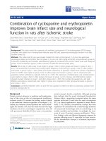
infarction (Fig. 1.1). The diagnosis of complete MCA
territory infarction based on clinical judgment is
unspecific and often requires neuroimaging studies
(Berrouschot et al. 1998).
1.2.1.1.2
Anterior Cerebral Artery
The ACA supplies the head of the caudate nucleus,
the anterior part of the internal capsule, the anterior
perforated substance and the paramedian frontal
lobe above the corpus callosum through the recurrent artery of Heubner (Ghika et al. 1990). Infarction of the paramedian frontal lobe causes weakness
of foot, leg and shoulder and represents a typical
pattern of neurological symptoms (Bogousslavsky
and Regli 1990). Also apraxia of the left arm (“anterior disconnection syndrome”) may be a typical
sign (Geschwind 1965). Transcortical motor and
sensory aphasia, urinary incontinence in bilateral
lesions occur. Abulia is seen in both unilateral and
bilateral frontal lobe infarctions, sometimes of fluctuating intensity. The “alien hand sign” is also found
in frontal lobe infarctions and may be another form
of disconnection syndrome (Geschwind et al. 1995).
Occasionally both ACA territories are supplied by
only one ACA in a unilateral hypoplastic or aplastic
A1 segment. Infarction of both ACA territories will
Fig. 1.1. Upper row, computed tomography of a patient with malignant right middle cerebral artery infarction on day one after
onset of symptoms. Lower row, on day two massive edema with midline shift in spite of hemicraniectomy
6
cause a sudden onset of abulia, paraparesis, apathy,
and incontinence, which may be misinterpreted as
sudden onset of dementia (Ferro 2001). Infarction
of the head of the caudate and the anterior internal capsule by occlusion of the Heubner artery will
cause slight motor weakness, dysarthria, behavioral
changes such as abulia or restlessness and hyperactivity. Cognitive and behavioral changes in these
patients resemble the clinical signs found in patients
with medial thalamic lesions (Bogousslavsky
1994).
1.2.1.1.3
Anterior Choroidal Artery
Blockade of the anterior choroidal artery may cause
infarction of the lateral geniculate body causing
hemianopia with preserved central vision and a
prominent sensory loss with hemiparesis (Bruno
et al. 1989).
1.2.1.2
Large Vessel Occlusive Disease of the Posterior
Circulation
1.2.1.2.1
Obstruction of the Subclavian Artery
In severe occlusive disease of the subclavian artery
(SCA) blood supply of the arm is mainly provided
by reversed flow through the vertebral artery (VA)
arising behind the obstruction. The so-called subclavian-steal syndrome consists of ischemic symptoms in the arm, especially after exercise, such as
pain or numbness or coolness (Reivich et al. 1961).
Consequently a diminished or delayed pulse in the
radial artery or decreased blood pressure on the
side of SCA stenosis can be palpated. Rarely neurological symptoms such as spells of dizziness may be
brought about by exercise of the arm. Even more rare
are ischemic brainstem strokes in subclavian-steal
syndrome (Bornstein and Norris 1986).
1.2.1.2.2
Obstruction of the Vertebral Arteries
The origin of the VA at the SCA is the most common
location of atherosclerotic VA disease. Dizziness,
accompanied by other brainstem symptoms, such
as diplopia, dysarthria, motor or sensory symptoms, suggest embolism towards the brainstem.
Also hemianopia may occur due to VA embolism
G. Gahn
when the emboli travel through the basilar artery
into the posterior cerebral arteries (PCA). The
pathophysiology of proximal VA obstruction is not
well understood, since the VA is rarely operated
on and therefore specimens as in carotid endarterectomy are rare (Caplan 1993). The mechanical
situation in VA is very different form the ICA since
it leaves the SCA at a 90° angle, whereas the ICA
takes off the CCA at an almost 180° angle (Brandt
et al. 2000).
VA obstruction causes hemodynamic problems
in approximately one third of patients with posterior circulation ischemia (Caplan et al. 2004).
Asymmetrical caliber of the two VAs is normal. In
the neck multiple nuchal and muscular branches
provide a network for potential collateral pathways,
that can be activated in VA obstruction.
Intracranial atherosclerotic VA obstruction is
mainly located at the origin of the posterior inferior
cerebellar artery (PICA) and less frequent at the site
of dura penetration. Consequently the most frequent
clinical syndrome in VA occlusive disease is the dorsolateral medullary syndrome (“Wallenberg’s” syndrome) consisting of dizziness, retroorbital pain,
facial numbness, dissociated sensory deficit, weakness, hoarseness, dysphagia and vomiting, nystagmus, Horner’s syndrome and failure of autonomic
respiration (Vuilleumier et al. 1995).
With involvement of the cerebellar hemisphere
supplied by the PICA, subsequent edema may cause
obstruction of the 4th ventricle, hydrocephalus or
compression of the medulla oblongata. Clinically,
involvement of the entire cerebellar hemisphere
can not be distinguished from partial cerebellar
infarction (Amarenco and Hauw 1990). Therefore
patients with neurological symptoms suggesting
infarction within the PICA territory require neuroimaging studies and close clinical monitoring.
In blockade of the anterior spinal artery, ischemia of the medial medulla may occur with contralateral hemiparesis, ipsilateral tongue weakness and
contralateral loss of posterior column sensation (Ho
and Meyer 1981).
Isolated cerebellar infarction without involvement
of the medulla is often difficult to identify, since gait
ataxia, vomiting and dizziness may not be accompanied by typical brainstem symptoms (Barth et al.
1994). Cerebellar edema may compress the medulla
and the pons leading to conjugate eye deviation to
the side opposite the lesion without contralateral
hemiparesis. This sign is probably pathognomonic
for severe cerebellar mass effect and requires immediate intervention.
Stroke Syndromes
1.2.1.2.3
Basilar Artery Obstruction
Basilar artery (BA) occlusive disease is a highly lifethreatening condition first described by Kubik and
Adams (1946). Atherosclerotic changes are mostly
located at the origin of the BA, sometimes extending from the VAs. Often these patients experience
brainstem TIAs such as diplopia, dizziness, weakness of both legs, or occipital headaches (von Campe
et al. 2003). Obstruction of the BA often interrupts
blood supply of the basis of the pons by the superior
cerebellar arteries (SCA). Pseudobulbar paralysis
by interruption of descending tracts to the bulbar
nuclei is often seen. The spinothalamic tract and
the cerebellar hemispheres are often spared from
infarction. Disturbance of eye movements occur
because of infarction of the lateral gaze centers in
the paramedian pontine tegmentum, e.g. the medial
longitudinal fasciculus (internuclear ophthalmoplegia), the parapontine reticular formation (PPRF),
which generates lateral gazes, or the combination
of both, resulting in the so called “one and a halfsyndrome” (Mehler 1989; Voetsch et al. 2004).
Infarction of the medial pontine tegmentum will
cause coma and is a poor prognostic sign (Fisher
1977; Kataoka et al. 1997).
In most patients with BA thrombosis, obstruction is limited to the mid portion of the basilar
artery (Fig. 1.2) (Voetsch et al. 2004). Embolic
occlusion rather than thrombotic occlusion mainly
blocks the distal part of the BA when it divides into
the PCAs. The distal BA supplies the midbrain and
7
the diencephalon by small perforating arteries.
Signs of dysfunction in the rostral brainstem are
a variety of pupillary abnormalities (e.g. anisocoria, afferent pupillary deficit) (Martin et al. 1998;
Mehler 1989). Vertical gaze palsy or skew deviation also point to the midbrain causing the “Top
of the basilar” syndrome (Caplan 1980). Memory
loss may occur in thalamic infarction as well as
agitation, hallucinations mimicking frontal lobe
disorders (Biller et al. 1985; Ghika-Schmid and
Bogousslavsky 2000). The triad of hypersomnia,
supranuclear vertical-gaze defect, and amnesia (the
so-called “paramedian diencephalic syndrome”)
is typically due to bilateral paramedian thalamic
strokes in the territory of the anterior thalamic/subthalamic (or thalamoperforating) arteries (“en ailes
de papillon”) (Meissner et al. 1987).
1.2.1.2.4
Posterior Cerebral Artery (PCA)
Historically, the French neurologist Charles Foix
in 1923 first described the syndrome of infarction in
the PCA territory as a thalamocapsular deficit (Foix
and Masson 1923). The PCAs arise from the BA, but
about 30% of patients have a hypo- or aplastic P1
segment with the PCA nourished by the ICA through
the posterior communicating artery (Margolis et
al. 1971).
Headache in patients with PCA disease is often
retro-orbital or above the eye reflecting innervation of the upper surface of the tentorium by the
first division of the trigeminal nerve (Brandt et
Fig. 1.2. Digital subtraction angiography of a patient with basilar artery thrombosis before (left) and after (right) thrombolytic
therapy. (Courtesy of Prof. von Kummer)
G. Gahn
8
al. 2000; Ferro et al. 1995). Infarction in the PCA
territory usually causes visual symptoms such as
homonymous hemianopia or quadrantanopia and
sensory deficits but seldom paralysis. Patients with
hemianopia due to infarction of the striate cortex
are fully aware of their deficit. In contrast patients
with infarction in the parietal lobe within the MCA
territory have visual neglect and are unaware of
their deficit (Ferber and Karnath 2001). Proximal PCA-occlusion may simulate MCA-infarction
because of thalamic involvement (Chambers et al.
1991). Optokinetic nystagmus is normal in patients
with hemianopia but reduced towards the side of the
visual defect in those with visual neglect (Morrow
and Sharpe 1993).
Some neuropsychological syndromes can be present in PCA infarction (Ferro 2001):
Alexia without agraphia in left occipital lobe
infarction and the splenium of the corpus callosum. Transfer of read words from the functional
right visual cortex to the left sided language center
is impossible due to interruption of the splenium.
Transfer of primary language information for writing or speech is not impaired.
Transcortical sensory aphasia appears in patients
with left PCA infarctions and displays difficulties in
naming objects but no problems in repeating without
understanding. Gerstmann’s syndrome with infarction of the angular gyrus consists of inability for
right-left differentiation, finger anomia, constructional apraxia, agraphia and acalculia. In associative visual agnosia after left PCA infarctions visual
but not tactile recognition of objects is impaired.
Prosopagnosia is a problem with recognizing faces
and occurs in right PCA infarctions. Cortical blindness occurs in bilateral PCA infarctions; however,
pupillary reflexes are preserved (also see Chap. 14).
1.2.2
Lacunar Infarction
Cerebral microangiopathy accounts for 20%–30 %
of all ischemic strokes and is mainly due to long
lasting arterial hypertension. Narrowing of arterioles is caused by so called lipohyalinosis, a process
collecting hyaline substances in the media of the
small cerebral arteries (Fisher 1965c). These small
arteries typically supply exclusively small territories
of less than 1 or 2 cm in diameter. With progression of arterial narrowing blood flow towards the
nourished territory diminishes until oligemia or
ischemia occur. Eventually thrombotic mechanisms
may contribute to the blockade of the artery at this
point. Finally a small infarction, called a “lacune”,
will occur. Especially in the basilar artery atheromatous changes in the arterial wall may occlude the origins of the small penetrating arteries or be the origin
of an expanding thrombosis adherent to the arterial
wall (“microatheroma”) (Fisher and Caplan 1971).
Usually this will cause blockade of several perforating arteries and subsequent small deep infarcts
in contrast to lacunar infarcts which result from
single perforating artery disease. An animal model
to study cerebral microangiopathy in a standardized
matter is presently not available (Caplan 1993).
Conversely we rely on clinical and imaging data in
humans to understand this disease.
Lacunar infarcts are typically located in the basal
ganglia, the deep white matter and in the brainstem
(Fisher 1965a, 1998). Depending on their location
and their size circumscribed neurological symptoms will occur. C. Miller Fisher described the
four classical lacunar syndromes:
•
•
•
•
Pure motor stroke (Fisher and Curry 1964)
Pure sensory stroke (Fisher 1965b)
Ataxic hemiparesis (Fisher 1978)
Dysarthria-clumsy-hand-syndrome
(Fisher 1967; Fisher and Curry 1964)
All these syndromes are a consequence of small
lacunes interrupting pathways in the white matter.
Lacunar strokes almost never cause cortical symptoms such as aphasia or apraxia. Depending again
on the location of the infarctions, patients may
present with combined symptoms sometimes with
preceding stereotyped TIAs. Lacunes within the
internal capsule may cause dense hemiplegia, but
never combined with impaired consciousness or
conjugate eye deviation as in territorial infarcts
with mass effect. At presentation, symptoms may be
subtle or mild and may fluctuate or progress. This is
probably related to the hemodynamic aspect in the
pathogenesis of the disease. Many patients experience progression of the neurological deficits during
the first 24 h after onset of ischemic symptoms, the
so-called capsular warning syndrome (Donnan
et al. 1993; Staaf et al. 2004). Often these patients
wake up in the morning with neurological deficits
which occurred sometimes during sleep and with
comparatively low blood pressure (Chaturvedi et
al. 1999). In contrast embolic strokes tend to occur
after getting up, when activation of the cardiovascular system provokes plaque disruption or increased
cardiac contractility. Notably, recent MRI studies
Stroke Syndromes
showed no differences in stroke subtypes between
waking strokes and strokes occurring during sleep
(Donnan et al. 1993; Fink et al. 2002). Lacunar
syndromes do not specifically help to localize the
infarct to a certain territory in the brain and are
not highly specific for the diagnosis of a “lacune”
(Baumgartner et al. 2003; Gan et al. 1997).
1.2.3
Borderzone Infarction
Borderzone infarcts develop at the junction between
different arterial territories (Adams et al. 1966).
Pathophysiologically two different classes of borderzone infarcts can be identified: infarction between
two arterial territories with a connecting arteriolar collateral network, so-called watershed infarcts,
and infarcts between two arterial territories without
arteriolar collaterals, so-called end-zone infarcts
(Bogousslavsky and Moulin 1995). This classification is based on the anatomic distribution of the
cerebral vascular supply consisting of two main systems: first, superficial arteries surround the brain
parenchyma with an anastomotic network and send
off perforating centripetal branches that do not anastomose (Moody et al. 1990). Second, deep branches
originating from the major arterial branches penetrating the brain without anastomoses. Clinically
relevant borderzones are the anterior borderzone
between the MCA and the ACA and the posterior
borderzone between the MCA and the PCA. They
represent watershed areas and cause mainly cortical
infarction. The subcortical borderzones between the
deep and the superficial perforators represent endzone areas and cause subcortical infarctions (Read
et al. 1998). In the posterior circulation both watershed and end-zone areas exist between the PICA
and the SCA territories. The penetrating branches of
the basilar artery are a potential source of end-zone
infarctions comparable to the lenticulostriate arteries (Bogousslavsky and Moulin 1995).
The underlying mechanism of borderzone
infarcts is the low flow situation (Ringelstein et
al. 1983) in the most distal fields supplied by the
cerebral circulation (“last meadows”) (also see
Chap. 15). Hemodynamic impairment will consequently first cause ischemia in these areas. Typical
clinical situations are a prolonged drop in systemic
blood pressure, e.g. during cardiac surgery causing
bilateral borderzone infarcts or severe occlusive
disease of the internal carotid artery (Bladin and
Chambers 1994). This may only lead to unilateral
9
borderzone infarction in the case of poor collateral
supply through the circle of Willis (Powers 1991).
Clinically stereotyped TIAs or the opticocerebral
symptom with simultaneous amaurosis and contralateral hemiparesis due to critically low blood supply
through an occluded or almost occluded ICA herald
a pending borderzone ischemia (Tsiskaridze et al.
2001). Rarely so-called limb shaking TIAs may point
to a hemodynamic ischemia in ICA occlusive disease
(Baquis et al. 1985).
1.3
Particular Etiological Stroke Syndromes
1.3.1
Cardioembolic Stroke
Thromboembolic stroke mainly derives from cardiac
thrombus formation (Schneider et al. 2004). Less
frequently the source is intra-arterial, from the distal
end of a thrombus within the lumen of an obstructed
carotid or vertebral artery or from an atheromatous
plaque in the cervical arteries or in the aortic arch.
The cardiac embolus usually arises in the anterior
circulation through the internal carotid artery up
into the middle cerebral artery. At a site of sudden
lumen reduction either at the origin of the middle
cerebral artery or more distally at the bifurcation
into the middle cerebral artery branches, it gets
stuck and blocks the lumen of the artery (Caplan
1993). Embolic infarction often turns into hemorrhagic transformation. Most often the middle cerebral artery, especially its inferior branch, is the site
of embolic obstruction (Bogousslavsky et al. 1989).
The embolic material may remain arrested and plug
the artery solidly. It also may break into fragments
and spread into smaller branches more peripherally.
The phenomenon of a clot first lodging in the internal carotid artery, producing profound symptoms of
hemispheric ischemia, and then migrating distally
to a MCA pial branch has been called the “spectacular shrinking deficit” (Minematsu et al. 1992). The
dramatic initial deficit diminishes to a minor deficit
corresponding to the terminal branch artery.
At total of 75% of cardiac emboli reach the
brain. Non-valvular atrial fibrillation with thrombus formation within the left atrial appendix or
the left atrium is the most common reason for cardiac emboli (Ferro 2003). Cardio-embolic infarcts
within the MCA territory carry a high risk for hemorrhagic transformation after reperfusion. Hemor-
10
rhagic transformation is a natural consequence of
cerebral infarction, occurring in up to 65% of stroke
patients and in up to 90% of patients with cardioembolic stroke within the first week after symptom
onset (Molina et al. 2001). Hemorrhagic transformation does not impair neurological outcome after
embolic stroke (Fiorelli et al. 1999). It may even
suggest favorable outcome indicating early reperfusion of the blocked MCA (Molina et al. 2002).
Paradoxical embolism can occur in a patent foramen ovale with a right to left shunt. Embolic material
arising from the pelvic or leg veins or elsewhere in
the venous system may bypass the pulmonary system
and reach the cerebral arteries (Braun et al. 2004).
1.3.2
Dissection
Spontaneous dissection of the internal carotid or
the vertebral artery is an important cause of ischemic stroke in young adults (Fig. 1.3). In the late
1970s Fisher et al. (1978) and Mokri et al. (1979)
described dissections of carotid and vertebral arteries
as detected by modern diagnostic techniques rather
than by post-mortem examination. This may occur
G. Gahn
after both major or trivial traumatic head or neck
injury (Schievink 2001). Many patients have preceding warning symptoms: the typical patient with carotid
artery dissection presents with pain on one side of the
head, face, or neck accompanied by a partial Horner’s
syndrome and followed hours or days later by cerebral
or retinal ischemia (Schievink 2001). Final infarction
may arise mostly due to embolic and seldom due to
hemodynamic mechanism (Benninger et al. 2004).
In carotid artery dissection, Horner’s syndrome
develops in less than half of patients as well as vagal,
hypoglossal or accessorial nerve palsy. The underlying mechanism could be nerve compression, stretching or occlusion of small nourishing branches within
an arterial wall by the intramural hematoma (Mokri
et al. 1996). The pathogenesis of dissection remains
obscure except in patients with obvious collagen tissue
disease such as fibromuscular dysplasia. Other types
of connective tissue alterations may also be associated
with cervical artery dissections (Hausser et al. 2004).
The site of dissection in adults is mainly the internal
carotid artery at its distal extracranial course above
the carotid bulb. In children the site of dissection is
predominately intracranially (Fullerton et al. 2001).
The risk for recurrent dissections is very low (Touze
et al. 2003).
Fig. 1.3. Computed tomography (left), digital subtraction angiography (DSA, middle) and MRI (right) of a patient with internal
carotid artery dissection. Note diffuse swelling of the frontal-parietal cortex on CT on day three after onset of symptoms. At this
time point the patient suffered from a severe left hemiparesis. DSA shows classical “string sign” of cervical artery dissection
with continuous narrowing of arterial lumen. MRI: ADC map (upper images) on day three after onset of symptoms shows small
area of ischemia (dark). Time-to-peak parameter image (lower images) shows delayed contrast inflow to the entire right MCA
territory. The patient underwent stent protected dilatation of the arterial stenosis 6 days after symptom onset and recovered
almost completely from the severe hemiparesis. (Courtesy of Prof. von Kummer)
Stroke Syndromes
1.3.3
Cerebral Venous Thrombosis
Thrombosis of the cerebral veins or sinuses may
develop secondary to infections of the ear or the
paranasal sinuses, to coagulation disorders or spontaneously (Bousser et al. 1985) (also see Chap. 18).
Occlusion of the cerebral venous system may cause
venous infarction stroke. The clinical signs are often
unspecific and may be mainly caused by an obscure
increase in intracranial pressure (Higgins et al.
2004). Fluctuating or permanent focal neurological deficits combined with headache and confusion
may lead to the correct diagnosis. Chemosis and
proptosis with cranial nerve III, IV and VI, and the
ophthalmic division of 5th cranial nerve palsy are
characteristic signs for thrombosis of the anterior
cavernous sinus. Seizures and hemiparesis, predominantly of the leg, are suggestive of the sagittal
sinus (Fig. 1.4). Involvement of the caudal cranial
nerves indicate thrombosis of the posterior part of
the cavernous sinus or the inferior petrous sinus.
Bilateral thalamic infarction should raise the question of straight sinus thrombosis (Herrmann et
al. 2004).
1.3.4
Migraine
Classical migraine with typical visual symptoms
preceding unilateral headache are seldom a differential diagnosis with ischemic stroke. Increased
awareness of patients of ischemic symptoms and
rapid presentation to emergency rooms with immediate initiation of thrombolytic therapy may chal-
11
lenge the physician to identify an ongoing migraine
attack with spreading depression mimicking cerebral ischemia. Accompanying vegetative symptoms
are unspecific. Familiar hemiplegic migraine is a
rare and even more challenging disorder due to its
dramatic course in young patients (Ducros et al.
2001). The association between migraine and stroke
is a dilemma for neurologists. Migraine is associated
with an increased stroke risk and it is considered
an independent risk factor for ischemic stroke in
a particular subgroup of patients. The pathogenesis is not known, but several studies report some
common biochemical mechanisms between the two
diseases. A classification of migraine-related stroke
that encompasses the full spectrum of the possible
relationship between migraine and stroke has been
proposed. It includes three main entities: coexisting
stroke and migraine, stroke with clinical features of
migraine, and migraine-induced stroke. The concept
of migraine-induced stroke is well represented by
migrainous infarction; it is described in the revised
classification of the International Headache Society
(IHS), and it represents the strongest demonstration of the relationship between ischemic stroke and
migraine (Fig. 1.5). An interesting common condition in stroke and migraine is a patent foramen
ovale which could play a pathogenetic role in both
disorders. The association between migraine and
cervical artery dissection is reported in recent studies. Migraine is more frequent in patients with cervical artery dissection (Tzourio et al. 2002). This
supports the hypothesis that an underlying arterial
wall disease could be a predisposing condition for
migraine.
Basilar artery or vertebrobasilar migraine is not
an uncommon type of migraine. Often young woman
Fig. 1.4. Patient with cerebral venous thrombosis. Venous MRA demonstrates occlusion of the sagittal sinus. MRI shows an
intracranial hemorrhage, the typical complication of cerebral venous thrombosis. (Courtesy of Prof. von Kummer)
12
G. Gahn
Fig. 1.5. Patient with hemiplegic migraine. Left, diffusion weighted MRI during migraine attack with severe right hemiparesis.
Note the slight diffusion changes in the left temporal cortex suggesting ischemia. Middle and right, follow-up MRI (protondensity- and T2-weighted) 1 year after migraine attack show no structural changes in the left temporal cortex. Clinically the
patient recovered completely. (Courtesy of Prof. von Kummer)
experience visual disturbance similar to those in typical migraine but involving both visual fields. These
symptoms may be accompanied by vertigo, ataxia,
dysarthria, and sensory disturbances in both arms
or legs bilaterally (Evans and Linder 2002).
1.3.5
Coma
In animals, destruction of the ascending reticular
activating system (ARAS) induces a state of coma
(Moruzzi and Magoun 1949). In men the ARAS is
located in the paramedian tegmentum of the dorsal
pons and the midbrain extending as a complex polysynaptic system from the upper half of the pons
through the midbrain to the dorsal part of the hypothalamus and to the thalamic reticular formation
(Vincent 2000).
In close vicinity of the ARAS the medial longitudinal fasciculus (MLF) and the oculomotor and
trochlear nuclei are situated. Combined coma and
oculomotor disturbances points to a brainstem
lesion (Parvizi and Damasio 2003). Other clinical
symptoms like respiratory pattern, pupillary reflex,
and position or movement patterns of the limbs may
help localize the site of the lesion.
Abnormal respiratory breathing patterns are of
limited practical value, since a comatose patient due
to cerebrovascular disease often requires immediate airway protection and mechanical ventilation.
Urgent diagnostic imaging will provide the appro-
priate diagnostic information (Brazis et al. 1990).
“Cheyne-Stokes respiration” consists of brief periods of hyperventilation regularly combined with
short episodes of apnea. During hyperventilation
periods the patients may become more alert. The
cause of Cheyne-Stokes respiration can be a large
bilateral cortical lesion, bilateral thalamic lesions,
as well as metabolic disturbances in uremia, anoxia,
heart failure (Cherniack and Longobardo 1973).
“Hyperventilation” may occur in midbrain or pontine lesions and is often accompanied by severe respiratory distress. It can also be found in brainstem
tumors leading to local pH lowering because of their
high metabolism and thereby providing a breathing
stimulus to the medullary respiratory center (Plum
1972). A lesion in the lateral tegmentum of the lower
pons may cause a “apneustic breathing” with long
inspiratory pauses (Plum and Alvord 1964). Low
pontine and medullary lesions may cause “cluster breathing” with irregular breathing sequences.
“Ataxic breathing” displays completely irregular breathing patterns, often seen in terminally ill
patients with impairment of the dorsomedial respiratory centers (Brazis et al. 1990).
The pupillary light reflex may help differentiating
metabolic cause from structural brainstem lesion in
comatose patients (Tokuda et al. 2003). The light
reflex is very resistant to metabolic dysfunction. An
abnormal light reflex, especially when unilateral,
points to a midbrain lesion. Bilateral diencephalic
lesions or metabolic coma may cause bilateral small
pupils well reacting to light (“diencephalic pupils”).
Stroke Syndromes
Midbrain lesions abolish the light reflex when
located in the tectum or the pretectum and thereby
disrupting the posterior commissure. Hippus and
the ciliospinal reflex may be preserved. Tegmental
lesions damaging the oculomotor nuclei may cause
an irregular shape of the pupils, anisocoria and loss
of light reflex. Tegmental lesions in the pons cause
miosis by disruption of the descending sympathetic
fibers (pinpoint pupils, minimally reacting to light).
Lateral pontine or medullary lesions cause Horner’s
syndrome (Brazis et al. 1990).
1.3.6
Eye Movement Abnormalities
In comatose patients evaluation of the oculomotor
system relies on evaluation and observation of involuntary eye movements. The oculocephalic and the
oculovestibular reflexes disappear in deep coma.
“Periodic alternating gaze” (Ping-Pong gaze)
with alternating eye movements from one extreme
of horizontal gaze to the other lasting from 2 to 5 s
indicate bilateral cerebral damage with preserved
brainstem but may also occur in brainstem hemorrhage (Masucci et al. 1981).
“Repetitive divergence” consists of slow divergence of the eyes followed by rapid return to mid
position. This rare phenomenon may be observed in
metabolic coma (Noda et al. 1987).
Nystagmoid jerking of one eye may occur in midto lower pontine lesions (Plum and Posner 1980).
Ocular bobbing consists of sudden bilateral downward movement of both eyes followed by slow return
to mid position. Pontine and cerebellar lesions as
well as metabolic and encephalitic disorders may
cause ocular bobbing. Inverse ocular bobbing
(“ocular dipping”) may occur in hypoxic encephalopathy (Stark et al. 1984).
Conjugate gaze palsy or forced eye deviation may
point to hemispheric lesions when looking towards
the side of the lesion (Tijssen et al. 1991) and will
point to a brainstem lesion when looking away from
the side of lesion. Damage to the MLF will cause disconjugated gazes, e.g. failure of adduction of the eye
on the side of lesion or, as in damage of the PPRF
and the MLF preservation of only abduction of the
contralateral eye (Wall and Wray 1983).
Abnormalities of vertical gazes may occur in both
unilateral and bilateral midbrain and diencephalic
lesions and can be evaluated by the doll’s eye maneuver or alternatively by irrigation of warm water in
both ears causing upward deviation or bilateral cold
13
water causing downward deviation (Bogousslavsky
et al. 1994; Hommel and Bogousslavsky 1991).
“Skew deviation” may be seen by various brainstem lesions and in increased intracranial pressure as well as in hepatic coma. Skew deviations are
ipsiversive (ipsilateral eye undermost) with caudal
pontomedullary lesions and contraversive (contralateral eye lowermost) with rostral pontomesencephalic lesions. They are associated with concomitant ocular torsion and tilts of the subjective visual
vertical toward the undermost eye (Brandt and
Dieterich 1993).
“Decorticate rigidity” may occur unilaterally
with hemispheric and diencephalic lesions contralateral to the lesion. It consists of adduction of the
arm, flexion in the elbow, and pronation and flexion
of the wrist. “Decerebrate rigidity” displays extension and pronation of the arms and forced plantar
flexion of the feet. It occurs in upper pontine and
midbrain destruction. Extension of the arms and
weak flexion of the legs suggest tegmental pontine
damage (Bogousslavsky et al. 1994; Brazis et al.
1990).
1.4
Summary
We gave a short overview of the most important
stroke syndromes in the clinical setting. Knowledge
of these syndromes helps to understand the complex
pathophysiology of cerebral ischemia. Combination
of clinical findings with the data from the new and
evolving imaging techniques certainly facilitates
and improves care for stroke patients.
References
Adams JH, Brierley JB, Connor RC et al (1966) The effects of
systemic hypotension upon the human brain. Clinical and
neuropathological observations in 11 cases. Brain 89:235–
268
Amarenco P, Hauw JJ (1990) Cerebellar infarction in the territory of the anterior and inferior cerebellar artery. A clinicopathological study of 20 cases. Brain 113:139–155
Baquis GD, Pessin MS, Scott RM (1985) Limb shaking – a
carotid TIA. Stroke 16:444–448
Barth A, Bogousslavsky J, Regli F (1994) Infarcts in the territory of the lateral branch of the posterior inferior cerebellar artery. J Neurol Neurosurg Psychiatry 57:1073–1076
Baumgartner RW, Sidler C, Mosso M et al (2003) Ischemic lacunar stroke in patients with and without potential mechanism other than small-artery disease. Stroke 34:653–659
14
Beis JM, Keller C, Morin N et al (2004) Right spatial neglect
after left hemisphere stroke: qualitative and quantitative
study. Neurology 63:1600–1605
Benninger DH, Georgiadis D, Kremer C et al (2004) Mechanism of ischemic infarct in spontaneous carotid dissection.
Stroke 35:482–485
Berrouschot J, Barthel H, von Kummer R et al (1998) 99m
technetium-ethyl-cysteinate-dimer single-photon emission CT can predict fatal ischemic brain edema. Stroke
29:2556–2562
Biller J, Sand JJ, Corbett JJ et al (1985) Syndrome of the paramedian thalamic arteries: clinical and neuroimaging correlation. J Clin Neuroophthalmol 5:217–223
Bladin CF, Chambers BR (1994) Frequency and pathogenesis
of hemodynamic stroke. Stroke 25:2179–2182
Bogousslavsky J (1994) Frontal stroke syndromes. Eur Neurol
34:306–315
Bogousslavsky, Moulin T (1995) Borderzone Infarcts. In:
Bogousslavsky J, Caplan LR (eds) Stroke syndromes, 1st
edn. Cambridge University Press, Cambridge, pp 358–
365
Bogousslavsky J, Regli F (1990) Anterior cerebral artery territory infarction in the Lausanne Stroke Registry. Clinical
and etiologic patterns. Arch Neurol 47:144–150
Bogousslavsky J, van Melle G, Regli F (1989) Middle cerebral
artery pial territory infarcts: a study of the Lausanne Stroke
Registry. Ann Neurol 25:555–560
Bogousslavsky J, Maeder P, Regli F et al (1994) Pure midbrain
infarction: clinical syndromes, MRI, and etiologic patterns.
Neurology 44:2032–2040
Bornstein NM, Norris JW (1986) Subclavian steal: a harmless
haemodynamic phenomenon? Lancet 2:303–305
Bousser MG, Chiras J, Bories J et al (1985) Cerebral venous
thrombosis-a review of 38 cases. Stroke 16:199–213
Brandt T, Dieterich M (1993) Skew deviation with ocular torsion: a vestibular brainstem sign of topographic diagnostic
value. Ann Neurol 33:528–534
Brandt T, Steinke W, Thie A et al (2000) Posterior cerebral
artery territory infarcts: clinical features, infarct topography, causes and outcome 1. Cerebrovasc Dis 10:170–182
Braun M, Gliech V, Boscheri A et al (2004) Transcatheter closure of patent foramen ovale (PFO) in patients with paradoxical embolism. Periprocedural safety and mid-term
follow-up results of three different device occluder systems.
Eur Heart J 25:424–430
Brazis PW, Masdeu JC, Biller J (1990) Localization in clinical
neurology, 3rd edn. Little Brown, Boston
Bruno A, Graff-Radford NR, Biller J et al (1989) Anterior choroidal artery territory infarction: a small vessel disease.
Stroke 20:616–619
Busch HJ, Buschmann IR, Mies G et al (2003) Arteriogenesis in hypoperfused rat brain. J Cereb Blood Flow Metab
23:621–628
Caplan LR (1980) “Top of the basilar” syndrome. Neurology
30:72–79
Caplan LR (1993) Stroke. A clinical approach, 2nd edn. Butterworth-Heinemann, Newton
Caplan LR, Wityk RJ, Glass TA et al (2004) New England
Medical Center Posterior Circulation registry. Ann Neurol
56:389–398
Chambers BR, Brooder RJ, Donnan GA (1991) Proximal posterior cerebral artery occlusion simulating middle cerebral
artery occlusion. Neurology 41:385–390
G. Gahn
Chaturvedi S, Adams HP Jr, Woolson RF (1999) Circadian
variation in ischemic stroke subtypes. Stroke 30:1792–
1795
Cherniack NS, Longobardo GS (1973) Cheyne-stokes breathing. An instability in physiologic control. N Engl J Med
288:952–957
Coyle P, Heistad DD (1991) Development of collaterals in the
cerebral circulation. Blood Vessels 28:183–189
De Groot E, Hovingh GK, Wiegman A et al (2004) Measurement of arterial wall thickness as a surrogate marker for
atherosclerosis. Circulation 109:III33–III38
Donnan GA, Bladin PF, Berkovic SF et al (1991) The stroke
syndrome of striatocapsular infarction. Brain 114:51–70
Donnan GA, O’Malley HM, Quang L et al (1993) The capsular warning syndrome: pathogenesis and clinical features.
Neurology 43:957–962
Ducros A, Denier C, Joutel A et al (2001) The clinical spectrum
of familial hemiplegic migraine associated with mutations
in a neuronal calcium channel. N Engl J Med 345:17–24
Evans RW, Linder SL (2002) Management of basilar migraine.
Headache 42:383–384
Feldmann E, Daneault N, Kwan E et al (1990) Chinese-white
differences in the distribution of occlusive cerebrovascular
disease. Neurology 40:1541–1545
Ferber S, Karnath HO (2001) Size perception in hemianopia
and neglect. Brain 124:527–536
Ferro JM (2001) Hyperacute cognitive stroke syndromes. J
Neurol 248:841–849
Ferro JM (2003) Cardioembolic stroke: an update. Lancet
Neurol 2:177–188
Ferro JM (2004) Patterns of ischaemic cerebral diseases. J
Neurol 251:1–10
Ferro JM, Melo TP, Oliveira V et al (1995) A multivariate study
of headache associated with ischemic stroke. Headache
35:315–319
Fink JN, Kumar S, Horkan C et al (2002) The stroke patient
who woke up: clinical and radiological features, including
diffusion and perfusion MRI. Stroke 33:988–993
Fiorelli M, Bastianello S, von Kummer R et al (1999) Hemorrhagic transformation within 36 hours of a cerebral infarct:
relationships with early clinical deterioration and 3-month
outcome in the European Cooperative Acute Stroke Study I
(ECASS I). Stroke 30:2280–2284
Fisher CM (1951) Occlusion of the internal carotid artery. Arch
Neurol Psychiatry 65:346–377
Fisher CM (1965a) Lacunes: small, deep cerebral infarcts. Neurology 15:774–784
Fisher CM (1965b) Pure sensory stroke involving face, arm,
and leg. Neurology 15:76–80
Fisher CM (1965c) The vascular lesion in lacunae. Trans Am
Neurol Assoc 90:243-5:243–245
Fisher CM (1967) A lacunar stroke. The dysarthria-clumsy
hand syndrome. Neurology 17:614–617
Fisher CM (1977) Bilateral occlusion of basilar artery branches.
J Neurol Neurosurg Psychiatry 40:1182–1189
Fisher CM (1978) Ataxic hemiparesis. A pathologic study. Arch
Neurol 35:126–128
Fisher CM (1998) Lacunes: small, deep cerebral infarcts. 1965.
Neurology 50:841
Fisher CM, Caplan LR (1971) Basilar artery branch occlusion:
a cause of pontine infarction. Neurology 21:900–905
Fisher CM, Curry HB (1964) Pure motor hemiplegia. Trans Am
Neurol Assoc 89:94-7:94–97
Stroke Syndromes
Fisher CM, Ojemann RG, Roberson GH (1978) Spontaneous
dissection of cervico-cerebral arteries. Can J Neurol Sci
5:9–19
Foix C, Masson A (1923) Le syndrome de l’atère cérebrale postérieur. Presse méd 31:261–365
Fullerton HJ, Johnston SC, Smith WS (2001) Arterial dissection
and stroke in children. Neurology 57:1155–1160
Gan R, Sacco RL, Kargman DE et al (1997) Testing the validity
of the lacunar hypothesis: the Northern Manhattan Stroke
Study experience. Neurology 48:1204–1211
Geschwind N (1965) Disconnexion syndromes in animals and
man. II. Brain 88:585–644
Geschwind N (1975) The apraxias: neural mechanisms of disorders of learned movement. Am Sci 63:188–195
Geschwind DH, Iacoboni M, Mega MS et al (1995) Alien hand
syndrome: interhemispheric motor disconnection due to a
lesion in the midbody of the corpus callosum. Neurology
45:802–808
Ghika JA, Bogousslavsky J, Regli F (1990) Deep perforators
from the carotid system. Template of the vascular territories. Arch Neurol 47:1097–1100
Ghika-Schmid F, Bogousslavsky J (2000) The acute behavioral
syndrome of anterior thalamic infarction: a prospective
study of 12 cases. Ann Neurol 48:220–227
Hacke W, Schwab S, Horn M et al (1996) ‘Malignant’ middle
cerebral artery territory infarction: clinical course and
prognostic signs. Arch Neurol 53:309–315
Hausser I, Muller U, Engelter S et al (2004) Different types
of connective tissue alterations associated with cervical
artery dissections. Acta Neuropathol (Berl) 107:509–514
Heinsius T, Bogousslavsky J, van Melle G (1998) Large infarcts
in the middle cerebral artery territory. Etiology and outcome patterns. Neurology 50:341–350
Hennerici MG (2004) The unstable plaque. Cerebrovasc Dis
17 [Suppl 3]:17–22
Herrmann KA, Sporer B, Yousry TA (2004) Thrombosis of the
internal cerebral vein associated with transient unilateral
thalamic edema: a case report and review of the literature.
AJNR Am J Neuroradiol 25:1351–1355
Higgins JNP, Gillard JH, Owler BK et al (2004) MR venography in idiopathic intracranial hypertension: unappreciated and misunderstood. J Neurol Neurosurg Psychiatry
75:621–625
Ho KL, Meyer KR (1981) The medial medullary syndrome.
Arch Neurol 38:385–387
Hochachka PW, Buck LT, Doll CJ et al (1996) Unifying theory
of hypoxia tolerance: molecular/metabolic defense and
rescue mechanisms for surviving oxygen lack. Proc Natl
Acad Sci USA 93:9493–9498
Hollenhorst RW (1958) Ocular manifestations of insufficiency
or thrombosis of the internal carotid artery. Trans Am Ophthalmol Soc 56:474–506
Hommel M, Bogousslavsky J (1991) The spectrum of vertical
gaze palsy following unilateral brainstem stroke. Neurology 41:1229–1234
Hossmann KA (1999) The hypoxic brain. Insights from ischemia research. Adv Exp Med Biol 474:155–169
Karnath HO, Himmelbach M, Rorden C (2002) The subcortical anatomy of human spatial neglect: putamen, caudate
nucleus and pulvinar. Brain 125:350–360
Kataoka S, Hori A, Shirakawa T et al (1997) Paramedian pontine infarction. Neurological/topographical correlation.
Stroke 28:809–815
15
Kennedy J, Buchan AM (2004) Acute neurovascular syndromes:
hurry up, please, it’s time. Stroke 35:360–362
Kertesz A (1993) Clinical forms of aphasia. Acta Neurochir
Suppl (Wien) 56:52–58
Kubik C, Adams R (1946) Occlusion of the basilar artery – a
clinical and pathological study. Brain 69:73–121
Margolis MT, Newton TH, Hoyt WF (1971) Cortical branches
of the posterior cerebral artery. Anatomic-radiologic correlation. Neuroradiology 2:127–135
Markus HS (2004) Cerebral perfusion and stroke. J Neurol
Neurosurg Psychiatry 75:353–361
Martin PJ, Chang HM, Wityk R et al (1998) Midbrain infarction: associations and aetiologies in the New England
Medical Center Posterior Circulation Registry. J Neurol
Neurosurg Psychiatry 64:392–395
Masucci EF, Fabara JA, Saini N et al (1981) Periodic alternating
ping-pong gaze. Ann.Ophthalmol 13:1123–1127
Mehler MF (1989) The rostral basilar artery syndrome: diagnosis, etiology, prognosis. Neurology 39:9–16
Meissner I, Sapir S, Kokmen E et al (1987) The paramedian
diencephalic syndrome: a dynamic phenomenon. Stroke
18:380–385
Minematsu K, Yamaguchi T, Omae T (1992) ‘Spectacular
shrinking deficit’: rapid recovery from a major hemispheric syndrome by migration of an embolus. Neurology
42:157–162
Mokri B, Sundt TM Jr, Houser OW (1979) Spontaneous internal carotid dissection, hemicrania, and Horner’s syndrome.
Arch Neurol 36:677–680
Mokri B, Silbert PL, Schievink WI et al (1996) Cranial nerve
palsy in spontaneous dissection of the extracranial internal
carotid artery. Neurology 46:356–359
Molina CA, Montaner J, Abilleira S et al (2001) Timing of Spontaneous Recanalization and Risk of Hemorrhagic Transformation in Acute Cardioembolic Stroke. Stroke 32:1079–1084
Molina CA, Alvarez-Sabin J, Montaner J et al (2002) Thrombolysis-related hemorrhagic infarction: a marker of early
reperfusion, reduced infarct size, and improved outcome
in patients with proximal middle cerebral artery occlusion.
Stroke 33:1551–1556
Moody DM, Bell MA, Challa VR (1990) Features of the cerebral vascular pattern that predict vulnerability to perfusion
or oxygenation deficiency: an anatomic study. AJNR Am J
Neuroradiol 11:431–439
Morrow MJ, Sharpe JA (1993) Retinotopic and directional deficits of smooth pursuit initiation after posterior cerebral
hemispheric lesions. Neurology 43:595–603
Moruzzi G, Magoun HW (1949) Brain stem reticular formation and activation of the EEG. Electroencephalogr Clin
Neurophysiol 1:455
Noda S, Ide K, Umezaki H et al (1987) Repetitive divergence.
Ann Neurol 21:109–110
Olsen TS (1991) Outcome following occlusion of the middle
cerebral artery. Acta Neurol Scand 83:254–258
Parvizi J, Damasio AR (2003) Neuroanatomical correlates of
brainstem coma. Brain 126:1524–1536
Pedersen PM, Vinter K, Olsen TS (2004) Aphasia after stroke:
type, severity and prognosis. The Copenhagen aphasia
study. Cerebrovasc Dis 17:35–43
Plum F (1972) Hyperpnea, hyperventilation, and brain dysfunction. Ann Intern Med 76:328
Plum F, Alvord EC (1964) Apneustic breathing in man. Arch
Neurol 10:101–112
16
Plum F, Posner JB (1980) The diagnosis of stupor and coma,
3rd edn. Davis, Philadelphia
Powers WJ (1991) Cerebral hemodynamics in ischemic cerebrovascular disease. Ann Neurol 29:231–240
Read SJ, Pettigrew L, Schimmel L et al (1998) White matter
medullary infarcts: acute subcortical infarction in the centrum ovale. Cerebrovasc Dis 8:289–295
Reivich M, Holling HE, Roberts B et al (1961) Reversal of blood
flow through the vertebral artery and its effect on cerebral
circulation. N Engl J Med 265:878–885
Ringelstein EB, Zeumer H, Angelou D (1983) The pathogenesis
of strokes from internal carotid artery occlusion. Diagnostic and therapeutical implications. Stroke 14:867–875
Ringelstein EB, Biniek R, Weiller C et al (1992) Type and extent
of hemispheric brain infarctions and clinical outcome in
early and delayed middle cerebral artery recanalization.
Neurology 42:289–298
Russmann H, Vingerhoets F, Ghika J et al (2003) Acute infarction limited to the lenticular nucleus: clinical, etiologic, and
topographic features. Arch.Neurol 60:351–355
Schievink WI (2001) Spontaneous dissection of the carotid
and vertebral arteries. N Engl J Med 344:898–906
Schneider AT, Kissela B, Woo D et al (2004) Ischemic stroke
subtypes: a population-based study of incidence rates
among blacks and whites. Stroke 35:1552–1556
Spinazzola L, Cubelli R, Della SS (2003) Impairments of trunk
movements following left or right hemisphere lesions: dissociation between apraxic errors and postural instability.
Brain 126:2656–2666
Staaf G, Geijer B, Lindgren A et al (2004) Diffusion-weighted
MRI findings in patients with capsular warning syndrome.
Cerebrovasc Dis 17:1–8
Stark SR, Masucci EF, Kurtzke JF (1984) Ocular dipping. Neurology 34:391–393
G. Gahn
Tijssen CC, van Gisbergen JA, Schulte BP (1991) Conjugate
eye deviation: side, site, and size of the hemispheric lesion.
Neurology 41:846–850
Tokuda Y, Nakazato N, Stein GH (2003) Pupillary evaluation for
differential diagnosis of coma. Postgrad Med J 79:49–51
Touze E, Gauvrit JY, Moulin T et al (2003) Risk of stroke and
recurrent dissection after a cervical artery dissection: a
multicenter study. Neurology 61:1347–1351
Tsiskaridze A, Devuyst G, de Freitas GR et al (2001) Stroke with
internal carotid artery stenosis. Arch Neurol 58:605–609
Tzourio C, Benslamia L, Guillon B et al (2002) Migraine and
the risk of cervical artery dissection: a case-control study.
Neurology 59:435–437
Urban PP, Wicht S, Vukurevic G et al (2001) Dysarthria in acute
ischemic stroke: lesion topography, clinicoradiologic correlation, and etiology. Neurology 56:1021–1027
Vincent SR (2000) The ascending reticular activating systemfrom aminergic neurons to nitric oxide. J Chem Neuroanat
18:23–30
Voetsch B, DeWitt LD, Pessin MS et al (2004) Basilar artery
occlusive disease in the New England Medical Center Posterior Circulation Registry. Arch Neurol 61:496–504
Von Campe G, Regli F, Bogousslavsky J (2003) Heralding manifestations of basilar artery occlusion with lethal or severe
stroke. J Neurol Neurosurg Psychiatry 74:1621–1626
Vuilleumier P, Bogousslavsky J, Regli F (1995) Infarction of
the lower brainstem. Clinical, aetiological and MRI-topographical correlations. Brain 118:1013–1025
Wall M, Wray SH (1983) The one-and-a-half syndrome-a
unilateral disorder of the pontine tegmentum: a study
of 20 cases and review of the literature. Neurology
33:971–980
Wray SH (1993) The management of acute visual failure. J
Neurol Neurosurg Psychiatry 56:234–240
Clinical Efficacy of MRI in Stroke
2
17
Clinical Efficacy of MRI in Stroke
Rüdiger von Kummer
CONTENTS
2.1
2.2
2.2.1
2.2.2
2.2.3
2.2.4
2.2.5
2.2.6
2.3
Introduction 17
Hierarchy of Efficacy Levels for Diagnostic
Imaging 17
Feasibility and Technical Capacity of Stroke
MRI 18
Diagnostic Accuracy 19
Diagnostic Impact 19
Therapeutic Impact 20
Impact on Patients’ Clinical Outcome 20
Impact on Health Care Costs 20
Summary 20
References 21
2.1
Introduction
It is well established that computed tomography
(CT) identifies patients with acute cerebral ischemia
among stroke syndrome patients and thus enables
effective thrombolytic therapy (The ATLANTIS, ECASS, and NINDS rt-PA Study Group
Investigators 2004). It is a matter of debate,
however, whether information provided by imaging other than the exclusion of hemorrhage, e.g. the
assessment of ischemic edema, arterial pathology,
or perfusion deficit, can really improve the clinical
outcome of acute ischemic stroke patients and can
thus reduce health costs (Powers 2000; Powers and
Zivin 1998; Hacke and Warach 2000) Moreover,
new imaging technology like magnetic resonance
imaging (MRI) offers new insights into acute stroke
pathology that may result in improved treatment for
more patients. This book will provide arguments for
the question of whether MRI should be implemented
in acute stroke management or not. This chapter
will outline the theoretical background needed to
understand under which conditions vascular and
R. von Kummer, MD
Department of Neuroradiology, University of Technology
Dresden, Fetscherstrasse 74, 01307 Dresden, Germany
brain pathology depicted by MRI will be clinically
effective. Subsequent chapters will describe the
stroke pathology depicted by MRI in more detail
and discuss its impact on stroke treatment and clinical outcome.
2.2
Hierarchy of Efficacy Levels for
Diagnostic Imaging
In theory, MRI can be clinically effective in acute
stroke patients on six different levels (Fryback and
Thornbury 1991; Kent and Larson 1992; Sunshine
and Applegate 2004) (Table 2.1): (1) MRI will reduce
health care costs, if it enables treatment that prevents disability and death in stroke victims. (2) MRI
will improve the clinical outcome of stroke patients
if it can identify patients who will benefit from an
effective treatment, e.g. thrombolysis, and exclude
others who will not benefit. (3) To identify patients
who will benefit from a specific treatment, MRI
must provide relevant information for the choice of
treatment not available from other sources. (4) This
could include MRI sequences that make it possible
to exclude brain hemorrhage and other diseases that
mimic ischemic stroke, and assess ischemic edema,
perfusion disturbance, mass effect, arterial wall
pathology, and obstruction. (5) The MRI sequence
should be sensitive and specific for stroke pathology
early after symptom onset. (6) This requires that
the MRI sequence is technically capable of reliably
detecting the relevant stroke pathology and can be
feasibly performed in acute stroke patients.
It is important to bear in mind that diagnostic
imaging is only clinically effective if an effective
treatment is available, and the information provided
by imaging identifies conditions where such treatment is beneficial. The clinical efficacy at any level
in this hierarchy is a precondition for the efficacy
of a higher level, but is not sufficient to guarantee
improved clinical outcome. For example, the capac-