Chronic inflammation mechanisms and regulation
Bạn đang xem bản rút gọn của tài liệu. Xem và tải ngay bản đầy đủ của tài liệu tại đây (19.54 MB, 679 trang )
Masayuki Miyasaka · Kiyoshi Takatsu
Editors
Chronic
Inflammation
Mechanisms and Regulation
Chronic Inflammation
Masayuki Miyasaka • Kiyoshi Takatsu
Editors
Chronic Inflammation
Mechanisms and Regulation
Editors
Masayuki Miyasaka
Interdisciplinary Program for Biomedical
Sciences
Institute for Academic Initiatives
Osaka University
Suita, Japan
WPI Immunology Frontier Center
Osaka University
Suita, Japan
MediCity Research Laboratory
University of Turku
Turku, Finland
Kiyoshi Takatsu
Department of Immunobiology and
Pharmacological Genetics
Graduate School of Medicine and
Pharmaceutical Sciences
University of Toyama
Toyama, Japan
Toyama Prefectural Institute for Pharmaceutical
Research
Toyama, Japan
ISBN 978-4-431-56066-1
ISBN 978-4-431-56068-5
DOI 10.1007/978-4-431-56068-5
(eBook)
Library of Congress Control Number: 2016945256
© Springer Japan 2016
This work is subject to copyright. All rights are reserved by the Publisher, whether the whole or part of
the material is concerned, specifically the rights of translation, reprinting, reuse of illustrations,
recitation, broadcasting, reproduction on microfilms or in any other physical way, and transmission
or information storage and retrieval, electronic adaptation, computer software, or by similar or
dissimilar methodology now known or hereafter developed.
The use of general descriptive names, registered names, trademarks, service marks, etc. in this
publication does not imply, even in the absence of a specific statement, that such names are exempt
from the relevant protective laws and regulations and therefore free for general use.
The publisher, the authors and the editors are safe to assume that the advice and information in this
book are believed to be true and accurate at the date of publication. Neither the publisher nor the
authors or the editors give a warranty, express or implied, with respect to the material contained
herein or for any errors or omissions that may have been made.
Printed on acid-free paper
This Springer imprint is published by Springer Nature
The registered company is Springer Japan KK
Preface
Inflammation, a reaction characterized by redness, fever, swelling, and pain, has
been considered a homeostatic tissue repair mechanism, which is evoked by the
body in response to infections and/or tissue injury. However, accumulating evidence indicates that, when inflammation becomes chronic, it acts as a strong
disease-promoting factor in a number of pathological disorders including arteriosclerosis, obesity, cancer, and Alzheimer disease. Chronic inflammation also promotes aging. Despite such importance, the dismaying fact is that we know very
little about why inflammatory reactions that would usually subside continue and
become chronic. More specifically, we do not know precisely what type of factors
induce chronic inflammation and promote its prolongation. Also we have little
knowledge about how chronic inflammation causes tissue degeneration and other
disorders. Furthermore, we have no effective treatment against chronic inflammation at present.
Realizing these situations, a key funding body of the Government of Japan, the
Japan Science and Technology Agency (JST), launched two major research programs (CREST and PRESTO) on chronic inflammation in 2010; CRESTO is a
funding program for team-oriented research, whereas PRESTO is for independent
research by young investigators. From 2010 until now, in the research area of
chronic inflammation, 17 teams were selected for CRESTO and conducted research
for 5 years (each team receiving 150–500 million yen in total), and 37 researchers
were selected and conducted research for 3–5 years in PRESTO (each scientist
receiving 30–40 million yen for 3-year research and 50–100 million yen for 5-year
research).
This book represents a compendium of such research efforts. Members of the
CREST and PRESTO projects contributed a chapter on their own work, and
research supervisors of the CRESTO and PRESTO projects (M.M. and K.T.,
respectively) edited the book. As you see in this book, thanks to the painstaking
and persistent hard work by the CRESTO and PRESTO members, we are now
beginning to understand what induces and maintains the chronicity of inflammation, and what kinds of mechanisms chronic inflammation utilizes to induce specific
v
vi
Preface
diseases including cancer, degenerative neurological disorders, and arteriosclerotic
diseases. We have also succeeded in creating novel technologies that allow for the
early detection and quantitative assessment of chronic inflammation.
Producing this book required the efforts of many people who deserve credit and
thanks. First, we would like to thank all the CRESTO and PRESTO investigators,
who worked strenuously on the subject of chronic inflammation and contributed a
nice chapter for the book. Second, our special thanks go to research officers of JST
and AMED (Japan Agency for Medical Research and Development) (CREST
“Chronic Inflammation” is now under the supervision of AMED since 2015),
particularly to Shinichi Kato (JST-CREST), Akihiko Kasahara (AMED-CREST),
and Isao Serizawa (PRESTO), who kept the projects organized and meticulously
prepared a number of research meetings for the members. Third, we are indebted to
the editorial assistance by Yuko Matsumoto and Yasutaka Okazaki of Springer
Japan. Fourth, we wish to acknowledge the constant support and understanding of
our wives, Chieko Takatsu and Etsuko Miyasaka. Finally, we thank you, the reader,
for your interest in this research field. We will be more than happy if our efforts are
successful in providing you with useful and stimulating information that will lead to
new developments in the field of chronic inflammation.
Suita, Japan
Toyama, Japan
September 2015
Masayuki Miyasaka
Kiyoshi Takatsu
Contents
Part I
Basic Mechanisms Underlying Induction, Progression, and
Resolution of Chronic Inflammation
1
Prostaglandins in Chronic Inflammation . . . . . . . . . . . . . . . . . . . .
Tomohiro Aoki and Shuh Narumiya
2
Cellular and Molecular Mechanisms of Chronic InflammationAssociated Organ Fibrosis . . . . . . . . . . . . . . . . . . . . . . . . . . . . . . .
Tatsuya Tsukui, Shigeyuki Shichino, Takeshi Shimaoka, Satoshi Ueha,
and Kouji Matsushima
3
19
3
Sema4A and Chronic Inflammation . . . . . . . . . . . . . . . . . . . . . . . .
Daisuke Ito and Atsushi Kumanogoh
37
4
MicroRNAs in Chronic Inflammation . . . . . . . . . . . . . . . . . . . . . . .
Y. Ito, S. Mokuda, K. Miyata, T. Matsushima, and H. Asahara
49
5
Genetic Dissection of Autoinflammatory Syndrome . . . . . . . . . . . .
Koji Yasutomo
63
6
Structural Biology of Chronic Inflammation-Associated Signalling
Pathways: Toward Structure-Guided Drug Development . . . . . . . .
Reiya Taniguchi and Osamu Nureki
7
Lipid Signals in the Resolution of Inflammation . . . . . . . . . . . . . . .
Makoto Arita
8
Regulation of Chronic Inflammation by Control of Macrophage
Activation and Polarization . . . . . . . . . . . . . . . . . . . . . . . . . . . . . .
Junko Sasaki and Takehiko Sasaki
9
77
89
97
Clarification of the Molecular Mechanisms That Negatively
Regulate Inflammatory Responses . . . . . . . . . . . . . . . . . . . . . . . . . 109
Takashi Tanaka
vii
viii
Contents
10
The Drosophila Toll Pathway: A Model of Innate Immune Signalling
Activated by Endogenous Ligands . . . . . . . . . . . . . . . . . . . . . . . . . 119
Takayuki Kuraishi, Hirotaka Kanoh, Yoshiki Momiuchi, Hiroyuki
Kenmoku, and Shoichiro Kurata
Part II
Imaging Analyses of Chronic Inflammation
11
Macrophage Dynamics During Bone Resorption and Chronic
Inflammation . . . . . . . . . . . . . . . . . . . . . . . . . . . . . . . . . . . . . . . . . 133
Junichi Kikuta, Keizo Nishikawa, and Masaru Ishii
12
Visualization of Localized Cellular Signalling Mediators in Tissues
by Imaging Mass Spectrometry . . . . . . . . . . . . . . . . . . . . . . . . . . . 147
Yuki Sugiura, Kurara Honda, and Makoto Suematsu
13
Tracking of Follicular T Cell Dynamics During Immune Responses
and Inflammation . . . . . . . . . . . . . . . . . . . . . . . . . . . . . . . . . . . . . . 161
Takaharu Okada
Part III
Chronic Inflammation and Cancer
14
The Role of Chronic Inflammation in the Promotion of Gastric
Tumourigenesis . . . . . . . . . . . . . . . . . . . . . . . . . . . . . . . . . . . . . . . . 173
Hiroko Oshima, Kanae Echizen, Yusuke Maeda, and Masanobu Oshima
15
Cellular Senescence as a Novel Mechanism of Chronic Inflammation
and Cancer Progression . . . . . . . . . . . . . . . . . . . . . . . . . . . . . . . . . 187
Naoko Ohtani
16
Establishment of Diagnosis for Early Metastasis . . . . . . . . . . . . . . . 201
Sachie Hiratsuka
17
Non-autonomous Tumor Progression by Oncogenic
Inflammation . . . . . . . . . . . . . . . . . . . . . . . . . . . . . . . . . . . . . . . . . 211
Shizue Ohsawa and Tatsushi Igaki
18
Inflammation-Associated Carcinogenesis Mediated by the
Impairment of microRNA Function in the Gastroenterological
Organs . . . . . . . . . . . . . . . . . . . . . . . . . . . . . . . . . . . . . . . . . . . . . . 223
Motoyuki Otsuka
19
Roles of Epstein–Barr Virus Micro RNAs in Epstein–Barr
Virus-Associated Malignancies . . . . . . . . . . . . . . . . . . . . . . . . . . . . 235
Ai Kotani
Part IV
20
Chronic Inflammation and Obesity/Environmental Stress
Chronicity of Immune Abnormality in Atopic Dermatitis:
Interacting Surface Between Environment and Immune System . . . 249
Takanori Hidaka, Eri H. Kobayashi, and Masayuki Yamamoto
Contents
ix
21
Role of Double-Stranded RNA Pathways in Immunometabolism
in Obesity . . . . . . . . . . . . . . . . . . . . . . . . . . . . . . . . . . . . . . . . . . . . 277
Takahisa Nakamura
22
Molecular Mechanisms Underlying Obesity-Induced Chronic
Inflammation . . . . . . . . . . . . . . . . . . . . . . . . . . . . . . . . . . . . . . . . . 291
Takayoshi Suganami, Miyako Tanaka, and Yoshihiro Ogawa
23
Roles of Mitochondrial Sensing and Stress Response in the
Regulation of Inflammation . . . . . . . . . . . . . . . . . . . . . . . . . . . . . . 299
Kohsuke Takeda, Daichi Sadatomi, and Susumu Tanimura
24
Oxidative Stress Regulation by Reactive Cysteine Persulfides
in Inflammation . . . . . . . . . . . . . . . . . . . . . . . . . . . . . . . . . . . . . . . 309
Tomohiro Sawa
Part V
Chronic Inflammation and Innate Immunity
25
Posttranscriptional Regulation of Cytokine mRNA Controls the
Initiation and Resolution of Inflammation . . . . . . . . . . . . . . . . . . . 319
Osamu Takeuchi
26
Roles of C-Type Lectin Receptors in Inflammatory Responses . . . . 333
Shinobu Saijo
27
Elucidation and Control of the Mechanisms Underlying Chronic
Inflammation Mediated by Invariant Natural Killer T Cells . . . . . . 345
Hiroshi Watarai
28
Understanding of the Role of Plasmacytoid Dendritic Cells in the
Control of Inflammation and T-Cell Immunity . . . . . . . . . . . . . . . . 357
Katsuaki Sato
29
Mechanisms of Lysosomal Exocytosis by Immune Cells . . . . . . . . . 369
Ji-hoon Song and Rikinari Hanayama
30
Potential Therapeutic Natural Products for the Treatment of
Obesity-Associated Chronic Inflammation by Targeting TLRs and
Inflammasomes . . . . . . . . . . . . . . . . . . . . . . . . . . . . . . . . . . . . . . . . 379
Yoshinori Nagai, Hiroe Honda, Yasuharu Watanabe,
and Kiyoshi Takatsu
Part VI
31
Chronic Inflammation and Adaptive Immunity
Human and Mouse Memory-Type Pathogenic Th2 (Tpath2) Cells in
Airway Inflammation . . . . . . . . . . . . . . . . . . . . . . . . . . . . . . . . . . . 401
Yusuke Endo, Kiyoshi Hirahara, Kenta Shinoda, Tomohisa Iinuma,
Heizaburo Yamamoto, Shinichiro Motohashi, Yoshitaka Okamoto,
and Toshinori Nakayama
x
Contents
32
Controlling the Mechanism Underlying Chronic Inflammation
Through the Epigenetic Modulation of CD4 T Cell Senescence . . . 417
Masakatsu Yamashita, Makoto Kuwahara, Junpei Suzuki,
and Takeshi Yamada
33
Adrenergic Control of Lymphocyte Dynamics and Inflammation . . . 429
Kazuhiro Suzuki
34
The Multifaceted Role of PD-1 in Health and Disease . . . . . . . . . . . 441
Mohamed El Sherif Gadelhaq Badr, Kikumi Hata, Masae Furuhata,
Hiroko Toyota, and Tadashi Yokosuka
35
The Role of Lysophospholipids in Immune Cell Trafficking and
Inflammation . . . . . . . . . . . . . . . . . . . . . . . . . . . . . . . . . . . . . . . . . 459
Masayuki Miyasaka, Akira Takeda, Erina Hata, Naoko Sasaki,
Eiji Umemoto, and Sirpa Jalkanen
Part VII
Chronic Inflammation and Autoimmune Diseases
36
Devising Novel Methods to Control Chronic Inflammation Via
Regulatory T Cells . . . . . . . . . . . . . . . . . . . . . . . . . . . . . . . . . . . . . 475
James B. Wing, Atsushi Tanaka, and Shimon Sakaguchi
37
Control of Chronic Inflammation Through Elucidation of
Organ-Specific Autoimmune Disease Mechanisms . . . . . . . . . . . . . 489
Mitsuru Matsumoto
38
Lysophosphatidylserine as an Inflammatory Mediator . . . . . . . . . . 501
Kumiko Makide, Asuka Inoue, and Junken Aoki
39
Aberrant Activation of RIG-I–Like Receptors and Autoimmune
Diseases . . . . . . . . . . . . . . . . . . . . . . . . . . . . . . . . . . . . . . . . . . . . . . 511
Hiroki Kato and Takashi Fujita
40
Elucidation of the Exacerbation Mechanism of Autoimmune
Diseases Caused by Disruption of the Ion Homeostasis . . . . . . . . . . 525
Masatsugu Oh-hora
Part VIII
Chronic Inflammation and Ageing
41
Pathophysiological Role of Chronic Inflammation in AgeingAssociated Diseases . . . . . . . . . . . . . . . . . . . . . . . . . . . . . . . . . . . . . 541
Yuichi Ikeda, Hiroshi Akazawa, and Issei Komuro
42
Uterine Cellular Senescence in the Mouse Model of Preterm
Birth . . . . . . . . . . . . . . . . . . . . . . . . . . . . . . . . . . . . . . . . . . . . . . . . 555
Yasushi Hirota
Contents
Part IX
xi
Chronic Inflammation and Bowel Diseases
43
Physiological and Pathological Inflammation at the Mucosal
Frontline . . . . . . . . . . . . . . . . . . . . . . . . . . . . . . . . . . . . . . . . . . . . . 567
Yosuke Kurashima and Hiroshi Kiyono
44
Control of Intestinal Regulatory T Cells by Human Commensal
Bacteria . . . . . . . . . . . . . . . . . . . . . . . . . . . . . . . . . . . . . . . . . . . . . 591
Koji Atarashi
45
Roles of the Epithelial Autophagy in the Intestinal Mucosal
Barrier . . . . . . . . . . . . . . . . . . . . . . . . . . . . . . . . . . . . . . . . . . . . . . 603
Koji Aoki and Manabu Sugai
46
Development of Sentinel-Cell Targeted Therapy for Inflammatory
Bowel Diseases . . . . . . . . . . . . . . . . . . . . . . . . . . . . . . . . . . . . . . . . 617
Kenichi Asano and Masato Tanaka
47
Identification of Long Non-Coding RNAs Involved in Chronic
Inflammation in Helicobacter Pylori Infection and Associated
Gastric Carcinogenesis . . . . . . . . . . . . . . . . . . . . . . . . . . . . . . . . . . 627
Reo Maruyama
Part X
Chronic Inflammation and Central Nervous System Disease
48
The Research for the Mechanism of Chronically Intractable Pain
Based on the Functions of Microglia as Brain Immunocompetent
Cell . . . . . . . . . . . . . . . . . . . . . . . . . . . . . . . . . . . . . . . . . . . . . . . . . 641
Kazuhide Inoue and Makoto Tsuda
49
The Role of Innate Immunity in Ischemic Stroke . . . . . . . . . . . . . . 649
Takashi Shichita, Minako Ito, Rimpei Morita, and Akihiko Yoshimura
50
Chronic Neuroinflammation Underlying Pathogenesis of
Alzheimer’s Disease . . . . . . . . . . . . . . . . . . . . . . . . . . . . . . . . . . . . 661
Takashi Saito
Part XI
Chronic Inflammation and Cardiovascular Diseases
51
The Roles of Hypoxic Responses During the Pathogenesis of
Cardiovascular Diseases . . . . . . . . . . . . . . . . . . . . . . . . . . . . . . . . . 675
Norihiko Takeda
52
Prevention and Treatment of Heart Failure Based on the Control
of Inflammation . . . . . . . . . . . . . . . . . . . . . . . . . . . . . . . . . . . . . . . 685
Motoaki Sano
Index . . . . . . . . . . . . . . . . . . . . . . . . . . . . . . . . . . . . . . . . . . . . . . . . . . . 697
Part I
Basic Mechanisms Underlying Induction,
Progression, and Resolution of Chronic
Inflammation
Chapter 1
Prostaglandins in Chronic Inflammation
Tomohiro Aoki and Shuh Narumiya
Abstract Chronic inflammation underlies various chronic diseases including autoimmune diseases, cancer, neurodegenerative diseases, vascular diseases, and metabolic syndrome. Inasmuch as aspirin-like nonsteroidal anti-inflammatory drugs
exert their effects by inhibiting prostaglandin (PG) biosynthesis, PGs have been
traditionally thought to function only as mediators of acute inflammation by
regulating short-lived events such as vasodilation, pain and fever. However, recent
studies using mice deficient in PG receptor in various models of chronic inflammation have demonstrated that, in addition to their short-lived actions in acute
inflammation, PGs exert long-term inflammatory actions by acting on mesenchymal, epithelial and immune cells and critically regulating gene expression at the
transcription level. In these actions, PGs often cooperate with various cytokines and
innate immunity molecules and amplify their actions. Through these studies,
evidence now accumulates that PGs function in various aspects of chronic inflammation such as conversion to immune inflammation, amplification of inflammation
by a positive feedback loop, sustained inflammatory cell infiltration, and tissue
remodelling. Here we review these findings and discuss their relevance to human
disease.
Keywords Prostaglandin • Cyclooxygenase (COX) • Cytokine • Pathogenassociated molecular pattern (PAMP) • NFκB • Helper T cell (Th) subset •
Autoimmune disease • Intracranial aneurysm • Colorectal cancer • Angiogenesis
T. Aoki • S. Narumiya (*)
AMED-CREST, Kyoto 606-8501, Japan
Center for Innovation in Immunoregulation Technologies and Therapeutics, Kyoto University
Graduate School of Medicine, Kyoto 606-8501, Japan
e-mail:
© Springer Japan 2016
M. Miyasaka, K. Takatsu (eds.), Chronic Inflammation,
DOI 10.1007/978-4-431-56068-5_1
3
4
1.1
T. Aoki and S. Narumiya
Introduction
Chronic inflammation is inflammation of prolonged duration (weeks to months to
years) in which active inflammation, tissue injury, and healing proceed simultaneously. It is histologically characterised by infiltration of mononuclear cells
including macrophages, lymphocytes and plasma cells, tissue destruction by products of the inflammatory cells, and repair involving angiogenesis and fibrosis
(Kumar et al. 2007). Given that chronic inflammation underlies various chronic
diseases including autoimmune diseases, cancer, neurodegenerative diseases, vascular diseases, and metabolic syndrome (Ben-Neriah and Karin 2011; Libby
et al. 2011), understanding mechanisms of chronic inflammation is important not
only for human health but also for social economy. Supposedly, there are distinct
mechanisms to make inflammatory response long-lasting and maintained chronically, and they include (1) conversion of acute inflammation to immune inflammation by acquired immunity, (2) amplification and continuation of inflammatory
processes by positive feedback mechanism or suppression of negative feedback
mechanism, (3) progression of inflammation by a chain of changes in active cell
populations at the inflammatory site, (4) tissue remodelling, and (5) epigenetic
changes associated with the above processes to sustain inflammation.
Prostaglandins (PGs) including PGD2, PGE2, PGF2α, PGI2, and thromboxane
(TX) A2 are cyclooxygenase (COX) metabolites of C20-unsaturated fatty acids
such as arachidonic acid, which are produced and released in response to extrinsic,
often noxious, stimuli. PGs exert their actions through a family of G proteincoupled receptors (GPCRs) specific for each PG, PGD receptor (DP), EP1 to EP4
subtypes of PGE receptor, PGF receptor (FP), PGI receptor (IP), and TXA receptor
(TP), and CRTH2/DP2 for PGD, another GPCR in a different family (Hirata and
Narumiya 2011). Because COX, an enzyme initiating PG biosynthesis, is the target
of aspirin-like nonsteroidal anti-inflammatory drugs (NSAIDs) with antiinflammatory, antipyretic and analgesic actions, PGs have been traditionally
thought as mediators of acute inflammation. Recent studies, however, have
revealed that PGs play important roles in the above-mentioned mechanisms of
chronic inflammation, its transition from acute inflammation, progression, and
maintenance. Here, we summarise these findings and discuss their implications in
chronic inflammation in humans.
1.1.1
PGs as an Amplifier of Cytokines and Innate Immunity
Molecules (Fig. 1.1)
Innate immunity molecules such as pathogen- or damage-associated molecular
patterns (PAMPs and DAMPs) are now recognised as a trigger of inflammation.
Because PAMPs such as lipopolysaccharide (LPS) and proinflammatory cytokines
1 Prostaglandins in Chronic Inflammation
5
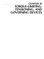
Fig. 1.1 Amplification of inflammatory signalling by the crosstalk between PGs and cytokines.
Cytokines induce expression of cyclooxygenase and PGE synthase, and PGs thus formed induce
expression of cytokines and cytokine receptor, therefore these two groups of inflammatory
mediators form an amplification loop to exacerbate inflammation
induced by these molecules such as interleukin (IL)-1β and IL-6 can induce an
inducible isoform of COX, COX-2, and initiate PG biosynthesis, it is generally
thought that PGs function as terminal mediators of inflammation to elicit acute
inflammation symptoms such as vasodilation and fever downstream of innate
immunity. However, PGs can amplify the actions of PAMPs and cytokines, and
there is bidirectional crosstalk between the two. For example, Honda et al. reported
that PGI2-IP signalling amplifies actions of IL-1β in collagen-induced arthritis
(CIA) of mice (Honda et al. 2006). They found that IP deficiency significantly
reduced the severity of arthritis assessed by synovial cell proliferation, inflammatory cell infiltration, and joint destruction in this model, which were accompanied
by significant reduction in the content of IL-6 in arthritic paws. They then showed
that indomethacin, a COX inhibitor, significantly reduced the IL-1β-induced IL-6
production in cultured synovial fibroblasts and the addition of an IP agonist,
cicaprost, restored the IL-6 production. Microarray analysis revealed that in addition to IL-6, PGI2-IP signalling amplified expression of various genes induced by
IL-1β in these cells, including those related to inflammation such as IL-11 and
CXCL7, those related to cell proliferation such as fibroblast growth factor (FGF),
and vascular and endothelial growth factor (VEGF), and those related to tissue
remodelling such as RANKL and the ADAM family molecules. Inasmuch as PGI2
alone did not induce expression of these genes, these results suggest that PGI2 can
function as an amplifier of IL-1β signalling. Intriguingly, PGI2-IP signalling augmented expression of the IL-1 receptor (IL1R1) itself. Therefore, this study
revealed induction of the receptor for relevant cytokine as one mechanism of
PG-mediated amplification of cytokine action. This mechanism combined with
cytokine-induced COX-2 expression makes a self-amplification loop for inflammation (Fig. 1.1). As described below, induction of the relevant cytokine receptor is
seen also in PG-mediated facilitation of differentiation and expansion of Th subsets
in acquired immunity (see Sect. 1.1.2). Amplification by PGs is not limited either to
actions of cytokines or to receptor induction. For example, Oshima et al. found that
LPS induced expression of COX-2, IL-1β, and IL-6 in cultured macrophages, and
6
T. Aoki and S. Narumiya
that this induction was ameliorated by the addition of celecoxib, a selective COX-2
inhibitor, or RQ-00015986, an EP4 antagonist (Oshima et al. 2011). These findings
suggest that endogenously formed PGE2 acts on the EP4 receptor and amplifies
actions by LPS. Similar amplification of TLR actions by PGE2 was reported for
induction of the p19 subunit of IL-23 (Il23a) in dendritic cells (DCs; see
Sect. 1.1.2). As reviewed below, the action of PGs as an amplifier of cytokines
and PAMPs/DAMPs constitutes one of the basic mechanisms whereby PGs function in chronic inflammation.
1.1.2
PGs in Conversion of Acute Inflammation to Immune
Inflammation (Fig. 1.2)
One possible mechanism by which inflammation becomes chronic is conversion of
acute inflammation to immune inflammation by acquired immunity. Acquired
immunity is initiated by presentation of antigen to naı¨ve T cells by DCs and
activated T cells differentiate to distinct helper T cell (Th) subsets under the
influence of specific cytokine milieu. Among Th subsets, Th1 and Th17,
characterised by production of interferon-γ (IFN-γ) and IL-17, respectively, play
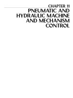
Fig. 1.2 PGE2 in conversion of acute inflammation to immune inflammation. PGE2 facilitates
Th1 differentiation and Th17 expansion via EP2 and EP4 synergistically with respective cytokines
through induction of their receptor IFN-γR1, IL-12Rβ2, and IL-23R PGE2 also promote IL-23
production from dendritic cells (DCs) synergistically with TLR ligands and CD40 stimulation to
further facilitate Th17 cell expansion
1 Prostaglandins in Chronic Inflammation
7
the crucial role in immune inflammation. Th1 differentiation is induced by IL-12
and facilitated by IFN-γ. Th17 differentiation and expansion are induced by TGF-β/
IL-6 and IL-23, respectively. Although PGs were previously considered as an
immunosuppressor (Harris et al. 2002), there is now substantial in vitro and
in vivo evidence that PGs act as an immune-activator under many circumstances.
Yao et al. found that under the Th1 skewing condition and with strengthened TCR
stimulation, PGE2 enhanced IL-12–mediated Th1 differentiation from mouse naı¨ve
T cells in a concentration-dependent manner (Yao et al. 2009). Facilitation of Th1
differentiation by PGE2 was mimicked by an EP2 or EP4 selective agonist and
abolished in T cells deficient in EP2 or EP4, suggesting that PGE2-EP2/EP4
signalling enhances Th1 differentiation. They further clarified the underlying
mechanism that PGE2-EP2/4 signalling activates the cAMP-PKA pathway, induces
expression of IL-12Rβ2 and IFN-γR1 genes via activating CREB and its
coactivator CRTC2, and amplifies the actions of IL-12 and IFN-γ on Th1 differentiation (Yao et al. 2013; Fig. 1.2). Notably, in addition to Th1 differentiation, PGE2
also facilitates IL-23-induced Th17 expansion. This is mediated redundantly via
EP2 and EP4 receptors in mouse (Yao et al. 2009), and preferentially via EP2 in
humans (Chizzolini et al. 2008; Boniface et al. 2009; Napolitani et al. 2009). In
human Th17 cells, PGE2-EP2 signalling exerts this effect apparently by
upregulating expression of IL-23 receptor and IL-1 receptor (Boniface
et al. 2009; Fig. 1.2). These studies thus provide further examples for the cytokine
amplifying action of PGE2 through receptor induction. Intriguingly, PGE2-EP2/4
signalling facilitates not only IL-23-induced Th17 expansion but also enhances
production of IL-23 from DCs. Ganea’s group showed that PGE2 potently enhances
expression of IL-23 p19 induced by various TLR ligands such as LPS, Poly-I-C,
CpG, and proteoglycan from DCs, and that this action is via EP2 and EP4
(Sheibanie et al. 2004; Khayrullina et al. 2008). They further demonstrated that
this PGE2 action is exerted by interaction of NFκB activated by TLR pathway and
CREB and C/EBP-β activated by PGE2-EP4-cAMP signalling (Kocieda
et al. 2012). Yao et.al found this PGE2-mediated enhancement of IL-23 production
also in DCs stimulated with anti-CD40 antibody and further found that the treatment with indomethacin or an EP4 antagonist almost completely suppressed the
IL-23 production (Yao et al. 2009), suggesting the presence of the PGE2-mediated
self-amplification cycle for IL-23 production in activated DCs. Interestingly,
whereas the PGE2-enhanced IL-23 mRNA expression by TLR ligands or TNF-α
is transient, peaking at 1 h, that by CD40 stimulation is of long duration lasting over
36 h, in which the early phase is mediated canonical and the late phase is mediated
by non-canonical NFκB signalling, both being similarly enhanced by the PGE2-EP4
signalling (Ma et al. 2016).
Consistently with these in vitro findings on Th1 and Th17 cells, genetic loss or
pharmacological antagonism of EP2 and EP4 significantly ameliorated disease
progression in mouse contact hypersensitivity (CHS), experimental autoimmune
encephalomyelitis (EAE), and transfer colitis, which are all Th1- and Th17mediated disease conditions (Yao et al. 2009, 2013). This amelioration was accompanied by remarkable suppression of antigen-induced proliferation and IFN-γ and
8
T. Aoki and S. Narumiya
IL-17 production of lymph node cells. Pharmacological antagonism of EP4 also
ameliorated CIA progression with concomitant suppression of IFN-γ and IL-17
production from lymph node cells (Chen et al. 2010). Conversely, Sheibanie
et al. demonstrated that intraperitoneal administration of PGE2 or an EP3/EP4
agonist, misoprostol, exacerbated CIA and 2,4,6-trinitrobenzene sulfonic acid
(TNBS)-induced colitis accompanied by the increase of IL-23 p19 and IL-17 in
the lesions (Sheibanie et al. 2007a, b). Such an immune activating function of PGs
is not limited to PGE2 but also seen in PGI2-IP signalling, which couples to cAMP
elevation like EP2/EP4. Nakajima et al. demonstrated that PGI2-IP signalling
facilitates Th1 differentiation in vitro and that IP deficiency impairs CHS
(Nakajima et al. 2010). These results suggest that PG signalling functions to
facilitate Th1 differentiation and Th17 expansion in vivo in mouse models of
various autoimmune diseases. Consistently with these mouse studies, recent
genome-wide association studies (GWAS) have identified Ptger4 (EP4) as a susceptible locus in a number of autoimmune diseases including Crohn’s disease (Glas
et al. 2012), multiple sclerosis (International Multiple Sclerosis Genetics Consortium; Wellcome Trust Case Control Consortium2 2011), and allergy (Hinds
et al. 2013). Furthermore, causal single nucleotide polymorphism (SNPs) related
to these autoimmune diseases are enriched in active enhancer region labelled with
acetylated H3K27 in Ptger4 locus and well correlated with EP4 expression (Farh
et al. 2015), supporting the clinical relevance of the findings on PGE2-EP4 signalling in mouse. In addition, Kofler et al. recently reported that EP2 undergoes
RORC-dependent silencing in T cells of healthy individuals, but is overexpressed
in T cells of patients of multiple sclerosis and simulation of EP2 in these patients’ T
cells induces a highly pathogenic phenotype expressing both IL-17 and IFN-γ
(Kofler et al. 2014), providing clinical relevance of the finding on EP2 and Th17
in mouse studies.
1.1.3
PGs in the Positive Feedback Loop to Amplify
Inflammatory Responses (Fig. 1.3)
The formation of a positive feedback loop to amplify and sustain inflammatory
responses can be another mechanism whereby inflammation becomes chronic.
Indeed, several studies have demonstrated the formation of such positive feedback
loops involving PG and their contribution to chronic inflammation. Intracranial
aneurysm (IA) is a regional bulging of intracranial arteries at bifurcation sites and a
major cause of subarachnoid hemorrhage (van Gijn et al. 2007). IA is chronic
inflammation of the artery histologically characterised by degenerative changes of
arterial walls and inflammatory cell infiltration mainly of macrophages (Chyatte
et al. 1999). High wall shear stress loaded on bifurcation sites of intracranial arteries
by blood flow is believed to trigger IA formation (Turjman et al. 2014). Aoki
et al. demonstrated induction of COX-2 and EP2 in endothelial cells at a
1 Prostaglandins in Chronic Inflammation
9
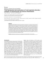
Fig. 1.3 PGE2 in positive
feedback loop for
inflammation. In IA, a
positive feedback loop
consisting of COX-2-PGE2EP2-NFκB is formed in
arterial endothelial cells
upon high wall shear stress.
Macrophages are recruited
by NFκB-dependent MCP-1
induction in this loop and
also form a similar loop for
further amplification of
inflammation
prospective site of IA formation in an animal model of IA, which was mimicked
in vitro in cultured endothelial cells under high shear stress (Aoki et al. 2011). It is
important to note that COX-2 inhibition by celecoxib significantly suppressed EP2
expression and EP2 deficiency suppressed COX-2 induction, and both suppressed
inflammation in IA walls and prevented IA formation in vivo. PGE2-EP2 signalling
activates NFκB and stimulates NFκB-mediated expression of various
proinflammatory genes including MCP-1 in cultured endothelial cells in vitro
(Aoki et al. 2011), which is consistent with the previous finding that IA formation
is dependent on NFκB (Aoki et al. 2007b). These findings together with the finding
that NFκB transcriptionally regulates COX-2 expression (Newton et al. 1997)
suggest that high wall shear stress triggers COX-2 expression through NFκB
activation in endothelial cells, which triggers a positive feedback loop of COX-2PGE2-EP2-NFκB to amplify inflammatory responses (Fig. 1.3). The same feedback
loop is formed in macrophages recruited by MCP-1 in IA walls for further amplification (Aoki et al. 2009, 2011; Kanematsu et al. 2011).
Chronic inflammation also underlies cancer development, and is typically
characterised by COX-2 expression in tumor lesion (Chulada et al. 2000). Pharmacological inhibition of COX by NSAIDs is long known to reduce incidence of
colorectal cancer (CRC) in humans (Rothwell et al. 2010; Janne and Mayer 2000),
and genetic deletion of COX-2 in mice reduced intestinal adenoma formation in an
animal model of human familial adenomatous polyposis coli (Oshima et al. 1996).
Sonoshita et al. used mice deficient in each EP subtype with ApcΔ716 mutation and
found that EP2 deficiency selectively reduced the number and size of adenomas in
this model (Sonoshita et al. 2001). They also demonstrated that COX-2 and EP2
10
T. Aoki and S. Narumiya
were strongly expressed in the stromal region of adenomas and further that EP2
deficiency almost completely abolished COX-2 expression (Sonoshita et al. 2001),
suggesting the presence of a positive feedback loop between PGE2, EP2, and
COX-2, as in IA (Aoki et al. 2011). To analyse the mode and the role of inflammation in CRC further, Ma et al. used azoxymethane-dextran sodium sulfate
treatment as a model of colitis-associated colon cancer (Ma et al. 2015). They
found that EP2 deficiency remarkably reduced inflammatory infiltrates and
suppressed the number of colon tumors in this model (Ma et al. 2015). Notably,
EP2 was expressed in both neutrophils, the major infiltrating cells in lesions, and
tumor-associated fibroblasts surrounding tumor cells, and functioned synergistically with TNF-α to produce various cytokines and chemokines through the selfamplification loop of COX-2-PGE2-EP2 to promote tumorigenesis.
1.1.4
Role of PGs in the Sustained Infiltration
of Inflammatory Cells to Affected Sites (Fig. 1.4)
Although inflammatory cell infiltration is transient in acute inflammation, chronic
inflammation exhibits sustained infiltration of inflammatory cells, which is crucial
for progression and maintenance of inflammation in various diseases. For example,
sustained infiltration of macrophages plays a crucial role in the pathogenesis of IA,
as administration of chlodronate liposome to deplete macrophages, gene deletion of
MCP-1, a macrophage chemokine, or expression of its dominant negative form all
significantly suppressed macrophage infiltration and prevented IA formation (Aoki
et al. 2009; Kanematsu et al. 2011). PG signalling plays a critical role in this
process, because MCP-1 expression is induced and amplified by a positive feedback
loop of the COX-2-PGE2-EP2-NFκB pathway formed in endothelial cells at the
prospective site of IA formation in the cerebral artery (Aoki et al. 2009, 2011).
Recruited macrophages then form this amplification loop, and produce MCP-1 by
themselves in addition to various cytokines and tissue-destructive proteinases, thus
making an autocrine loop for sustained macrophage accumulation and further
exacerbation of inflammation in the lesion (Aoki et al. 2007a, 2009; Fig. 1.4).
Oshima et al. (2011) reported a similar augmentation of MCP-1-mediated macrophage recruitment by PG signalling. They used Helicobacter pylori-infected gastric
tumor as a model and found that bacterial colonisation and PGE2-EP4 signalling
cooperatively induced MCP-1 expression to recruit macrophages to promote gastric
tumors (Oshima et al. 2011). On the other hand, Ma et al. (2015) found the
involvement of PGE2-EP2 signalling in sustained neutrophil recruitment in the
AOM-DSS model of colitis-associated colon cancer. They found extensive neutrophil infiltration and significant expression of CXCL1, a neutrophil chemokine, in
the tumor lesion in this model (Ma et al. 2015). Intriguingly, infiltrating neutrophils
expressed EP2 and CXCL1 as well, and EP2 deficiency suppressed neutrophil
1 Prostaglandins in Chronic Inflammation
11
Fig. 1.4 PGs in sustained infiltration of inflammatory cells. PGE2 induces production of
chemokines such as MCP-1 and CXCL1 from various types of cells via EP2 or EP4 and recruits
relevant inflammatory cells to affected sites. The recruited cells then produce the chemokines by
their own, which forms an autocrine/paracrine loop (blue colour) in the affected region and
sustains inflammatory cell infiltration
infiltration and CXCL1 expression. Furthermore, EP2 stimulation of primary culture of neutrophils augmented CXCL1 expression synergistically with TNF-α.
These results suggest that neutrophils self-amplify their recruitment through the
PGE2-EP2-CXCL1 pathway, which critically contributes to tumorigenesis in their
model.
These findings clearly show that PG signalling sustains infiltration of inflammatory cells under different inflammatory settings through induction of various
chemoattractants in a positive feedback manner and makes inflammation longlasting (Fig. 1.4).
1.1.5
Role of PGs in Tissue Remodelling (Fig. 1.5)
In chronic inflammation, destruction and repair of affected tissues simultaneously
occur and these processes lead to tissue remodelling including tissue metaplasia,
fibrosis, angiogenesis, and granulation. PGs either facilitate or suppress tissue
remodelling in a context-dependent manner (Fig. 1.5). For example, the airway
undergoes extensive remodelling in bronchial asthma. In the ovalbumin (OVA)induced allergic asthma model, OVA challenge induced expression of genes
12
T. Aoki and S. Narumiya
Fig. 1.5 PGs in tissue
remodelling. PGs either
promote or suppress tissue
remodelling, including
metaplasia, fibrosis,
angiogenesis, or granulation
in affected tissues
depending on the
microenvironment. Red
or blue colour indicates
examples of PG
contribution to promotion
or suppression of tissue
remodelling, respectively
involved in tissue remodelling such as Gob-5, Munc5ac, MMP-12, and ADAM-8,
and stimulation of PGE2-EP3 potently suppresses this induction (Kunikata
et al. 2005). On the other hand, the same PGE2-EP3 signalling facilitates angiogenesis associated with chronic inflammation and tumor. Amano et al. implanted a
Matrigel sponge or tumor cells in mice, and found induction of angiogenesis in
these implants in wild-type mice (Amano et al. 2003). This angiogenesis was
suppressed in EP3À/À mice with reduced VEGF expression in the stroma. Bone
marrow transfer experiment by the same group indicates that bone marrow cells
bearing EP3 is responsible for VEGF expression in the stroma around the implant,
and recruitment of VEGFR-1+/VEGFR-2+ cells there (Ogawa et al. 2009). Perhaps
in relation to these findings, Wang et al. found that PGE2 induced expression of
CXCL1, a chemokine for endothelial cells, from CRC cells in vitro, and that
administration of PGE2 in vivo augmented CXCL1 expression in LS-174 T cells
transplanted in immune-compromised mice and enhanced angiogenesis around the
xenograft, which was abolished by the injection of anti-CXCL1 antibody (Wang
et al. 2006). They suggested clinical relevance of their findings by showing
correlation between CXCL1 expression and PGE2 content in specimens of human
CRC tumors (Wang et al. 2006). For granulation, Katoh et al. (2010) used implants
of tumor cells and micropore chamber and found expression of CXCL12 around the
implants, which was sensitive to COX-2 inhibitor, augmented by PGE2 and absent
in EP3À/À or EP4À/À mice. These authors further found that this chemokine recruits
CXCR4+S100A4+ fibroblasts from bone marrow to the site for granulation. This
PGE2-EP3/4 signalling at the site of implant functions not only in stroma formation
but also in lymphangiogenesis (Katoh et al. 2010). Matrigel implants containing
FGF-2 induced proliferative inflammation and associated lymphangiogenesis,
which was suppressed by COX-2 inhibitor, augmented by PGE2 and absent again
in EP3À/À or EP4À/À mice (Katoh et al. 2010). Notably, agonists selective to EP3 or
EP4 induced VEGF-3 and VEGF-4 from macrophages and/or fibroblast in culture,
suggesting that PGE2 induces growth factors for lymph endothelial cells for
1 Prostaglandins in Chronic Inflammation
13
lymphangiogenesis (Hosono et al. 2011). Lastly, PGs also regulate tissue fibrosis.
Tissue fibrosis is characterised by proliferation of fibroblasts and excessive deposition of extracellular matrix proteins, and disrupts normal tissue architecture and
functions. Oga et al. (2009) used bleomycin-induced pulmonary fibrosis as a model
of idiopathic pulmonary fibrosis in humans, and demonstrated that the fibrosis in
this model was attenuated in FPÀ/À mice. Intriguingly, the loss of FP did not affect
inflammatory responses in lesions but decreased collagen synthesis independently
of TGF-β (Oga et al. 2009). Consistently, PGF2α enhanced collagen synthesis in
lung fibroblasts in vitro in an additive way to TGF-β. These results indicate that
PGF2α-FP signalling exerts action on its own in fibrosis. In contrast to this
profibrotic effect of PGF2α, Lovgren et al. (2006) demonstrated that the loss of IP
augmented lung fibrosis in a bleomycin-induced pulmonary fibrosis model. Such
anti-fibrotic action of PGI2-IP signalling was also reported in the heart in mice
subjected to pressure overload (Hara et al. 2005). Francois et al. (2005) also
reported that IPÀ/À mice developed cardiac fibrosis, which was suppressed
completely by coincidental deletion of TP, suggesting IP signalling and TP signalling antagonise in cardiac fibrosis. Protective action of PGs against tissue fibrosis
was also reported for the PGE2-EP4 signalling, which functions against
tubulointerstitial fibrosis in the kidney of mice subjected to unilateral ureteral
obstruction (Nakagawa et al. 2012).
1.2
Conclusions
As reviewed, substantial evidence now accumulates that PGs function in various
aspects of chronic inflammation from immune inflammation to tissue remodelling,
at least in animal models. Although we have not discussed the role of PGs in
epigenetic changes associated with chronic inflammation in this review, we believe
it will be uncovered soon. It is therefore important now to extrapolate the findings in
animal experiments to human disease and to identify the context-dependent action
of each PG and its receptor in chronic inflammation associated with various human
diseases. Given development of agonists and antagonists selective to each subtype
of PG receptors and given the potential and adverse effects of traditional NSAIDs
and COX-2 inhibitors, it is the time to examine the potential of receptor-selective
drugs to manipulate chronic diseases such as cancer, autoimmune, neurodegenerative, and vascular diseases.
References
Amano H, Hayashi I, Endo H, Kitasato H, Yamashina S, Maruyama T, Kobayashi M, Satoh K,
Narita M, Sugimoto Y, Murata T, Yoshimura H, Narumiya S, Majima M (2003) Host
prostaglandin E2-EP3 signalling regulates tumor-associated angiogenesis and tumor growth.
J Exp Med 197(2):221–232
14
T. Aoki and S. Narumiya
Aoki T, Kataoka H, Morimoto M, Nozaki K, Hashimoto N (2007a) Macrophage-derived matrix
metalloproteinase-2 and -9 promote the progression of cerebral aneurysms in rats. Stroke 38
(1):162–169. doi:10.1161/01.STR.0000252129.18605.c8
Aoki T, Kataoka H, Shimamura M, Nakagami H, Wakayama K, Moriwaki T, Ishibashi R,
Nozaki K, Morishita R, Hashimoto N (2007b) NFκB is a key mediator of cerebral aneurysm
formation. Circulation 116(24):2830–2840. doi:10.1161/CIRCULATIONAHA.107.728303
Aoki T, Kataoka H, Ishibashi R, Nozaki K, Egashira K, Hashimoto N (2009) Impact of monocyte
chemoattractant protein-1 deficiency on cerebral aneurysm formation. Stroke 40(3):942–951.
doi:10.1161/STROKEAHA.108.532556
Aoki T, Nishimura M, Matsuoka T, Yamamoto K, Furuyashiki T, Kataoka H, Kitaoka S,
Ishibashi R, Ishibazawa A, Miyamoto S, Morishita R, Ando J, Hashimoto N, Nozaki K,
Narumiya S (2011) PGE2-EP2 signalling in endothelium is activated by haemodynamic stress
and induces cerebral aneurysm through an amplifying loop via NFκB. Br J Pharmacol 163
(6):1237–1249. doi:10.1111/j.1476-5381.2011.01358.x
Ben-Neriah Y, Karin M (2011) Inflammation meets cancer, with NFκB as the matchmaker. Nat
Immunol 12(8):715–723. doi:10.1038/ni.2060
Boniface K, Bak-Jensen KS, Li Y, Blumenschein WM, McGeachy MJ, McClanahan TK,
McKenzie BS, Kastelein RA, Cua DJ, de Waal MR (2009) Prostaglandin E2 regulates Th17
cell differentiation and function through cyclic AMP and EP2/EP4 receptor signalling. J Exp
Med 206(3):535–548. doi:10.1084/jem.20082293
Chen Q, Muramoto K, Masaaki N, Ding Y, Yang H, Mackey M, Li W, Inoue Y, Ackermann K,
Shirota H, Matsumoto I, Spyvee M, Schiller S, Sumida T, Gusovsky F, Lamphier M (2010) A
novel antagonist of the prostaglandin E2 EP4 receptor inhibits Th1 differentiation and Th17
expansion and is orally active in arthritis models. Br J Pharmacol 160(2):292–310. doi:10.
1111/j.1476-5381.2010.00647.x
Chizzolini C, Chicheportiche R, Alvarez M, de Rham C, Roux-Lombard P, Ferrari-Lacraz S,
Dayer JM (2008) Prostaglandin E2 synergistically with interleukin-23 favors human Th17
expansion. Blood 112(9):3696–3703. doi:10.1182/blood-2008-05-155408
Chulada PC, Thompson MB, Mahler JF, Doyle CM, Gaul BW, Lee C, Tiano HF, Morham SG,
Smithies O, Langenbach R (2000) Genetic disruption of Ptgs-1, as well as Ptgs-2, reduces
intestinal tumorigenesis in Min mice. Cancer Res 60(17):4705–4708
Chyatte D, Bruno G, Desai S, Todor DR (1999) Inflammation and intracranial aneurysms.
Neurosurgery 45(5):1137–1146
Farh KK, Marson A, Zhu J, Kleinewietfeld M, Housley WJ, Beik S, Shoresh N, Whitton H, Ryan
RJ, Shishkin AA, Hatan M, Carrasco-Alfonso MJ, Mayer D, Luckey CJ, Patsopoulos NA, De
Jager PL, Kuchroo VK, Epstein CB, Daly MJ, Hafler DA, Bernstein BE (2015) Genetic and
epigenetic fine mapping of causal autoimmune disease variants. Nature 518(7539):337–343.
doi:10.1038/nature13835
Francois H, Athirakul K, Howell D, Dash R, Mao L, Kim HS, Rockman HA, Fitzgerald GA,
Koller BH, Coffman TM (2005) Prostacyclin protects against elevated blood pressure and
cardiac fibrosis. Cell Metab 2(3):201–207. doi:10.1016/j.cmet.2005.08.005
Glas J, Seiderer J, Czamara D, Pasciuto G, Diegelmann J, Wetzke M, Olszak T, Wolf C, MullerMyhsok B, Balschun T, Achkar JP, Kamboh MI, Franke A, Duerr RH, Brand S (2012)
PTGER4 expression-modulating polymorphisms in the 5p13.1 region predispose to Crohn’s
disease and affect NFκB and XBP1 binding sites. PLoS One 7(12):e52873. doi:10.1371/
journal.pone.0052873
Hara A, Yuhki K, Fujino T, Yamada T, Takayama K, Kuriyama S, Takahata O, Karibe H,
Okada Y, Xiao CY, Ma H, Narumiya S, Ushikubi F (2005) Augmented cardiac hypertrophy
in response to pressure overload in mice lacking the prostaglandin I2 receptor. Circulation 112
(1):84–92. doi:10.1161/CIRCULATIONAHA.104.527077
Harris SG, Padilla J, Koumas L, Ray D, Phipps RP (2002) Prostaglandins as modulators of
immunity. Trends Immunol 23(3):144–150
1 Prostaglandins in Chronic Inflammation
15
Hinds DA, McMahon G, Kiefer AK, Do CB, Eriksson N, Evans DM, St Pourcain B, Ring SM,
Mountain JL, Francke U, Davey-Smith G, Timpson NJ, Tung JY (2013) A genome-wide
association meta-analysis of self-reported allergy identifies shared and allergy-specific susceptibility loci. Nat Genet 45(8):907–911. doi:10.1038/ng.2686
Hirata T, Narumiya S (2011) Prostanoid receptors. Chem Rev 111(10):6209–6230. doi:10.1021/
cr200010h
Honda T, Segi-Nishida E, Miyachi Y, Narumiya S (2006) Prostacyclin-IP signalling and prostaglandin E2-EP2/EP4 signalling both mediate joint inflammation in mouse collagen-induced
arthritis. J Exp Med 203(2):325–335. doi:10.1084/jem.20051310
Hosono K, Suzuki T, Tamaki H, Sakagami H, Hayashi I, Narumiya S, Alitalo K, Majima M (2011)
Roles of prostaglandin E2-EP3/EP4 receptor signalling in the enhancement of
lymphangiogenesis during fibroblast growth factor-2-induced granulation formation.
Arterioscler Thromb Vasc Biol 31(5):1049–1058. doi:10.1161/ATVBAHA.110.222356
International Multiple Sclerosis Genetics Consortium, Wellcome Trust Case Control Consortium2
(2011) Genetic risk and a primary role for cell-mediated immune mechanisms in multiple
sclerosis. Nature 476(7359):214–219. doi:10.1038/nature10251
Janne PA, Mayer RJ (2000) Chemoprevention of colorectal cancer. N Engl J Med 342
(26):1960–1968. doi:10.1056/NEJM200006293422606
Kanematsu Y, Kanematsu M, Kurihara C, Tada Y, Tsou TL, van Rooijen N, Lawton MT, Young
WL, Liang EI, Nuki Y, Hashimoto T (2011) Critical roles of macrophages in the formation of
intracranial aneurysm. Stroke 42(1):173–178. doi:10.1161/STROKEAHA.110.590976
Katoh H, Hosono K, Ito Y, Suzuki T, Ogawa Y, Kubo H, Kamata H, Mishima T, Tamaki H,
Sakagami H, Sugimoto Y, Narumiya S, Watanabe M, Majima M (2010) COX-2 and prostaglandin EP3/EP4 signalling regulate the tumor stromal proangiogenic microenvironment via
CXCL12-CXCR4 chemokine systems. Am J Pathol 176(3):1469–1483. doi:10.2353/ajpath.
2010.090607
Khayrullina T, Yen JH, Jing H, Ganea D (2008) In vitro differentiation of dendritic cells in the
presence of prostaglandin E2 alters the IL-12/IL-23 balance and promotes differentiation of
Th17 cells. J Immunol 181(1):721–735
Kocieda VP, Adhikary S, Emig F, Yen JH, Toscano MG, Ganea D (2012) Prostaglandin
E2-induced IL-23p19 subunit is regulated by cAMP-responsive element-binding protein and
C/AATT enhancer-binding protein beta in bone marrow-derived dendritic cells. J Biol Chem
287(44):36922–36935. doi:10.1074/jbc.M112.402958
Kofler DM, Marson A, Dominguez-Villar M, Xiao S, Kuchroo VK, Hafler DA (2014) Decreased
RORC-dependent silencing of prostaglandin receptor EP2 induces autoimmune Th17 cells. J
Clin Invest 124(6):2513–2522. doi:10.1172/JCI72973
Kumar V, Abbas A, Fausto N, Mitchell R (2007) Robbins basic pathology, 8th edn. Elsevier,
Missouri
Kunikata T, Yamane H, Segi E, Matsuoka T, Sugimoto Y, Tanaka S, Tanaka H, Nagai H,
Ichikawa A, Narumiya S (2005) Suppression of allergic inflammation by the prostaglandin E
receptor subtype EP3. Nat Immunol 6(5):524–531. doi:10.1038/ni1188
Libby P, Ridker PM, Hansson GK (2011) Progress and challenges in translating the biology of
atherosclerosis. Nature 473(7347):317–325. doi:10.1038/nature10146
Lovgren AK, Jania LA, Hartney JM, Parsons KK, Audoly LP, Fitzgerald GA, Tilley SL, Koller
BH (2006) COX-2-derived prostacyclin protects against bleomycin-induced pulmonary fibrosis. Am J Physiol Lung Cell Mol Physiol 291(2):L144–L156. doi:10.1152/ajplung.00492.2005
Ma X, Aoki T, Tsuruyama T, Narumiya S (2015) Definition of prostaglandin E2-EP2 signals in the
colon tumor microenvironment which amplify inflammation and tumor growth. Cancer Res 75
(14):2822–2832
Ma X, Aoki T, Narumiya S (2016) Prostaglandin E2-EP4 signalling persistently amplifies CD40mediated induction of IL-23 p19 expression through canonical and non-canonical NFκB
pathways. Cell Mol Immunol 13(2):240–250