Principles of critical care in obstetrics
Bạn đang xem bản rút gọn của tài liệu. Xem và tải ngay bản đầy đủ của tài liệu tại đây (17.16 MB, 358 trang )
Principles of
Critical Care in
Obstetrics
Volume I
Alpesh Gandhi
Narendra Malhotra
Jaideep Malhotra
Nidhi Gupta
Neharika Malhotra Bora
Editors
123
Principles of Critical Care in Obstetrics
Alpesh Gandhi • Narendra Malhotra
Jaideep Malhotra • Nidhi Gupta
Neharika Malhotra Bora
Editors
Principles of Critical
Care in Obstetrics
Volume I
Editors
Alpesh Gandhi
Arihant Women’s Hospital
Ahmedabad
Gujarat
India
Narendra Malhotra
Global Rainbow Healthcare
Agra
India
Jaideep Malhotra
Art Rainbow-IVF
Agra
India
Nidhi Gupta
SN Medical College
Obstetrics and Gynecology
Agra
India
Neharika Malhotra Bora
Bharti Vidya Peethmedical College
Pune
India
ISBN 978-81-322-2690-1
ISBN 978-81-322-2692-5
DOI 10.1007/978-81-322-2692-5
(eBook)
Library of Congress Control Number: 2015960281
Springer New Delhi Heidelberg New York Dordrecht London
© Springer India 2016
This work is subject to copyright. All rights are reserved by the Publisher, whether the whole or
part of the material is concerned, specifically the rights of translation, reprinting, reuse of
illustrations, recitation, broadcasting, reproduction on microfilms or in any other physical way,
and transmission or information storage and retrieval, electronic adaptation, computer software,
or by similar or dissimilar methodology now known or hereafter developed.
The use of general descriptive names, registered names, trademarks, service marks, etc. in this
publication does not imply, even in the absence of a specific statement, that such names are
exempt from the relevant protective laws and regulations and therefore free for general use.
The publisher, the authors and the editors are safe to assume that the advice and information in
this book are believed to be true and accurate at the date of publication. Neither the publisher nor
the authors or the editors give a warranty, express or implied, with respect to the material
contained herein or for any errors or omissions that may have been made.
Printed on acid-free paper
Springer (India) Pvt. Ltd. is part of Science+Business Media (www.springer.com)
Contents
Part I
Introduction to Critical Care
1
Epidemiology of Critical Illness in Obstetrics. . . . . . . . . . . . . .
Shikha Singh and Narendra Malhotra
2
Pregnancy-Induced Alterations in Physiology
and Laboratory Reports . . . . . . . . . . . . . . . . . . . . . . . . . . . . . . .
C.N. Purandare, Madhuri Patel, and Surekha Tayade
3
9
3
Ethics in the Setting Up of Obstetric HDU and ICU . . . . . . . .
K. Muhunthan and Sabaratnam Arulkumaran
4
Organisation and Role of Critical Care Units:
Obstetric HDU/ICU . . . . . . . . . . . . . . . . . . . . . . . . . . . . . . . . . . .
Alpesh Gandhi
21
Cardiopulmonary Resuscitation in the Pregnant
Woman . . . . . . . . . . . . . . . . . . . . . . . . . . . . . . . . . . . . . . . . . . . . .
Amita Gandhi and Alpesh Gandhi
35
5
Part II
6
7
15
Procedures and Monitoring in the HDU/ICU Unit
Role of Imaging in Noninvasive Monitoring
in Obstetric Intensive Care Unit. . . . . . . . . . . . . . . . . . . . . . . . .
Santosh Singhal, Rishabh Bora, Narendra Malhotra,
and Jaideep Malhotra
Basic Hemodynamic and Cardiac Monitoring
in Obstetrics . . . . . . . . . . . . . . . . . . . . . . . . . . . . . . . . . . . . . . . . .
Pratima Mittal, Jyotsna Suri, and Pradeep K. Verma
51
59
8
Respiratory Monitoring and Blood Gas Physiology . . . . . . . . .
Shivakumar Iyer and Jignesh Shah
69
9
Obstetric Monitoring in Critically Ill Pregnant Women . . . . .
Narendra Malhotra, Anupama Suwal, Jaideep Malhotra,
and Neharika Malhotra Bora
81
10
Fetal Surveillance in Critically Ill Obstetric Patient . . . . . . . .
Neharika Malhotra, Rishabh Bora, and Keshav Malhotra
85
v
Contents
vi
11
12
13
14
15
Infection Prevention and Control Policy
in Obstetric HDU and ICU . . . . . . . . . . . . . . . . . . . . . . . . . . . . .
Jayam Kannan
95
Transfusion of Blood Components and Derivatives
in the Obstetric Patients . . . . . . . . . . . . . . . . . . . . . . . . . . . . . . .
Lakhbir Dhaliwal and Rakhi Rai
105
Fluid and Electrolyte Balance in Critically Ill
Obstetric Patient . . . . . . . . . . . . . . . . . . . . . . . . . . . . . . . . . . . . .
Ruchika Garg and Rekha Rani
119
Mechanical Ventilation in Critically Ill
Obstetric Patient . . . . . . . . . . . . . . . . . . . . . . . . . . . . . . . . . . . . .
Mohammed Azam Danish
133
Nutrition in the Critically Ill Obstetric Patient. . . . . . . . . . . . .
Kamini A. Rao and Smitha Avula
Part III
16
17
18
143
Clinical Shock Syndromes
Post-partum Haemorrhage: Prevention, Medical
and Mechanical Methods of Management. . . . . . . . . . . . . . . . .
Ruchika Garg
153
Conservative and Nonconservative Surgical
Management of Postpartum Hemorrhage. . . . . . . . . . . . . . . . .
V.P. Paily and Vasanthi Jayaraj
159
The Lower Segment of Uterus – A Critical Area
in Childbirth and Resulting Trauma . . . . . . . . . . . . . . . . . . . .
Ajit C. Rawal
175
19
Ruptured Ectopic Pregnancy . . . . . . . . . . . . . . . . . . . . . . . . . . .
Abdul Vahab and P. Mumtaz
199
20
Cardiogenic Shock in Pregnancy . . . . . . . . . . . . . . . . . . . . . . . .
Sourya Acharya
207
21
The Recognition and Management of Maternal Sepsis . . . . . .
Karen Orr, Damien Hughes, Claire Jamison,
and Paul Fogarty
215
22
Anaphylactic Shock in a Pregnant Woman . . . . . . . . . . . . . . . .
Veena Agrawal
237
23
Sudden Obstetric Collapse . . . . . . . . . . . . . . . . . . . . . . . . . . . . .
Lisa M. Nathan and Asha Rijhsinghani
253
24
Disseminated Intravascular Coagulation (DIC)
and Thrombocytopenia in Pregnancy . . . . . . . . . . . . . . . . . . . .
Alka Saraswat, Jaideep Malhotra, Narendra Malhotra,
and Neharika Malhotra Bora
259
Contents
vii
Part IV
HDP and It’s Problems Requiring Critical Care
25
Hypertensive Crisis in Pregnancy . . . . . . . . . . . . . . . . . . . . . . .
Girija Wagh
271
26
Eclampsia . . . . . . . . . . . . . . . . . . . . . . . . . . . . . . . . . . . . . . . . . . .
Sanjay Gupte
277
27
Antepartum Hemorrhage . . . . . . . . . . . . . . . . . . . . . . . . . . . . . .
Nidhi Gupta
281
28
HELLP Syndrome . . . . . . . . . . . . . . . . . . . . . . . . . . . . . . . . . . . .
P.K. Shah, Mayoor Daigavane, and Natasha DSouza
303
Part V
Critical Conditions in LR/OT
29
Amniotic Fluid Embolism and Pulmonary Embolism . . . . . . .
Nidhi Patel and Ajesh Desai
313
30
Management of Critical Cord Accidents . . . . . . . . . . . . . . . . . .
A.K. Debdas
327
31
Acute Inversion of the Uterus . . . . . . . . . . . . . . . . . . . . . . . . . . .
Gokul Chandra Das and Gitanjali Deka
335
32
Rupture of the Gravid Uterus. . . . . . . . . . . . . . . . . . . . . . . . . . .
Ashis Kumar Mukhopadhyay
339
33
Shoulder Dystocia . . . . . . . . . . . . . . . . . . . . . . . . . . . . . . . . . . . .
Madhu Nagpal
347
34
Difficulty in the Delivery of a Baby During LSCS . . . . . . . . . .
Parul J. Kotdawala and Munjal J. Pandya
355
Part I
Introduction to Critical Care
1
Epidemiology of Critical Illness
in Obstetrics
Shikha Singh and Narendra Malhotra
Critical illness in pregnancy as a morbidity outcome is difficult to define and therefore difficult to
measure and study precisely. As stated by Harmer,
“Death represents the tip of the morbidity iceberg,
the size of which is unknown” [1]. The stage at
which any condition becomes severe enough to be
classified as a critical illness has not been clearly
defined. However, it may be helpful to consider
critical illness as impending, developing, or established significant organ dysfunction, which may
lead to long-term morbidity or death. This allows
some flexibility in the characterization of disease
severity since it recognizes condition that can
deteriorate rather quickly in pregnancy.
It has been suggested that most women suffering a critical illness in pregnancy are likely to be
in an intensive care unit. These cases have been
described by some as “near-miss” mortality
cases. There are many conditions in pregnancy
that occur frequently and require special medical
care, but do not actually become critical illness.
Most women with these complications have relatively uneventful pregnancies that result in good
outcome. Nevertheless, each of these conditions
can be associated with significant complications
that have the potential for serious morbidity, disability, and mortality.
The successful epidemiologic evaluation of any
particular disease or condition has several prerequisites. Two of the most important prerequisites
are that the condition should be accurately defined
and that there should be measurable outcomes of
interest. Another requirement is that these must be
some systematic way of data collection or surveillance that will allow the measurement of the outcomes of interest and associated risk factors.
Historically, surveillance of pregnancy-related
critical illness has focused on the well-defined
outcome of maternal mortality in order to identify illnesses or conditions that might have led to
maternal death. Maternal mortality data collection is well established in many places, but specific surveillance systems that track severe
complications of pregnancy not associated with
maternal mortality are rare. Examination of complicating conditions associated with maternal
hospitalization can provide information on the
types of conditions requiring hospitalized case.
ICU Admissions and Maternal
Mortality
S. Singh (*)
Department of Obstetrics and Gynaecology,
S.N. Medical College, Agra, India
e-mail:
N. Malhotra, MD
Director, Rainbow Hospitals, Agra, India
Evaluation of obstetric admissions to intensive
care units (ICUs) may be one of the best ways to
approach surveillance of critical illness in pregnancy. Unfortunately, there is no publicly available
© Springer India 2016
A. Gandhi et al. (eds.), Principles of Critical Care in Obstetrics: Volume I,
DOI 10.1007/978-81-322-2692-5_1
3
S. Singh and N. Malhotra
4
population-based database for obstetric admissions to ICU that provides sufficiently detailed
information to allow in-depth study of these
conditions.
The prevalence of obstetric patients requiring
critical care ranges from 100 to 900 per 100,000
gestations [2–4]. The maternal mortality due to
critical illness is 12–20 % but varies significantly
between developing and developed countries
(440/10,000 deliveries in India vs. 12/100,000
deliveries in the USA) [5].
A review of 33 studies between 1990 and
2006 by Ananth and Smulian [6], involving 19,
55, and 111 deliveries, found an overall obstetric
admission rate to ICU of 0.07–0.89 %.
According to the study, reported maternal
mortality for critically ill obstetric patients admitted to an ICU is approximately 8.4 % with range
of 0–33 % in different setups. These reports are
from developed countries and less developed
countries have much higher mortality rates. In a
study on obstetric admissions to ICU of King
Edward Memorial Hospital (KEMH), Mumbai,
by Munnur et al. [5], the maternal mortality was
as high as 25 % in Indian patients. Factors leading to adverse outcomes in Indian subjects were
lack of antenatal care, delayed presentation,
higher severity of illness at presentation, and lack
of an aggressive obstetric approach. Organization
of health care services and social customs also
contributed to low antenatal care and lack of
aggressive obstetric approach. Panchal et al. [7],
in a retrospective analysis of 1,023 ICU admissions, showed that age, race, hospital type, volume of deliveries, and source of admission were
all associated with risk of admission to the ICU
in obstetrics.
Illnesses Responsible for Obstetric
ICU Admissions
Data pooled by Munnur et al. [5] provides sufficient detail about the primary indication for the
obstetrics ICU admission (Table 1.1). It is no surprise that hypertensive disease and obstetric hemorrhage were responsible for over 50 % of the
primary admitting diagnoses. Specific organ
Table 1.1 Medical disorders requiring intensive care
unit (ICU) admission [5]
Medical disorders
Community-acquired
pneumonia
Urinary tract infection
Malaria
Hematological disorder
Congenital heart
disease
Rheumatic heart
disease
Aspiration pneumonia
Diabetes mellitus
Chronic renal failure
Trauma
Drug abuse
Rheumatological
disorders
Anaphylaxis
Asthma
DVT/pulmonary
embolism
Malignancy
Acute abdomen
CNS infection
Viral hepatitis
Bacteremia
Attempted suicide
(poisoning/drug
overdose)
Transfusion reaction
Cardiac arrest prior to
ICU admission
Endocrine
Arterial disease
Intracranial
hemorrhage
Cerebral venous
thrombosis
Tetanus
Typhoid
Leptospirosis
Cerebral infarction
King Edward
Memorial
Hospital
(n = 754)
23 (3.1 %)
Ben Taub
General
Hospital
(n = 174)
5 (2.9 %)
2 (0.3 %)
75 (10.0 %)
12 (1.6 %)
2 (0.3 %)
18 (10.3 %)
0
1 (0.6 %)
2 (1.2 %)
16 (2.1 %)
2 (1.2 %)
23 (3.1 %)
16 (2.1 %)
4 (0.5 %)
0
0
2 (0.3 %)
6 (3.5 %)
4 (2.3 %)
1 (0.6 %)
1 (0.6 %)
5 (2.9 %)
2 (1.2 %)
0
1 (0.1 %)
5 (0.7 %)
2 (1.2 %)
5 (2.9 %)
2 (1.2 %)
1 (0.1 %)
6 (0.8 %)
6 (0.8 %)
47 (6.2 %)
13 (1.7 %)
13 (1.7 %)
6 (3.5 %)
10 (5.7 %)
0
0
8 (4.6 %)
1 (0.6 %)
2 (0.3 %)
21 (2.8 %)
1 (0.6 %)
1 (0.6 %)
8 (1.1 %)
1 (0.1 %)
9 (1.2 %)
1 (0.6 %)
1 (0.6 %)
1 (0.6 %)
26 (3.5 %)
0
2 (0.3 %)
1 (0.1 %)
2 (0.3 %)
2 (0.3 %)
0
0
0
0
system dysfunction was responsible for the majority of remaining admissions. Of those, pulmonary,
cardiac, and infectious complications had the
1
Epidemiology of Critical Illness in Obstetrics
5
Table 1.2 Obstetric conditions requiring intensive care unit (ICU) admission [5]
Medical disorders
Preeclampsia/eclampsia
Postpartum hemorrhage
IUFD
Postabortal/puerperal sepsis
HELLP syndrome
Abruptio placentae
Acute fatty liver of pregnancy
Antepartum hemorrhage
Chorioamnionitis
Abortions
Abnormal adherence of placenta
Peripartum cardiomyopathy
Uterine rupture
Amniotic fluid embolism
King Edward Memorial
Hospital (n = 754)
343 (45.5 %)
115 (15.3 %)
94 (12.5 %)
49 (6.5 %)
42 (5.6 %)
43 (5.7 %)
33 (4.4 %)
27 (3.6 %)
7 (0.9 %)
18 (2.4 %)
8 (1.1 %)
4 (0.5 %)
6 (0.8 %)
4 (0.5 %)
greatest frequency. It was also clear from these
reports that both obstetric and medical complications of pregnancy were responsible for the
obstetric ICU admissions (Tables 1.1 and 1.2).
Causes of Mortality in Obstetric ICU
Admissions
When specific causes of mortality for the obstetric
ICU admissions were reviewed by Ananth et al.,
26 studies gave sufficient data to assign a primary
etiology for maternal death (Table 1.3) [8].
Of a total of 138 maternal deaths, over 57 %
were related to complications of hypertensive
diseases, pulmonary illnesses, and cardiac diseases. Other deaths were commonly related to
complications of hemorrhage, bleeding into the
central nervous system (CNS), malignancy, and
infection. More importantly, despite identified
primary etiology for the maternal deaths, nearly
all cases were associated with multiple organ
dysfunction score (MODS), which again emphasizes the complex condition of these critically ill
women.
In a retrospective analysis by Munnur et al. of
10-year data (1992–2001) pertaining to 928 critically ill obstetric patients from King Edward
Memorial Hospital (KEMH), Mumbai, being
compared to a similar patient population at
Ben Taub General Hospital
(n = 174)
74 (42.5 %)
32 (18.4 %)
8 (4.6 %)
26 (14.9 %)
31 (17.8 %)
15 (8.6 %)
3 (1.7 %)
4 (2.3 %)
22 (12.6 %)
6 (3.5 %)
9 (5.2 %)
10 (5.8 %)
3 (1.7 %)
1 (0.5 %)
Houston County Hospital, the mean age of Indian
patients was 25.4 ± 4.6 years, of which only 26 %
had received prenatal care (at least two prenatal
visits) as compared to 86 % of Western patients;
only 60 % of Indian patients went for admission
within 24 h of onset of illness (vs. 90 % for
Western patients), with mean APACHE II score
of 16 on Day 1 (vs. ten for Western patients),
with altered mental status (50 %), bleeding
(40 %), seizures (30 %), fever (27 %), dyspnea
(23 %), and jaundice (21 %) being the most common manifestations in this subset (vs. fever 55 %,
bleeding 53 %, dyspnea 44 % in Western
population).
In both ICUs, 70 % of critically ill pregnant
patients were admitted with obstetric disorders.
The incidence of preeclampsia/eclampsia (45 %),
PPH (15 %), abruptio placentae (6 %), acute fatty
liver of pregnancy (4 %), and APH (4 %) in
Indian patients was similar to their Western counterparts. Medical disorders were responsible for
only 30 % of ICU admissions.
The incidence of organ dysfunction in Indian
subjects in the abovementioned study was
reported as follows: neurological (63 %), hematologic (58 %), renal (50 %), respiratory (46 %),
cardiovascular (38 %), and hepatic (36 %).
DIC was seen in 23 % of subjects, while the maximum MODS score was 5 [3–7]. The major causes
of CNS dysfunction in Indian subjects were
S. Singh and N. Malhotra
6
Table 1.3 Identified primary causes of mortality in
obstetric admissions to ICUs [8]
Identified etiology
Hypertensive diseases
Hypertensive crisis with
renal failure
HELLP syndrome
complications
Eclampsia complications
Other hypertensive disease
complications
Pulmonary
Pneumonia complications
Amniotic fluid embolus
Adult respiratory distress
syndrome
Pulmonary embolus
Cardiac
Eisenmenger’s complex
Myocardial infarction
Arrhythmia
cardiomyopathy
Unspecified
Hemorrhage
Central nervous system
hemorrhage
Arteriovenous
malformation
Brain stem hemorrhage
Intracranial hemorrhage
Infection
Sepsis
Tuberculosis meningitis
Malignancy
Hematologic
Thrombotic
thrombocytopenic
purpura
Gastrointestinal
Acute fatty liver of
pregnancy
Poisoning/overdose
Anesthesia complication
Trauma
Unspecified
Total
Number
Percentage
36
26.1
27
19.6
16
11.6
14
10
10.1
7.2
11
8.0
sia, DIC, PPH, hemorrhagic shock, severe
malaria, leptospirosis, and acute fatty liver of
pregnancy. Hematological failure was predominantly due to bacterial sepsis and DIC. Respiratory
failure was due to community-acquired pneumonia, acute asthma, and ARDS due to abdominal
sepsis. Cardiovascular failure was due to obstetric
shock and rheumatic heart disease. Hepatic dysfunction was predominantly due to acute viral
hepatitis in Indian subjects and due to HELLP
syndrome in Western subjects.
To conclude, understanding the nature of critical illness in pregnancy is an important and
evolving process. However, our currently available tools and databases for examining these
patients still need improvement [9]. As our
understanding of critical illnesses continues to
mature, we will hopefully gain greater insight
into the specific nature of these conditions that
will lead to improved prevention strategies and
better therapies for the diseases when they occur.
These data will improve our ability to plan and
allocate the necessary resources to adequately
care for these often complex and severe illnesses.
A multidisciplinary approach to manage these
patients is required, and it can also be well guided
by epidemiology statistics.
References
8
2
5.8
1.5
1
0.7
2
1
1
9
138
1.5
0.7
0.7
6.5
100 %
eclampsia, cerebral malaria, CNS infections,
hepatic coma, and cerebral venous thrombosis.
Important causes of renal failure were preeclamp-
1. Harmer M. Maternal mortality – is it still relevant?
Anaesthesia. 1997;52:99–100.
2. Baskett TF, Sternadel J. Maternal intensive care and
near – miss mortality in obstetrics. Br J Obstet
Gynaecol. 1998;105:981–4.
3. Kilpatrick SJ, Matthay MA. Obstetric patients requiring critical care. A five-year review. Chest.
1992;101:1407–12.
4. Naylor DF, Olson MM. Critical care obstetrics and
gynecology. Crit Care Clin. 2003;19:127–49.
5. Munnur U, Karnad DR, Bandi VDP, Lapsia V, Suresh
MS, Ramshesh P, Gardner MA, Longmire S,
Guntupalli KK. Critically ill obstetric patients in an
American and an Indian public hospital: comparison
of case – mix, organ dysfunction, intensive care
requirements, and outcomes. Intensive Care Med.
2005;31:1087–94.
6. Ananth CV, Smulian JC. Epidemiology of critical illness in pregnancy. In: Belfort M, Saade G, Foley M,
Phelan J, Dildy G, editors. Critical care obstetrics. 5th
ed. Boston: Blackwell Publishing Ltd; 2010. p. 1–10.
1
Epidemiology of Critical Illness in Obstetrics
7. Panchal S, Arria AM, Harris AP. Intensive care utilization during hospital admission for delivery.
Anesthesiology. 2000;92:1537–44.
8. Ananth CV. Epidemiology of critical illnesses and
outcomes in pregnancy. In: Belfort MA, Dildy GA,
7
Saade GR et al editors. Critical Care Obstetrics. 4th
ed. Boston: Blackwell Publishing Ltd; 2004. p. 11.
9. Soubra HS, Guntupalli KK. Critical illness in pregnancy: an overview. Crit Care Med. 2005;33(10
Suppl):S248–55.
2
Pregnancy-Induced Alterations
in Physiology and Laboratory
Reports
C.N. Purandare, Madhuri Patel,
and Surekha Tayade
Introduction
Pregnancy in the human female is a unique state
in which virtually all maternal systems are dramatically altered to permit the sustenance and
growth of the intrauterine conceptus. Major physiological changes include cardiovascular, hematologic, metabolic, renal, and respiratory changes,
most of which begin soon after conception and
continue throughout pregnancy till until late gesC.N. Purandare (*)
President FIGO, President FOGSI 2009, The Dean
Indian College of Obstetricians and Gynaecologists,
Mumbai, India
Editor Emieritus Journal FOGSI, Consultant Obst. and
Gyn. St. Elizabeth, Saifee and BSES Hospitals,
Mumbai, India
Ex. Hon. Professor OBGYN, Grant Medical
College and J.J. Hospital, Mumbai, India
M. Patel
Treasurer FOGSI, Joint Secretary FOGSI – 2009,
First Assistant Editor, Journal OBGYN of India, Hon.
Clinical Associate, Nowrosjee Wadia Maternity
Hospital, Mumbai, India
Consultant Obst. and Gyn. St. Elizabeth and
Cumballa Hill Hospitals, Mumbai, India
Ex. Professor and HOD OBGY, ESIC-PGIMSR,
MGMH, Mumbai, India
Ex. Associate Professor, Grant Medical College and
J.J Group of Hospitals, Mumbai, India
S. Tayade
Professor Obstetrics and Gynecology, Mahatma
Gandhi Institute of Medical Sciences, Sewagram,
Wardha, Maharashtra, India
tation. These changes affect various patient laboratory test results. The body can generally
compensate for these changes [1]. However, in the
presence of conditions such as anemia, clotting
disorders, bleeding during pregnancy, preeclampsia, and trauma caused by motor vehicle accident,
the body may not be able to compensate for the
changes. At this point, laboratory values can
become significantly skewed from the values normally noted during pregnancy. In caring for pregnant women and their unborn infants, it is
important for the healthcare provider to understand the normal physiologic changes that occur
during pregnancy. The provider can utilize various laboratory tests and diagnostic tools to assess
the magnitude of these changes and to identify
abnormal changes. It is imperative that they
should be aware of both the normal and abnormal
laboratory values to be able to make decisions
about clinical management of the woman.
However, very few laboratories provide clinicians
with normal reference ranges during pregnancy.
This chapter makes an attempt to discuss the
physiological changes and alterations in the laboratory values that occur during pregnancy.
Changes in Hematological System
Maternal blood volume increases during pregnancy, and this involves an increase in plasma
volume as well as in red cell and white cell volumes [2]. The plasma volume increases by
© Springer India 2016
A. Gandhi et al. (eds.), Principles of Critical Care in Obstetrics: Volume I,
DOI 10.1007/978-81-322-2692-5_2
9
C.N. Purandare et al.
10
40–50 %, whereas the red cell volume goes up by
only 15–20 %, which causes a situation that is
described as “physiological anemia of pregnancy” (normal hemoglobin, 12 g/dL; hematocrit, 35 %) [3]. Because of this apparent
hemodilution, blood viscosity decreases by
approximately 20 %. The exact mechanism of
this increase in plasma volume is unknown.
However, several hormones such as reninangiotensin-aldosterone, atrial natriuretic peptide, estrogen, and progesterone may be involved
in this interesting phenomenon. Two current
hypotheses attribute the increase to:
1. An underfill state caused by initial vasodilatation, which stimulates hormones such as
renin, angiotensin, and aldosterone.
2. An overfill state characterized by an early
increase in sodium retention (due to an
increase in mineralcorticoids) that retains
fluid, causing an increase in blood volume.
Levels of clotting factors I, VII, VIII, IX, X,
and XII and the fibrinogen count are elevated
during pregnancy as well. At present, the
majority of observers report a statistically significant fall in platelet count as pregnancy progresses [4]. A recent study that observed an
increase in thrombopoietin with the advancement of the gestational age also confirmed this
finding [4]. Systemic fibrinolysis also may
increase slightly.
White blood cell (WBC) counts, especially
neutrophils, increase naturally during pregnancy. During active labor, there may be
another normal increase, even in the absence of
infection. In nonpregnant patients, a normal
WBC count is somewhere between 5 and 10
(5000–10,000 cells/mm3), but for pregnancy,
those normal values can be between 6 and 16 in
the third trimester and may reach 20–30 in
labor and early postpartum. When evaluating
for infection, therefore, you need to look for
other clinical indicators, such as increased temperature, bacteriuria, WBC in urine, uterine
tenderness, and fetal tachycardia, and document them [5, 6].
Normal hematologic values
Nonpregnant
Hemoglobin (HGB) 12–16 g/dl
Hematocrit (HCT)
36–48 %
Red blood cells
4–5.3 × 10⁶/cu
mm
(RBC)
White blood cells
4–10.6 ×
(WBC)
103/cu mm
Pregnant
11.5–15 g/day
32–36.5 %
2.81–4.49 ×
106/cu mm
6–20 ×
103/cu mm
To evaluate the genesis of anemia, the following laboratory values are taken into consideration:
If anemia is from low iron, you will see the
following results: [1]
• Microcytic/hypochromic red blood cells
(smaller/paler than normal)
• Serum ferritin <11 ng/ml (mg/L)
• Transferrin saturation level <16 %
• Serum iron <30 mcg/dl
• Mean corpuscular hemoglobin concentration
(MCHC) <30 g/dl
• Iron-binding capacity increased (>400 mcg/dl)
Pregnancy is typically considered a hypercoagulable state—meaning that most pregnant
women clot more readily than normal and are
predisposed to deep-vein thrombosis or other
clot-related conditions. During pregnancy, there
is an increase in certain factors in the clotting
cascade due to normal adaptation (see table).
Platelets are usually unchanged in pregnancy,
and increased levels of platelets are rare. Normal
levels should be 140,000–300,000 per mm3.
Normal levels for clotting factors
Nonpregnant
(%)
Factor V
50–147
Protein S
54–160
Antithrombin
80–130
Pregnant
Increased
30–70 %
Should remain
stable (a decrease
indicates increased
thrombosis risk)
Clinical Implications
The increased blood volume serves several important functions: (1) It takes care of the increased
circulatory need of the enlarging uterus as well as
2
Pregnancy-Induced Alterations in Physiology and Laboratory Reports
the needs of the fetoplacental unit. (2) It fills the
ever-increasing venous reservoir. (3) It protects the
parturient from the bleeding at the time of delivery.
(4) Parturients become hypercoaguable as the gestation progresses. It takes about 6 weeks after
delivery for the blood volume to return to normal.
Values for disseminated intravascular coagulation
Normal
Fibrinogen (factor
170–470 mg/dl
I)
Platelets
150,000–400,000 per
mm3
<10 mcg/ml
Fibrin split
productsa
0–0.5 mcg/ml
D-dimerb
DIC
↓
↓
↑
↑
a
Also called fibrin degradation products (FSP or FDP)
when clots are broken down
b
D-dimer is made when clots are broken down
Changes in the Cardiovascular
System
An increase in cardiac output is one of the most
important changes of pregnancy. Cardiac output
increases by 30–40 % during pregnancy, and the
maximum increase is attained around 30 weeks’
11
gestation. The increase in heart rate lags behind
the increase in cardiac output initially and then
ultimately rises by 10–15 beats per minute by
28–32 weeks’ gestation. The increase in cardiac
output initially depends mainly on the rise in
stroke volume, and later the increase in heart rate
also becomes an important factor. With Doppler
and M-mode echocardiography technique,
increases in end-diastolic chamber size and total
left ventricular wall thickness have been observed
in recent years. Cardiac output can vary depending on the uterine size as well as on the maternal
position at the time of measurement. The enlarged
gravid uterus can cause aortocaval compression
while the pregnant woman is in the supine position, and this will lead to reduced venous return
and ultimately maternal hypotension. This effect
will be exaggerated in parturients with polyhydramnios or multiple gestations.
Cardiac output increases further during labor
and may show values 50 % higher than prelabor
values. In the immediate postpartum period, cardiac output increases maximally and can rise
80 % above prelabor values and approximately
100 % above nonpregnant measurements. The
increase in stroke volume as well as in heart rate
maintains the increased cardiac output [7].
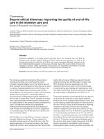
Hemodynamic parameters during pregnancy
Cardiac
output
Percent change
50
40
30
Heart rate
20
me
olu
v
ke
o
Str
10
0
0
8
16
24
Weeks of gestation
32
Clinical Implications
An increased cardiac output might not be well
tolerated by pregnant women with valvular heart
disease (e.g., aortic or mitral stenosis) or coronary arterial disease.
40
Pregnant
Nonpregnant
Changes in the Respiratory System
Changes in the respiratory parameters start as
early as the 4th week of gestation. Minute ventilation is increased at term by about 50 % above nonpregnant values. The increase in minute ventilation
is mainly due to an increase in tidal volume (40 %)
C.N. Purandare et al.
12
and, to a lesser extent, to an increase in the respiratory rate (15 %) [8]. Alveolar ventilation is greatly
increased as the tidal volume increases without
any change in the anatomic dead space. At term
the PCO2 value is decreased (32–35 mmHg).
Increased progesterone concentrations during
pregnancy decrease the threshold of the medullary
respiratory center to carbon dioxide [9, 10].
Clinical Implications
A decreased functional residual capacity as well
as increased oxygen consumption can cause a
rapid development of maternal hypoxemia.
Changes in the Renal System
The glomerular filtration rate is increased during
pregnancy because of increased renal plasma flow
[11]. A rise in the filtration rate decreases plasma
blood urea nitrogen (BUN) and creatinine concentrations by about 40–50 %. Tubular reabsorption
of sodium is increased. However, glucose and
amino acids might not be absorbed as efficiently;
hence, glycosuria and aminoaciduria may develop
in normal gestation [12, 13]. The renal pelvis and
ureters are dilated, and peristalsis is decreased.
Normal values for renal function
Nonpregnant
Serum
0.6–1.4 mg/dl
creatinine
Serum BUN
7–31 mg/dl
Serum uric
acid
Urine Cr
clearance
Urine uric
acid
Urine glucose
2.4–8.2 mg/dl
Pregnant
0.53–0.9 mg/dl
decrease
8–10 mg/dl
decrease
2–5.8 mg/dl
90–130 mL/min
150–200 mL/min
150–
990 mg/24 h
60–115 mg/dl
Increases
Increases
Clinical Implications
Normal parturients’ BUN (8–9 mg/dl) and creatinine (0.4 mg/dl) values are 40 % less than in nonpregnant women. So nonpregnant values in
parturients will suggest abnormal kidney function.
Physiological diuresis during the postpartum
period occurs between the 2nd and 5th days. The
glomerular filtration rate and BUN concentration
slowly return to nonpregnant values by the 6th
postpartum week [13].
Changes in the Gastrointestinal System
Gastrointestinal motility, food absorption, and
lower esophageal sphincter pressure are decreased
during pregnancy, probably due to an increased
level of plasma progesterone [14]. Lower esophageal sphincter pressure is decreased during pregnancy; on the other hand, intragastric pressure is
increased during the last trimester. Heartburn during pregnancy is the result of reduced barrier pressure [15]. The gastric emptying time of solid as
well as liquid material is not changed during pregnancy. Because of decreased plasma gastrin concentration during pregnancy, there is reduction in
the total acid content of the stomach. Gastric emptying time is significantly slower during labor, and
hence, gastric volume is increased.
In addition to increased production of lipids and
certain clotting factors, some enzymes found within
the liver are also increased without indicating
pathology. It is important to distinguish a normal
rise in these levels from a pathologic change caused
by organ damage or destruction arising, for example, from preeclampsia or hepatitis. In preeclampsia, microclots in the liver and capsular edema are
danger signs, and if clotting factors become affected,
the patient is at a high risk for disseminated intravascular coagulation (DIC). Diagnoses are not
based upon a single abnormal value [16].
Normal hepatic values
Liver enzymes
Nonpregnant
Alanine
14–67 U/L
transaminase
(ALT)
Aspartate
6–58 U/L
aminotransferase
(AST)
Alkaline
38–150 lMU/ml
phosphatase
(ALP)
Lactate
117–224 U/L
dihydrogenase
(LDH)
Pregnant
Unchanged
Unchanged
> up to 2–4
times
Upper end
of normal to
700 U/L
2
Pregnancy-Induced Alterations in Physiology and Laboratory Reports
Changes in the Musculoskeletal
System
The hormone relaxin is responsible for both the
generalized ligamentous relaxation and the softening of collagenous tissues.
Clinical Implications
Relaxation of ligaments and collagen tissue of
the vertebral column is the main cause of lordosis
during pregnancy.
Changes in the Dermatological
System
Hyperpigmentation of certain parts of the body
such as the face, neck, and midline of the abdomen is not uncommon during pregnancy.
Melanocyte-stimulating hormone is responsible
for this change. Enlargement of the breasts is an
integral part of the physiological changes of
pregnancy [17].
Changes in the Ocular System
Intraocular pressure has been shown to decrease
during pregnancy; this is related to (1) increased
progesterone levels, (2) the presence of relaxin,
and (3) decreased production of aqueous humor
due to increased secretion of human chorionic
gonadotropin [18].
Maternal Physiological Changes [11]
Enlarged breasts, especially in parturients with
short necks, may make intubation extremely difficult. A short-handled laryngoscope as described
by Datta and Briwa may be helpful in such cases
[19]. Changes in intraocular pressure in parturients may produce visual disturbances as well as
contact lens intolerance.
Normally, pregnant women require calories
additional to the normal daily requirement. These
13
recommendations, varying from country to country, also suggest the addition of protein, iron, and
other mineral and vitamin supplements to provide
the necessary materials for fetal and maternal
welfare throughout the pregnancy. However, it is
understood that appropriate nutrition is important
for maximizing the possibility of healthy offspring. Hytten and Leitch [12] and others [14]
have pointed out that it is difficult to focus on
nutrition alone as a factor in the growth and development of normal babies. Women who are appropriately nourished during the course of pregnancy
are economically better and educated and have
greater access to the medical antepartum care that
seems to be associated with improved pregnancy
outcomes. Antepartum nutrition, however, continues to be an area of great interest because the balanced intake of food in pregnant women is a
simple intervention that may have a significant
impact on the outcome of reproduction. The current recommendations proposed by the Food and
Nutrition Board of the US National Science
Foundation are listed in Table 2.1.
Skin Changes
A lot of changes in the skin are observed during
pregnancy. Hyperpigmentation is seen in the areolae, the perineal skin, the anal region, the inner
thighs, and the linea nigra, which appears on the
abdominal wall. Melasma or chloasma is a
blotchy, sharply marginated hyperpigmentation
that occurs on the face of dark-haired and darkcomplexioned women. It is most often centrally
distributed on the face.
By the third trimester of pregnancy, vascular
“spiders” due to circulating estrogens occur in
Table 2.1 Recommended dietary allowances (Revised in
2005)
Nutrient
Protein
Calories
Calcium
Iron
Folic acid
Ascorbic
acid
For nonpregnant
woman
45 g/day
2100
1000 mg/day
18 mg/day
400 μg/day
75 mg/day
For pregnant woman
+30 g/day
+300
+1000 mg/day
+9 mg/day
+200 μg/day
+10 mg/day
C.N. Purandare et al.
14
about 67 % of white patients and 11 % of black
patients. These lesions occur on the neck, throat,
face, and arms.
Striae are common among women in late pregnancy. There seems to be a familial tendency in
the occurrence of these lesions. When they occur,
they first appear during the 6th and 7th months of
gestation on the abdominal skin; they then occur
on the breasts, upper arms, lower back, buttocks,
and thighs. They have been related to a combination of stretching of the skin and increased levels
of corticosteroids and estrogen in pregnancy.
2.
3.
4.
5.
6.
7.
Summary
The myriad changes that occur during the pregnant state have to be well understood by the health
provider to analyze the condition of the patient.
An understanding of some of the major mechanisms that produce these changes is helpful in the
analysis of symptoms and problems that arise during the course of a normal gestation. When associated disease is present, understanding of these
alterations becomes more important in that they
must be distinguished from pathophysiologic
changes brought by the disease process. The
interaction between disease and gestational physiology may make the appropriate diagnosis and
management of the pregnant woman difficult.
When a pregnant woman requires medical or surgical therapy, the consultative services of an
obstetrician or clinician trained in the complexities of maternal physiology are absolutely critical
to the proper management of clinical problems.
8.
9.
10.
11.
12.
13.
14.
15.
16.
17.
18.
References
1. Cunnigham FG. Normal reference ranges for laboratory
values in pregnancy. McGraw-Hill Publishing Co./Elsevier
Publishing Co; 2015. />
19.
normal-reference-ranges-for-laboratory-values-in-pregnancy
Lund CJ, Donovan JC. Blood volume during pregnancy. Am J Obstet Gynecol. 1967;98:393.
Ueland K. Maternal cardiovascular hemodynamics.
VII Intrapartum blood volume changes. Am J Obstet
Gynecol. 1976;126:671.
Fay RA, et al. Platelets in pregnancy: hyperdestruction in pregnancy. Obstet Gynecol. 1983;61:238.
Mashini IS, et al. Serial noninvasive evaluation of cardiovascular hemodynamics during pregnancy. Am
J Obstet Gynecol. 1987;156:1208.
Ueland K, et al. Maternal cardiovascular dynamics.
III Labor and delivery under local and caudal analgesia. Am J Obstet Gynecol. 1969;103:8.
Rosenfeld CR, et al. Effect of estradiol-17b on blood
flow to reproductive and nonreproductive tissues in
pregnant ewes. Am J Obstet Gynecol. 1976;124:618.
Prowse CM, Gaenster EA. Respiratory and acid-base
changes during pregnancy. Anesthesiology. 1965;26:381.
Tyler JM. The effects of progesterone on the respiration of patients with emphysema and hypercapnea.
J Clin Invest. 1960;39:34.
Reid DHS. Respiratory changes in labour. Lancet.
1966;1:784.
Christensen PJ, et al. Amino acids in blood plasma
and urine during pregnancy. Scand J Clin Lab Invest.
1957;9:54.
Welsh GW, Sims EAH. The mechanism of renal glycosourea in pregnancy. Diabetes. 1960;9:363.
Lind LJ, et al. Lower esophageal sphincter pressures
in pregnancy. Can Med Assoc J. 1968;98:571.
Cohen SE. Why is the pregnant patient different?
Semin Anesth. 1982;1:73.
Palahniuk RJ, et al. Pregnancy decreases the requirement for inhaled anesthetic agent. Anesthesiology.
1974;41:82.
Steinbrook RA, et al. Dissociation of plasma and
cerebrospinal fluid beta-endorphin-like immunoactivity levels during pregnancy and parturition. Anesth
Analg. 1982;61:893.
Bader AM, et al. Acute effect of progesterone on conduction blockade in the isolated rabbit nerve. Anesth
Analg. 1990;71:545.
Butterworth JF, et al. Pregnancy increases median
nerve susceptibility to lidocaine. Anesthesiology.
1990;72:962.
Weinreb RN, et al. Maternal ocular adaptations during
pregnancy. Obstet Gynecol Surv. 1987;42:471.
3
Ethics in the Setting
Up of Obstetric HDU and ICU
K. Muhunthan and Sabaratnam Arulkumaran
Introduction
Childbirth is a major life event for women and
their families. However, in a small proportion,
severe and sometimes life-threatening complications occur during pregnancy. Such critically ill
women should receive the same standard of care
for both their pregnancy-related and critical care
needs, delivered by professionals with the same
level of competences irrespective of whether
these are provided in a maternity or general critical care setting [1].
Maternal critical care is an area which is less
discussed than other parts of obstetric care.
However, there has been a growing need to
address this area from a national and international
point of view: to collate, to standardise, to share
and to learn. Maternal morbidity and mortality
has been analysed by different methods in majority of countries. What has become apparent is that
there is still a significant number of morbidity and
mortality associated with suboptimal care [2].
Critical care in pregnancy poses a major challenge to clinicians as it requires consideration of
the physiological changes associated with preg-
K. Muhunthan (*)
Senior Lecturer and Head of Obstetrics and
Gynaecology, University of Jaffna, SriLanka
e-mail:
S. Arulkumaran
Professor Emeritus of Obstetrics and Gynaecology,
St George’s University of London, UK
nancy and the need to reassure the well-being of
the foetus [3].
In order to safeguard the right of the woman to
live, to have good health and to minimise unacceptable outcome of obstetric morbidity and
mortality, it is important to address essential and
ethical aspects in planning and setting up an
obstetric HDU and ICU.
Implementing a Standardised
System on Recognising the Level
of Care Needed
It is imperative that all carers understand the terminology used in setting up and organising HDU
and ICU to provide care for critically ill patients
in the peripartum period.
Maternal critical care, high dependency care
and high-risk maternity care are not interchangeable, the term critical care having a more precise
definition. It is also recommended that the terms
‘high dependency’ and ‘intensive care’ be
replaced by the term ‘critical care’ [4].
It is important to define the level of critical
care required by the mother depending on the
number of organs requiring support and the type
of support required. Such accepted definitions
will provide a platform for the woman to receive
the needed treatment. Prioritisation of patients
based on the needed care is an important key for
proper communication and timely admission.
Often these facilities are in high demand, and
© Springer India 2016
A. Gandhi et al. (eds.), Principles of Critical Care in Obstetrics: Volume I,
DOI 10.1007/978-81-322-2692-5_3
15
K. Muhunthan and S. Arulkumaran
16
ethical practice demands only those who need
care in these facilities are admitted.
It is ethical to term facilities as HDU and ICU
only if the service provided to mothers meets
with the expected level of care [5].
Worldwide several definitions are adopted,
and the four levels of critical care as defined by
the Intensive Care Society are as follows [6]:
Level 0: Patients whose needs can be met through
normal ward care
Level 1: Patients at risk of their condition deteriorating and needing a higher level of observation or those recently relocated from higher
levels of care
Level 2: Patients requiring invasive monitoring/
intervention that include support for a single
failing organ system (excluding advanced
respiratory support)
Level 3: Patients requiring advanced respiratory
support (mechanical ventilation) alone or
basic respiratory support along with support
of at least one additional organ
Thus, maternal critical care can be distinguished
from ‘high-risk’ obstetrics because the foetal issues
are excluded. The maternal risk factors or obstetric
complications that require closer observations or
intervention, but, not support of an organ system,
are outside the levels 2 and 3 of critical care.
Predicting the Population
Requirement for Obstetric Critical
Care Beds
Provision of critical care through HDU and ICU
to women in the peripartum period needs to meet
the requirement of a country or region.
Birth rates are measured in various ways in
various countries, and using these figures and
Non Clinical Staff
Recorder
Recogniser
the maternal mortality and morbidity data, the
health care provider must be able to estimate
scientifically the numbers of adult critical care
beds required [7]. Another approach of projecting the needed number of facilities is to
audit pregnant women and those who were
recently pregnant and were admitted to adult
general critical care units during the previous
years.
A substantial portion of critical care may have
been and could be provided through high dependency, rather than intensive care set-up. In order
to plan the setting up of such facilities, it is
important to differentiate the level of critical care
required by any population.
Workforce Development and Staff
Competences
Lead professionals in maternity services have a
responsibility to ensure staffs are competent in
the early recognition of acutely ill and deteriorating patients and are able to perform the initial
resuscitation of such patients. This can be
achieved by regular certified courses at acute illness management.
Whichever the training modality is practised, assessment of competences is essential.
Multidisciplinary scenario-based training in
the form of skills and drills has been found to
be valuable, particularly when developing team
drills for life-threatening clinical situations.
In addition it is imperative that the staffs who
are involved in the care of acutely ill patients in
the hospital are competent with regard to knowledge, skills and attitudes required for safe and
effective treatment and care along a chain of
response. Figure 3.1 and the Table 3.1 give the
example of safe and effective treatment and care
along the chain of response [8].
Primary
Responder
Communication and Handover
Fig. 3.1 Safe and effective treatment and care along the chain of response
Secondary
Responder
Tertiary
Responder
(Critical Care)
3
Ethics in the Setting Up of Obstetric HDU and ICU
Table 3.1 Safe and effective treatment of care along the
chain of response
Non-clinical supporter who may also be the ‘alerter’
and may include the woman or visitor
The recorder who takes designated measurements and
records observations and information. In maternity
services this could be a maternity support worker,
health care assistant or midwife
The recogniser who monitors the patient’s condition;
interprets designated measurements, observations and
information and adjusts the frequency of observations
and level of monitoring. In the maternity setting this
could be a midwife, recovery or other nurse working
within the unit or foundation doctor
The primary responder who goes beyond recording
and further observation by interpreting the
measurements and initiating a clinical management
plan, e.g. commencing oxygen therapy, insertion of
airway adjuncts and selection and administration of a
bolus of intravenous fluids. This would be a junior
doctor or specialist trainee or foundation doctor with
appropriate competencies
The secondary responder who is likely to be called to
attend when the patient fails to respond to the primary
intervention or continues to ‘trigger’ or ‘retrigger’ a
response. This individual will assess the clinical effect
of the primary intervention, formulate a diagnosis,
refine the management plan, initiate a secondary
response and have the knowledge to recognise when
referral to critical care is indicated. This would be an
obstetric or anaesthetic specialist trainee
Tertiary responder: This role encompasses the acute
care competencies, such as advanced airway
management, resuscitation, clinical assessment and
interpretation of acutely ill obstetric patients. In the
maternity unit, this role is routinely provided by
consultant anaesthetists with certified training in
obstetric anaesthesia. The acute care competencies
required focus primarily on the clinical and technical
aspects of care and the delivery of effective patient
management. They assume the possession and
application at every level of complementary generic
competencies such as record-keeping, team working,
interpersonal skills and clinical decision-making. Of
particular note in this context is the ability to rapidly
access hospital information systems and retrieve
patient information, such as blood results and x-rays
Care of the Critically Ill Obstetric
Patient in Regular Settings
Before a decision is taken that a woman would
need critical care in HDU or ICU, it is obvious
that they may have been cared in a less intense
set-up like a regular obstetric ward. Hence it is
ethical that consultant-led obstetric services
17
should have adequate facilities, expertise,
capacity and back-up for timely and comprehensive obstetric emergency care, including the
possibility of transfer to intensive care.
It is therefore important that maternity and
critical care services design pathways at a local
level which ensure that a critically ill parturient
accesses equitable care when needed. Such pathways should facilitate mother and baby remaining together unless precluded by a clinical reason.
Such arrangements should detail defined escalation arrangements for bringing critical care, midwifery and obstetric competences into the
maternity or critical care unit. These arrangements need to take into account local configuration, size and complexity of maternity and critical
care services.
Transfer to Critical Care Area
from a Maternity Ward
Women may require transfer to a critical care
area for a higher level of care (both level 2 and
level 3) either pre-delivery or postpartum. Such
transfers need to satisfy an accepted standard
similar to the ICS Standards’ ‘Guidelines for the
transport of the critically ill adult’ and need to be
accompanied by an additional plan addressing
the maternal, foetal and postnatal needs of the
patient [9]. The plan should also indicate whether
or not pre-delivery shared care between obstetrics and critical care is essential.
All maternity units must have the facilities and
staff to resuscitate, stabilise and transfer critical
care patients [10]. The transfer should take place
with an appropriately trained practitioner.
Although this is generally an anaesthetist, it can be
a specific ‘transfer’ clinician or an intensivist.
Positioning of the pregnant patient poses additional
risks in the avoidance of aortocaval compression.
Transfer to Maternity Ward
from Critical Care Area
After the decision to transfer a patient from a
critical care area to the maternity ward has been
made, she should be transferred as early as possible during the day.
K. Muhunthan and S. Arulkumaran
18
Transfer from critical care areas to the maternity ward between 22.00 and 07.00 should be
avoided whenever possible [9]. Both the critical
care and receiving maternity ward teams should
take shared responsibility for the care of the
patient being transferred [11].
They should jointly ensure that:
• There is continuity of care through a formal
structured handover from critical care staff to
ward staff.
• There should be a supported written care plan
with instructions to medical and nursing staff.
• The receiving staff, with support from critical
care staff if required, should deliver the agreed
plan.
• The formal structured handover of care should
include:
I. A summary of critical care stay, including
diagnosis, treatment and outstanding
investigations
II. A monitoring plan detailing the frequency
of observations
III. A plan for ongoing treatment including
drugs and therapies, nutrition plan, infection status and any agreed limitations of
treatment
IV. Physical and rehabilitation needs
V. Psychological and emotional needs
VI. Specific communication or language
needs
The Maternity and General Critical
Care Area Interface
The pregnant woman being cared for in a general critical care area requires daily review by a
multidisciplinary team including a named obstetric consultant and named senior midwife.
The individualised patient management plan
should include care during the antepartum, intrapartum and postpartum periods with significant
midwifery input for normal midwifery care.
The role of the maternity team includes discussing any specific obstetric conditions with the
critical care team, for example pre-eclampsia,
which may be obscured by the woman’s current
medical emergency. A neonatologist may also be
required to advise on management of prematurity
if a preterm delivery is a possibility.
As these women are critically ill, there should
be regular communication between midwives,
obstetricians and neonatologists as more complex aspects of obstetric care are considered.
Whilst the general critical care staff are experienced in communicating and updating family
members, it has to be understood that there are
different needs and information that the family
requires from the midwife, e.g. emotional and
social support, potential preparation for premature delivery, a baby in special care, etc.
Intensive care may be physically, emotionally,
mentally and financially very taxing to the
woman and her family. Awareness and monitoring
of mental health is important as these women are
more vulnerable due to the impact of increased
risk for adverse outcome. The health care team
should have good education to provide the needed
support to the woman and her family due to the
longer recovery period.
Conclusion
Wherever a pregnant woman is receiving critical
care in an HDU or ICU set-up, there must be a
fundamental principle that her pregnancy care is
continued and integrated into overall care plans
and that this continues through to the postnatal
period.
The multiple caregivers have to ensure that the
needs of the critical care do not overshadow the
needs of the woman and her family in regard to
midwifery or obstetric care.
It is ethical for us to respect basic human
rights, i.e. to preserve an individual’s life and
health and to provide the care with dignity,
self-respect and confidentiality. It is also
important that the patient and her relative
get all the information and are involved in
decision-making. The women should be provided the best of care available. These aspects
of care based on basic ethical principles are
tested to the extreme when critical care is
3
Ethics in the Setting Up of Obstetric HDU and ICU
delivered in the intensive care setting. In many
parts of the world, such care is available to
those who could pay as such care is available
only in paying centres and not in government
hospitals. Pregnant mothers are young and are
in their prime of life. Their health and life is
compromised due to an obstetric or pre-existing medical complication. If the transient
severe illness is overcome by providing critical care, then these mothers will continue to
serve as the nucleus of the family and their
society. Hence the health community should
try and establish critical care for these young
women with equal access despite their socioeconomic standards and capabilities rather
than stretching the health budget to less significant issues.
References
1. Association OA: providing equity of critical and
maternity care for the critically ill pregnant or recently
pregnant woman – July 2011.
2. Special issue: saving mothers’ lives: reviewing maternal deaths to make motherhood safer: 2006–2008.
The eighth report of the confidential enquiries into
maternal deaths in the United Kingdom. BJOG.
2011;118(Suppl S1):1–203).
19
3. Sultan P, Arulkumaran N, Rhodes A. Provision of
critical care services for the obstetric population. Best
Pract Res Clin Obstet Gynaecol. 2013;27(6):803–9.
4. Comprehensive critical care – a review of adult
critical care services. London: DH; 2000. www.
dh.gov.uk/en/Publicationsandstatistics/Publications/
PublicationsPolicyAndGuidance/DH_4006585.
5. Wheatly S. Maternal critical care: what’s in a name?
J Obstet Anesth. 2010;19:353–5.
6. Levels of critical care for adult patients. Standards and
guidelines. London: ICS; 2009. www.ics.ac.uk/intensive_care_professional/standards_and_guidelines/
levels_of_critical_care_for_adult_patients.
7. Lyons RA, Wareham K, Hutchings HA, Major E,
Ferguson B. Population requirement for adult criticalcare beds: a prospective quantitative and qualitative
study. Lancet. 2000;355(9204):595–8.
8. Competencies for recognising and responding
to acutely Ill patients in Hospital. London: DH;
2008, www.dh.gov.uk/en/Publicationsandstatistics/
Publications/PublicationsPolicyAndGuidance/
DH_096989.
9. Guidelines for the transport of the critically ill adult.
London: ICS; 2002. www.ics.ac.uk/intensive_care_
professional/standards_and_guidelines/transport_of_
the_critically_ill_2002 .
10. Safer childbirth: minimum standard for the organization and delivery of care in labour, https://
www.rcm.org.uk/sites/default/files/WPRSafer
ChildbirthReport2007.
11. Improving patient handover (Good Practice
No.12). London: RCOG; 2010. www.rcog.
org.uk/womens-health/clinical-guidance/
improving-patient-handover-good-practice-no-12.