Strategies to enhance the therapeutic ratio of radiation as a cancer treatment
Bạn đang xem bản rút gọn của tài liệu. Xem và tải ngay bản đầy đủ của tài liệu tại đây (8.42 MB, 308 trang )
Mitchell S. Anscher · Kristoffer Valerie
Editors
Strategies to
Enhance the
Therapeutic Ratio
of Radiation as a
Cancer Treatment
Strategies to Enhance the Therapeutic
Ratio of Radiation as a Cancer Treatment
Mitchell S. Anscher • Kristoffer Valerie
Editors
Strategies to Enhance
the Therapeutic Ratio
of Radiation as a Cancer
Treatment
Editors
Mitchell S. Anscher
Department of Radiation Oncology
Massey Cancer Center
Virginia Commonwealth University
Richmond, VA, USA
Kristoffer Valerie
Department of Radiation Oncology
Massey Cancer Center
Virginia Commonwealth University
Richmond, VA, USA
ISBN 978-3-319-45592-1
ISBN 978-3-319-45594-5 (eBook)
DOI 10.1007/978-3-319-45594-5
Library of Congress Control Number: 2016954313
© Springer International Publishing Switzerland 2016
This work is subject to copyright. All rights are reserved by the Publisher, whether the whole or part of
the material is concerned, specifically the rights of translation, reprinting, reuse of illustrations, recitation,
broadcasting, reproduction on microfilms or in any other physical way, and transmission or information
storage and retrieval, electronic adaptation, computer software, or by similar or dissimilar methodology
now known or hereafter developed.
The use of general descriptive names, registered names, trademarks, service marks, etc. in this publication
does not imply, even in the absence of a specific statement, that such names are exempt from the relevant
protective laws and regulations and therefore free for general use.
The publisher, the authors and the editors are safe to assume that the advice and information in this book
are believed to be true and accurate at the date of publication. Neither the publisher nor the authors or the
editors give a warranty, express or implied, with respect to the material contained herein or for any errors
or omissions that may have been made.
Printed on acid-free paper
This Springer imprint is published by Springer Nature
The registered company is Springer International Publishing AG
The registered company address is: Gewerbestrasse 11, 6330 Cham, Switzerland
Contents
1
Mechanisms of Normal Tissue Response ..............................................
Jolinta Y. Lin, Isabel L. Jackson, and Zeljko Vujaskovic
1
2
The Role of Hypoxia in Radiation Response ........................................
Monica M. Olcina, Ryan Kim, and Amato J. Giaccia
29
3
The Role of Cancer Stem Cells in Tumour Radioresponse .................
Annett Linge, Anna Dubrovska, Michael Baumann,
and Mechthild Krause
43
4
Novel Strategies to Prevent, Mitigate or Reverse
Radiation Injury and Fibrosis ...............................................................
Pierre Montay-Gruel, Gael Boivin, and Marie-Catherine Vozenin
75
5
Technology Based Strategies to Enhance
the Therapeutic Ratio ............................................................................. 109
David V. Fried and Shiva K. Das
6
Nitric Oxide Synthase Uncoupling in Tumor
Progression and Cancer Therapy .......................................................... 139
Ross B. Mikkelsen, Vasily A. Yakovlev, Christopher S. Rabender,
and Asim Alam
7
Aiming the Immune System to Improve the Antitumor
Efficacy of Radiation Therapy ............................................................... 159
Chunqing Guo, Timothy Harris, and Xiang-Yang Wang
8
The Role of MicroRNAs in Modulating
Tissue Response to Radiation ................................................................ 183
Rebecca J. Boohaker and Bo Xu
v
Chapter 12
Radiosensitizing Glioma by Targeting ATM
with Small Molecule Inhibitors
Amrita Sule and Kristoffer Valerie
Abstract Malignant glioma is a devastating and incurable brain cancer. Current
standard treatment of malignant glioma is surgery followed by chemotherapy and
radiation. Progress during the past few decades in improving long-term survival has
been painfully slow with a median overall survival currently at a little more than 1
year. New strategies targeting the DNA damage response, including the ATM (ataxia
telangiectasia mutated) kinase, are currently being pursued. ATM is a master regulator of cell cycle checkpoints, DNA repair, and cell death in response to radiation.
Pre-clinical studies using novel small molecule inhibitors of the ATM kinase are in
progress and results from these look promising for future testing in humans. In fact,
one ATM kinase inhibitor is currently in a Phase I trial in combination with chemotherapy of advanced solid cancers. This chapter focuses on discussing recent
advances in developing and testing highly specific inhibitors targeting the ATM
kinase for cancer therapy with focus on malignant glioma.
Keywords Ataxia telangiectasia mutated (ATM) • Convection-enhanced delivery
(CED) • DNA damage response (DDR) • Glioblastoma multiforme (GBM) •
Ionizing radiation (IR) • Malignant glioma • Mitotic catastrophe • Phosphatidylinositol
3-kinase-related kinase (PIKK) • p53 • Radiosensitizer • Radiotherapy •
Temozolomide (TMZ)
12.1
Introduction
Nearly 80,000 new cases of malignant glioma (classified by the World Health
Organization (WHO) as Grade III and IV glioma) are diagnosed each year in the
United States with 17,000 people dying from the disease. Grade IV is also referred
A. Sule • K. Valerie (*)
Department of Radiation Oncology, Massey Cancer Center, Virginia Commonwealth
University, Richmond, VA, USA
e-mail:
© Springer International Publishing Switzerland 2016
M.S. Anscher, K. Valerie (eds.), Strategies to Enhance the Therapeutic Ratio
of Radiation as a Cancer Treatment, DOI 10.1007/978-3-319-45594-5_12
289
290
A. Sule and K. Valerie
to as glioblastoma multiforme (GBM). GBM is a highly lethal brain tumor presented as one of two subtypes with distinct clinical histories and molecular profiles. Hallmark characteristics of GBM include uncontrolled cell proliferation,
diffuse infiltration, and resistance to apoptosis. These features account for GBM’s
poor prognosis and resistance toward radio- and chemotherapy, and a median
patient survival of only 12–15 months [1]. In older individuals, the most common
form is of the primary subtype which arises de novo with no prior symptoms or
evidence of progression from low-grade tumors. The secondary subtype of GBM
occurs in younger patients from lower grade glioma. GBMs are classified into four
different subgroups based on gene expression profiling; (1) classical, (2) mesenchymal, (3) neural, and (4) pro-neural [2, 3]. Primary GBM is mostly found in the
classical subgroup with EGFR mutation/amplifications and mutations in CDKN2A
and PTEN. On the other hand, secondary GBMs are usually found in the pro-neural subgroup with frequent mutations in PDGFR, IDH1/2, and p53 [2]. The frequency of p53 mutation in this sub-group is 65 % or greater whereas classical
GBM harbors p53 mutations 30 % of the time [4, 5]. Recently, a new more reliable
molecular classification based on IDH status and specific TERT promoter mutations was proposed [6, 7].
Standard treatment of GBM is surgery followed by temozolomide (TMZ), an
alkylating drug, and radiation [8, 9]. However, little improvement has been seen
in the long-term survival of patients with GBM during the last several decades.
Thus, new treatments and approaches are urgently needed. As the understanding
of the molecular mechanisms associated with GBM continues to expand, and
more specific and potent drugs are developed, efficient delivery of therapeutic
agents to the brain becomes very important and remains a challenging clinical
problem. In particular, both the blood–brain barrier (BBB) and blood–tumor barrier hamper the successful treatment of brain tumors by severely limiting access
of therapeutic agents to the brain and tumor [10, 11]. These obstacles have made
the efficient delivery of anticancer drugs to the brain a major technical hurdle,
and therefore this area of research is lagging behind the development of the
drugs themselves. Because surgery is standard treatment for GBM, the delivery
of therapeutic agents directly to the brain during surgery, e.g. GLIADEL® wafers,
or post-surgery by convection-enhanced delivery (CED) via a cannula and positive pressure does not deviate significantly from current treatment practice. For
the obvious reasons of being easier to administer and lower cost, an orally bioavailable and BBB-penetrable ATM inhibitor would be preferable over CEDbased delivery. However, specific circumstances might favor the latter route of
drug administration, e.g. if radiomimetic drugs, such as etoposide, and camptothecin, etc., that either are too toxic when administered systemically or are BBBimpermeable, CED could be the most efficacious and appropriate mode of
delivery [11].
There have been significant advances in the development and pre-clinical testing
of radiosensitizers for high grade gliomas during the past few years with focus on
targeting the DNA damage response (DDR) (see [12–14] for recent reviews).
Despite the identification of exciting new targets and the development of drugs
12
Radiosensitizing Glioma by Targeting ATM with Small Molecule Inhibitors
291
against these targets, their clinical use is still under evaluation. One of the earliest
targets identified and pursued is the protein mutated in ataxia telangiectasia (ATM)
and its intrinsic protein kinase [15]. ATM is mutated in the human autosomal
recessive disorder, ataxia-telangiectasia (A-T) [16]. The extreme radiosensitivity of
cells from A-T patients has been known since the 1970s [17]. ATM, a serine-threonine kinase and member of the phosphatidylinositol 3-kinase-related kinase (PIKK)
family, is a major regulator of the DDR. ATM is activated in response to DNA
double strand breaks (DSBs) induced by DNA damage such as ionizing radiation
(IR) or spontaneously during replication and cell growth. Once activated, ATM
phosphorylates numerous proteins involved in cell cycle regulation, DNA repair,
apoptosis, etc. [18, 19]. ATM-mediated phosphorylation and other subsequent posttranslational modifications affect the stability, sub-cellular localization, and the
interaction of proteins involved in these processes, thereby masterminding the DDR
[20]. ATM is also known to regulate insulin and other growth factor signaling
responses resulting from the stimulation with non-classical DDR agents suggesting
a much broader role for ATM in regulating cell growth and homeostasis in addition
to the DDR [16].
During the past 10 years the ATM inhibitor KU-55933 has extensively been
used in tissue culture experiments by numerous laboratories to demonstrate the
involvement of the ATM kinase in various capacities. KU-55933 was developed
by KuDOS Pharmaceuticals, Ltd, in the United Kingdom, and shown to be a
highly specific ATM kinase inhibitor competitively binding to the ATP-pocket
[21]. The KU-55933 IC50 for ATM (13 nM) is at least 200-fold lower than for the
other PIKKs, including DNA-PKcs and ATR. Around 2007, at the time KuDOS
was acquired by AstraZeneca, we were offered an improved analog, KU-60019,
to test as a radiosensitizer in our mouse glioma models. We extensively characterized KU-60019 in vitro with glioma cells to assess its impact on the DDR [22].
Briefly, in addition to the improved radiosensitization seen with KU-60019, we
documented high specificity toward the ATM kinase with no effect on 229 other
kinases in vitro. Radiosensitization was observed with all cell lines tested,
whether tumor or normal, except for A-T cells, strongly suggesting that the ATM
kinase was the target for KU-60019. Furthermore, KU-60019 has high stability
and is quickly reversible in vitro in wash out experiments. Additionally, we carried out limited in vitro combination testing of KU-60019, temozolomide (TMZ),
and radiation [23]. When U87 glioma cells were co-treated with KU-60019 and
TMZ a slight increase in radiation-induced cell killing was noted although TMZ
alone was unable to radiosensitize the cells. In addition, without radiation,
KU-60019 with or without TMZ reduced glioma cell growth but had no significant effect on the survival of human astrocytes [23]. Another study showed a
beneficial interaction of KU-55933 and TMZ in vitro but only with inherently
TMZ-sensitive glioma cell lines [24]. Thus, there is no reason to believe that an
ATM kinase inhibitor would be counter-effective with current standard care of
glioma. Other ATM inhibitors, such as CP466722 [25] and KU-59403 [26], have
been developed with only the latter evaluated in a pre-clinical setting and neither
one tested against glioma.
292
A. Sule and K. Valerie
Fig. 12.1 Potential impact of an ATM inhibitor in combination with a DNA damaging agent on
cell cycle checkpoints, DNA repair, and cell death. The ATM kinase phosphorylates >700 proteins,
some at multiple sites, that is necessary for fully triggering the DDR [27]. Blocking the DDR
including G1 and G2 arrest, DNA repair, and apoptosis/cell death with an ATM inhibitor is
expected to affect many cellular responses to radiation and chemotherapy and kill tumor cells.
Descriptors; →, activation/phosphorylation (Ⓟ); inhibition, ⊥
12.2
12.2.1
Rationale for Targeting the ATM Kinase
Advantages of ATM Kinase-Directed Therapy
It was realized early on that an ATM inhibitor would likely serve as an excellent
radiosensitizer based on the radiosensitivity of A-T patients [17]. The basic idea
behind this notion is that an ATM kinase inhibitor, such as KU-55933 or KU-60019,
would be expected to block cell cycle checkpoints and DNA repair so that tumor cells
would die from apoptosis or other cell death (Fig. 12.1). Many proteins regulating
cell cycle checkpoints (e.g., p53, MDM2, and CHK2), DNA repair (BRCA1, NBS1),
cell death/apoptosis (cABL) are directly phosphorylated by ATM [16, 27, 28], so an
ATM inhibitor would effectively block signaling and prevent all downstream DDRassociated processes from taking place with fatal consequences to the tumor cell.
Cancer-specific targeting is a long-sought-after goal in cancer therapy. We demonstrated for the first time that a small molecule ATM kinase inhibitor, KU-60019,
efficiently radiosensitized orthotopic gliomas with a much greater response seen
with mutant p53 relative to matched glioma with normal p53 [29]. Briefly, human
glioma U87 cells (p53 wild type) transduced with a retrovirus expressing a
p53-281G mutant were grown intra-cranially in nude mice in parallel with mice
12
Radiosensitizing Glioma by Targeting ATM with Small Molecule Inhibitors
293
injected with parental U87 cells. The mutant p53 acts a dominant-negative in this
situation and imposes a mutant p53 phenotype on the cells. Treatment with
KU-60019 prior to radiation repeated three times 3 days apart resulted in a significantly improved (p = 0.00011) survival of U87-281G mice, whereas mice with
parental U87 p53 wild type gliomas did not respond under these conditions of relatively low total radiation dose (Fig. 12.2). The radiation dose was purposely set low
so that KU-60019 radiosensitization would be more easily discerned. With parental
U87 tumors, radiation alone and KU-60019 alone showed a trend toward longer
survival, whereas a significant effect of KU-60019 alone versus untreated was
noted with U87-281G tumors [29].
The U87 parental and U87-281G cells were analyzed in vitro in their response to
KU-60019 with or without radiation (Fig. 12.3). We found that indeed the U87281G cells had a compromised G1/S checkpoint, as expected, grew substantially
faster and were more responsive to KU-60019 treatment alone in growth assays.
Additionally, the cells were more radiosensitive, and responded more robustly to
KU-60019 and radiation, resulting in more cell death than with U87 parental cells.
The results from this work laid the foundation for the notion that mutant p53 gliomas might respond to ATM inhibitor radiosensitization more robustly than p53 wild
type gliomas.
It has been reported that high grade glioma cells show signs of elevated replicative stress compared to lower grade brain tumor cells [30], perhaps favoring a
highly responsive phenotype to radiation and inhibition of ATM, which is then
enhanced by mutant p53. Our own work suggests that mutant p53 glioma cells die
by increased mitotic catastrophe (apoptosis in or subsequent to mitosis) when challenged by radiation in the presence of an ATM inhibitor [31]. It is likely that the
consequence of ATM inhibition and interference with DDR signaling in p53 mutant
glioma cells occur at multiple levels, e.g., abrogation of cell cycle checkpoints and
inhibition of DNA repair, blocking signaling through the TAO kinases and p38MK2-CDC25A, ultimately leading to mitotic catastrophe [32, 33]. The more proximal mechanism causing mitotic catastrophe is possibly through PLK1 and Aurora
A controlling the G2/M transition into M resulting in elevated mitotic failure in p53
mutant glioma cells exposed to radiation in the presence of an ATM inhibitor [34–
36]. Since p53 is mutated in about a third of all gliomas and the notion that mutant
p53 gliomas are more responsive to ATM inhibitor-based radiation therapy suggests
that an ATM inhibitor could be a promising adjuvant therapy that would fit well
with current standard of care [9].
The molecular weight of KU-60019 is >500 Da and does not cross the
BBB. Therefore, in our initial attempts to radiosensitize gliomas KU-60019 was
administered intra-tumorally by CED or osmotic pump in order to document inhibition of the DDR in tumor and surrounding brain tissue resulting in a survival benefit
to mice transplanted with p53 mutant gliomas [29]. Consequently, an orally bioavailable ATM inhibitor would simplify and reduce the technical aspects of ensuring efficient glioma radiosensitization. Further in this chapter we will discuss efforts
that our group and others have made toward bringing an ATM inhibitor closer to
clinical testing.
294
A. Sule and K. Valerie
Survival (%)
100
U87-p53281G
50
p = 0.00011
CED
0
No Treatment
3 x 3 Gy
3 x KU-60019
3 x (KU-60019 + 3 Gy)
0 3 6 9 12 20 40 60 80 100 120 140 160 180 200
3 x 3 Gy
Time (Days)
Survival (%)
100
U87-p53WT
50
CED
0
No Treatment
3 x 3 Gy
3 x KU-60019
3 x (KU-60019 + 3 Gy)
0 3 6 9 12 20 40 60 80 100 120 140 160 180 200
3 x 3 Gy
Time (Days)
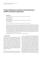
Fig. 12.2 KU-60019 radiosensitizes p53 mutant but not p53 wild type intra-cranial U87 tumors.
Human glioma U87 cells were transduced with a retrovirus expressing mutant p53-281G. ATM
inhibitor was administered by CED immediately followed by 3 Gy radiation on day 6, 9, and 12
(3 × 3 Gy) after intracranial injection of tumor cells. (Top) U87-281G tumors are highly responsive
to KU-60019 radiosensitization whereas parental U87 tumors (bottom) are not (p = 0.00011).
Whereas mice injected with parental U87 cells survived 60–80 days regardless of treatment, 50 %
of the mice injected with U87-281G cells and treated with both KU-60019 and radiation survived
for at least 160 days. Radiation dose was purposely set at 3 × 3 Gy in order to see survival benefits
with KU-60019 and radiation. Survival is plotted as Kaplan-Meier curves. Adapted from
Biddlestone-Thorpe et al. with permission [29]
12
Radiosensitizing Glioma by Targeting ATM with Small Molecule Inhibitors
a
U87
U87281G
b
U87
p53
U87-281G
10 Gy – 16 hr
Untreated
295
28.2%
10 Gy – 16 hr
Untreated
14.4%
25.1%
23.2%
BrdU
GAPDH
c
p=0.035
d
Control
KU60019
20.0
e
Luciferase
10
1X
0.9X
10.0
0.4X
7.5
5.0
Surviving Fraction
15.0
Surviving Fraction
Cell Number (x104)
G1:
S:
G2/M:
%
38.6
9.7
46.6
CFA
0
1.0
17.5
12.5
%
G1:
65.9
S:
16.2
G2/M: 12.1
p=0.014
1X
22.5
%
G1:
59.8
S:
6.3
G2/M: 28.8
%
G1:
68.8
S:
10.3
G2/M: 16.1
7-AAD
0.8
0.6
0.4
U87
U87/p53(281G)
0.2
-1
U87
U87-281G
U87 + KU-60019
U87-281G + KU-60019
U87
U87-281G
2.5
0.0
10
0
3
6
Dose, Gy
9
10
-2
0
1
2
Dose, Gy
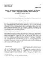
Fig. 12.3 Genetically matched glioma cells differing in p53 status demonstrate significantly different responses to ATM inhibitor and radiation. (a) Over-expression of mutant p53-281G from a
retrovirus in p53 wild type human U87 glioma cells produces a dominant p53 effect on cell cycle
checkpoints and DNA repair. Western blotting with anti-p53 antibody of extracts from U87 or
U87-281G cells shows expression of mutant p53 whereas endogenous wild-type p53 is undetectable in these unirradiated cells (b) U87-281G cells have a defective radiation-induced G1/S checkpoint and an intact G2/M checkpoint. U87 and U87-281G cells were irradiated with 10 Gy and
BrdU added immediately for 16 h to detect cells entering S to accumulate in G2/M. Both U87 and
U87-281G cells entered S after radiation with 10 Gy but whereas parental U87 showed a 50 %
decrease, U87-281G traversed into S unperturbed relative to unirradiated cells suggesting that the
G1/S checkpoint is compromised in the latter cells. Additionally, more U87-281G cells arrest in
G2/M than U87 cells because more cells go through the G1/S checkpoint. Altogether, U87-281G
cells have a compromised G1/S checkpoint whereas the G2/M checkpoint is still relatively intact
(c) In line with the finding that U87-281G cells lack the G1/S checkpoint, the cells demonstrate
higher proliferation rate and are more responsive to KU-60019 growth inhibition (d) U87-281G
cells are more radiosensitive than parental U87 by luciferase assay (e) In a colony-forming assay,
U87-281G cell are more radiosensitive and responsive to KU-60019 than parental U87 cells.
Adapted from Biddlestone-Thorpe et al. with permission [29]. Colony-forming assay, CFA
12.2.1.1
ATM Regulates Pro-survival Signaling at Multiple Levels
Interestingly, in our initial studies we found that KU-60019 not only inhibited the
DDR, but also reduced AKT phosphorylation and pro-survival signaling, and inhibited migration and invasion in vitro [22]. AKT needs to be phosphorylated on both
296
A. Sule and K. Valerie
INS-R
INS
EGFR
IGF
IGF
KINASE
KINASE
KINASE
KINASE
KINASE
KINASE
ATM
RAS
?
RAF
AKT
Cytoplasm
EGF
EGF
KINASE
KINASE
KINASE
KINASE
KINASE
KINASE
Plasma
membrane
INS
IGF-1R
MEK
PP2A
ERK
ATM
DSB repair/
radioresistance
Nucleus
ATM
DNA
Fig. 12.4 AKT and MEK/ERK signaling are subsets of the ATM signaling network. ATM is
known to interact with insulin, IGF-1R, as well as EGF growth factor signaling [22, 38, 39, 68].
Also central to ATM activation and regulation is the yin-yang relationship with PP2A, a phosphatase known to bind to ATM and intimately partake in the reversal of the DDR by dephosphorylating
many proteins phosphorylated by ATM and other kinases [69–71]
S473 and T308 in order to become fully activated and able to phosphorylate downstream target proteins necessary for eliciting a proliferative response. Since the
AKT S473 and T308 residues are not followed by an asparagine (-S/Q- or -T/Q-),
i.e., consensus ATM phosphorylation sites, and thus would not likely be direct ATM
kinase targets suggests that ATM might regulate AKT phosphorylation/activation
indirectly [22]. On the other hand, the more promiscuous DNA-PKcs is known to
phosphorylate AKT S473 in response to DNA damage [37]. We favor a mechanism
by which DNA-PKcs directly phosphorylates AKT and ATM negatively regulates
AKT dephosphorylation in response to radiation and growth factor signaling thus
implicating a critical role for ATM in AKT signaling [22].
The fact that insulin-mediated AKT S473 phosphorylation is substantially
reduced (~50 %) by ATM inhibition suggests a role for ATM in AKT pro-survival
signaling and tumor growth that overlaps with the DDR [22, 38]. Interestingly, it
has been shown that overexpression of insulin growth factor 1 (IGF-1) receptor in
A-T cells increases radioresistance [39]. Growth factor receptors such as insulin,
IGF-1R, and EGFR, are intimately associated with ATM and the DDR (Fig. 12.4).
It is possible that ATM interacts with receptor signaling at multiple levels including
the plasma membrane, cytoplasm, and nucleus. The observation that both RASRAF-MEK-ERK as well as AKT signaling are affected by ATM manipulation has
been reported by a number of laboratories including ours [22, 40–42]. From these
studies it is clear that ATM exerts its control at multiple levels including growth factor receptors, cytoplasmic signal transduction, and dephosphorylation of AKT. It
makes sense that a key DDR regulator such as ATM would serve as the gate-keeper
12
Radiosensitizing Glioma by Targeting ATM with Small Molecule Inhibitors
297
between cell survival and death. Importantly, one would expect ATM inhibitors to
act a multiple levels to enhance tumor radiosensitivity and inhibit tumor growth.
In addition to controlling the DDR, ATM also seems to regulate glioma migration and invasion which is not surprising given its association with ERK and AKT
signaling. We first demonstrated that an ATM inhibitor reduced the migration and
invasion of human glioma cells in vitro [22]. In support of this early finding, other
groups have since shown that reducing the ATM protein by genetic means generates
a blockade to AKT phosphorylation/activation downstream of HER2 which, if left
unperturbed, promotes breast cancer dispersal [43]. Thus, ATM promotes HER2dependent tumorigenicity and its expression correlates with reduced time of recurrence diagnosed with invasive HER2+ breast cancer. This suggest that HER2+
tumors have a selective advantage in retaining ATM expression and, therefore, ATM
inhibition might counter metastatic potential of HER2+ breast cancers. In a separate
study, it was demonstrated that ATM acts via IL-8 to enhance breast cancer metastasis to the lung [44]. The induction of IL-8 occurred as a consequence of oxidative
stress which is known to activate ATM [45]. Knocking down ATM or inhibiting with
KU-55933 resulted in reduced oxidative stress and IL-8 expression suggesting that
IL-8 was under control of ATM [44]. Most importantly, blocking ATM reduced
breast cancer migration and invasion in vitro and in vivo. Thus, ATM might play a
role in breast cancer metastasis and progression in addition to its well-established
role in tumor suppression [44].
Our own follow-up studies that an ATM kinase inhibitor reduces glioma cell
migration and invasion in vitro [22], have now provided evidence that glioma dispersal in mouse brain is under control of ATM (in preparation). Briefly, a matched
human glioma cell pair, one with ATM levels reduced by shRNA and the other a
mock knockdown, showed significant reduced ability of the shATM glioma cells to
migrate and invade in vivo when presented as intra-cranial tumors. Therefore, an
ATM inhibitor might prevent glioma dispersal in between radiation fractions as well
as enhancing the killing of tumor cells when irradiated as we have proposed previously [23]. Altogether, ATM might exert control over cell growth, survival, and
death signaling at multiple levels; distal via growth factor receptors at the plasma
membrane and more proximal at the level of cytoplasmic signal transduction via the
ERK and AKT pathways, and a more direct involvement in the dephosphorylation
of AKT resulting in reduced tumor cell growth. In summary, an ATM inhibitor may
limit tumor growth, migration, and invasion by inhibiting ERK and AKT signaling
in addition to acting as a very potent radiosensitizer.
12.2.1.2
ATM-EGFR-ERK and ATM-AKT Signaling Modulate DNA
DSB Repair
As expected, there is a close relationship between pro-survival signaling and proficient DNA repair—at low levels of DNA damage occurring during clinically relevant
radiation doses, DNA repair is operating optimally whereas apoptosis and related
death mechanisms are suppressed [46, 47]. Using KU-55933, we demonstrated that
298
A. Sule and K. Valerie
ATM was critically involved in regulating homologous recombination repair (HRR)
in human glioma cells via MAPK signaling and that the ERK pathway appears to
form a regulatory feed-back loop with ATM [40]. We and others have shown that the
stimulation of cellular growth with growth factors such as epidermal growth factor
(EGF), or, alternatively, blocking growth signaling by small molecule inhibitors or by
genetic means modulate DSB repair [48–53]. In regards to GBM, it is particularly
relevant that EGFRviii-mediated signaling promoted DSB repair both via both nonhomologous end joining (NHEJ) and HRR [48, 54]. EGFRviii is a mutant form of
EGFR that acts in a ligand-independent and auto-stimulatory manner through multiple pathways including ERK and AKT in many primary GBMs [55]. Altogether, an
ATM inhibitor will directly inhibit DSB repair resulting from radiation.
12.2.1.3
ATM Is Required for Neuronal Cell Death
The brain consists mostly of non-proliferating, terminally differentiated cells such as
neurons, astrocytes, and oligodendrocytes. Proliferating cells are mostly limited to
neural progenitors and stem cells able to reconstitute in part cell populations after
traumatic brain injury. Since ATM is required for radiation-induced neuronal apoptosis [56], transiently inhibiting ATM in the brain is expected to protect neurons
from cell death. However, this has yet to be demonstrated. Almost 20 years ago
using mouse genetics it was elegantly demonstrated in a string of very significant
reports that ATM-dependent apoptosis in the CNS, and specifically in neurons, is
mediated by p53 since p53−/− mice showed a similar lack of radiation-induced cell
death in the developing nervous system as ATM−/− mice [56]. In addition, the ATMdependent apoptotic pathway in neurons required BAX, a p53-dependent effector
and critical participant in apoptosis [57]. Furthermore, ATM- and BAX-dependent
apoptosis also required caspase-3 activation. However, in contrast to radiosensitive
ATM−/− fibroblasts and radioresistant ATM−/− neurons, survival of ATM−/− astrocytes after irradiation was similar to wild-type astrocytes suggesting that in this type
of CNS cells, ATM functions in controlling cellular growth and radiosensitivity by
distinct mechanisms [58].
Altogether, based on these earlier findings and our own unpublished results, we
speculate that an ATM inhibitor would have a substantial protective effect on irradiated CNS (p53 wild type) at low radiation doses that would result in cell cycle arrest
rather than apoptosis in cells with proliferative capacity such as neural progenitors
and stem cells, and prevent p53-dependent apoptosis in terminally differentiated
neurons that would also require BAX and caspase 3. In contrast, gliomas, and in
particular those with defective p53 signaling, would die by mitotic catastrophe
when exposed to ATM inhibitor and radiation (Fig. 12.5). However, it is currently
unclear whether a small molecule ATM kinase inhibitor would result in the same
phenotype as the complete absence of ATM in the mouse (as in ATM−/− mice) [59],
and whether the mouse phenotype can be recapitulated in humans and be fully
applied to the human situation during cancer therapy. Future clinical trials will
address these issues.
Cell cycle
arrest
Mitotic
Catastrophe
p53 Mut
+ ATM Inhibitor
RADIATION
Glioma cell death
Fig. 12.5 Proposed model for the enhanced response of mutant p53 gliomas to ATM inhibitor radiosensitization and the protection of normal healthy brain.
The model is based on the findings by McKinnon and co-workers using ATM, p53, BAX, and ARF KO mice [56–58]. Healthy brain not exposed to an ATM
inhibitor is expected to undergo apoptosis in a radiation dose-dependent manner. In the presence of an ATM inhibitor a shift from apoptosis to cell cycle arrest
occurs after radiation due to an intact p53 response. On the other hand, in a glioma p53 mutant environment ATM inhibition and radiation would kill cells by
mitotic catastrophe
Apoptosis
p53 WT
p53 WT
Cell cycle
arrest
+ ATM Inhibitor
RADIATION
Radioprotection
- ATM Inhibitor
RADIATION
Apoptosis
Healthy Brain
CNS Cell death
p53 Mutant Glioma
12
Radiosensitizing Glioma by Targeting ATM with Small Molecule Inhibitors
299
300
12.2.2
A. Sule and K. Valerie
Limitations of ATM Kinase-Directed Therapy
The adverse effect of an ATM inhibitor on HRR and triggering of carcinogenesis
has been put forward cautioning against its use in patients [60, 61]. However, it is
important to realize that any chemo- or radiotherapy regimen will potentially have
the unfortunate side-effect of causing secondary cancers. Clinical dose-findings will
reveal whether benefits outweigh toxicity of ATM inhibitor-based therapies. Clearly,
one of the advantages with combining an ATM inhibitor with radiation therapy is
the ability to reduce systemic toxicity by applying conformal radiation only targeting the tumor. Chemotherapy in combination with an ATM inhibitor would not have
that benefit since drugs alone would potentially show increased systemic toxicity to
organs such as liver, kidneys, and blood. Thus, a strong case for using an ATM
inhibitor in combination with conformal radiation can be made.
12.2.3
Cancer-Specific Targeting
Almost 50 % of all cancers have mutated or defective p53 [62]. For gliomas the
overall proportion is about 30 % with slower-progression secondary GBM having
much more frequent p53 alterations than primary GBM [5]. Since p53 is mutated in
about a third of all gliomas and our discovery that mutant p53 gliomas are more
responsive, ATM inhibitor-based radiation therapy could be a promising adjuvant
therapy that would fit well with current standard of care to treat this subset of
patients [9]. It would be very exciting to see whether the results from our pre-clinical testing of an ATM inhibitor translate into a greater response in patients with
p53-mutated gliomas. If so, a third of all GBM patients might have a greater chance
of survival past 1 year.
12.3
Pre-clinical Testing
Our studies showing that orthotopic human xenografts are responsive to ATM
inhibitor radiosensitization are supported by similar findings by several other
groups. It is now well-established that glioma stem cells or glioma-initiating cells
(GICs) are the important tumor population responsible for treatment failure [63]. In
fact, CD133+ GICs isolated from both human glioma xenografts and primary
patient GBM preferentially activate the DDR in response to radiation, and the repair
of radiation-induced DNA damage was more effective than in CD133- tumor. In
addition, the radioresistance of CD133+ cells was reversed with a specific inhibitor
of the CHK1 and CHK2 checkpoint kinases. An ATM kinase inhibitor would probably result in the same effect as the CHK inhibitors. In fact, we have shown that
mouse glioma cells isolated from a spontaneous tumor from a genetically
12
Radiosensitizing Glioma by Targeting ATM with Small Molecule Inhibitors
301
engineered mouse with high incidence of glioma-formation responded well to
KU-60019 radiosensitization in vitro [29]. In support of these findings, Vecchio
et al. showed a p53-dependent response to ATM inhibitor and radiation in that low
expressing p53 GICs responded better to KU-60019 and radiation than higher
expressing cells [64], in agreement with our earlier report using matched laboratory
glioma cell lines only differing in p53 [29]. It was also demonstrated that KU-60019
appeared to be safe when administered alone without resulting in any detectable
toxicity or mutation in the various mouse tissues examined [65]. Furthermore, similar radiosensitization was also seen with pediatric GICs suggesting that an ATM
kinase inhibitor could be a safe and effective radiosensitizer of both adult and pediatric gliomas. In another study, Lim et al. demonstrated that KU-55933 was able to
radiosensitize GICs and prolong the survival of mice with orthotopic gliomas [66].
However, it appears as if this study pretreated the GICs with the ATM inhibitor
prior to intra-cranial injection and radiation. Nevertheless, the authors’ conclusion
was that HRR was the predominant type of DSB repair in the GICs which was
reduced by the ATM inhibitor and, in turn, resulted in radiosensitization. On the
other hand, using a small molecule inhibitor of DNA-PKcs, important for classical
NHEJ, did not affect DSB resolution and radiosurvival in vitro suggesting that
NHEJ is less critical for DSB repair in GICs and might be less effective for therapeutic intervention toward GBM.
As all these studies have indicated, KU-60019 does not cross the BBB. Therefore,
new more BBB-penetrable ATM inhibitors are needed. We have tested one such
orally bioavailable ATM inhibitor and presented preliminary data from several
mouse orthotopic glioma models [67]. Briefly, both a mouse syngeneic glioma
grown in immune-competent mice as well as human orthotopic xenografts in nude
mice were radiosensitized after the mice were given oral gavage of the ATM inhibitor (manuscript in preparation). Continued research on the efficacy and safety in the
next year or so will demonstrate whether any such ATM inhibitor could move forward toward clinical testing.
12.4
Clinical Testing
AZD0156, a clinical ATM inhibitor compound developed by AstraZeneca, is currently undergoing testing in patients with advanced malignancies (ClinicalTrials.
gov ID: NCT02588105). The goal of this trial is preliminary assessment of the antitumor activity of AZD0156 either as monotherapy or in combination with Olaparib
(PARP inhibitor), cytotoxic chemotherapies, or novel anti-cancer agents. Planned
enrollment in this multi-national trial is 225 patients. A more effective, BBB penetrating ATM inhibitor for glioma is currently being tested in pre-clinical models. It
will be important to soon as possible also test AZD0156 together with radiation
since the conformal nature of this modality is expected to result in a greater therapeutic index than any combination with DNA damaging drugs.
302
12.5
A. Sule and K. Valerie
Conclusions
The development and testing of ATM inhibitors for brain cancer therapy is proceeding and is expected to enter the clinical testing arena in the next few years or so. The
potential benefits of an ATM inhibitor as an adjuvant to radiotherapy go beyond just
killing the tumor with a radiosensitizer. It is possible that significant clinical benefit
might be seen in patients with mutant p53 gliomas with cancer-specific killing
whereas normal, healthy brain tissue is protected or at least a reduction in toxicity
seen. Regardless, one should be able to lower the total radiation dose to the brain in
combination with an ATM inhibitor thereby reducing long-term sequela and cognitive impairment of patients. In fact, today with a median survival of only little more
than 1 year for GBM patients the long-term consequences of surgery and chemoradiation are not fully considered because of the anticipated short life expectancy of
these patients. Once long-term survival rate improves treatment side-effects would
have to be addressed to also increase the quality of life. In fact, it is possible that
ATM inhibitors could also be beneficial in the recovery of radiation damage to neurons and the brain as a whole when provided post-treatment.
Acknowledgement We thank present and former members of the Valerie lab for advice and guidance in preparing this chapter. We also want to thank Nicholas Valerie for providing suggestions.
Funded by the National Institutes of Health (NCI and NINDS), and generous funding provided by
AstraZeneca.
References
1. Dunn GP, Rinne ML, Wykosky J, Genovese G, Quayle SN, Dunn IF, et al. Emerging insights
into the molecular and cellular basis of glioblastoma. Genes Dev. 2012;26(8):756–84.
2. Verhaak RG, Hoadley KA, Purdom E, Wang V, Qi Y, Wilkerson MD, et al. Integrated genomic
analysis identifies clinically relevant subtypes of glioblastoma characterized by abnormalities
in PDGFRA, IDH1, EGFR, and NF1. Cancer Cell. 2010;17(1):98–110.
3. Phillips HS, Kharbanda S, Chen R, Forrest WF, Soriano RH, Wu TD, et al. Molecular subclasses of high-grade glioma predict prognosis, delineate a pattern of disease progression, and
resemble stages in neurogenesis. Cancer Cell. 2006;9(3):157–73.
4. Ohgaki H, Kleihues P. Genetic alterations and signaling pathways in the evolution of gliomas.
Cancer Sci. 2009;100(12):2235–41.
5. Zheng H, Ying H, Yan H, Kimmelman AC, Hiller DJ, Chen AJ, et al. p53 and Pten control neural
and glioma stem/progenitor cell renewal and differentiation. Nature. 2008;455(7216):1129–33.
6. Killela PJ, Pirozzi CJ, Healy P, Reitman ZJ, Lipp E, Rasheed BA, et al. Mutations in IDH1,
IDH2, and in the TERT promoter define clinically distinct subgroups of adult malignant gliomas. Oncotarget. 2014;5(6):1515–25.
7. Killela PJ, Reitman ZJ, Jiao Y, Bettegowda C, Agrawal N, Diaz Jr LA, et al. TERT promoter
mutations occur frequently in gliomas and a subset of tumors derived from cells with low rates
of self-renewal. Proc Natl Acad Sci U S A. 2013;110(15):6021–6.
8. Stupp R, Mason WP, van den Bent MJ, Weller M, Fisher B, Taphoorn MJ, et al. Radiotherapy
plus concomitant and adjuvant temozolomide for glioblastoma. N Engl J Med.
2005;352(10):987–96.
9. Stupp R, Hegi ME, Gilbert MR, Chakravarti A. Chemoradiotherapy in malignant glioma: standard of care and future directions. J Clin Oncol. 2007;25(26):4127–36.
12
Radiosensitizing Glioma by Targeting ATM with Small Molecule Inhibitors
303
10. Lesniak MS, Brem H. Targeted therapy for brain tumours. Nat Rev Drug Discov.
2004;3(6):499–508.
11. Biddlestone-Thorpe L, Marchi N, Guo K, Ghosh C, Janigro D, Valerie K, et al. Nanomaterialmediated CNS delivery of diagnostic and therapeutic agents. Adv Drug Deliv Rev.
2012;64(7):605–13.
12. Morgan MA, Lawrence TS. Molecular pathways: overcoming radiation resistance by targeting
DNA damage response pathways. Clin Cancer Res. 2015;21(13):2898–904.
13. Dasgupta T, Haas-Kogan DA. The combination of novel targeted molecular agents and radiation in the treatment of pediatric gliomas. Front Oncol. 2013;3:110.
14. Raleigh DR, Haas-Kogan DA. Molecular targets and mechanisms of radiosensitization using
DNA damage response pathways. Future Oncol. 2013;9(2):219–33.
15. Savitsky K, Bar-Shira A, Gilad S, Rotman G, Ziv Y, Vanagaite L, et al. A single ataxia telangiectasia gene with a product similar to PI-3 kinase. Science. 1995;268(5218):1749–53.
16. Shiloh Y, Ziv Y. The ATM protein kinase: regulating the cellular response to genotoxic stress,
and more. Nat Rev Mol Cell Biol. 2013;14(4):197–210.
17. Taylor AM, Harnden DG, Arlett CF, Harcourt SA, Lehmann AR, Stevens S, et al. Ataxia telangiectasia: a human mutation with abnormal radiation sensitivity. Nature. 1975;258(5534):427–9.
18. Valerie K, Yacoub A, Hagan MP, Curiel DT, Fisher PB, Grant S, et al. Radiation-induced cell
signaling: inside-out and outside-in. Mol Cancer Ther. 2007;6(3):789–801.
19. Valerie K, Povirk LF. Regulation and mechanisms of mammalian double-strand break repair.
Oncogene. 2003;22(37):5792–812.
20. Lavin MF. Ataxia-telangiectasia: from a rare disorder to a paradigm for cell signalling and
cancer. Nat Rev Mol Cell Biol. 2008;9(10):759–69.
21. Hickson I, Zhao Y, Richardson CJ, Green SJ, Martin NM, Orr AI, et al. Identification and
characterization of a novel and specific inhibitor of the ataxia-telangiectasia mutated kinase
ATM. Cancer Res. 2004;64(24):9152–9.
22. Golding SE, Rosenberg E, Valerie N, Hussaini I, Frigerio M, Cockcroft XF, et al. Improved
ATM kinase inhibitor KU-60019 radiosensitizes glioma cells, compromises insulin, AKT and
ERK prosurvival signaling, and inhibits migration and invasion. Mol Cancer Ther.
2009;8(10):2894–902.
23. Golding SE, Rosenberg E, Adams BR, Wignarajah S, Beckta JM, O’Connor MJ, et al. Dynamic
inhibition of ATM kinase provides a strategy for glioblastoma multiforme radiosensitization
and growth control. Cell Cycle. 2012;11(6):1167–73.
24. Nadkarni A, Shrivastav M, Mladek AC, Schwingler PM, Grogan PT, Chen J, et al. ATM inhibitor KU-55933 increases the TMZ responsiveness of only inherently TMZ sensitive GBM cells.
J Neurooncol. 2012;110(3):349–57.
25. Rainey MD, Charlton ME, Stanton RV, Kastan MB. Transient inhibition of ATM kinase is sufficient to enhance cellular sensitivity to ionizing radiation. Cancer Res. 2008;68(18):7466–74.
26. Batey MA, Zhao Y, Kyle S, Richardson C, Slade A, Martin NM, et al. Preclinical evaluation of
a novel ATM inhibitor, KU59403, in vitro and in vivo in p53 functional and dysfunctional
models of human cancer. Mol Cancer Ther. 2013;12(6):959–67.
27. Matsuoka S, Ballif BA, Smogorzewska A, McDonald 3rd ER, Hurov KE, Luo J, et al. ATM
and ATR substrate analysis reveals extensive protein networks responsive to DNA damage.
Science. 2007;316(5828):1160–6.
28. Bensimon A, Schmidt A, Ziv Y, Elkon R, Wang SY, Chen DJ, et al. ATM-dependent and -independent dynamics of the nuclear phosphoproteome after DNA damage. Sci Signal.
2010;3(151):rs3.
29. Biddlestone-Thorpe L, Sajjad M, Rosenberg E, Beckta JM, Valerie NC, Tokarz M, et al. ATM
kinase inhibition preferentially sensitizes p53-mutant glioma to ionizing radiation. Clin Cancer
Res. 2013;19(12):3189–200.
30. Bartkova J, Hamerlik P, Stockhausen MT, Ehrmann J, Hlobilkova A, Laursen H, et al.
Replication stress and oxidative damage contribute to aberrant constitutive activation of DNA
damage signalling in human gliomas. Oncogene. 2010;29(36):5095–102.
31. Beckta JM, Ahmad SF, Yang H, Valerie K. Revisiting p53 for cancer-specific chemo- and
radiotherapy: ten years after. Cell Cycle. 2014;13(5):710–3.
304
A. Sule and K. Valerie
32. Raman M, Earnest S, Zhang K, Zhao Y, Cobb MH. TAO kinases mediate activation of p38 in
response to DNA damage. EMBO J. 2007;26(8):2005–14.
33. Reinhardt HC, Aslanian AS, Lees JA, Yaffe MB. p53-deficient cells rely on ATM- and ATRmediated checkpoint signaling through the p38MAPK/MK2 pathway for survival after DNA
damage. Cancer Cell. 2007;11(2):175–89.
34. van Vugt MA, Bras A, Medema RH. Polo-like kinase-1 controls recovery from a G2 DNA
damage-induced arrest in mammalian cells. Mol Cell. 2004;15(5):799–811.
35. van Vugt MA, Smits VA, Klompmaker R, Medema RH. Inhibition of Polo-like kinase-1 by DNA
damage occurs in an ATM- or ATR-dependent fashion. J Biol Chem. 2001;276(45):41656–60.
36. Smits VA, Klompmaker R, Arnaud L, Rijksen G, Nigg EA, Medema RH. Polo-like kinase-1 is
a target of the DNA damage checkpoint. Nat Cell Biol. 2000;2(9):672–6.
37. Bozulic L, Surucu B, Hynx D, Hemmings BA. PKBalpha/Akt1 acts downstream of DNA-PK
in the DNA double-strand break response and promotes survival. Mol Cell.
2008;30(2):203–13.
38. Viniegra JG, Martinez N, Modirassari P, Losa JH, Parada Cobo C, Lobo VJ, et al. Full activation of PKB/Akt in response to insulin or ionizing radiation is mediated through ATM. J Biol
Chem. 2005;280(6):4029–36.
39. Peretz S, Jensen R, Baserga R, Glazer PM. ATM-dependent expression of the insulin-like
growth factor-I receptor in a pathway regulating radiation response. Proc Natl Acad Sci U S A.
2001;98(4):1676–81.
40. Golding SE, Rosenberg E, Neill S, Dent P, Povirk LF, Valerie K. Extracellular signal-related
kinase positively regulates ataxia telangiectasia mutated, homologous recombination repair,
and the DNA damage response. Cancer Res. 2007;67(3):1046–53.
41. Wei F, Xie Y, Tao L, Tang D. Both ERK1 and ERK2 kinases promote G2/M arrest in etoposidetreated MCF7 cells by facilitating ATM activation. Cell Signal. 2010;22(11):1783–9.
42. Li Y, Yang DQ. The ATM inhibitor KU-55933 suppresses cell proliferation and induces apoptosis by blocking Akt in cancer cells with overactivated Akt. Mol Cancer Ther.
2010;9(1):113–25.
43. Stagni V, Manni I, Oropallo V, Mottolese M, Di Benedetto A, Piaggio G, et al. ATM kinase
sustains HER2 tumorigenicity in breast cancer. Nat Commun. 2015;6:6886.
44. Chen WT, Ebelt ND, Stracker TH, Xhemalce B, Van Den Berg CL, Miller KM. ATM regulation of IL-8 links oxidative stress to cancer cell migration and invasion. Elife. 2015;4.
45. Guo Z, Kozlov S, Lavin MF, Person MD, Paull TT. ATM activation by oxidative stress.
Science. 2010;330(6003):517–21.
46. Khalil A, Morgan RN, Adams BR, Golding SE, Dever SM, Rosenberg E, et al. ATM-dependent
ERK signaling via AKT in response to DNA double-strand breaks. Cell Cycle.
2011;10(3):481–91.
47. Hawkins AJ, Golding SE, Khalil A, Valerie K. DNA double-strand break—induced prosurvival signaling. Radiother Oncol. 2011;101(1):13–7.
48. Golding SE, Morgan RN, Adams BR, Hawkins AJ, Povirk LF, Valerie K. Pro-survival AKT
and ERK signaling from EGFR and mutant EGFRvIII enhances DNA double-strand break
repair in human glioma cells. Cancer Biol Ther. 2009;8(8):730–8.
49. Kao GD, Jiang Z, Fernandes AM, Gupta AK, Maity A. Inhibition of phosphatidylinositol3-OH kinase/Akt signaling impairs DNA repair in glioblastoma cells following ionizing radiation. J Biol Chem. 2007;282(29):21206–12.
50. Mukherjee B, McEllin B, Camacho CV, Tomimatsu N, Sirasanagandala S, Nannepaga S, et al.
EGFRvIII and DNA double-strand break repair: a molecular mechanism for radioresistance in
glioblastoma. Cancer Res. 2009;69(10):4252–9.
51. Toulany M, Kehlbach R, Florczak U, Sak A, Wang S, Chen J, et al. Targeting of AKT1
enhances radiation toxicity of human tumor cells by inhibiting DNA-PKcs-dependent DNA
double-strand break repair. Mol Cancer Ther. 2008;7(7):1772–81.
52. Kriegs M, Kasten-Pisula U, Rieckmann T, Holst K, Saker J, Dahm-Daphi J, et al. The epidermal growth factor receptor modulates DNA double-strand break repair by regulating nonhomologous end-joining. DNA Repair. 2010;9(8):889–97.
12
Radiosensitizing Glioma by Targeting ATM with Small Molecule Inhibitors
305
53. Fraser M, Harding SM, Zhao H, Coackley C, Durocher D, Bristow RG. MRE11 promotes
AKT phosphorylation in direct response to DNA double-strand breaks. Cell Cycle.
2011;10(13):2218–32.
54. Mukherjee B, Choy H, Nirodi C, Burma S. Targeting nonhomologous end-joining through
epidermal growth factor receptor inhibition: rationale and strategies for radiosensitization.
Semin Radiat Oncol. 2010;20(4):250–7.
55. Furnari FB, Fenton T, Bachoo RM, Mukasa A, Stommel JM, Stegh A, et al. Malignant astrocytic glioma: genetics, biology, and paths to treatment. Genes Dev. 2007;21(21):2683–710.
56. Herzog KH, Chong MJ, Kapsetaki M, Morgan JI, McKinnon PJ. Requirement for Atm in ionizing radiation-induced cell death in the developing central nervous system. Science.
1998;280(5366):1089–91.
57. Chong MJ, Murray MR, Gosink EC, Russell HR, Srinivasan A, Kapsetaki M, et al. Atm and
Bax cooperate in ionizing radiation-induced apoptosis in the central nervous system. Proc Natl
Acad Sci U S A. 2000;97(2):889–94.
58. Gosink EC, Chong MJ, McKinnon PJ. Ataxia telangiectasia mutated deficiency affects astrocyte growth but not radiosensitivity. Cancer Res. 1999;59(20):5294–8.
59. Choi S, Gamper AM, White JS, Bakkenist CJ. Inhibition of ATM kinase activity does not
phenocopy ATM protein disruption: implications for the clinical utility of ATM kinase inhibitors. Cell Cycle. 2010;9(20):4052–7.
60. White JS, Choi S, Bakkenist CJ. Irreversible chromosome damage accumulates rapidly in the
absence of ATM kinase activity. Cell Cycle. 2008;7(9):1277–84.
61. White JS, Choi S, Bakkenist CJ. Transient ATM kinase inhibition disrupts DNA damageinduced sister chromatid exchange. Sci Signal. 2010;3(124):ra44.
62. Kandoth C, McLellan MD, Vandin F, Ye K, Niu B, Lu C, et al. Mutational landscape and significance across 12 major cancer types. Nature. 2013;502(7471):333–9.
63. Bao S, Wu Q, McLendon RE, Hao Y, Shi Q, Hjelmeland AB, et al. Glioma stem cells promote
radio resistance by preferential activation of the DNA damage response. Nature.
2006;444(7120):756–60.
64. Vecchio D, Daga A, Carra E, Marubbi D, Baio G, Neumaier CE, et al. Predictability, efficacy
and safety of radiosensitization of glioblastoma-initiating cells by the ATM inhibitor
KU-60019. Int J Cancer. 2014;135(2):479–91.
65. Vecchio D, Daga A, Carra E, Marubbi D, Raso A, Mascelli S, et al. Pharmacokinetics, pharmacodynamics and efficacy on pediatric tumors of the glioma radiosensitizer KU60019. Int
J Cancer. 2015;136(6):1445–57.
66. Lim YC, Roberts TL, Day BW, Stringer BW, Kozlov S, Fazry S, et al. Increased sensitivity to
ionizing radiation by targeting the homologous recombination pathway in glioma initiating
cells. Mol Oncol. 2014;8(8):1603–15.
67. Karlin JD, Tokarz M, Beckta J, Farhan A, Pike K, Barlaam B, MacFaul P, Patel B, Thomason
A, Tudge E, Wilson J, Lau A, Cadogan E, Durant S, Valerie K. A novel ATM kinase inhibitor
effectively radiosensitizes glioblastoma in mice. Int J Radiat Oncol Biol Phys.
2014;90((Supplement 1)):S35.
68. Gueven N, Keating KE, Chen P, Fukao T, Khanna KK, Watters D, et al. Epidermal growth factor sensitizes cells to ionizing radiation by down-regulating protein mutated in ataxiatelangiectasia. J Biol Chem. 2001;276(12):8884–91.
69. Lu J, Kovach JS, Johnson F, Chiang J, Hodes R, Lonser R, et al. Inhibition of serine/threonine
phosphatase PP2A enhances cancer chemotherapy by blocking DNA damage induced defense
mechanisms. Proc Natl Acad Sci U S A. 2009;106(28):11697–702.
70. Chowdhury D, Keogh MC, Ishii H, Peterson CL, Buratowski S, Lieberman J. gamma-H2AX
dephosphorylation by protein phosphatase 2A facilitates DNA double-strand break repair. Mol
Cell. 2005;20(5):801–9.
71. Goodarzi AA, Jonnalagadda JC, Douglas P, Young D, Ye R, Moorhead GB, et al.
Autophosphorylation of ataxia-telangiectasia mutated is regulated by protein phosphatase
2A. EMBO J. 2004;23(22):4451–61.