The next generation in membrane protein structure determination
Bạn đang xem bản rút gọn của tài liệu. Xem và tải ngay bản đầy đủ của tài liệu tại đây (6.81 MB, 187 trang )
Advances in Experimental Medicine and Biology 922
Isabel Moraes Editor
The Next
Generation
in Membrane
Protein Structure
Determination
Advances in Experimental Medicine
and Biology
Volume 922
Editorial Board
IRUN R. COHEN, The Weizmann Institute of Science, Rehovot, Israel
N.S. ABEL LAJTHA, Kline Institute for Psychiatric Research, Orangeburg,
NY, USA
JOHN D. LAMBRIS, University of Pennsylvania, Philadelphia, PA, USA
RODOLFO PAOLETTI, University of Milan, Milan, Italy
More information about this series at />
Isabel Moraes
Editor
The Next Generation
in Membrane Protein
Structure Determination
123
Editor
Isabel Moraes
Membrane Protein Laboratory
Diamond Light Source/Imperial College London
Harwell Campus
Didcot, Oxfordshire, UK
ISSN 0065-2598
ISSN 2214-8019 (electronic)
Advances in Experimental Medicine and Biology
ISBN 978-3-319-35070-7
ISBN 978-3-319-35072-1 (eBook)
DOI 10.1007/978-3-319-35072-1
Library of Congress Control Number: 2016950435
© Springer International Publishing Switzerland 2016
This work is subject to copyright. All rights are reserved by the Publisher, whether the whole
or part of the material is concerned, specifically the rights of translation, reprinting, reuse of
illustrations, recitation, broadcasting, reproduction on microfilms or in any other physical way,
and transmission or information storage and retrieval, electronic adaptation, computer software,
or by similar or dissimilar methodology now known or hereafter developed.
The use of general descriptive names, registered names, trademarks, service marks, etc. in this
publication does not imply, even in the absence of a specific statement, that such names are
exempt from the relevant protective laws and regulations and therefore free for general use.
The publisher, the authors and the editors are safe to assume that the advice and information in
this book are believed to be true and accurate at the date of publication. Neither the publisher
nor the authors or the editors give a warranty, express or implied, with respect to the material
contained herein or for any errors or omissions that may have been made.
Printed on acid-free paper
This Springer imprint is published by Springer Nature
The registered company is Springer International Publishing AG Switzerland
Preface
Over the years membrane proteins have fascinated scientists for playing
a fundamental role in many critical biological processes. Located across
the native cell membrane or mitochondria wall, integral membrane proteins
perform a large diversity of vital functions including energy production,
transport of ions and/or molecules across the membrane and signaling.
Mutations or improper folding of these proteins are associated with many
known diseases such as Alzheimer’s, Parkinson’s, depression, heart disease,
cystic fibrosis, obesity, cancer and many others. It is estimated that more than
one quarter of the human genome codes for integral membrane proteins and
it is therefore imperative to investigate the role of these proteins in human
health and diseases. Today, around 60 % of the drugs on the market target
membrane proteins. Although most of the commercially available drugs have
been facilitated by conventional drug discovery methods, it is the information
provided by the protein atomic structures that discloses details regarding
the binding mode of drugs. In addition, atomic structures contribute to a
better understanding of the protein function, mechanism, and regulation at
the molecular level. Consequently, membrane protein structural information
plays a significant role not just in medicine but also in many pharmaceutical
drug discovery programs.
More than 30 years have passed since the first atomic structure of an integral membrane protein was solved (Deisenhofer et al. 1985). Nevertheless,
the number of membrane protein structures available, when compared with
soluble proteins is still very low ( and
the main reason for this has been the many technical challenges associated
with protein expression, purification, and the growth of well-ordered crystals
for X-ray structure determination. In the last few years, developments
in recombinant methods for overexpression of membrane proteins; new
detergents/lipids for more efficient extraction and solubilisation; protein
engineering through mutations, deletions, fusion partners and monoclonal
antibodies to promote diffraction quality crystals; automation/miniaturization
and synchrotron/beamline developments have been crucial to recent successes
in the field. In addition, developments in computational approaches have been
of extremely valuable importance to the link between the protein structure and
its physiological function. Molecular dynamics simulations combined with
homology modeling has become a powerful tool in the development of novel
pharmacological drug targets.
v
vi
Preface
The chapters presented in this book provide a unique coverage of different
methods and developments essential to the field of membrane proteins
structural biology. The contributor authors are all experts in their respective
fields and it is our hope that the material found within the book will provide
valuable information to all the researchers whether experts or new.
Oxfordshire, UK
February 2016
Isabel Moraes
Acknowledgments
The editor wish to thank to all the authors who enthusiastically have agreed
to be part of this volume. The research of the editor is supported by the
Wellcome Trust grant 099165/Z/12/Z and by the EU Marie Curie FP7PEOPLE-2011-ITN NanoMem.
vii
Contents
1
2
Expression Screening of Integral Membrane Proteins
by Fusion to Fluorescent Reporters . . . . . . . . . . . . . . . . . . . . . . . .
Louise E. Bird, Joanne E. Nettleship, Valtteri Järvinen,
Heather Rada, Anil Verma, and Raymond J. Owens
Detergents in Membrane Protein Purification
and Crystallisation . . . . . . . . . . . . . . . . . . . . . . . . . . . . . . . . . . . . . .
Anandhi Anandan and Alice Vrielink
3
NMR of Membrane Proteins: Beyond Crystals . . . . . . . . . . . . . .
Sundaresan Rajesh, Michael Overduin, and Boyan B. Bonev
4
Characterisation of Conformational and Ligand Binding
Properties of Membrane Proteins Using Synchrotron
Radiation Circular Dichroism (SRCD) . . . . . . . . . . . . . . . . . . . . .
Rohanah Hussain and Giuliano Siligardi
5
6
7
1
13
29
43
Membrane Protein Crystallisation: Current Trends
and Future Perspectives . . . . . . . . . . . . . . . . . . . . . . . . . . . . . . . . . .
Joanne L. Parker and Simon Newstead
61
Crystal Dehydration in Membrane Protein
Crystallography . . . . . . . . . . . . . . . . . . . . . . . . . . . . . . . . . . . . . . . . .
Juan Sanchez-Weatherby and Isabel Moraes
73
Nonlinear Optical Characterization of Membrane Protein
Microcrystals and Nanocrystals . . . . . . . . . . . . . . . . . . . . . . . . . . .
Justin A. Newman and Garth J. Simpson
91
8
Exploiting Microbeams for Membrane Protein Structure
Determination . . . . . . . . . . . . . . . . . . . . . . . . . . . . . . . . . . . . . . . . . . 105
Anna J. Warren, Danny Axford, Neil G. Paterson,
and Robin L. Owen
9
Applications of the BLEND Software to Crystallographic
Data from Membrane Proteins . . . . . . . . . . . . . . . . . . . . . . . . . . . . 119
Pierre Aller, Tian Geng, Gwyndaf Evans, and James Foadi
10
Serial Millisecond Crystallography of Membrane Proteins . . . 137
Kathrin Jaeger, Florian Dworkowski, Przemyslaw Nogly,
Christopher Milne, Meitian Wang, and Joerg Standfuss
ix
x
Contents
11
Serial Femtosecond Crystallography of Membrane Proteins . . 151
Lan Zhu, Uwe Weierstall, Vadim Cherezov, and Wei Liu
12
Beyond Membrane Protein Structure: Drug Discovery,
Dynamics and Difficulties . . . . . . . . . . . . . . . . . . . . . . . . . . . . . . . . 161
Philip C. Biggin, Matteo Aldeghi, Michael J. Bodkin,
and Alexander Heifetz
Index . . . . . . . . . . . . . . . . . . . . . . . . . . . . . . . . . . . . . . . . . . . . . . . . . . . . . . . 183
1
Expression Screening of Integral
Membrane Proteins by Fusion
to Fluorescent Reporters
Louise E. Bird, Joanne E. Nettleship, Valtteri Järvinen,
Heather Rada, Anil Verma, and Raymond J. Owens
Abstract
The production of recombinant integral membrane proteins for structural
and functional studies remains technically challenging due to their
relatively low levels of expression. To address this problem, screening
strategies have been developed to identify the optimal membrane sequence
and expression host for protein production. A common approach is to
genetically fuse the membrane protein to a fluorescent reporter, typically
Green Fluorescent Protein (GFP) enabling expression levels, localization
and detergent solubilisation to be assessed. Initially developed for
screening the heterologous expression of bacterial membrane proteins
in Escherichia coli, the method has been extended to eukaryotic hosts,
including insect and mammalian cells. Overall, GFP-based expression
screening has made a major impact on the number of membrane protein
structures that have been determined in the last few years.
Keywords
Integral membrane protein • Green fluorescent protein • Insect cells •
Escherichia coli • Saccharomyces cerevisiae • Pichia pastoris • HEK 293
cells
L.E. Bird • J.E. Nettleship • V. Järvinen • H. Rada
A. Verma • R.J. Owens ( )
OPPF-UK, The Research Complex at Harwell,
Rutherford Appleton Laboratory Harwell, Oxford, UK
Division of Structural Biology, Henry Wellcome Building
for Genomic Medicine, University of Oxford, Roosevelt
Drive, Oxford, UK
e-mail: ;
;
;
;
;
1.1
Introduction
The production of recombinant integral membrane proteins (IMPs) for structural and functional studies is technical challenging due to low
levels of expression often limited by toxicity
to the expression host cells. To overcome these
limitations screening of sequence variants either
engineered or exploiting the natural sequence
diversity of orthologues, has been successfully
used to improve the production of many mem-
© Springer International Publishing Switzerland 2016
I. Moraes (ed.), The Next Generation in Membrane Protein Structure Determination,
Advances in Experimental Medicine and Biology 922, DOI 10.1007/978-3-319-35072-1_1
1
2
L.E. Bird et al.
155
98
63
3C protease
40
32
GFP
3C protease
21
GFP
N/C terminal GFP vectors
Parallel expression screening
of DDM lysates using in-gel
fluorescence as a read-out
Analysis of solubilisation in four detergents
(DM, DDM, LDAO, Cymal-6) using FSEC
11
N-GFP
C-GFP
GFP
control
Scale up of selected constructs to
0.1 -1.0 L and preparation of washed total membranes
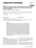
Fig. 1.1 Schematic diagram of workflow for screening for expression of integral membrane proteins
brane proteins. This approach has been greatly
facilitated by genetic fusion to a fluorescent reporter protein, typically Green fluorescent protein
(GFP). This enables rapid expression screening
and hence identification of proteins that are stably
inserted into the membrane without the need to
purify the membrane protein (Drew et al. 2005).
Once a well expressed stable protein is identified
the GFP moiety can also be used to monitor
purification and for pre-crystallization screening
(Drew et al. 2006; Kawate and Gouaux 2006).
A generic workflow for this method is shown
in Fig. 1.1. In this chapter the use of GFP as a
reporter for the expression of membrane proteins
in different heterologous hosts will be reviewed.
1.2
Bacteria
Escherichia coli is the most commonly used
prokaryotic host for overexpression of IMPs, followed by the Gram positive bacterium, Lactococ-
cus lactis (Kunji et al. 2003; Drew et al. 2006;
Gordon et al. 2008; Frelet-Barrand et al. 2010;
Chen 2012; King et al. 2015). Bacterial hosts
have obvious advantages for the over-expression
of recombinant proteins with rapid growth rates,
inexpensive growth media and the ease of genetic
manipulation. Moreover, the biology of transcription, translation and insertion into membranes are
also well characterised, allowing manipulation of
the host cell to facilitate heterologous expression
of proteins. Nevertheless, the expression of membrane proteins in bacteria can be problematical
for a number of reasons. The expressed protein
may prove to be toxic to the host cell (Kunji
et al. 2003) or saturate the membrane insertion
machinery (Loll 2003; Wagner et al. 2006). Rare
codons in the protein or insufficient amino acid
availability (Angov et al. 2008; Marreddy et al.
2010; Bill et al. 2011) or insufficient membrane
capacity (Arechaga et al. 2000) may all limit
the expression of membrane proteins in bacteria.
Therefore, screening for correctly folded protein
1 Expression Screening of Integral Membrane Proteins by Fusion to Fluorescent Reporters
3
is critical, with fusion to GFP at either the N or
C-terminus now being widely used as a reporter
of insertion into the bacterial membrane (Drew
et al. 2001; Sonoda et al. 2011; Lee et al. 2014a,
b, c). The combination of (1) high-throughput
cloning strategies to construct fusion GFP fusion
vectors with (2) screening in E. coli using in gelfluorescence of detergent lysates of whole cells,
enables the expression of large numbers of IMPs
to be evaluated at small scale (Sonoda et al.
2011; Schlegel et al. 2012; Lee et al. 2014a; Bird
et al. 2015). For example, in one study, 47 orthologues of bacterial SEDS (shape, elongation, division, and sporulation) proteins were cloned and
candidate proteins rapidly identified for further
analysis (Bird et al. 2015). Typically an affinity
purification tag, for example octa-histidine, is included with the GFP reporter so that fluorescence
can be used to monitor the mono-dispersity and
integrity of the membrane proteins during purification by size exclusion chromatography (Fluo-
rescence detected Size Exclusion Chromatography, FSEC) (Drew et al. 2006; Bird et al. 2015).
Thus, fusion to GFP has facilitated purification
to homogeneity and subsequent crystallization of
many IMPs expressed in E. coli, for example,
Pseudomonas aeruginosa lysP, E. coli sodiumproton NhaA and the Streptococcus thermophilus
peptide transporter PepTSt (Lee et al. 2014b; Nji
et al. 2014).
Fusion of IMPs to GFP is useful for comparing expression in different strains of bacteria (see Fig. 1.2 for an example). The E. coli
strain BL21(DE3) and related strains are most
commonly used for heterologous protein production. In these strains, the bacteriophage T7 RNA
polymerase is expressed from the mutant lacUV5
promoter resulting in high-level expression of
a polymerase that is more processive than the
native E. coli RNA polymerase (Iost et al. 1992).
Driving transcription generally leads to higher
levels of heterologous protein production. How-
Fig. 1.2 Screening expression in E.coli of 47 SED
(Sporulation Elongation Division) proteins from a wide
range of bacteria, by in-gel fluorescence. Strains were
grown in Powerbroth (Molecular Dimensions) and expression induced at 20 ı C overnight. (a) C41(DE3) plysS,
induced with 1 mM IPTG. (b) Lemo21 (DE3), grown in
the presence of 0.625 mM rhamnose and induced with
1 mM IPTG. (c) KRX, induced with 2.5 mM rhamnose
and 1 mM IPTG. Detergent lysates of E. coli cells were
analysed by SDS-PAGE and gels imaged using Blue Epi
illumination and a 530/28 filter. A GFP control is shown
in lane F3 and the numbers to the left refer to the sizes in
kDa of molecular weight markers run in parallel
4
ever, for membrane proteins this can result in
saturation of the Sec translocon and subsequent
misfolding of much of the expressed membrane
protein (Wagner et al. 2006, 2007; Klepsch et al.
2011). To avoid this problem, Miroux and Walker
isolated strains of BL21(DE3) that survived the
over-expression of membrane proteins by an unknown mechanism (Miroux and Walker 1996).
These strains, C41(DE3) and C43(DE3), known
as the Walker strains, are used pragmatically to
express a membrane proteins, though high levels
of expression are not seen for all membrane
proteins (Miroux and Walker 1996; Wagner et al.
2008). Analyses of the Walker strains, using the
bacterial membrane protein YidC fused to GFP
(Wagner et al. 2007), showed that mutations in
the lacUV5 promoter are responsible for the often
improved membrane protein expression (Drews
et al. 1973; Wagner et al. 2008). The mutations that were found, result in lower levels of
mRNA production and hence a slower rate of
protein synthesis. This presumably ensures that
membrane protein translocation machinery is not
saturated.
These data suggested that to optimize expression levels of folded and functional inserted
IMPs, it is important to match the rate of transcription /translation with the capacity of the Sec
translocon. The Lemo21(DE3) strain has been
specifically engineered according to this principal
and incorporates the gene for T7 lysozyme on a
plasmid under the control of the highly titratable
rhamnose promoter (Giacalone et al. 2006; Wagner et al. 2008). T7 lysozyme is an inhibitor of
T7RNA polymerase, and Schlegel et al. showed
that the expression level of a number of membrane proteins could be optimised by varying the
level of rhamnose in the cell media (Schlegel
et al. 2012). However, not all IMPs express well
in Lemo21(DE3) and screening E. coli strains
with different expression kinetics is important for
achieving expression (Schlegel et al. 2012; Bird
et al. 2015).
Fusion of IMPs with GFP at the C-terminus
of the protein in tandem with the erythromycin
resistance protein (23S ribosomal RNA adenine
N-6 methyl transferase, ErmC) has been used
to evolve both E coli and L. lactis strains for
L.E. Bird et al.
improved production of membrane proteins
(Linares et al. 2010; Gul et al. 2014). In both
cases the protein is under the regulation of
a titratable promoter, the arabinose inducible
pBAD promoter in E. coli and the NICE (nisininducible controlled gene expression) promoter
in L. lactis. In this approach, the optimum inducer
concentration, induction time and temperature of
induction are established using readout from
the GFP reporter. The cells are then exposed,
under these conditions, to increasing levels of
erythromycin, since the GFP and ErmC are
at the C-terminus, cells that have evolved to
express higher levels of the functional protein
will be resistant to a higher concentration of
erythromycin. The strains are then plated on
erythromycin at the highest concentration used
and the most fluorescent colonies are analysed.
The strains can be cured of the selection plasmid
and it was shown that expression is increased
for proteins other than the test plasmid (Linares
et al. 2010; Gul et al. 2014). The evolved E. coli
when compared with the parental strain showed
up to a tenfold increase in fluorescence levels
and when compared to the Walker strains had
increased levels of expression per unit of biomass
(Gul et al. 2014). Interestingly, deep sequencing
of four evolved E. coli strains revealed that all
had mutations were in the gene encoding DNAbinding protein, H-NS, which is involved in
chromosome organization and transcriptional
silencing, although the exact mechanism causing
the elevated expression is unclear (Gul et al.
2014). In L. lactis the strain selection led to a two
to eightfold increases in the expression levels of
a variety of proteins. In contrast to E. coli, deep
sequencing of the genome of the evolved strains
identified point mutations in a single gene, nisK,
which is the histidine kinase sensor protein of
the two component regulatory system that directs
nisin-A mediated expression. It seems likely that
the mutations enhance phosphoryl transfer to
NisR and increase transcription from the nisin-A
promoter (Linares et al. 2010).
Most IMPs have been produced in E. coli,
which reflects its popularity as a host for heterologous expression of soluble proteins. However
other bacterial species may be more suitable
1 Expression Screening of Integral Membrane Proteins by Fusion to Fluorescent Reporters
for IMP production. For example, Gram positive
bacteria, such as L. lactis, express two copies
of the IMP chaperone YiDC and thus may be
better than E. coli at translocating heterologous
proteins and hence may be less susceptible to
saturation of the integration machinery (Zweers
et al. 2008; Funes et al. 2009; Funes et al. 2011;
Schlegel et al. 2014) A number of other features
of L. lactis, like the slower growth rate and
reduced proteolytic activity when compared to E.
coli, may also facilitate IMP production in this
bacterium (Schlegel et al. 2014).
1.3
Yeast
Like E. coli, yeast require relatively low cost
of media, have fast growth rates and can
be easily genetically modified, making them
attractive expression host for IMP production.
Moreover, the post translational modifications
and lipid environment of yeast cells may
be more appropriate for the expression of
eukaryotic IMPs. The two yeast strains that
have been widely used for IMP production are
Saccharomyces cerevisiae and Pichia pastoris
and less commonly, Schizosaccharomyces pombe
(Yang and Murphy 2009; Yang et al. 2009; He
et al. 2014). It is important to note that protein
5
glycosylation in yeast is not typical of higher
eukaryotic cells with N-linked glycosylation sites
in S. cerevisiae hyper-glycosylated with high
mannose glycoforms. In P. pastoris, the N-linked
glycans are shorter than in S. cerevisiae and
strains have been engineered that add glycoforms
more typical of human glycoproteins (Hamilton
et al. 2006; Darby et al. 2012).
The GFP screening pipeline used with E. coli
has been adapted to both S. cerevisiae and P.
pastoris (Drew et al. 2008; Drew and Kim 2012b;
Brooks et al. 2013; Scharff-Poulsen and Pedersen 2013). There are, however, some differences,
for example, as part of the screening process it
can be useful to include a confocal microscope
image to confirm the localization of the IMPGFP fusion protein (Newstead et al. 2007; Drew
et al. 2008) (Fig. 1.3). Additionally, S. cerevisiae
cloning can be carried out by in vivo homologous
recombination of PCR products into 2 based
episomal vectors (Drew and Kim 2012a; ScharffPoulsen and Pedersen 2013). The inducible GAL1
promoter is often used to drive expression as the
yields are generally higher compared to constitutive promoters (Newstead et al. 2007). The induction of the IMP-GFP fusion can be optimized
by varying parameters, such as, the timing of
induction, using non-selective media, the addition
of chemical chaperones such as DMSO, glycerol
Fig. 1.3 S. cerevisiae expressing a recombinant Candida albicans TOK1 GFP fusion protein observed under (a) white
light (b) fluorescence optics (Image courtesy of Prof. Per Pedersen, University of Copenhagen)
6
and histidine and also by lowering the temperature (Drew and Kim 2012c). Furthermore, the
levels of expression of IMP-GFP fusions can be
improved by the choice of strain and by plasmid
engineering (Pedersen et al. 1996; Drew and
Kim 2012a; Scharff-Poulsen and Pedersen 2013;
Molbaek et al. 2015). For example, Molbaek et
al. produced functional full-length human ERG
KC -GFP fusions by utilizing the strain PAP1500,
which overexpresses the GAL4 transcriptional
activator. This was combined with a vector that
has a strong hybrid CYC-GAL promoter and the
compromised leu2-d gene, which elevates the
episomal copy number to between 200 and 400
plasmids per cell in response to leucine starvation
(Romanos et al. 1992; Molbaek et al. 2015).
For P. pastoris, strain development is more
complicated. Since genes to be expressed have to
be integrated into the yeast genome using a resistance marker such as zeocin and typically use the
methanol inducible AOX1 promoter (Logez et al.
2012). This means that a shuttle vector has to
be constructed and different P. pastoris transformants have to be characterised to identify the best
recombinant strain for IMP expression. Again,
fusion to GFP enables the expression screening
of integrated clones using a plate based assay. For
example, using this methodology Brooks et al.
isolated a clone of mouse PEMT (ER associated
phosphatidyl ethanolamine N-methyl transferase)
that gave a final yield of 5 mg/L of purified
protein (Brooks et al. 2013). In an interesting
development, Parcej et al. reported the use of
fusions to different fluorophores to monitor the
expression of the human heterodimeric ATP binding cassette (ABC) transporter associated with
antigen processing (TAP) in P. pastoris. The subunits were tagged with either monomeric venus
and a HIS10 tag or monomeric cerulean with a
strepII tag, dual wavelength monitoring was then
used to monitor expression of individual subunits
and purification of the complex (Parcej et al.
2013). This approach could clearly be applied to
the expression of multi-subunit IMPs in other cell
hosts.
Yeast is clearly a very useful host for expression of IMPs, however in a study of 43
eukaryotic membrane proteins Newstead et al.
L.E. Bird et al.
showed that while 25 out of 29 yeast membrane
proteins were produced to greater than 1 mg/L in
S. cerevisiae, only 4 of the 14 membrane proteins
from higher eukaryotic organisms were produced
at this level, suggesting that a higher eukaryotic
heterologous expression systems is often necessary for higher eukaryotic proteins (Newstead
et al. 2007).
1.4
Insect and Mammalian Cells
Insect cells are widely used for the production
of eukaryotic recombinant proteins, including
IMPs. The cells are easy to handle and in general
give higher yields of recombinant proteins than
transfected mammalian cells. The main cell lines
in use are from Spodoptera frugiperda (Sf9 and
Sf21) and Trichoplusia ni (High Five) with the
gene of interest typically introduced using the
baculovirus expression vector system (BEVS)
(Zhang et al. 2008; Mus-Veteau 2010; Milic
and Veprintsev 2015). Transient transfection
with plasmid vectors has also been reported
for rapid screening of IMP expression using
GFP fusion proteins (Chen et al. 2013). In
addition, Drosophilia melanogaster S2 cells in
combination with inducible plasmid vectors have
been used for the expression of recombinant
IMPs (Brillet et al. 2010). However, it is
important to note that the lipid composition
of insect cell membranes differs from those of
mammalian and bacterial cells. For example, the
main sterol in mammalian cells is cholesterol,
whereas it is ergosterol in insect cells (and yeast):
there are no sterols in bacterial cell membranes
(Lagane et al. 2000; Eifler et al. 2007). In
addition, N-glycosylation in insect cells consists
of short so-called pauci-mannose glycoforms,
which are not found on mammalian IMPs.
GFP-tagging can be used for expression
screening in insect cells in the same way as for
E. coli and yeast. However in contrast to E. coli
cells, there is evidence of GFP-tagged proteins
produced in insect cells that are misfolded but
still show GFP fluorescence (Thomas and Tate
2014). Fusion to GFP remains a convenient
way for screening many constructs in parallel at
1 Expression Screening of Integral Membrane Proteins by Fusion to Fluorescent Reporters
7
Fig. 1.4 Fluorescence detected size exclusion profiles
(FSEC) and in-gel fluorescence (inset) of detergent extracts of the total membrane fraction from SF9 insect
cells expressing Caenorhabditis elegans GTG1 fused to
GFP. Membranes were extracted in the following detergents (1 % final concentration plus 0.2 % cholesterol): n-
Decyl-“-D-Maltoside (DM: lane 1, dark blue trace); nDodecyl-“-D-Maltoside (DDM: lane 2, dark green trace);
Lauryldimethylamine-N-Oxide (LDAO: lane 3, yellow
trace); 6-Cyclohexyl-1-Hexyl-“-D-Maltoside (cymal-6:
lane 4, blue trace); n-Dodecylphosphocholine (FC12; lane
5, green trace)
small scale, particularly different orthologues, in
order to identify the best expressed candidate for
purification and crystallization (Lee and Stroud
2010; He et al. 2014; Hu et al. 2015). Analysis
of the subsequent products by FSEC (see Fig. 1.4
for an example) enables the optimal detergent for
solubilisation to be identified and any misfolded
fusion proteins to be detected.
Transient expression in Human Embryonic
Kidney cells (HEK293) provides a rapid way
of screening protein expression, including IMPs
and has become the system of choice for the
production of secreted/cell surface glycoproteins
for structural biology (Aricescu and Owens
2013). In particular HEK-293 cells deficient in
N-acetylglucosamine tranferase I (HEK Gnt1
/ ) are used to produce proteins containing
only a high mannose glycoform, which can be
removed by endoglycosidase treatment following
purification. Simplifying the N-glycosylation of
proteins appears to favour crystallization since
sample heterogeneity is reduced (Chang et al.
2007). This approach is equally relevant for
modifying the N-glycans of IMPs which may
in turn aid crystallization.
The use of GFP fusions in combination with
transient expression in HEK cells was introduced
by Gouaux and co-workers (Kawate and Gouaux
2006) for optimizing the expression of the ATPgated ion channel P2X4. Protein production for
crystallization was subsequently transferred to
insect cells (Kawate et al. 2009). For IMP production in mammalian cells, inducible stable cell
lines are usually required to generate sufficient
biomass without the problem of toxicity from
constitutive expression (Chaudhary et al. 2011,
2012). Although this requires more time and effort than using insect cells, there are now a number of structures of membrane proteins produced
in this way. In all cases, multiple constructs were
initially screened by transient expression using
fusion to GFP as a reporter of protein expression
and stability by FSEC analysis. Although recombinant protein yields from mammalian cells are
generally lower than either microbial or insect
cell over-expression systems, there may be a sig-
8
L.E. Bird et al.
nificant advantage in using mammalian cells for
the production of human/mammalian IMPs. The
proteins will be produced in a cellular context
with native post-translational modifications and
lipid environment, it is becoming increasingly
apparent that this leads to improved protein quality due to lower levels of misfolded aggregates
(Yamashita et al. 2005; Chaudhary et al. 2011).
An alternative to the production of stable
cell lines for IMP production is the use of
baculovirus mediated gene transduction for
large-scale production of IMPs in mammalian
cells, typically HEK Gnt1
/
(Goehring
et al. 2014). The so-called BacMam system
(Dukkipati et al. 2008) involves the inclusion of
a mammalian cell transcription unit(s) within a
baculovirus transfer vector so that on generation
of a recombinant virus, the inserted gene can
be expressed in mammalian cells. The same
plasmid vector can be used for small-scale
transient transfection of HEK cells to identify
the optimal construct and then to generate a
BacMam baculovirus for scaling up of protein
production by bulk transduction of HEK cells for
further characterization (Goehring et al. 2014).
Using this protocol, sample preparation can be
accomplished in 4–6 weeks, which is at least
half the time required to generate and scale-up
stable cell lines. The approach has been used by
the Gouaux group to produce a number of IMPs
for structural determination (Althoff et al. 2014;
Baconguis et al. 2014; Dürr et al. 2014; Lee et al.
2014c; Wang et al. 2015).
1.5
Summary and Conclusions
Initially developed for screening the expression
of bacterial membrane proteins in Escherichia
coli, the use of GFP fusions has been successfully
extended to eukaryotic hosts, including insect and
mammalian cells. Although E. coli and yeast are
useful tools for the over-expression of recombinant membrane proteins, there is a marked difference in the lipid compositions of membranes
from prokaryotes and eukaryotes. This in turn
may affect the quality and quantity of heterologous proteins inserted into the host membrane.
Given that the host cell determines the nature of
post-translational modifications, such as glycosylation and phosphorylation, in choosing an expression host for screening, it may the appropriate
to match the host cell to the recombinant product
for example, human IMPs in mammalian cells.
Acknowledgments The OPPF-UK is funded by the
Medical Research Council, UK (grant MR/K018779/1).
References
Althoff T, Hibbs RE, Banerjee S, Gouaux E (2014) X-ray
structures of GluCl in apo states reveal a gating mechanism of Cys-loop receptors. Nature 512(7514):333–
337
Angov E, Hillier CJ, Kincaid RL, Lyon JA (2008) Heterologous protein expression is enhanced by harmonizing
the codon usage frequencies of the target gene with
those of the expression host. PLoS ONE 3(5):e2189
Arechaga I, Miroux B, Karrasch S, Huijbregts R, de
Kruijff B, Runswick MJ, Walker JE (2000) Characterisation of new intracellular membranes in Escherichia
coli accompanying large scale over-production of
the b subunit of F(1)F(o) ATP synthase. FEBS Lett
482(3):215–219
Aricescu AR, Owens RJ (2013) Expression of recombinant glycoproteins in mammalian cells: towards an
integrative approach to structural biology. Curr Opin
Struct Biol 23(3):345–356
Baconguis I, Bohlen CJ, Goehring A, Julius D, Gouaux
E (2014) X-ray structure of acid-sensing ion channel
1-snake toxin complex reveals open state of a Na(C)selective channel. Cell 156(4):717–729
Bill RM, Henderson PJF, Iwata S, Kunji ERS, Michel H
et al (2011) Overcoming barriers to membrane protein
structure determination. Nat Biotechnol 29(4):335–
340
Bird LE, Rada H, Verma A, Gasper R, Birch J, Jennions
M et al (2015) Green fluorescent protein-based expression screening of membrane proteins in Escherichia
coli. J Vis Exp 95:e52357
Brillet K, Pereira CA, Wagner R (2010) Expression of
membrane proteins in Drosophila Melanogaster S2
cells: production and analysis of a EGFP-fused G
protein-coupled receptor as a model. Methods Mol
Biol 601:119–133
Brooks CL, Morrison M, Joanne Lemieux M (2013) Rapid
expression screening of eukaryotic membrane proteins
in Pichia pastoris. Protein Sci 22(4):425–433
Chang VT, Crispin M, Aricescu AR, Harvey DJ, Nettleship JE et al (2007) Glycoprotein structural genomics: solving the glycosylation problem. Structure
15(3):267–273
Chaudhary S, Pak JE, Pedersen BP, Bang LJ, Zhang
LB et al (2011) Efficient expression screening of
1 Expression Screening of Integral Membrane Proteins by Fusion to Fluorescent Reporters
human membrane proteins in transiently transfected
human embryonic kidney 293S cells. Methods 55(4):
273–280
Chaudhary S, Pak JE, Gruswitz F, Sharma V, Stroud RM
(2012) Overexpressing human membrane proteins in
stably transfected and clonal human embryonic kidney
293S cells. Nat Protoc 7(3):453–466
Chen R (2012) Bacterial expression systems for recombinant protein production: E. coli and beyond. Biotechnol Adv 30(5):1102–1107
Chen H, Shaffer PL, Huang X, Rose PE (2013) Rapid
screening of membrane protein expression in transiently transfected insect cells. Protein Expr Purif
88(1):134–142
Darby RA, Cartwright SP, Dilworth MV, Bill RM (2012)
Which yeast species shall I choose? Saccharomyces
cerevisiae versus Pichia pastoris (review). Methods
Mol Biol 866:11–23
Drew D, Kim H (2012a) Preparation of Saccharomyces
cerevisiae expression plasmids. Methods Mol Biol
866:41–46
Drew D, Kim H (2012b) Screening for high-yielding
Saccharomyces cerevisiae clones: using a green fluorescent protein fusion strategy in the production of
membrane proteins. Methods Mol Biol 866:75–86
Drew D, Kim H (2012c) Optimizing Saccharomyces cerevisiae induction regimes. Methods Mol Biol 866:191–
195
Drew D, von Heijne G, Nordlund P, de Gier JWL (2001)
Green fluorescent protein as an indicator to monitor
membrane protein overexpression in Escherichia coli.
FEBS Lett 507(2):220–224
Drew D, Slotboom DJ, Friso G, Reda T, Genevaux P et al
(2005) A scalable, GFP-based pipeline for membrane
protein overexpression screening and purification. Protein Sci 14(8):2011–2017
Drew D, Lerch M, Kunji E, Slotboom DJ, de Gier JW
(2006) Optimization of membrane protein overexpression and purification using GFP fusions. Nat Methods
3(4):303–313
Drew D, Newstead S, Sonoda Y, Kim H, von Heijne
G, Iwata S (2008) GFP-based optimization scheme
for the overexpression and purification of eukaryotic
membrane proteins in Saccharomyces cerevisiae. Nat
Protoc 3(5):784–798
Drews J, Grasmuk H, Unger FM (1973) Peptide chain initiation with chemically formylated Met-tRNAs from
E. coli and yeast. Biochem Biophys Res Commun
51(3):804–812
Dukkipati A, Park HH, Waghray D, Fischer S, Garcia
KC (2008) BacMam system for high-level expression of recombinant soluble and membrane glycoproteins for structural studies. Protein Expr Purif 62(2):
160–170
Dürr KL, Chen L, Stein RA, De Zorzi R, Folea IM, Walz
T, Gouaux E (2014) Structure and dynamics of AMPA
receptor GluA2 in resting, pre-open, and desensitized
states. Cell 158(4):778–792
Eifler N, Duckely M, Sumanovski LT, Egan TM, Oksche
A et al (2007) Functional expression of mammalian
9
receptors and membrane channels in different cells. J
Struct Biol 159(2):179–193
Frelet-Barrand A, Boutigny S, Kunji ER, Rolland N
(2010) Membrane protein expression in Lactococcus
lactis. Methods Mol Biol 601:67–85
Funes S, Hasona A, Bauerschmitt H, Grubbauer C,
Kauff F et al (2009) Independent gene duplications
of the YidC/Oxa/Alb3 family enabled a specialized
cotranslational function. Proc Natl Acad Sci U S A
106(16):6656–6661
Funes S, Kauff F, van der Sluis EO, Ott M, Herrmann JM
(2011) Evolution of YidC/Oxa1/Alb3 insertases: three
independent gene duplications followed by functional
specialization in bacteria, mitochondria and chloroplasts. Biol Chem 392(1–2):13–19
Giacalone MJ, Gentile AM, Lovitt BT, Berkley NL, Gunderson CW, Surber MW (2006) Toxic protein expression in Escherichia coli using a rhamnose-based tightly
regulated and tunable promoter system. Biotechniques
40(3):355–364
Goehring A, Lee CH, Wang KH, Michel JC, Claxton DP
et al (2014) Screening and large-scale expression of
membrane proteins in mammalian cells for structural
studies. Nat Protoc 9(11):2574–2585
Gordon E, Horsefield R, Swarts HG, de Pont JJH, Neutze
R, Snijder A (2008) Effective high-throughput overproduction of membrane proteins in Escherichia coli.
Protein Expr Purif 62(1):1–8
Gul N, Linares DM, Ho FY, Poolman B (2014)
Evolved Escherichia coli strains for amplified, functional expression of membrane proteins. J Mol Biol
426(1):136–149
Hamilton SR, Davidson RC, Sethuraman N, Nett JH,
Jiang Y et al (2006) Humanization of yeast to produce
complex terminally sialylated glycoproteins. Science
313(5792):1441–1443
He Y, Wang K, Yan N (2014) The recombinant expression systems for structure determination of eukaryotic
membrane proteins. Protein Cell 5(9):658–672
Hu NJ, Rada H, Rahman N, Nettleship JE, Bird L et al
(2015) GFP-based expression screening of membrane
proteins in insect cells using the baculovirus system.
Methods Mol Biol 1261:197–209
Iost I, Guillerez J, Dreyfus M (1992) Bacteriophage T7
RNA polymerase travels far ahead of ribosomes in
vivo. J Bacteriol 174(2):619–622
Kawate T, Gouaux E (2006) Fluorescence-detection
size-exclusion chromatography for precrystallization
screening of integral membrane proteins. Structure
14(4):673–681
Kawate T, Michel JC, Birdsong WT, Gouaux E (2009)
Crystal structure of the ATP-gated P2X(4) ion channel
in the closed state. Nature 460(7255):592–598
King MS, Boes C, Kunji ER (2015) Membrane protein
expression in Lactococcus lactis. Methods Enzymol
556:77–97
Klepsch MM, Persson JO, De Gier JWL (2011) Consequences of the overexpression of a eukaryotic membrane protein, the human KDEL receptor, in Escherichia coli. J Mol Biol 407(4):532–542
10
Kunji ER, Slotboom DJ, Poolman B (2003) Lactococcus
lactis as host for overproduction of functional membrane proteins. Biochim Biophys Acta 1610(1):97–
108
Lagane B, Gaibelet G, Meilhoc E, Masson JM, Cézanne
L, Lopez A (2000) Role of sterols in modulating the
human mu-opioid receptor function in Saccharomyces
cerevisiae. J Biol Chem 275(43):33197–33200
Lee JK, Stroud RM (2010) Unlocking the eukaryotic
membrane protein structural proteome. Curr Opin
Struct Biol 20(4):464–470
Lee C, Kang HJ, Hjelm A, Qureshi AA, Nji E, Choudhury H et al (2014a) MemStar: a one-shot Escherichia coli-based approach for high-level bacterial
membrane protein production. FEBS Lett 588(20):
3761–3769
Lee C, Yashiro S, Dotson DL, Uzdavinys P, Iwata S
et al (2014b) Crystal structure of the sodium-proton
antiporter NhaA dimer and new mechanistic insights.
J Gen Physiol 144(6):529–544
Lee CH, Lü W, Michel JC, Goehring A, Du J, Song X,
Gouaux E (2014c) NMDA receptor structures reveal
subunit arrangement and pore architecture. Nature
511(7508):191–197
Linares DM, Geertsma ER, Poolman B (2010) Evolved
Lactococcus lactis strains for enhanced expression
of recombinant membrane proteins. J Mol Biol
401(1):45–55
Logez C, Alkhalfioui F, Byrne B, Wagner R (2012) Preparation of Pichia pastoris expression plasmids. Methods
Mol Biol 866:25–40
Loll PJ (2003) Membrane protein structural biology: the
high throughput challenge. J Struct Biol 142(1):144–
153
Marreddy RK, Geertsma ER, Permentier HP, Pinto JP,
Kok J, Poolman B (2010) Amino acid accumulation
limits the overexpression of proteins in Lactococcus
lactis. PLoS ONE 5(4):e10317
Milic D, Veprintsev DB (2015) Large-scale production and protein engineering of G protein-coupled
receptors for structural studies. Front Pharmacol
6:66
Miroux B, Walker JE (1996) Over-production of proteins
in Escherichia coli: mutant hosts that allow synthesis
of some membrane proteins and globular proteins at
high levels. J Mol Biol 260(3):289–298
Molbaek K, Scharff-Poulsen P, Helix-Nielsen C, Klaerke
DA, Pedersen PA (2015) High yield purification of
full-length functional hERG KC channels produced in
Saccharomyces cerevisiae. Microb Cell Fact 14:15
Mus-Veteau I (2010) Heterologous expression of membrane proteins for structural analysis. Methods Mol
Biol 601:1–16
Newstead S, Kim H, von Heijne G, Iwata S, Drew D
(2007) High-throughput fluorescent-based optimization of eukaryotic membrane protein overexpression
and purification in Saccharomyces cerevisiae. Proc
Natl Acad Sci U S A 104(35):13936–13941
L.E. Bird et al.
Nji E, Li D, Doyle DA, Caffrey M (2014) Cloning, expression, purification, crystallization and preliminary
X-ray diffraction of a lysine-specific permease from
Pseudomonas aeruginosa. Acta Crystallogr F Struct
Biol Commun 70(10):1362–1367
Parcej D, Guntrum R, Schmidt S, Hinz A, Tampé
R (2013) Multicolour fluorescence-detection sizeexclusion chromatography for structural genomics
of membrane multiprotein complexes. PLoS ONE
8(6):e67112
Pedersen PA, Rasmussen JH, Jørgensen PL (1996) Expression in high yield of pig alpha 1 beta 1 Na, KATPase and inactive mutants D369N and D807N in
Saccharomyces cerevisiae. J Biol Chem 271(5):2514–
2522
Romanos MA, Scorer CA, Clare JJ (1992) Foreign
gene expression in yeast: a review. Yeast 8(6):
423–488
Scharff-Poulsen P, Pedersen PA (2013) Saccharomyces
cerevisiae-based platform for rapid production and
evaluation of eukaryotic nutrient transporters and transceptors for biochemical studies and crystallography.
PLoS ONE 8(10):e76851
Schlegel S, Löfblom J, Lee C, Hjelm A, Klepsch M et al
(2012) Optimizing membrane protein overexpression
in the Escherichia coli strain Lemo21(DE3). J Mol
Biol 423(4):648–659
Schlegel S, Hjelm A, Baumgarten T, Vikström D, de Gier
JW (2014) Bacterial-based membrane protein production. Biochim Biophys Acta 1843(8):1739–1749
Sonoda Y, Newstead S, Hu NJ, Alguel Y, Nji E et al (2011)
Benchmarking membrane protein detergent stability
for improving throughput of high-resolution X-ray
structures. Structure 19(1):17–25
Thomas J, Tate CG (2014) Quality control in eukaryotic membrane protein overproduction. J Mol Biol
426(24):4139–4154
Wagner S, Bader ML, Drew D, de Gier JW (2006) Rationalizing membrane protein overexpression. Trends
Biotechnol 24(8):364–371
Wagner S, Baars L, Ytterberg AJ, Klussmeier A, Wagner
CS et al (2007) Consequences of membrane protein
overexpression in Escherichia coli. Mol Cell Proteomics 6(9):1527–1550
Wagner S, Bader ML, Drew D, de Gier JW (2008) Tuning
Escherichia coli for membrane protein overexpression.
Proc Natl Acad Sci U S A 105(38):14371–14376
Wang KH, Penmatsa A, Gouaux E (2015) Neurotransmitter and psychostimulant recognition by the dopamine
transporter. Nature 521(7552):322–327
Yamashita A, Singh SK, Kawate T, Jin Y, Gouaux E
(2005) Crystal structure of a bacterial homologue
of NaC/Cl–dependent neurotransmitter transporters.
Nature 437(7056):215–223
Yang H, Murphy AS (2009) Functional expression
and characterization of Arabidopsis ABCB, AUX 1
and PIN auxin transporters in Schizosaccharomyces
pombe. Plant J 59(1):179–191
1 Expression Screening of Integral Membrane Proteins by Fusion to Fluorescent Reporters
Yang Y, Hu Z, Liu Z, Wang Y, Chen X, Chen G (2009)
High human GLUT1, GLUT2, and GLUT3 expression
in Schizosaccharomyces pombe. Biochemistry (Mosc)
74(1):75–80
Zhang F, Manzan MA, Peplinski HM, Thiem SM et
al (2008) A new Trichoplusia ni cell line for membrane protein expression using a baculovirus expres-
11
sion vector system. Vitro Cell Dev Biol Anim 44(7):
214–223
Zweers JC, Barák I, Becher D, Driessen AJ, Hecker
M et al (2008) Towards the development of Bacillus subtilis as a cell factory for membrane proteins and protein complexes. Microb Cell Fact
7:10
2
Detergents in Membrane Protein
Purification and Crystallisation
Anandhi Anandan and Alice Vrielink
Abstract
Detergents play a significant role in structural and functional
characterisation of integral membrane proteins (IMPs). IMPs reside in
the biological membranes and exhibit a great variation in their structural
and physical properties. For in vitro biophysical studies, structural and
functional analyses, IMPs need to be extracted from the membrane lipid
bilayer environment in which they are found and purified to homogeneity
while maintaining a folded and functionally active state. Detergents are
capable of successfully solubilising and extracting the IMPs from the
membrane bilayers. A number of detergents with varying structure and
physicochemical properties are commercially available and can be applied
for this purpose. Nevertheless, it is important to choose a detergent that is
not only able to extract the membrane protein but also provide an optimal
environment while retaining the correct structural and physical properties
of the protein molecule. Choosing the best detergent for this task can be
made possible by understanding the physical and chemical properties of
the different detergents and their interaction with the IMPs. In addition,
understanding the mechanism of membrane solubilisation and protein
extraction along with crystallisation requirements, if crystallographic
studies are going to be undertaken, can help in choosing the best detergent
for the purpose. This chapter aims to present the fundamental properties
of detergents and highlight information relevant to IMP crystallisation.
The first section of the chapter reviews the physicochemical properties
of detergents and parameters essential for predicting their behaviour in
solution. The second section covers the interaction of detergents with the
biologic membranes and proteins followed by their role in membrane
A. Anandan • A. Vrielink ( )
School of Chemistry and Biochemistry, University of
Western Australia, 35 Stirling Highway, Crawley, WA
6009, Australia
e-mail: ;
© Springer International Publishing Switzerland 2016
I. Moraes (ed.), The Next Generation in Membrane Protein Structure Determination,
Advances in Experimental Medicine and Biology 922, DOI 10.1007/978-3-319-35072-1_2
13
14
A. Anandan and A. Vrielink
protein crystallisation. The last section will briefly cover the types of
detergent and their properties focusing on custom designed detergents for
membrane protein studies.
Keywords
Detergents • Lipids • Micelles • Membrane proteins • Protein purification • Crystallisation
2.1
Physicochemical Properties
of Detergents
Detergents are surfactants (surface acting
reagents) that decrease the interfacial tension
between two immiscible liquids. The overall
molecular structure of detergents consists of a
hydrophilic polar head group and a hydrophobic
non-polar tail group (Fig. 2.1a) that renders
them amphiphilic. The polar head group of a
detergent can be ionic, non-ionic or zwitterionic
and usually has a strong attraction for aqueous
solvent molecules whereas the detergent nonpolar tail is generally repelled from the aqueous
solvent. Consequently, in an aqueous medium,
the hydrophobic tail of detergent molecules
usually orients itself to minimize contact with
water while the hydrophilic head interacts with
the water molecules. As a result, the detergent
monomers align themselves as a single layer at
the hydrophilic-hydrophobic interface, reducing
the surface tension of the solvent (Fig. 2.1b). This
alignment not only reduces the interaction of the
hydrophobic tail with water molecules, it also
allows the interaction between the detergent head
group and the solvent, facilitating the detergent
molecules to stay soluble in aqueous media
(Rosen and Kunjappu 2012).
Detergent molecules persist as monomers in
solution up to a particular concentration. As
the detergent concentration increases, detergent
molecules assemble into complex structures
called micelles. The hydrophobic tails of the
detergent molecules pack together, forming the
core of the micelle and reducing their interaction
with the water molecules. In contrast, the
polar head groups orient themselves outwards
from the micelle core, enabling interaction
with the aqueous solvent (Fig. 2.1c). The
minimal detergent concentration required for
the formation of micelles is called the ‘critical
micelle concentration ’ (CMC) and the number of
detergent monomers required to form a micelle
is called the ‘aggregation number’ (Helenius
Fig. 2.1 (a) Schematic representation of the overall
molecular structure of detergents. (b) Alignment of detergent molecules at hydrophobic and hydrophilic interface
and (c) detergent micelles at CMC
2 Detergents in Membrane Protein Purification and Crystallisation
15
Fig. 2.2 A general phase diagram showing the various phases and their boundaries at varying detergent concentration
and temperature. KP represents the Krafft point and CP represents the cloud point
et al. 1979; Neugebauer 1990). The CMC
is of great importance when extracting and
solubilising membrane proteins for structural
and functional studies. The detergent CMC
is dependent on the detergent alkyl chain
size and its saturation. For example, the
CMC value decreases with the length of the
alkyl chain and increases with the addition
of double bonds. It is thus understandable
that it is the CMC value that determines the
micelle size of a detergent. While detergents
with lower CMC values form large micelles
and exchanging them with other detergents
is difficult, detergents with high CMC values
require a higher concentrations for extraction
and purification (Keyes et al. 2003). Detergents
with a CMC between 0.5 and 50 mM have been
reported to be suitable for IMP solubilisation
and purification. Finally, experimental conditions
such as buffer composition and temperature also
have a profound influence on the CMC and
aggregation number of the detergent.
Above the CMC the detergent molecules coexist as both monomers and micelles in solution. Detergent solutions are also dynamic sys-
tems undergoing a constant exchange of detergent molecules between monomeric and micellar
state (le Maire et al. 2000). A further increase
in the detergent concentration might result in
aggregation of the detergent micelles leading to
phase separation. The two phase-system comprises a detergent rich phase and detergent poor
phase (Arnold and Linke 2007). In addition to
the influence of the detergent concentration on
micelle formation and phase separation, the temperature, pH, ionic strength and type of detergent
also play an important role. The temperature
at which detergent monomers form micelles is
called the Krafft point or upper consolute temperature (Gu and Sjöblom 1992). The temperature at
which phase separation occurs is called the cloud
point or lower consolute temperature (Arnold
and Linke 2007). Figure 2.2 shows a simplified
general phase diagram of a detergent displaying
the solubility of the detergent as a function of
concentration and temperature.
Detergent micelles are asymmetric in structure
with rough surfaces and disorganised clumps of
alkyl tails within the hydrophobic core region
(Garavito and Ferguson-Miller 2001). Micelle