Systems for drug delivery
Bạn đang xem bản rút gọn của tài liệu. Xem và tải ngay bản đầy đủ của tài liệu tại đây (4.62 MB, 205 trang )
Saurabh Bhatia
Systems
for Drug
Delivery
Safety, Animal, and Microbial
Polysaccharides
Systems for Drug Delivery
Saurabh Bhatia
Systems for Drug Delivery
Safety, Animal, and Microbial
Polysaccharides
Saurabh Bhatia
Assistant Professor
School of Medical and Allied Sciences
GD Goenka University
Gurgaon, India
ISBN 978-3-319-41925-1
ISBN 978-3-319-41926-8
DOI 10.1007/978-3-319-41926-8
(eBook)
Library of Congress Control Number: 2016944155
© Springer International Publishing Switzerland 2016
This work is subject to copyright. All rights are reserved by the Publisher, whether the whole or part of
the material is concerned, specifically the rights of translation, reprinting, reuse of illustrations, recitation,
broadcasting, reproduction on microfilms or in any other physical way, and transmission or information
storage and retrieval, electronic adaptation, computer software, or by similar or dissimilar methodology
now known or hereafter developed.
The use of general descriptive names, registered names, trademarks, service marks, etc. in this publication
does not imply, even in the absence of a specific statement, that such names are exempt from the relevant
protective laws and regulations and therefore free for general use.
The publisher, the authors and the editors are safe to assume that the advice and information in this book
are believed to be true and accurate at the date of publication. Neither the publisher nor the authors or the
editors give a warranty, express or implied, with respect to the material contained herein or for any errors
or omissions that may have been made.
Printed on acid-free paper
This Springer imprint is published by Springer Nature
The registered company is Springer International Publishing AG Switzerland
Author Bio
Saurabh Bhatia, is currently working as an Assistant Professor at the School of
Medical and Allied sciences, GD Goenka University, Gurgaon, Haryana, India. He
has several years of academic experience, teaching such specialized subjects as
Natural product science, nanotechnology, biotechnology, parasitology, polymeric
sciences, biomaterials. He has promoted several marine algae and their derived
polymers throughout India. He has written more than 30 international publications
in these areas and has been an active participant of more than 35 national and international conferences. So far he has successfully finished nine books in pharma and
its allied sciences. His published books include Modern Applications of Plant
Biotechnology in Pharmaceutical Sciences, Academic press, Elsevier, 2015;
Nanotechnology in Drug Delivery: Fundamentals, Design, and Applications, Apple
Academic Press 2016; Leishmaniasis: Biology, Control and New Approaches for Its
Treatment, Apple Academic Press 2016; Natural polymer drug delivery systems:
Nanoparticles, plants and algae, Springer, 2016, Natural polymer drug delivery
systems: Nanoparticles, Mammals and microbes, Springer, 2016. Dr. Bhatia has
graduated from Kurushetra University followed by M. Pharm from Bharati
Vidyapeeth University, Pune, India. He has received his Ph.D. degree from Jadavpur
University, Kolkata, India.
v
Contents
1
Mammalian Polysaccharides and Its Nanomaterials ............................
1.1 Introduction ......................................................................................
1.1.1 Polysaccharide-Based Nanoparticles .................................
1.2 Hydrophobically Modified Hyaluronic Acid ...................................
1.3 Chemically Crosslinked Hyaluronic Acid Semi-IPN ......................
1.4 Photopolymerized Hyaluronic Acid IPNS .......................................
1.5 Hydrophobically Modified Hyaluronic Acid ...................................
1.6 Hydrophobically Modified Heparin .................................................
1.7 Chondroitin Sulfate, Heparin and Hyaluronic Acid:
pH/Ion-Responsive Networks ..........................................................
1.8 Chondroitin Sulfate and Hyaluronic Acid:
Electrical Field-Responsive Network ..............................................
1.8.1 Chondroitin Sulfate and Hyaluronic Acid..........................
1.9 Heparin & Hyaluronic Acid: Anti-Adhesivesurfaces ......................
1.9.1 Hyaluronic Acid .................................................................
1.9.2 Heparin ...............................................................................
1.10 Hyaluronic Acid and Chondroitin Sulfate
(Polysaccharides of Human Origin): Biodegradable Polymers
as Biomaterials .................................................................................
1.10.1 Hyaluronic Acid .................................................................
1.10.2 Chondroitin Sulfate ............................................................
1.11 Natural–Origin Polymers as Carriers and Scaffolds
for Biomolecules and Cell Delivery in Tissue Engineering
Applications .....................................................................................
1.11.1 Hyaluronan .........................................................................
1.11.2 Chondroitin Sulphate .........................................................
1.12 Rationale for the Use of HA in Drug Delivery ................................
1.13 Chondroitin Sulfate-Based Nanocarriers
for Drug/Gene Delivery ...................................................................
1.14 Chondroitin Sulphate: Colon-Specific Drug Delivery .....................
1
1
2
2
4
6
6
7
7
8
8
8
9
9
10
10
12
13
13
14
15
17
19
vii
viii
Contents
1.15 Hyaluronan and Its Medical and Esthetic Applications ...................
1.15.1 Aging and Hyaluronan .......................................................
1.16 Polysaccharides Based Composites .................................................
1.16.1 Heparin-Based Composites ................................................
1.16.2 Hyaluronan-Based Composites ..........................................
20
21
21
21
22
2
Microbial Polysaccharides as Advance Nanomaterials .........................
2.1 Introduction ......................................................................................
2.2 Microbial Polysaccharides: General Applications ...........................
2.3 Microbial Polysaccharides Production ............................................
2.4 Biosynthesis of Polysaccharides ......................................................
2.5 Polysaccharides Recovery................................................................
2.6 Microbial Polysaccharides vs Plant Polysaccharides ......................
2.7 Microbial Polysaccharides: General Features ..................................
2.7.1 Xanthan ..............................................................................
2.7.2 Dextrans .............................................................................
2.7.3 Bacterial Alginate...............................................................
2.7.4 Scleroglucan .......................................................................
2.7.5 Gellan .................................................................................
2.7.6 Pullulan ..............................................................................
2.7.7 Curdlan ...............................................................................
2.7.8 Levan Polysaccharides .......................................................
2.7.9 Bacterial Polysaccharides...................................................
2.7.10 Gellam, Guar and Xanthan Gums ......................................
29
29
33
34
34
34
34
35
35
36
41
42
43
43
47
48
48
49
3
Chitosan Based Nanomaterials and Its Applications.............................
3.1 Introduction ......................................................................................
3.2 Chitin................................................................................................
3.3 Chitosan and Chitooligosaccharides ................................................
3.4 Chitin Nanoparticles ........................................................................
3.5 Chitosan Nanoparticles ....................................................................
3.6 Chitooligosaccharide Nanoparticles ................................................
3.7 Chitosan Applications ......................................................................
3.7.1 Thermosensitive Gels .........................................................
3.7.2 Chitosan Nanoparticles and Gene Therapy:
Chitosan-DNA Conjugated ................................................
3.7.3 Chitosan in Gene Therapy: Bio-Conjugated
Nano Applications..............................................................
3.7.4 Chitosan Based Amnioacid Polymer Conjugate ................
3.7.5 Chitosan Based Quantum Dots ..........................................
3.7.6 Chitosan Based Ceramic Glass Nanopaticles ....................
3.7.7 Chitosan Based Metallic Nanoparticles .............................
3.7.8 Chitosan Based Cationic-Cationic Polymer:
Macromolecule Grafted NPs ..............................................
3.7.9 Chitosan Based Functionalized Nanoparticles ...................
3.7.10 Chitosan Based Self Assembled/Amphiphillic NPs ..........
55
55
56
56
57
58
61
62
62
62
65
69
69
69
69
71
71
72
Contents
ix
3.7.11
3.7.12
3.7.13
3.7.14
3.7.15
3.7.16
3.7.17
3.7.18
Chitosan Based Coacervative Nanoparticles ......................
Chemically Modified Chitosan NPs ...................................
Chitosan Based NPs for Poorly Soluble Drug ...................
Chitosan Based Quaternized Nanoparticles .......................
Chitosan Based Peg-Yalated Nanoparticles .......................
Chitosan Based Glycolated Nanoparticles .........................
Chitosan Based Nanoparticles............................................
Fluorescent Nanoparticles (C Dots or Core-Shell
Silica Nanoparticles) ..........................................................
3.7.19 Crosslinked Chitosan Polymers Based NPs .......................
3.7.20 Solid Lipid Nanoparticles (SLNPs) ...................................
3.7.21 Synthetic Nanoparticle: Chitosan B-Cyclodextrin NPs .....
3.7.22 Lecithin Polymer Conjugates .............................................
3.7.23 Glycolyated Chitosan Based NPs.......................................
3.7.24 Galactosylated Chitosan Based NPs ..................................
3.7.25 Phytochemicals Based Chitosan Nanoparticles .................
3.7.26 Glycoisyalated Chitosan Nanoparticles:
siRNA Chitosan Conjugate ................................................
3.7.27 Chitosan Based Microencapsulated NPs............................
3.7.28 Chitosan Based Monodisperse Nanoprticles......................
3.7.29 Improved Stable Conjugates ..............................................
3.7.30 Chitosan Based Coreshell Nanoparticles ...........................
3.7.31 Chitosan Based Surface Modified Nanoparticles ...............
3.7.32 Lipid Nanoparticles: Large Molecule Carrier
Nanoparticle .......................................................................
3.7.33 Chitosan Based Controlled Release Nanoparticles ............
3.7.34 Chitosan Based Bioadhesive Nanoparticles .......................
3.8 Targeted Applications ........................................................................
3.8.1 Chitosan Bio-Targeted Applications ..................................
3.9 Miscelleneous Applications ...............................................................
3.9.1 Food Industry .....................................................................
3.9.2 Immobilization ...................................................................
3.9.3 Chitosan as a Drug .............................................................
4
Advance Polymers and Its Applications .................................................
4.1 Introduction ........................................................................................
4.2 Polymers and Their Physically Crosslinked Hydrogels
by Freeze–Thaw Technique ...............................................................
4.3 Smart Polymers: Controlled Delivery of Drugs .................................
4.4 Auto-Associative Amphiphilic Polysaccharides
as Drug Delivery Systems ..................................................................
4.5 Supramolecular Hydrogels: Potential Mode of Drug Delivery .........
4.6 “Click” Reactions in Polysaccharide Modification..........................
4.7 Star Polymers: Advances in Biomedical Applications ....................
4.8 Ordered Polysaccharides: Stable Drug Carriers ..............................
73
74
74
76
77
77
78
80
80
82
82
83
84
84
84
84
85
86
87
87
88
88
88
89
89
91
94
94
97
97
119
119
121
122
124
127
128
130
131
x
Contents
4.9
Interpenetrating Polymer Networks Polysaccharide
Hydrogels for Drug Delivery and Tissue Engineering..................... 134
4.10 Polysaccharide-Based Antibiofilm Surfaces .................................... 135
4.11 Polymers, and Their Complexes Used as Stabilizers
for Emulsions ................................................................................... 139
5
6
Advanced Application of Natural Polysaccharides................................
5.1 Introduction ......................................................................................
5.2 Biodegradable Polymers as Bio-Materials.......................................
5.2.1 Biodegradable Polymers ....................................................
5.2.2 Hydrolytically Degradable Polymers as Biomaterials .......
5.3 Natural Polysaccharides as Carriers and Scaffolds
FOR Biomolecules and Cell Delivery in Tissue Engineering
Applications .....................................................................................
5.4 Natural and Synthetic Polysaccharides for Wounds
and Burns Dressing ..........................................................................
5.5 Present Research on the Blends of Natural and Synthetic
Polymersas New Biomaterials .........................................................
5.6 Applications of Synthetic Polymers in Clinical Medicine...............
5.7 Current Progress on Gelatin NPS in Drug and Vaccine
Delivery ............................................................................................
5.7.1 Drawbacks and Challenges ................................................
5.8 Current advancement of Chitosan-Based Polyelectrolyte
Complexes with Natural Polysaccharides for Drug Delivery ..........
5.9 Relevance of Chitosan and Chitosan Derivatives
as Biomaterials .................................................................................
5.10 Hyaluronic Acid for Anticancer Drug
and Nucleic Acid Delivery ...............................................................
5.11 Chondroitin Sulfate-Based Nanocarriers
for Drug/Gene Delivery ...................................................................
5.12 Nanoengineering of Vaccines Using Natural Polysaccharides ........
147
147
148
150
151
Modern Polysaccharides and Its Current Advancements .....................
6.1 Introduction ......................................................................................
6.2 Polysaccharide Colloidal Particles Delivery Systems......................
6.3 Polysaccharides Scaffolds: for Bone Regeneration .........................
6.4 Polysaccharides-Based Nanodelivery Systems ................................
6.5 Polysaccharides and Its Recent Advances In Delivering .................
6.6 Unexplored Potentials of Polysaccharide Composites ....................
6.7 Use of Microwave Irradiation in the Grafting Modification
of the Polysaccharides......................................................................
6.8 Cationization of Polysaccharides for Promoting Greener
Derivatives with Many Commercial Applications ...........................
6.9 What Could Be Greener Than Composites
Made from Polysaccharides? ...........................................................
171
171
172
172
173
175
176
151
154
155
157
158
158
159
160
161
164
165
177
179
180
Contents
6.10 The Use of Mucoadhesive Polymers in Buccal Drug Delivery .......
6.10.1 New Generation of Mucoadhesive Polymers .....................
6.10.2 Thiolated Mucoadhesive Polymers ....................................
6.10.3 Target-Specific, Lectin-Mediated Bioadhesive
Polymers.............................................................................
6.10.4 Mucoadhesive Polssacharides in the Design
of Nano-Drug Delivery Systems
for Non-Parenteral Administration ....................................
6.11 Polysaccharide Based Gene Transfection Agents ............................
6.12 Polymeric Micro/Nanoparticles: Particle Design
and Potential Vaccine Delivery Applications...................................
7
Toxicity of Nanodrug Delivery Systems ..................................................
7.1 Introduction ......................................................................................
7.2 Nanotoxicology ................................................................................
7.3 In Vitro and In Vivo Tests to Assess Oral Nanocarriers Toxicity ....
7.4 Toxicity of Nanocarriers for Oral Delivery ......................................
xi
180
181
181
181
182
183
184
189
189
190
193
194
Chapter 1
Mammalian Polysaccharides and Its
Nanomaterials
Abstract Mammalian polysaccharides based nanomaterials emerged as potential
drug delivery and diagnostic candidates for their wide applications in therapeutic
world. Recently a variety of mammalian polysaccharides have been explored with
their diverse derivatives and their role in drug delivery as nanomaterials. Recent
research has explored various mammalian polysaccharides such as hyaluronan,
chondroitin sulfate and heparin and their wide applications in biomedical research.
This chapter emphasized on the nanoapplications of mammalian polysaccharides
based nanomaterials with their applications in biomedical research.
Keywords Nanoparticles • Mammalian polysaccharides • Drug delivery • Heparin
• Hyaluronic acid
1.1
Introduction
In general polysaccharides are the polymers of monosaccharides. In natural world,
polysaccharides have a range of resources from microbial origin (e.g. dextran, xanthan gum), plant origin (e.g. pectin, guar gum), algal origin (e.g. alginate), and
animal origin (chitosan, chondroitin). Polysaccharides have variety of reactive
groups, broad rangeof molecular weight, unstable chemical composition, which
supply to their multiplicity in structure and in property. From the standpoint of polyelectrolyte, polysaccharides can be classified into polyelectrolytes and nonpolyelectrolytes, the previous can be further categorized into positively charged
polysaccharides (chitosan) and negatively charged polysaccharides (alginate, heparin, hyaluronic acid, pectin, etc.). Owing to the occurrence of different derivable
groups on molecular chains, polysaccharides can be effortlessly modified chemically and biochemically, lead to various types of polysaccharide derivatives. Since
natural biomaterials, polysaccharides are safe, highly stable, hydrophilic, non-toxic
and biodegradable. Moreover, polysaccharides have rich resources in environment
and low cost in their processing. Mainly, majority of natural polysaccharides have
hydrophilic groups e.g. carboxyl, hydroxyl, and amino groups, which could form
non-covalent bonds with biological tissues, forming bioadhesion. For an instance,
© Springer International Publishing Switzerland 2016
S. Bhatia, Systems for Drug Delivery, DOI 10.1007/978-3-319-41926-8_1
1
1
2
Mammalian Polysaccharides and Its Nanomaterials
alginate, chitosan, starch, and so on is excellent bioadhesive materials. Nanoparticle
carriers made of bioadhesive polysaccharides could extend the residence time and
thus enhance the absorbance of loaded drugs. All these merits provide polysaccharides a capable prospect as biomaterials. For the utilizing these naturally occurring
polysaccharides as drug carriers, concerns of toxicity, safety and availability are
really simplified. Recently, array of investigations have been performed on polysaccharides and their derivatives for their potential utilization as nanoparticle drug
delivery systems.
1.1.1
Polysaccharide-Based Nanoparticles
Since for polysaccharide-based nanoparticles, previous researchers have ever made
outstanding reviews in 2001 and 2005, correspondingly, spotting on the fabrication
and application of chitosan nanoparticle carriers. As time goes on, more polysaccharide based nanoparticles appear which significantly augment the adaptability of
nanoparticle carriers in terms of category and function. According to structural
characteristics, these nanoparticles are fabricated mainly by four different mechanisms, specifically covalent crosslinking, ionic crosslinking, polyelectrolyte complexation, and self-assembly of hydrophobically modified polysaccharides.
1.2
Hydrophobically Modified Hyaluronic Acid
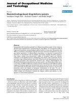
Hyaluronic acid (or hyaluronan, HA) (Fig. 1.1) is a linear, nonsulphated glycosaminoglycan composed of β-1,4-linked disaccharide units of β-1,3-linked glucuronic
acid and N-acetyl-D-glucosamine. HA is one of the main components of the extracellular matrix (ECM) and is present at high concentrations in all connective tissues
where it executes a rheological/structural function. In addition, owing to its capacity
to interact with some cell receptors, HA plays a significant role in processes such as
HO
O
HO
OH
O
HO
O
o
NH
H3C
O
HO
O
n
Fig. 1.1 Structure of hyaluronic acid
1.2
Hydrophobically Modified Hyaluronic Acid
3
cell migration, proliferation, and differentiation [1]. In the earlier period HA was
derived by extraction from rooster combs, however currently it is preferably derived
as product with superior features however with some impurities by fermentation of
Streptococcus strain. Recently, commercial HA has been obtained by recombinant
Bacillus subtilis sp. that is identified as a GRAS (safe) microorganism [2]. The features of HA can be improved and altered in various ways inorder to derive materials
with novelphysico-chemical and biologicalfeatures (hydrophobicity, amphiphilicity
& particular biological activities). Currently various HA derivatives are synthesized
and developed for different delivery system (Fig. 1.2)
The most commonly adopted chemical modifications of HA target three functional groups, specifically the glucuronic acid group, the primary and secondary
hydroxyl groups, and the amine group (after deacetylation of N-acetyl group)
(Table 1.1).
Particularly, carboxylates are commonly altered by esterification and amidation
reactions typically recognized using carbodiimide assisted coupling reactions. In
addition tobis-epoxide and divinylsulfone crosslinking, hydroxyl groups have been
altered by etherification and esterification reactions, resulting in linear and crosslinked HA-based products, respectively [3–5]. Owing to the outstanding biocompatibility and biodegradability, HA is one of the most commonly used biopolymers
used in the biomedical field and industry. In fact, numerous HA linear or cross-
Fig. 1.2 Hyaluronic acid derivatives
1
4
Mammalian Polysaccharides and Its Nanomaterials
Table 1.1 Modifications of hyaluronic acid
Polysaccharides
Hyaluronic acid
Modification
approaches
Esterification
Amidation
Ugi condensation
Description of reactions or
products
Esterification of HA by
alkylation using alkyl halides
(chlorides, iodides, bromide), by
using diazomethane, and by
using epoxides
Amidation of HA in water or of
HA’s TBA salt in organic
solvent with coupling agents,
e.g. EDC, NHS
Formation of diamide linkage
between polysaccharides chains
by using formaldehyde,
cyclohexylisocyanide and
diamine
Potential
applications
Cell carrier for skin
wounds, drug carrier
HA–drug conjugates
for controlling
release, target
specific delivery of
biomolecules
Controlled drug
delivery
linked derivatives have been fabricated that are utilized for tissue repair, treatment
of joint diseases, wound healing, anticancer drug delivery, and as scaffolds for tissue engineering. HA structure, elucidated by Karl Meyer [4] and revealed on
Fig. 1.3, consists of the reappearance of a disaccharide unit of an N-acetylglucosamine and a β-glucuronic acid. Its molecular weight is relatively high, above
a million. Its most significant physicochemical features are its capability to retain
water, a very high hydration ratio, and its viscoelasticity, these two features being
interdependent. Combined with its negative charge, HA plays a significant role in
the regulation of tissue hydration, permeability to small or large molecules and the
physicochemical features of tissues, as well as in several signaling pathways.
1.3
Chemically Crosslinked Hyaluronic Acid Semi-IPN
Earlier researchers [6] prepared a semi-IPN composed of HA and a network of
poly(2-hydroxy ethyl methacrylate-co-2-methacryloxyethyl trimethyl ammonium) (p(HEMA-co-METAC)) crosslinked by ethylene glycol dimethacrylate
(EGDMA). Owing to the incomplete neutralization of the positive charges of the
synthetic networks by HA, the water uptake of this IPN declined within rising
weight fraction of the polysaccharide in the matrix. This event was even stronger
by substituting HA with chondroitin sulphate, a polysaccharide with a higher
charge density due to the occurrence of sulphate groups. The p(HEMA-coMETAC)/HA semi-IPN demonstrated excellent cytocompatibility with mouse
fibroblasts and the net positive charge of the IPN gels developed the cell adhesion
in contrast to that of gels composed of only HA.A semi-IPN system appropriate
1.3 Chemically Crosslinked Hyaluronic Acid Semi-IPN
5
Polymer
OH
CH2OH
O
OH
HO
O
O
NH
C-O
CH3
CO 2
O
.
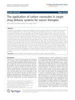
Fig. 1.3 Repeating disaccharide unit of HA and plan illustration presenting its space filling and
expanded configuration
for bioprinting was prepared by previous researchers [7] by means of a photopolymerizable dextran derivative, dex-HEMA (hydroxyethyl-methacrylate-derivatized dextran) as crosslinkable component and high molecular weight
HA. Dex-HEMA dissolved in an aqueous solution of Alg was crosslinked upon
UV exposure by means of Irgacure 2959 as photoinitiator. Mechanical characterization of these semi-IPN hydrogels with variable HA contents were carried out
proofing, specially, that the crosslinking kinetics were approximately
6
1
Mammalian Polysaccharides and Its Nanomaterials
instantaneous, as revealed by the sudden augment of the storage modulus G′ after
10 s of UV exposition. The system demonstrated excellent capability of chondrocytes after 3 days of incubation. Bioprinting [8] was approved by using a bioscaffolder pneumatic dispensing system. The polymer solution was extruded via
needle on a stationary platform subsequent to layer-by-layer deposition procedure, and stabilized by photocuring. The outcome demonstrated that the acquired
3D construct had a high porosity with well-defined strand spacing and that the
overall architecture could be simply tuned by regulating the procedure parameters, e.g. fiber spacing and orientation, representing the appropriateness of the
HA/dex-HEMA systems for bioprinting applications in tissue engineering.
1.4
Photopolymerized Hyaluronic Acid IPNS
The commercial attention for HA-based semi-IPNs and IPNs is established by a
world patent dated 1994 filed by the Italian Industry Fidia Farmaceutici SpA, that
demonstrates IPN biomaterials based on native HA or semi-synthetic HA derivatives and a non-carcinogenic, non-toxic synthetic polymer as second IPN component. The patent also declares HA derivatives with pharmacologically vigorous
molecules for IPN applications in a broad variety of sanitary fields, from urology,
dermatology, orthopaedics up to plastic and cardiovascular surgeries, in the form of
films, hydrogels, membranes, sponges, non-woven tissues, etc. [9]. The majority of
significant chemically modified HA polymers for the IPN formation are the methacrylated or acrylated derivatives, due to the mild conditions require for their synthesis [10, 11]. Methacrylic moieties can be conveniently incorporated on the
polysaccharide chains by exploiting the reactivity of carboxyl or hydroxyl groups of
HA, and the properties of the obtained networks can be suitably modified by altering the polysaccharide derivatization degree [12].
1.5
Hydrophobically Modified Hyaluronic Acid
In the previous work, hyaluronic acid was chemically bonded to dioleoylphosphatidylethanolamine (DOPE) in the presence of EDC chloride as a coupling agent for 24 h
at 37 8C. Ultrafiltration removes the coupling agent and the unreacted DOPE [13].
The ensuing product was used in the fabrication of cationic liposomes to yield lipoplexes used in gene therapy [14, 15].
1.7
1.6
Chondroitin Sulfate, Heparin and Hyaluronic Acid: pH/Ion-Responsive Networks
7
Hydrophobically Modified Heparin
Heparin is a natural sulfated polysaccharide containing units of sulfonated glucuronic acid and glucosamine derivatives (Fig. 1.4). From decades, heparin is used as
an anticoagulant and is also being studied as a potential agent to control complement activity and inflammation. In addition, heparins can intervene with the activity
of growth factors e.g. beta fibroblast growth factor (bFGF) and vascular endothelial
growth factor (VEGF), ensuing in the obstruction in angiogenesis and tumor development. Obviously these features, spherical and monodisperse heparin-based
nanoparticles that are chemically modified with deoxycholic acid were improved
with different DS (6.2, 8 and 10 %) [16]. Deoxycholic acid-bearing heparin nanoparticles were enclosed with negatively charged heparin shells, demonstrating zeta
potentials by 56 mV. Partition equilibrium constants for pyrene in the nanoparticles
showed that rising DS improved the hydrophobicity of the nanoparticle core. The
mean aggregation number of deoxycholic acid per hydrophobic microdomain, evaluated by the fluorescence quenching methods by means of cetylpyridinium chloride, showed that five to nine amphiphilic heparin chains contains a hydrophobic
domain in the conjugates [16].
1.7
Chondroitin Sulfate, Heparin and Hyaluronic Acid: pH/
Ion-Responsive Networks
These three animals-from polysaccharides are getting reticently growing consideration as elements of responsive crosslinked networks for the release of small drugs
and proteins. The acidic character of chondroitin sulfate makes it appropriate for
intermingling with positively charged molecules, including chitosan, and for being
used as polyanions in layer-by-layer (LbL) assemblies. Microcapsules of chitosan–
chondroitin sulfate crosslinked with glutaraldehyde are potentially useful for parenteral delivery of low molecular weight heparin [17] and oral administration of
5-fluorouracil [18]. Correspondingly, tablets of chitosan–chondroitin sulfate exhibiting pH-responsive release of indomethacin can be appropriate for colonic administration [19]. Microspheres of complexes of heparin and albumin crosslinked with
Fig. 1.4 Hydrophobically modified heparin
8
1
Mammalian Polysaccharides and Its Nanomaterials
glutaraldehyde have also revealed pH- and ionic strength-dependent swelling,
because of the ionic groups present in both albumin and heparin [20, 21]. The pH
dependent performance of hyaluronic acid hydrogels has been examined for photocrosslinked networks of a polymerizable derivative of hyaluronic acid. After loading with thrombin, the release occurs faster at pH 7 than at pH 1 [22].
1.8
1.8.1
Chondroitin Sulfate and Hyaluronic Acid: Electrical
Field-Responsive Network
Chondroitin Sulfate and Hyaluronic Acid
The release level of negatively charged macromolecules from hyaluronic acid
hydrogels crosslinked with EGDE was presented to decrease when an electric field
was switched on. Hyaluronic acid hydrogels speedily swell in water; however the
swelling is restricted in the presence of ions. Likewise, applying an electrical field
dramatically minimized the swelling and, therefore, the release rate of poly(styrene
sulfonic acid) and poly(glutamic acid, tyrosine) sodium salts. When the electric
field was removed, the release level augmented again. Therefore, these hydrogels
showed pulsate on–off release as the electric field was switched off–on [23].
Chondroitin sulfate hydrogels crosslinked with EGDE have been revealed as appropriate for electro-responsive administration of peptides and proteins, including
vasopressin, aprotinin, lysozyme and albumin. Chondroitin sulfate and albumin are
negatively charged at physiological pH, while vasopressin, aprotinin and lysozyme
have positive charges. The release of aprotinin and lysozyme could be regulated by
means of the voltage, while albumin and vasopressin were flaccidly released. The
performance of aprotinin and lysozyme could be explained owing to the fact that
positive charged macromolecules under electrical field incline to move from the
hydrogel to the cathode. While the similar performance was predictable for vasopressin, it did not ensue perhaps because its smaller size and low charge make its
passive diffusion easier. As albumin is negatively charged, variances in the release
rate owing to fluctuations in the electrical field are not anticipated [24]. These initial
outcomes propose that chondroitin sulfate hydrogels may be suitable for the development of electro-responsive implantable DDSs.
1.9
Heparin & Hyaluronic Acid: Anti-adhesive Surfaces
Prevention of bacterial adhesion on surfaces via anti-adhesive coatings is one of the
easiest, possibly cost-effective alternatives to ignore biofilm formation. Bacterial adhesion is a complex process which is influences by various factors involving — as stated
above — the physical and chemical features of material surface, nevertheless also
1.9
Heparin & Hyaluronic Acid: Anti-Adhesivesurfaces
9
bacterial cell properties and environmental causes e.g. the bulk medium composition
(ionic strength, presence of organic substances) and flow conditions. Adhesion of bacteria to negatively charged surfaces under physiological pH environment may be influenced by electrostatic repulsion forces as the net electrostatic charge of maximum
bacterial cell walls is negative at neutral pH [25]. It has also been frequently reported
that hydrophilic, low surface energy materials are less vulnerable to bacterial adhesion
than hydrophobic ones, however opposing outcomes do exist. It is usually accepted
that hydrophilic surfaces in connection with media encompassing organic molecules
e.g. proteins oppose the development of a conditioning film sheltering adhesion sites
for bacteria — restricting specific adhesion/attachment of bacteria and following biofilm development. Anionic polysaccharides with hydrophilic features have been subsequently acknowledged potential candidates to explain anti-adhesive surfaces.
1.9.1
Hyaluronic Acid
The most studied polysaccharides as a biofilm repelling coating is hyaluronic acid
[26–28]. In 1999, Morra and Cassineli [26] recognized non-fouling features of glass
surfaces modified with hyaluronic acid covalently bound to a first layer of
poly(ethyleneimine). Presenting hydrophilic features, this coating minimize adhesion of S. epidermidis and E. coli by some orders of extent, in contrast to the
unmodified glass slide. Harris and Richards [27] studied S. aureus adhesion on titanium surfaces, displaying differences in and grafted or not with hyaluronic acid.
Viewing no clear dependence on surface roughness, bacterial adhesion was considerably minimized by the coating. In the similar approach, adhesion of S. aureus on
Ti foils functionalized with hyaluronic acid-catechol was lesser than on pristine
substrates [28]. The bacteria-repelling features of hyaluronic acid have been presently demonstrated by a decline in adhesion of S. aureus cells to hyaluronic acidcoated Ti surfaces [29] and poly(methyl methacrylate) intraocular lenses [30] in
contrast to untreated surfaces. A graft copolymer derivative of hyaluronic acid bearing amino and carboxyl groups showed better prevention of S. aureus adhesion on
Ti disks than the pristine hyaluronic acid hydrogel [29]. However, many commercial hyaluronic acid-based coatings currently available.
1.9.2
Heparin
Heparin is another natural polysaccharide of animal origin whose anti-adhesive features have been widely studied. Heparin is usually used as an antithrombotic coating in implanted devices that are in contact with blood, in specific catheters and
stents. The Bioline Coating® from Maquet Cardiopulmonary GmbH, Rastatt,
Germany-a subsidiary of Getinge AB, Göteborg, Sweden, the Bioactive Surface
CBAS® from Carmeda AB, Upplands Väsby, Sweden – a subsidiary of W.L. Gore
1
10
Mammalian Polysaccharides and Its Nanomaterials
and Associates, Inc., Newark, Del., and the Trillium® biosurface from Medtronic,
Inc., Minneapolis, Minn., are some HP-based antithrombotic coatings available on
the market. This negatively charged, linear polysaccharide has been immobilized on
material surfaces via numerous physical or chemical approaches comprising electrostatic deposition, layer-by-layer self-assembly and covalent attachment [31].
Bacterial adherence to heparinized commercial devices, e.g., ureteral [32, 33] and
biliary [34] stents, central vein [35] and dialysis [36] catheters, has been assessed in
vitro or in vivo. Majority of the investigations demonstrated anti-adhesive effects of
heparin coatings though Lange et al. noted no important variation in the number of
bacteria adhered to heparin-coated stents and non-coated controls [37].
1.10
1.10.1
Hyaluronic Acid and Chondroitin Sulfate
(Polysaccharides of Human Origin): Biodegradable
Polymers as Biomaterials
Hyaluronic Acid
Hyaluronic acid (HA) was initially separated in 1934 from the vitreous humor of
the eye by Meyer and Palmer [38]. This biopolymer has gradually raised attention as a exclusive biomaterial since its discovery. Hyaluronic acid is a member
of the glycosaminoglycan family, which are linear polysaccharides consisting of
alternating units of N-acetyl-D-glucosamine and glucuronic acid, and are found
in virtually every tissue in vertebrates. HA can be considered to be the major
glycosaminoglycan having molecular weights up to numerous millions. In contrast with other members of the glycosaminoglycan family existing in the human
body, e.g. dermatan sulfate, keratin sulfate, chondroitin sulfate, and heparin sulfate, HA is not covalently bond to proteins. HA is water-soluble and forms
extremely viscous solutions with distinctive viscoelastic features. HA can form
3-D structures in solution with widespread intramolecular hydrogen bonding. It
has been reported to be present at high concentrations in synovial fluid and vitreous humor and considerably contributes to the viscoelastic features of these tissues. Additionally, HA plays a significant structural role in a variety of tissues
including articular cartilage, the nucleus pulposus, skin, the cervis, and the glycocalyx of endothelial cells. Reports have demonstrated that within the cells, HA
is manufactured on the cytosol surface of the plasma membrane under the direction of three glycosyltransferases: hyaluronan synthase-1 (Has-1, Has-2and
Has-3 [39]. Between these, Has-2 is the main enzyme accountable for HA production while embryogenesis; nevertheless, particular roles played by Has1 and
Has3 are not yet apparent [40]. The traditional sources for HA isolations are
rooster combs and bovine vitreous humor. Nevertheless, utilizing bioprocess
methodologies for HA fabrication is gaining attention and numerous bacterial
fermentation procedures are presently under progress. HA can experience
1.10 Hyaluronic Acid and Chondroitin Sulfate (Polysaccharides of Human Origin):…
11
degradation within the body by free radicals e.g. nitric oxide and MMPs found in
the extracellular matrix, trailed by endocytosis. It can also experience digestion
by lysosomal enzymes to form mono and disaccharides, which can be subsequently transformed into ammonia, carbon dioxide and water via the Krebs cycle
[41]. In previous investigations, HA was considered to be a passive structural
component of connective tissues; nevertheless, later investigations shown it to be
energetically elaborate various biological procedures e.g. modulating cell migration and differentiation during embryogenesis, regulating extra cellular matrix
organization and metabolism, in addition playing significant roles in wound healing, metastasis, and inflammation [42]. Since HA is synthesized by cells while
initial wound healing, this polymer has been widely studied for wound dressing
applications. Additional distinctive features of HA include its capability to
encourage angiogenesis, to control wound site inflammation by acting as a free
radical scavenger, and to be identified by receptors on a diversity of cells related
with tissue repair. Owing to the high functionality and charge density of HA, it
can be cross-linked by a different physical and chemical methods [43]. Improved
HA, e.g. esterified derivatives like ethyl/benzyl esters (HYAFFs) and crosslinked hyaluronic acid gels have been broadly studied for wound dressing application. These chemical alterations have also been found to significantly minimize
the degradation rate of the polymer. The benzyl esters (HYAFFs) experience
hydrolytic degradation via ester bonds in the absence of enzymatic activity with
degradation time’s varying from 1–2 weeks to 2–3 months, depending on the
degree of esterification. The de-esterified polymers are more hydrated and soluble and resemble native HA [44, 45]. HA also plays an important role in tissue
repair by encouraging mesenchymal and epithelial cell migration and differentiation, thus improving collagen deposition and angiogenesis. This character, in
addition to its immunoneutrality makes HA an ideal biomaterial for tissue engineering and drug delivery applications. Its aqueous solubility permits HA to be
synthesized into various kinds of porous and three-dimensional structures for
these applications. Therefore a viscous formulation of HA containing fibroblast
growth factor (OSSIGELs) is experiencing late stage clinical trial as a synthetic
bone graft to hasten bone fracture healing. Likewise HYAFFs 11 is presently
been utilized as a carrier vehicle for a different growth factors and morphogens
as well as bone marrow stromal cells. In an investigation that associated HYAFFs
11 with an absorbable collagen sponge as a carrier vehicle for osteoinductive
protein, recombinant human bone morphogenetic protein-2 (rhBMP-2) shown a
well healing response with HYAFFs11 carrier than collagen [46]. HA-based
materials have also replaced collagen-based materials as injectable soft tissue
fillers [47]. High molecular weight viscous HA solutions (AMVISCs and
AMVISCs PLUS) are being used as a vitreous humor substitute as well as to
shield the sensitive eye tissue through cataract extraction, corneal transplantation
and glaucoma surgery. Viscous HA solutions (SYNVISCs, ORTHOVISCs) are
clinically utilized as a synovial fluid substitute to relieve pain and improve join
mobility in osteoarthritis patients [48]. A recent animal investigation established
the merits of exogenous HA in treating vascular diseases [49].
1
12
1.10.2
Mammalian Polysaccharides and Its Nanomaterials
Chondroitin Sulfate
Reports have shown that a significant phase of wound healing includes the secretion of glycosaminoglycans by fibroblast cells to form a hydrophilic matrix
appropriate for remodeling while healing. A current investigation expending rat
embryonic fibroblast cells showed that the most of the glycosaminoglycan chains
produced were chondroitin sulfate, signifying the implication of this natural
polymer for its utilization in biomedical applications [50]. Chondroitin sulfate is
the main component of aggrecan, the most plentiful glycosaminoglycan found in
the proteoglycans of articular cartilage. Reports have revealed that CS can trigger the metabolic response of cartilage tissue and has antiinflammatory features
[51]. It is also elaborate cell recognition, intracellular signaling, and the connection of extracellular matrix components to cell-surface glycoproteins [52].
Chondroitin sulfate entails repeating unit formed by N-acetyl galactosamine
(GalNAc) and glucuronic acid (GlcA) modified by sulfation, where the location
of sulfation differs with the kind of CS [53]. In mammals chondroitin sulfate
disaccharides have been found to be monosulfated in the fourth or sixth position
of the GalNAc residue or disulfated in the second and sixth position of the GlcA
and GalNAcor in the four and six positions of GalNAc residue [54]. The enzymes
accountable for these alterations are chondroitin sulfotransferases. Owing to its
biocompatibility, non-immunogenicity and pliability, CS hydrogels have been
broadly studied for wound dressing applications [55]. Alike to HA, numerous
physical and chemical crosslinking techniques have been established for CS to
form hydrogels for biomedical applications [56]. As CS plays a significant role
in controlling the expression of the chondrocyte phenotype, it has been broadly
studied as a scaffolding material for cartilage tissue engineering. This is mainly
significant since investigations have revealed that effective cartilage regeneration
can be attained via the use of a tissue engineered implant, simply if the seeded
cells experience normal proliferation and phenotype development within the biodegradable scaffold together with the fabrication of a novel cartilage-specific
extracellular matrix. Numerous investigations have examined the efficiency of
utilizing composite scaffolds composed of CS and other biopolymers, e.g. collagen or synthetic biodegradable polymers, as scaffolds for cartilage tissue engineering. These investigations have explored a strong correlation between the use
of CS and the bioactivity of the seeded chondrocytes [57]. Additional natural
bioactive polysaccharides that are being acknowledged as potential biomaterials
for different biomedical applications comprise heparin sulfate, keratin sulfate
and dermatan sulfate.
1.11
Natural–Origin Polymers as Carriers and Scaffolds for Biomolecules and Cell…
1.11
1.11.1
13
Natural–Origin Polymers as Carriers and Scaffolds
for Biomolecules and Cell Delivery in Tissue
Engineering Applications
Hyaluronan
Hyaluronic acid is most often called as hyaluronan owing to the information that it
exists in vivo as a polyanion and not in the protonated acid form [58]. Hyaluronan
is a naturally occurring non-sulfated glycosaminoglycan and a main macromolecular component of the intercellular matrix of most connective tissues e.g. cartilage,
vitreous of the human eye, umbilical cord and synovial fluid [58]. Hyaluronic acid
is a linear polysaccharide that comprised of alternating disaccharide units of α-1,4Dglucuronic acid and β-1,3-N-acetyl-D-glucosamine, connected by β(1→3) bonds
[59]. Hyaluronan and its linked networks have various physiological functions that
comprise tissue and matrix water regulation, structural and space-filling properties,
lubrication, and a number of macromolecular functions [58]. Particularly for its
enhanced viscoelastic features, hyaluronan function as a lubricant and shock
absorber in synovial fluid. Hyaluronan has been extensively investigated for drug
delivery, for dermal, nasal, pulmonary, parenteral, liposome-modified, implantable
delivery devices and for gene delivery (reviewed in Liao et al. [58]). Hyaluronan
for tissue engineering has been intensive on cartilage, bone and osteochondral
applications, most probable owing to the information that it is a major macromolecular component of the extracellular matrix. Industrially available hyaluronan is
derived from various sources, chiefly by isolation from rooster comb, umbilical
cord, synovial fluid, or vitreous humor. In addition, hyaluronic acid can be simply
and controllably fabricated in large scales via microbial fermentation, from strains
of bacteria such as Streptococci [58], enabling the scale-up of derived products and
avoiding the risk of animal-derived pathogens. Hyaluronan is accessible for numerous applications, for lubrication and mechanical support for the joints in osteoarthritis (Artz® from Seikagaku Corporation in Japan; Hyalgan® and Hyalubrix®
from Fidia in Italy) as a viscoelastic gel for surgery and wound healing (Jossalind®
from Hexal in Germany; Bionect® from CSC Pharmaceutical in USA), for implantation of artificial intraocular lens (Healon® from OVD from Advanced Medical
Optics in USA, Opegan R® from Seikagaku in Japan, Opelead® from Shiseido in
Japan, Orthovisc® from Anika in USA) and as culture media for use in in vitro
fertilization (EmbryoGlue® from Vitrolife, USA) [58]. Hyaff® commercialized by
Fidia in Italy has been extensively employed as a biomaterial for biomedical applications. From a chemical viewpoint, Hyaff® is a benzyl ester of hyaluronic acid
and its key features are that HYAFF® preserves the biological features of the natural molecule from which it originates, the natural degradation of Hyaff® releases
hyaluronic acid, which is then degraded via well-known metabolic pathways and
that depends on the extent of esterification, it is likely to obtain polymers with various levels of hydrophobicity.
14
1
Mammalian Polysaccharides and Its Nanomaterials
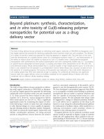
Fig. 1.5 HA cryogels and hydrogen bonding between –COOH groups
The word hydrogel explains 3-D network structures derived from a class of
synthetic and/or natural polymers which can absorb and retain considerable
amount of water or biological fluids (Fig. 1.5). Polysaccharides that are employed
to produce physical cryogels: carboxymethylated cellulose, xanthan, hyaluronan,
carboxymethylated curdlan, starch (amylose, amylopectin and their mixtures),
β-glucan, locust bean gum, maltodextrins and agarose. A variety of physically
crosslinked cryogels from polysaccharides with tunable mechanical, structural,
biological features as well as numerous applications is considered and the studies
of the fabrication mechanism for these cryogels are also explored. The accurate
forming method of HA cryogel has not been completely understood. The complication in gel formation method of HA cryogel might be primarily obtained from
its chemical structure, which includes not only massive –OH groups as in PVA
and galactomannan, but also –COO and –NHCH3 groups along with potential
hydrophobic regions. The intermolecular and intramolecular hydrogen bonding
induced from –COOH in HA chains may play a significant role in respect to the
network formation and stabilization of HA gel, and the probable example is
revealed in Fig. 1.5.
1.11.2
Chondroitin Sulphate
Extracellular matrix components are appreciated building elements for the fabrication of biomaterials involved in tissue engineering, particularly if their biological,
chemical and physical features can be regulated. An instance is chondroitin sulfate,
one of the best physiologically vital glycosaminoglycans. Glycosaminoglycans
(GAGs) are present in the lubricating fluid of the joints and as components of
1.12
Rationale for the Use of HA in Drug Delivery
15
cartilage, synovial fluid, bone, and heart valves. With the exception of hyaluronan,
these polysaccharides are covalently connected to a protein core, thus creating proteoglycans [60]. Bio-characteristics of GAGs comprise the binding and modulation
of growth factors and cytokines, proteases inhibition, and the participation in adhesion, migration, proliferation and entiation of cells [61]. Additionally, GAGs are virtually non-immunogenic and degrade to non-toxic oligosaccharides. These features
together with their well-defined physical and chemical properties make them very
fascinating materials for tissue engineering. Owing to its GAG nature, chondroitin
sulfate is a smart natural–origin polymer applied fundamentally in cartilage tissue
engineering. However, and owing to its biological characteristics, is frequently used
in other tissue engineering applications to valorize other polymers so as to interact
with cells and proteins modifying cell behavior of the developed materials.
Chondroitin sulfate comprised of repeating disaccharide units of D-glucuronic acid
and N-acetyl galactosamine sulfated at either 4- or 6-positions [62]. Chondroitin
sulfate can conjugate with core protein to harvest highly absorbent aggregan, which
is a chief structure inside cartilage and functions as a shock absorber or it can offer
syndecan, which is a cell receptor which can interact with adhesion proteins, cells
and the extracellular matrix (ECM) [62]. In vitro studies suggest that chondroitin
sulfate is also able to advance matrix component production by human chondrocytes
[63]. Additionally, chondroitin sulfate proteoglycans have a serious role in renewal
and plasticity in the central nervous system as suggested by Galtrey and Fawcett
[64]. However, the readily water-soluble behavior of chondroitin sulfate restricts its
application as a solid-state drug delivery vehicle. Accordingly, it is usual to carry out
a crosslinking behavior to tailor the properties of chondroitin sulfate as examined in
various researches [65] or to syndicate it with other polymers, e.g. chitosan, gelatin
and hyaluronan, collagen, poly(vinyl alcohol) or poly-(lacticco-glycolic acid) so as
to harvest more stable materials. Additionally, and meanwhile chondroitin sulfate in
negatively charged, interaction with positively charged molecules e.g. polymers or
growth factors is expected being an important concern to enable the design of delivery systems. For examples this is employed to harvest chondroitin sulfate–chitosan
sponges as delivery systems for platelet-derived growth factor BB (PDGF-BB) for
bone regeneration as evidenced by JeongPark et al. [66] where this communication
revealed to induce more sustained release of the growth factor. As earlier mentioned,
owing to its biofeatures, chondroitin sulfate has been used in some extent in the tissue engineering field, chiefly in cartilage applications.
1.12
Rationale for the Use of HA in Drug Delivery
For biomedical functions, HA is chiefly produced by microbial fermentation; it can
also be isolated from rooster combs and umbilical cords [67]. HA depolymerization
can be attained in batch cultures via either by enzymatic reaction or physical or
chemical degradations [68–70]. HA can be related chemically to drugs or to drug
carriers. The formation of HA drug conjugates, or the relationship of HA to