2008 anti oxidant status in embryonic post hatch and larval stages of asian seabass lates calcarifer
Bạn đang xem bản rút gọn của tài liệu. Xem và tải ngay bản đầy đủ của tài liệu tại đây (315.01 KB, 8 trang )
Fish Physiol Biochem (2008) 34:151–158
DOI 10.1007/s10695-007-9155-4
Anti-oxidant status in embryonic, post-hatch and larval
stages of Asian seabass (Lates calcarifer)
N. Kalaimani Æ N. Chakravarthy Æ R. Shanmugham Æ A. R. Thirunavukkarasu Æ
S. V. Alavandi Æ T. C. Santiago
Received: 11 October 2006 / Accepted: 2 July 2007 / Published online: 7 August 2007
Ó Springer Science+Business Media B.V. 2007
Abstract The concentrations of anti-oxidant
enzymes such as superoxide dismutase (SOD), catalase (CAT) and selenium-dependent glutathione
peroxidase (SeGPx), and low molecular weight
free-radical scavengers such as reduced glutathione
(GSH) and ascorbic acid (vitamin C) were evaluated
during the period from gastrulation (GS) to 25 days
post-hatch (dph) in the larvae of Asian Seabass, Lates
calcarifer. Oxidative damage due to lipid peroxidation (LPO) was also assessed, by evaluation of the
formation of malondialdehyde (MDA). All the three
anti-oxidant enzymes, SOD, CAT and GPx, showed
high activities during gastrulation, suggesting an
increased metabolic rate during the period of embryonic development. Though the SOD activity
apparently decreased progressively during 3–20 dph
of larval development, the difference was not significant. CAT showed high activity during gastrulation
and remained constant up to 3 dph, suggesting an
increased need to metabolise hydrogen peroxide
(H2O2) and organic peroxides. In contrast, SeGPx
activity increased progressively from 5 dph to 25 dph
during larval development, indicating an increased
need to detoxify lipid peroxides. This is evident from
N. Kalaimani (&) Á N. Chakravarthy Á
R. Shanmugham Á A. R. Thirunavukkarasu Á
S. V. Alavandi Á T. C. Santiago
Central Institute of Brackishwater Aquaculture, 75,
Santhome High Road, R.A. Puram, Chennai, Tamil Nadu
600028, India
e-mail:
the observation of increased lipid peroxidation from
10 dph to 25 dph during larval development. GSH
levels were low at gastrulation, indicating increased
metabolic rate and formation of lipid radicals during
this period, corresponding to the decrease in the level
of ascorbic acid, which is consumed for regeneration
of GSH.
Keywords Anti-oxidant enzymes Á
Ascorbic acid Á Eggs Á Embryos Á Gastrulation Á
Larvae Á Lates calcarifer Á Malondialdehyde Á
Reactive oxygen species
Introduction
Approximately 0.1% of all oxygen entering the
mitochondrial electron transport chain is released as
reactive oxygen species (ROS), which can cause
damage to lipids, proteins, and DNA (Fridovich
2004). ROS and other pro-oxidants are continually
detoxified and removed in cells by anti-oxidant
defence systems, which include anti-oxidant enzymes
such as superoxide dismutase (SOD), glutathione
peroxidase (GPx), catalase, glutathione reductase
(GR) and glutathione-S-transferase (GST), and by
non- enzymatic defences such as ubiquitous tripeptide glutathione, vitamins E, C and A, carotenes, and
ubiquinol (Wilhelm Filho 1996).
Knowledge of anti-oxidant enzyme expression in
early embryonic stages would be extremely important
123
152
in understanding the origin and formation of protective mechanisms during the life history of organisms.
Rudneva (1999) has studied the anti-oxidant enzymes
and low molecular weight scavengers in eggs and
larvae of various Black Sea species (molluscs,
crustacea and fish, including elasmobranchs and
teleosts). The levels of anti-oxidant enzymes during
larval development of Macrobrachium malcolmsonii,
Macrobrachium rosenbergii, and Dentex dentex have
been studied (Arun and Subramanian 1998; Dandapat
et al. 2003; Mourente et al. 1999). Developmental
aspects of detoxifying enzymes in the fish Salmo
iridaeus has been reported by Aceto et al. (1994).
Anti-oxidant enzyme activities in embryonic and
early larval stages of turbot and sprat (Sprattus
sprattus) larvae have also been studied (Peters and
Livingstone 1996; Peters et al. 2001). The activities
of the anti-oxidative enzymes in the cephalopods
Sepia officinalis and Lolliguncula brevis have been
reported by Zielinski and Portner (2000). The metamorphosis period in Senegal sole species is especially
critical, with high utilisation of energy reserves, due
to an elevated metabolism, which consumes more
exogenous oxygen to meet the metabolic demands
(Fernandez-Diaz et al. 2001; Yufera et al. 1999).
Studies have been made on anti-oxidant status in
other animals. Starrs et al. (2001) examined the
activities of catalase, SOD and GPx in the developing
lungs of two oviparous vertebrate species, the
chicken (Gallus gallus), and an agamid lizard (Pogona vitticeps), and in a metamorphosing vertebrate,
the anuran Limnodynastes terraereginae. They concluded that the anti-oxidant enzymes are
differentially regulated in different species and
appear to have evolved different levels of dependency on environmental variables, and the late
developmental increase in anti-oxidant enzymes
activity seen in mammals is not as pronounced in
oviparous and metamorphosing vertebrates. The
activity of the basic anti-oxidant enzymes superoxide
dismutases, catalase and glutathione peroxidase in
liver has already developed at early stages of
embryogenesis and is considerably enlarged in the
end embryogenesis of goose (Danchenko and Kalytka
2002). ‘‘Aging’’ was associated with increases in the
activity of red cell SOD and GPx, and significant
correlations amongst red cell GR, GPx and SOD
activities were found in old but not in younger adult
Japanese quails (Godin et al. 2001).
123
Fish Physiol Biochem (2008) 34:151–158
Sea bass, Lates calcarifer, is a euryhaline hardy
fish suitable for farming in marine, freshwater and
brackishwater ecosystems. It is a fast-growing fish,
suitable for culture in both ponds and cages. Technology for seed production of seabass under captivity
has been developed by the Central Institute of
Brackishwater Aquaculture (CIBA), Chennai, India.
It is also widely distributed in the Indo-Pacific region.
It has been selected because of the potential for
culture in this region. The oxidation process and
interaction and status of anti-oxidants will throw light
on the metabolic status of the early stages of Asian
seabass, Lates calcarifer, larvae, which will have
bearing on the health of the fish. In the present work,
the process of changes in anti-oxidant levels at
various stages of development and growth were
studied.
Materials and methods
Larval rearing
Lates calcarifer larvae were obtained by induced
spawning from captive brood stock at the facilities of
CIBA, Chennai, India. The different stages selected
were fertilised eggs, gastrulation stage, 3 days after
hatching (3 dph), 5, 10, 15, 20 and 25 dph. Larvae
were reared in 4–5-tonne capacity circular fibreglass
reinforced plastic (FRP) tanks in a salinity of
30 ± 1 ppt. Fertilised eggs of Lates calcarifer collected from the spawning tanks were incubated in the
incubation tanks. The hatched out larvae were
transferred to the rearing tanks. From the 3rd day,
larvae were fed with rotifers, Brachionus plicatilis,
up to the 9th day, with rotifer density maintained
around 20–50 individuals per millilitre. From the
10th day to the 15th day, larvae were co-fed with
rotifers and Artemia nauplii. From the 16th day,
feeding was done with Artemia nauplii, alone, up to
the 25th day. The Artemia nauplii density was
maintained at 2,000 to 6,000 individuals per litre.
Initial larval density ranged from 40 to 50 individuals
per litre. Depending upon age and size, the larval
density was reduced to 20–25 individuals per litre at
10 dph, and later, after 15 dph, the density was
maintained around 10–15 individuals per litre. For
the experiment, larvae and eggs were collected from
fish spawned through induced maturation at night. To
Fish Physiol Biochem (2008) 34:151–158
determine the total length (TL), and the status of
metamorphic development, we took samples of 20–
30 larvae periodically and examined them under a
light microscope. Two replicates were performed.
Biochemical analysis
Aliquots of larvae Lates calcarifer, weighing approximately 1 g, were homogenised in phosphatebuffered saline (PBS) solution with a pH of 7.6,
using a Potter homogeniser kept in an ice-water bath.
After centrifugation (13,000 g, 5°C, 15 min), the
supernatant was used immediately for determination
of enzyme activities. All assays were carried out in
duplicate at 25°C.
Superoxide dismutase (Superoxide: Superoxide
oxidoreductase, EC 1.15.1.1) was assayed in the
homogenate by the method of Marklund and Marklund (1974). The enzyme activity was expressed as
units per milligramme of protein in tissues. The
reaction was initiated by the addition of 0.5 ml
pyrogallol reagent, and the change in optical density
was measured at 480 nm for 3 min. Fifty percent
inhibition of pyrogallol by the enzyme was taken as
one enzyme unit. Catalase (Hydrogen peroxide:
Hydrogen-peroxide oxidoreductase, EC 1.11.1.6)
activity was assayed by the method of Sinha
(1972). Absorbance was read at 570 nm. Catalase
activity was expressed as micromoles of H2O2
consumed per minute per milligramme of protein.
Glutathione peroxidase (Glutathione: Hydrogen-peroxide oxidoreductase, EC 1.11.1.9) was assayed by
the method of Rotruck et al. (1973), with some
modifications. The inclusion of sodium azide in the
incubation medium inhibits catalase activity. Absorbance was measured at 420 nm. GPx activity was
expressed as microgrammes of glutathione utilised
per minute per milligramme of protein. Protein
content of tissue samples was estimated by the
method of Lowry et al. (1951). The blue complex
formed was measured at 640 nm after 15 min against
the blank.
Tissue lipid peroxidation (LPO) was measured by
the method of Devasagayam (1986). Malondialdehyde (MDA), an end product of LPO, reacts with
thiobarbituric acid (TBA) to form a pink chromogen
(allegedly an MDA adduct) and is measured by its
absorbance at 532 nm. Ascorbic acid was estimated
153
by the method of Omaye et al. (1979). Ascorbic acid
values were expressed as microgrammes per milligramme of protein. The disappearance of reduced
glutathione (GSH) was measured by its reaction with
5,50 -dithio-bis (2-nitro benzoic acid) (DTNB). GSH
in homogenate was measured according to the
method of Beutler and Kelley (1963), using Ellman’s
reagent. This method is based on the development of
a yellow complex upon the addition of 5,50 -dithio bis
(2-nitro benzoic acid) to compounds containing
sulphydryl groups.
Statistical analysis
Results are presented as mean ± SD (n = 4). Differences between mean values of enzyme activities,
levels of GSH, vitamin C and lipid peroxidation in
different stages of development, starting from fertilised eggs till 25 dph, were analysed by one-way
analysis of variance (ANOVA) followed by testing
for multiple range comparisons between means
(Duncan’s). The differences between means were
reported as statistically significant when P < 0.05.
Results
Fertilised eggs float on the surface of water immediately after spawning. Gastrulation occurs 6 h after
fertilisation. Eighteen hours after spawning, the eggs
hatch out (day 0). During the period up to 3 dph,
absorption of the yolk takes place. The size of the
eggs after fertilisation and at gastrulation was
observed to be 0.84 ± 0.3 mm and 0.87 ± 0.05 mm,
respectively. First feeding stage of larvae was studied
at 3 dph, when the mouth of the larvae opened, and
the larvae were fed with the rotifer Brachionus
plicatilis (strain S-1, O.F. Muller, size range 120–
300 lm). The length of the larvae on 3 dph was
2.4 ± 0.2 mm. The fifth day after hatching was
included in our evaluation so that we could observe
changes in anti-oxidant level corresponding to the
rotifer in the diet. During this period, the dorsal and
anal fins start appearing and the serration appears in
the pre-operculum. The length of the larvae on 5 dph
increased to 2.8 ± 0.5 mm. Anti-oxidant status at the
10th day of larval development (length
3.0 ± 0.2 mm) was observed when Artemia nauplii
123
154
Fish Physiol Biochem (2008) 34:151–158
(Commercial strain OSI PRO 80) were fed in addition
to rotifer. Morphologically, on the 15th day of larval
development, the dorsal and anal fins are separated
from the caudal fin and the pelvic fin appears. A
white band from the centre of the dorsal fin to the
anal fin is visible. The anti-oxidant status after the
inclusion of Artemia nauplii in the diet of seabass
larvae at 15 dph was evaluated. The length of larvae
was observed to be 4.5 ± 0.5 mm, 8.0 ± 0.5 mm and
10 ± 0.5 mm on 15, 20 and 25 dph, respectively. On
20 dph the number of spines and soft rays of the
dorsal and anal fins become constant. Scales appear
in the mid-lateral surface above the anal fin. The
body colour changes from black to pale brown.
Feeding was continued with Artemia nauplii. The
20th and 25th days of larval development representing the metamorphosis period were also included in
our study. The metamorphosis period in fish is
especially critical, with high utilisation of energy
reserves due to elevated metabolism, which consumes more exogenous oxygen to meet the metabolic
demands (Fernandez-Diaz et al. 2001; Yufera et al.
1999).
The activities of the anti-oxidant enzymes SOD,
catalase, selenium (Se)-glutathione peroxidase, level
of lipid peroxidation and the amounts of glutathione
and vitamin C during different development stages of
seabass larvae, including fertilised eggs and gastrulation, are presented in Table 1.
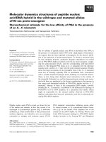
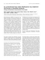
The activities of the enzymes SOD and catalase
are presented in Figs. 1 and 2, respectively. SOD
activity apparently found to be maximum at
gastrulation (GS) was observed to decline gradually,
and the decrease was not significant. The activity of
catalase was the highest in the gastrulation stage. The
minimum values were observed for SOD during
20 dph and for catalase at 15 dph. The decrease was
significant in catalase, and the activities increased
gradually on 20 dph and 25 dph. The increase in the
activity of catalase was significant at 25 dph compared to 15 dph.
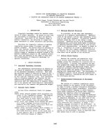
GPx activity (Fig. 3) was observed to be high
during gastrulation and minimal at 5 dph. GPx
activity was found to increase gradually after 5 dph
of larval development. This increase in GPx activity
directly correlates with the increase in lipid peroxidation observed in this period of larval development.
Selenium-dependent glutathione peroxidase (SeGPx)
activity in different stages varied significantly, except
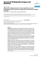
on 5 dph and 10 dph. The rate of lipid peroxidation
(Fig. 4) was very high at gastrulation and decreased
to a minimum at 5 dph. The significant differences in
GPx activities and lipid peroxidation during various
stages are presented in Table 1.
At gastrulation, GSH level (Fig. 5) was observed
to be low. A smaller increase in GSH level than that
found in fertilised eggs (FEs) was noted after
gastrulation up to 3 dph. After 3 dph, the level of
GSH was found to decline gradually. But again, GSH
showed a small increase at 25 dph. Ascorbic acid
concentration (Fig. 6) was very low at gastrulation,
and there was a drastic increase in vitamin C at
3 dph, when the larvae were fed with rotifers. After
that the level of ascorbic acid was found to decrease
Table 1 Status of anti-oxidant enzymes and anti-oxidants and
lipid peroxidation in different developmental stages of Lates
calcarifer. Results are mean ± SD (n = 4). Values within each
column bearing different superscript letters are significantly
different at P < 0.05 (SeGPx selenium-dependent glutathione
peroxidase, FE fertilised egg, GS gastrulation)
Different
SOD (units/mg Catalase (lmol H2O2
consumed/min
stages of
protein)
per mg protein
development
SeGpx GSH
LPO (nmol of
GSH
Vit. C
utilised/min
malondialdehyde
(lg/mg protein) (lg/mg protein)
per mg protein released/mg protein)
FE
43.93 ± 5.04a
16.63 ± 2.80a
50.25 ± 2.09a
34.56 ± 2.91a
95.25 ± 2.49a
27.72 ± 2.88a
GS
46.81 ± 5.9a
20.19 ± 3.56b
56.07 ± 2.05b
64.29 ± 3.02b
25.36 ± 3.13b
8.12 ± 1.46b
a
3 dph
5 dph
43.77 ± 5.3
43.38 ± 5.45a
10 dph
43.33 ± 5.23a
15 dph
43.27 ± 5.01
a
20 dph
25 dph
123
ab
c
31.07 ± 4.20
17.02 ± 1.34d
c
5.18 ± 1.49
4.10 ± 1.06c
c
102.72 ± 6.25
85.41 ± 6.24d
69.21 ± 2.39c
36.11 ± 3.37d
1.71 ± 1.00de
16.98 ± 1.67d
11.49 ± 1.36d
66.33 ± 4.67e
36.06 ± 3.78d
c
d
f
36.03 ± 2.63d
18.53 ± 1.90
6.59 ± 1.02c
d
1.24 ± 0.49
28.99 ± 1.82
11.02 ± 1.14
57.17 ± 2.37
43.17 ± 4.83a
3.47 ± 0.41de
35.06 ± 1.84e
17.19 ± 1.52e
20.23 ± 2.58b
a
ce
f
e
d
43.44 ± 4.91
4.30 ± 0.65
41.57 ± 1.24
18.09 ± 2.38
80.26 ± 3.48
12.09 ± 2.94be
13.40 ± 1.96e
FE - Fertilised eggs
GS - 6hrs after spawning
60
50
40
30
20
10
0
FE
GS 3dph 5dph 10dph15dph20dph25dph
SOD
Stage
FE - Fertilised eggs
GS - 6hrs after spawning
25
FE - Fertilised eggs
GS - 6hrs after spawning
70
60
50
LPO
40
30
20
10
0
FE
GS
3dph
15
10
5
0
FE
GS
3dph
5dph 10dph 15dph 20dph 25dph
Fig. 2 Catalase activity during different larval stages of Lates
calcarifer
Glutathione
120
FE - Fertilised eggs
GS - 6hrs after spawning
100
80
60
40
20
0
FE
GS
3dph 5dph 10dph 15dph 20dph 25dph
Glutathione Peroxidase
Stage
70
FE - Fertilised eggs
GS - 6hrs after spawning
60
50
5dph 10dph 15dph 20dph 25dph
Fig. 4 Lipid peroxidation during different larval stages of
Lates calcarifer
Catalase
20
Stage
µg glutathione utilised/min/mg protein
80
Stage
mg Glutathione/mg protein
µM H2O2 Consumed /min/mg
protein
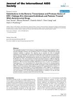
Fig. 1 Superoxide dismutase activity in different larval stages
of Lates calcarifer
nmoles malondialdehyde released/mg
protein
155
Fig. 5 Glutathione (reduced) levels in different larval stages
of Lates calcarifer
40
Vitamin C
30
80
20
10
0
FE
GS
3dph 5dph 10dph 15dph 20dph 25dph
Stage
Fig. 3 Glutathione peroxidase activity in different larval
stages of Lates calcarifer
gradually. From 20 dph to 25 dph the vitamin C
content remained constant. The significant differences are presented in Table 1.
µg Vitamin C/mg protein
units/mg protein
Fish Physiol Biochem (2008) 34:151–158
70
FE - Fertilised eggs
60
GS - 6hrs after spawning
50
40
30
20
10
0
FE
GS
3dph
5dph 10dph 15dph 20dph 25dph
Stage
Fig. 6 Vitamin C levels in different larval stages of Lates
calcarifer
Discussion
Measurable amounts of SOD, catalase (CAT) and
SeGPx were present in the fertilised eggs of Lates
calcarifer, suggesting that the eggs are well protected
against peroxidation, despite high levels of
polyunsaturated fatty acids (PUFAs) present in the
eggs, as observed in Dentex dentex (Mourente et al.
1999). SOD and CAT are responsible for the
inactivation of superoxide and hydrogen peroxide,
respectively. SOD, by converting superoxide to
123
156
hydrogen peroxide, provides substrate for CAT
(Miller et al. 1993). Contradicting results for SOD
activity during early development of fish have been
reported. The activity of SOD decreased throughout
larval development from egg in S. maximus (Peters
and Livingstone 1996) and in Dentex dentex (Mourente et al. 1999), and an increase in activity was
reported in other species (Aceto et al. 1994; Rudneva
1999). In our study the levels of SOD were not
significantly different with larval development.
According to Starrs et al. (2001), the response of
this enzyme to both natural and experimental oxygen
tension is difficult to interpret. In the lung of the
bearded dragon, SOD activity continued to decrease
throughout late development. While no change in
SOD activity occurred between days 14 and 18 in the
embryonic chicken lung, there was a significant
decrease between days 18 and 19.
Catalase activity was high at 3 dph, when seabass
larvae started feeding on rotifers, and this phase is
considered as a highly energy demanding phase in the
larval life-cycle, with a high requirement for oxygen
(Sole et al. 2004). Most of the fish species showed
strong enzymatic changes in anti-oxidant defences
when their larvae changed from endogenous feeding
to exogenous feeding (Mourentex et al. 1999; Peters
and Livingstone 1996; Rudneva 1999). In our study
significant increase was noted in CAT activity from
15 dph to 25 dph. During this period of metamorphosis the Artemia nauplii feed concentration was
increased. The result as of our study was similar to
that of Fernandez-Diaz et al. (2006). The activity
profile of anti-oxidant enzymes during larval development under an Artemia diet demonstrated a stagespecific compensatory mechanism to neutralise peroxides (Dandapat et al. 2003). Fernandez-Diaz et al.
(2006), studied the variation in larval development
and stress defences in Solea senegalensis larvae fed
on live and microencapsulated diets and observed
that CAT, SOD, GPx and lipid peroxidation levels in
the sole larvae showed diet and age dependence in
their responses. Larvae fed with an inert diet showed
similar biomarker activities and were different from
larvae fed with Artemia (P < 0.05).
At gastrulation, GPx level was high, which may be
due to high metabolic activity resulting in increased
H2O2 accumulation. From embryo to hatching, the
level of GPx gradually decreased and reached its
minimum at 5 dph. During this period, activities of
123
Fish Physiol Biochem (2008) 34:151–158
other anti-oxidant enzymes (SOD, CAT) were elevated, and after 5 dph the levels were stable and there
was a gradual increase in GPx up to 25 dph. These
observations indicate a progressive need to remove
H2O2 and lipid peroxides from the cells. This could
be due to increase in LPO from 5 dph to 25 dph. The
increase in GPx is apparently not due to production of
H2O2 from dismutation of OÀ
2 , since larval SOD and
CAT activity declined from the 5th day onwards. The
increase in catalytic activity of CAT and GPx after
the initiation of feeding with Artemia nauplii, corresponding with the increase in its density, may have
been a response to strong metabolic changes, such as
metamorphosis, occurring during this time (Mourente
et al. 1999; Sole et al. 2004). The increase in lipid
peroxidation with larval age with higher GPx activity
has been observed in Artemia fed sole larvae from
hatching to 28 dph (Sole et al. 2004). Our observation in Lates calcarifer is also in agreement with
earlier finding.
Lipid peroxidation was the highest at gastrulation
and gradually declined up to 5 dph. A similar result
was obtained in Dentex larvae (Mourente et al. 1999).
However, LPO increased gradually from 10 dph to
25 dph in seabass larvae. During metamorphosis at
20 dph and 25 dph, an increase in LPO was observed
that may have been due to altered metabolic rate.
Under an Artemia diet, co-evolution of lipid peroxidation and GPx is also expected, since the role of
GPx is to eliminate not only hydrogen peroxide but
also lipid peroxides (Fernandez-Diaz et al. 2006). At
the stage of metamorphosis only, an enhanced level
of LPO occurred. During this period the exogenous
feed is changed, both in quality and quantity, which,
in turn, leads to altered metabolic rate resulting in
oxidation of PUFAs.
Vitamin C level is high at 3 dph of development.
However, the levels of vitamin C on 5, 10 and 15 dph
were not significantly different. From 15 dph vitamin C level distinctly decreased. During early larval
development, vitamin C level counterbalances the
oxidative damage and lipid peroxidation, but, when
the concentration of feed was increased, the scavenging rate of vitamin C decreased because of the
high level of LPO. The decrease in vitamin C
indirectly suggests a high rate of lipid peroxidation.
Vitamin E reacts with lipid peroxy and alkoxy
radicals and donates its labile hydrogen, generating
vitamin E in its radical form. Vitamin E radical is
Fish Physiol Biochem (2008) 34:151–158
reduced back to vitamin E by vitamin C (McCay
1985). Thus, the gradual depletion of vitamin C level
relates to the gradual increase in vitamin E, which, in
turn, counterbalances the lipid radicals. Vitamin E
level could be strictly correlated to a metabolic
acceleration or to high rates of ROS generation, as
demonstrated in animal systems (Chamiec et al.
1996; Jialal et al. 2001). Thus, the increase and
decrease in vitamin C level has an inverse relation
with vitamin E. During marine animal embryogenesis, the activities of most of the anti-oxidant enzymes
examined tend to increase, especially in eggs and
hatching larvae, while the contents of low molecular
weight anti-oxidants decrease (Rudneva 1999). The
eggs of Lates were found to contain anti-oxidant
enzymes, and, during embryogenesis, the levels of
these enzymes tended to increase, while low molecular weight anti-oxidants, such as vitamin C and
GSH, were found to decrease. These results are
similar to those of Rudneva (1999), who suggested
that, in the early stages of embryogenic development
of marine organisms, the low molecular weight antioxidants play an important protective role against
oxygen damage. During embryogenesis, the concentration of low molecular weight scavengers
decreased, with an enhancement of anti-oxidant
enzyme activities, possibly as a result of gene
expression.
GSH is a water-soluble anti-oxidant, found in the
cytosol and mitochondria (Halliwell and Gutteridge
1989). Glutathione, a tripeptidethiol consisting of
glycine, cysteine and glutamic acid moieties, exists in
high concentration virtually in all types of living
cells. It has been demonstrated that glutathione plays
an important role in scavenging free radicals and
active oxygen species (Comporti 1987; McLennan
et al. 1991; Ogino et al. 1989). It occurs mainly by
detoxifying hydrogen peroxide and lipid peroxides
through reactions catalysed by glutathione peroxidase. In our study an inverse relationship with
peroxide decomposition of membrane poly-unsaturated fatty acids and GSH was clearly established,
which is in agreement with the earlier finding of Shen
et al. (1990). At gastrulation, GSH level was at its
minimum, which might be due to an increased
metabolic rate and increased formation of lipid
radicals. When the levels of free radicals are high,
more reduced glutathione (GSH) is converted to its
oxidised form [glutathione disulphide (GSSG)], and,
157
hence, there is a reduction in the level of GSH (Oup
et al. 1996). GSH level also shows an inverse
relationship with LPO decomposition. The levels of
GSH and vitamin C displayed a decreasing trend
from 5 dph to 20 dph of larval development. However, from 20 dph to 25 dph, the level of vitamin C
remained constant, and GSH showed a significant
increase, which may have been due to a small
increase in the activities of CAT and GPx during this
period. Story (1996) suggested that NADPH-dependent glutathione reductase (GR) replenishes the GSH
substrate for GPx and glutathione-S-transferase from
GSSG. This trend was observed during 20 dph to
25 dph, which may be an indication of the activity of
secondary enzymes in Lates calcarifer larvae during
this period.
Our study indicates that oxidant stress is high
during the embryonic and early larval stages, which
may be attributed to metamorphosis and the initiation
of external feeding, possibly linked to altered respiration rates at these stages. These results are in line
with those of the earlier studies confirming that the
initiation of external feeding and metamorphosis are
amongst the critical and most stressful periods for fish
larvae. The results of this study should pave the way
for further research on the effects of the enrichment
of live feed with PUFAs and other anti-oxidants for
the management of stress during larval rearing of
Lates calcarifer.
Acknowledgements The authors are grateful to A.G.
Ponniah, Director, CIBA, for critically going through the
manuscript and for his valuable suggestions. The help rendered
by R. Subburaj, Technical Officer, R. Thiagarajan, Technical
Assistant and V. Anuradha, Senior Research Fellow, is
gratefully acknowledged.
References
Aceto A, Amicarelli F, Sacchetta P, Dragan B, Bucciarelli T,
Masciocco L, Miranda M, Dillio C (1994) Developmental
aspects of detoxifying enzymes in fish (Salmo iridaeus).
Free Radic Res 21:285–294
Arun S, Subramanian P (1998) Antioxidant enzymes in fresh
water prawn Macrobrachium malcolmsonii during
embryonic and larval development. Comp Biochem
Physiol B 121:273–277
Beutler E, Kelley BM (1963) The effect of sodium nitrate on
red cell glutathione. Experientia 19:96–97
Chamiec T, Herbaezynskacedro K, Ceremuzynski L (1996)
Effects of antioxidant vitamin C and E on signal-over
123
158
aged electrocardiogram in acute myocardial infarction.
Am J Cardiol 77:237–241
Comporti M (1987) Glutathione depleting agents and lipid
peroxidation. Chem Phys Lipids 45:143–169
Danchenko OO, Kalytka VV (2002) Formation of antioxidant
defence system of geese in embryogenesis and early
postnatal ontogenesis. Ukr Biokhim Zh 74:120–124
Dandapat J, Chainy GBN, Rao KJ (2003) Lipid peroxidation
and antioxidant defence status during larval development
and metamorphosis of giant prawn, Macrobrachium rosenbergii. Comp Biochem Physiol C 135:221–233
Devasagayam TPA (1986) Lipid peroxidation in rat uterus.
Biochem Biophys Acta 876:507–514
Fernandez-Diaz C, Yufera M, Canavate JP, Moyano PJ,
Alarcon FJ, Diaz M (2001) Growth and physiological
changes during metamorphosis of Senegal Sole reared in
the laboratory. J Fish Biol 58:1086–1097
Fernandez-Diaz C, Kopecka I, Canavate JP, Sarasquete C, Sole
M (2006) Variations on development and stress defences
in Solea senegalensis larvae fed on live and microencapsulated diets. Aquaculture 251:573–584
Fridovich I (2004) Mitochondria: are they the seat of senescence? Aging Cell 3:13–16
Godin DV, Nichols CR, Hoekstra KA, Garnett ME, Cheng KM
(2001) Red cell and plasma antioxidant components and
atherosclerosis in Japanese quail: a time-course study. Res
Commun Mol Pathol Pharmacol 110:27–51
Halliwell B, Gutteridge JMC (1989) Protection against Oxyrad in biological systems: the superoxide theory of oxygen
toxicity. In: Halliwel B, Gutteridge JMC (eds) Free radicals in biology and medicine. Clarendon Press, Oxford
Jialal I, Devaraj S, Kaul N (2001) The effect of alphatocopherol on monocyte proatherogenic activity. J Nutr
131:3893–3943
Lowry OH, Rosebrough NJ, Farr AL, Randall RJ (1951) Protein measurement with Folin phenol reagent. J Biol Chem
193:265–275
Marklund S, Marklund G (1974) Involvement of the superoxide anion radical in the auto oxidation of pyrogallol and a
convenient assay for superoxide dismutase. Eur J Biochem 47:469–474
McCay PB (1985) Vitamin E: interactions with free radicals
and ascorbate. Annu Rev Nutr 5:323–340
McLennan SV, Heffeman S, Wright L, Rae C, Fisher E, Yue
DK, Turtle JR (1991) Changes in hepatic glutathione
metabolism in diabetes. Diabetes 40:340–344
Miller JK, Brzezinska-Slebodzinska E, Madsen FC (1993)
Oxidative stress, antioxidants and animal function. J Dairy
Sci 76:2812–2821
Mourente G, Tocher DR, Diaz E, Grau A, Pastor E (1999)
Relationship between antioxidants, antioxidant enzyme
activities and lipid peroxidation products during early
123
Fish Physiol Biochem (2008) 34:151–158
development in Dentax dentax eggs and larvae. Aquaculture 179:309–324
Ogino T, Kawabata T, Awadi M (1989) Stimulation of glutathione synthesis in iron loaded mice. Biochem Biophys
Acta 1006:131–135
Omaye S, Turnbull JD, Sauberlich HE (1979) Selected methods for the determination of ascorbic acid in animal cell,
tissues and fluids. Methods Enzymol 62:1–11
Oup P, Nourooz-Zadeh J, Tritschler HJ, Wolff S (1996)
Activation of aldose reductase in rat lens and metal–ion
chelation by aldose reductase inhibitors and lipoic acid.
Free Radic Res 25:337–346
Peters LD, Livingstone DR (1996) Antioxidant enzyme activities in embryologic and early larval stages of turbot. J
Fish Biol 49:986–997
Peters LD, Porte C, Livingstone DR (2001) Variation of antioxidant enzyme activities in sprat (Sprattus sprattus)
larvae in mixed zooplankton from the southern North Sea.
Mar Pollut Bull 42:1087–1095
Rotruck JT, Poe AL, Ganther HE, Swanson AB, Hafeman DG,
Hoekstra NG (1973) Selenium: biochemical role as a
component of glutathione peroxidase purification and
assay. Science 179:588–590
Rudneva II (1999) Antioxidant system of Black Sea animals in
early development. Comp Biochem Physiol C 122:265–
271
Shen X, Aw TY, Jones DP (1990) Glutathione dependent
protection against oxidative injury. Pharmacol Ther
47:61–71
Sinha SK (1972) Colorimetric assay of catalase. Ann Biochem
47:389–395
Sole M, Potrykus J, Fernandez-Diaz C, Blasco J (2004) Variations on stress defences and metallothionein levels in the
Senegal sole, Solea senegalenisis, during early larval
stages. Fish Physiol Biochem 30:57–66
Starrs AP, Orgeig S, Daniels CB, Davies M, Lopatko OV
(2001) Antioxidant enzymes in the developing lungs of
egg-laying and metamorphosing vertebrates. J Exp Biol
204:3973–3981
Story KB (1996) Oxidative stress: animal adaptations in nature.
Braz J Med Biol Res 29:1715–1733
Wilhelm Filho D (1996) Fish antioxidant defenses—a comparative approach. Braz J Med Biol Res 29:1735–1742
Yufera M, Parra G, Santiago R, Carrascosa M (1999) Growth,
carbon, nitrogen and caloric content of Solea snegalensis
(Pisces: Soleidae) from egg fertilization to metamorphosis. Mar Biol 134:43–49
Zielinski S, Portner HO (2000) Oxidative stress and antioxidative defense in cephalopods: a function of metabolic
rate or age? Comp Biochem Physiol B Biochem Mol Biol
125:147–160