1979 quantitative comparison of low and high output neuromuscular synapses from a motoneuron of the lobster homarus americanus
Bạn đang xem bản rút gọn của tài liệu. Xem và tải ngay bản đầy đủ của tài liệu tại đây (1.18 MB, 9 trang )
Cell Tissue Res. 198, 455-463 (1979)
Cell and Tissue
Research
9 by Springer-Verlag 1979
Quantitative Comparison
of Low- and High-output Neuromuscular Synapses
from a Motoneuron of the Lobster (Homarusamericanus)*
C.K. Govind and D.E. Meiss
Scarborough College, University of Toronto, West Hill, Ontario, Canada
Summary. Representative examples of low- and high-output neuromuscular
synapses between motoneuron and distal accessory flexor muscle of the lobster
were selected on the basis of their mean quantal content, and subsequently
analysed by serial section electron microscopy. The high-output terminal has
twice as m a n y synapses as the low-output terminal. However, since the mean
surface area of synapses is significantly smaller in the high-output terminal than
in the low-output one, the total synaptic surface area between the two types of
terminals is similar. Also, though the high-output terminal possesses a greater
number of presynaptic dense bodies than its low-output counterpart, the mean
number per synapse is similar for the two terminals. The terminals, however,
differ significantly in the size of their dense bodies. Thus both the mean and total
surface area of these bodies is greater in the high-output terminal than in the
low-output one. Moreover, the mean ratio of dense body area to synaptic area is
significantly greater for the high-output terminal than for its low-output
counterpart. This difference in dense body area parallels the difference in
quantal content of synaptic transmission between the low- and high-output
terminals and supports the hypothesis that presynaptic densities represent the
ultrastructural correlates of transmitter mobilization and/or release.
Key words: Neuromuscular synapses - Presynaptic density - Ultrastructure Serial sections - Crustaceans.
Crustacean neuromuscular synapses are often regarded as model systems for
synapses in the CNS of vertebrates because of the multiple converging inputs from a
single m o t o r axon and the interaction of both excitatory and inhibitory axons
Govind, Scarborough College, Universityof Toronto, West Hill, Ontario
M1C 1A4, Canada
* Supported by grants from the National Research Council and Muscular Dystrophy Association of
Canada to C.K. Govind. D.E. Meissis a post-doctoral fellowof the Muscular Dystrophy Association of
Canada. We thank Eva Yap-Chung for her expert and unfailing technical assistance
Send offprint requests to." C.K.
0302-766X/79/0198/0455/$01.80
456
C.K. Govind and D.E. Meiss
(Katz, 1966). The analogy may be extended further as the multiple terminals arising
from a single axon vary in their properties of transmitter release. Thus single
crustacean excitatory motoneurons such as those to the opener muscle in crayfish
(Bittner, 1968), the stretcher muscle in crabs (Sherman and Atwood, 1972; Govind
et al., 1973) and the accessory flexor muscle in lobsters (DeRosa and Govind, 1978;
Meiss and Govind, 1979a, b) give rise to a wide spectrum of synapses ranging from
low-quantal release to high-quantal release types. This diversity in physiological
properties of synapses from a single axon must have an underlying ultrastructural
basis.
Likely factors in this respect are the size and number of synaptic contacts which
have been shown to differ in terminals of the single axon to the stretcher muscle of
Hyas (Sherman and Atwood, 1972) where high-quantal release terminals have more
numerous and larger synapses than their low-quantal release counterparts. More
recently, however, a better correlation has been found between presynaptic dense
bodies and transmitter output with the use of serial section electron microscopy
which has permitted a quantitative analysis of the ultrastructural parameters. Thus
terminals that release large amounts of transmitter such as those formed by the fast
axon of Pachygrapsus (Atwood and Jahromi, 1978) invariably have presynaptic
dense bodies in all of their individual synapses whereas dense bodies are present in
only half of the synapses in a crab pyloric muscle (Atwood et al., 1978) which release
a relatively smaller amount of transmitter. Moreover, comparison of terminals
from two fibers of the proximal accessory flexor muscle of the lobster reveal
significant differences in the number and size of presynaptic dense bodies which
correlate with the different amplitudes of the excitatory postsynaptic potentials
(EPSPs) of these fibers (Govind and Chiang, 1979). A more rigorous test of the
relationship between presynaptic densities and transmitter output would be to
section serially terminals for which the quantal output of synaptic transmission had
been determined. The present paper reports on such a study in the distal accessory
flexor muscle of the lobster by comparing the quantitative ultrastructure of
physiologically identified low- and high-output terminals.
Materials and Methods
Adult lobsters (Homarusamericanus)weighingapproximately 500g each, were purchased locally and
held in Instant Ocean at 10-12~C. The first pair of walking legs was used. The posterior surface of the
distal accessoryflexormuscle (DAFM) was exposed in the meropodite and held in cool (10~C) lobster
saline (Meiss and Govind, 1979b). The excitor axon to the DAFM was stimulated with short duration
( < 0.1 msec)rectangular pulses via platinum wireelectrodesplaced on the main leg nerve. The resulting
excitatory post-synaptic potentials (EPSPs) were monitored intracellularly with 10-15Mr] microelectrodes filled with a saturated solution of the dye methylblue in 1 M KAc.At the end of the experiment the
dye was iontophoresedinto the fiber (Thomas and Wilson, 1966)for identification during fixation for
electron microscopy. Simultaneous with EPSP recordings, the synaptic currents were monitored
extracellularly at discrete fociwith 2-4 Mf~microelectrodesfilled with 2 M NaC1solution. Conventional
procedures were followedfor amplifying, displaying and photographing the electrical signals.
After the synapses were characterized physiologicallythe muscle was fixed at room temperature
(20-22~C) for electron microscopy.The entire muscle was immersed for 1.5 h in a 0.1 M Sorenson's
phosphate buffer solution containing3~ glutaraldehyde; 1% formaldehyde;3% NaC1;4% sucroseand
0.01 M CoC12.Next the musclewas washedfor 0.5 h in 0.1 M Sorenson'sbuffersolutioncontaining8%
Lobster Neuromuscular Synapses
457
sucrose (pH 7.4). During this period the dye-marked fiber was isolated and subsequentlypost-fixedfor
1h in 2% osmium tetroxide. Following a brief (5-10rain) wash in the buffer solution, the fiber was
embedded in an Epon-araldite mixture. Thin serialsectionsweremounted on Formvar coated singleslot
grids. They were stained with uranyl acetate and lead citrate and examinedwith a Zeiss9S or a Siemens
102 electron microscope. A record was kept of the thickness of each section. Measurements of the
number and surface area of synapseswere made from serial micrographs with a final magnification of
26,400. The surface area of dense bodies was measured from micrographs magnifiedto x 132,000 and
• 198,000. A detailed description of the methods for obtaining the surface area of synapses and dense
bodies cut in serial section appears elsewhere (Govind and Chiang, 1979).
Results
Selection of Low- and High-Output Synapses
The single excitatory axon to the D A F M gives rise to physiologically diverse
synapses which are regionally distributed such that those on proximal muscle fibers
release comparatively little transmitter (low-output types) whereas those on distal
muscle fibers release greater amounts (high-output types) (Meiss and Govind,
1979b). For the present study the quantal content of synaptic transmission was
obtained for terminals on a proximal and a distal muscle fiber. On each muscle
fiber, a focal synaptic site was located with an extracellular electrode, and 200--300
consecutive ERSPs were recorded at 1 Hz stimulation in trains of 20 impulses with
inter-train intervals of 2 min to minimize possible synaptic depression (Fig. 1). The
mean quantal content (m) calculated by means of the failure method (Del Castillo
and Katz, 1954; Dudel and Kuffler, 1961) was 0.27 and 5.29 at each of the synaptic
sites on the proximal and distal fibers respectively. These representative examples of
low- and high-output, synapses were subsequently fixed for electron microscopy
and serially sectioned.
Quantitative Comparison of Low- and High-Output Synapses
The fine structure of neuromuscular terminals and synapses on the proximal and
distal fibers o f the D A F M is shown in Figs. 2 4 . The terminal regions are in contact
with granular sarcoplasm of the muscle (Fig. 2), and this criterion is used to
distinguish them from axonal regions (Atwood and Morin, 1970). Within the
terminals, synapses were identified according to conventional criteria of densely
stained pre- and postsynaptic membranes, presynaptic vesicles, and dense bodies
(Gray, 1963). Analysis of single sections reveals no qualitative differences between
the low- (Figs. 2, 3) and high-output (Fig. 4) neuromuscular terminals. In fact, they
resemble other lobster (Govind and Chiang, 1979) and crustacean (Hoyle and
McNeil, 1968; Atwood et al., 1977, 1978) neuromuscular terminals. It is only when
these terminals are serially sectioned and quantitatively analysed that differences
between them are seen (Table 1).
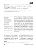
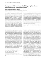
The low- and high-output terminals serially sectioned for a comparable length
(14.17 lam and 15.36 ~tm respectively), show that over this length the latter terminal
has twice as m a n y synapses as the former (37 versus 18; Fig. 5). This is also evident
from the fact that the high-output terminal has an average of 2.40 synapses for each
458
C.K. Govind and D.E. Meiss
Fig. 1. Electrical recordingsfrom high-output neuromuscularterminal of distal accessoryflexormuscle
of lobster at a fast time sweepA showinga singleresponse and at a slowtime sweepB showingmultiple
responses at 1 Hz stimulation. Upper trace, extracellularly recorded synaptic potentials (ERSPs) for
calculating quantal content; lower trace, intracellulady recorded excitatory post-synaptic potentials
(EPSPs) for monitoring nerve activity. Calibration: Vertical, upper trace 400 gV; lower trace 10mV:
Horizontal, (A)20ms; (B) 4s
micrometer of muscle fiber compared to 1.26 synapses for the low-output
counterpart. Despite the the two-fold difference in the number of synapses, their
total surface area is similar in the two types of terminals, the reason being that the
mean size of individual synapses is significantly smaller (about one half) in the highoutput terminal than in the low-output one. Consequently, the total synaptic area is
similar in the two types of terminals and cannot account for the differences in
quantal output between them.
However, comparison of the presynaptic dense bodies between low- and highoutput terminals reveals differences that correlate with the differences in quantal
output between the two terminals. There are fewer dense bodies in the low-output
terminal compared to the high-output one (9 versus 22; Fig. 5). However, since
synaptic number follows a similar unequal ratio between the two types of terminals,
the mean number of dense bodies per synapse is not significantly different. The
distribution of the dense bodies is also similar in both types of terminals (Fig. 5),
with about a third of the synapses having one dense body and more than half not
possessing a dense body. The only difference in dense body distribution is that the
high-output terminal has more synapses with two or more dense bodies than its
low-output counterpart (Fig. 5).
Fig. 2. Low-outputterminal (T) with single long synapse (between long arrows) associated dense body
(D) and synapticvesicles(V). Terminalin contactwith granular sarcoplasm(G) ofmuscleand connective
tissue (C). Scale mark: 0.25~tm. Magnification: x 26,400
Fig. 3. High power micrograph of low-output synapse (between long arrows) showingdarkly stained
pre- and post-synaptic membranes, synaptic vesicles (V) and bar-type presynaptic density (D). T
terminal; G granular sarcoplasm. Scale mark: 0.25gin. Magnification: x 198,000
Fig. 4. High-outputterminal (T) showingsynapses(between long arrows) with presynapticdensities (D)
with lateral (bar-type) and cross-sectionalconfigurations. V synaptic vesicles; G granular sarcoplasm.
Scale mark: 0.25~un. Magnification: x 112,000
Lobster Neuromuscular Synapses
459
Table 1. Quantitative comparison of synapses and presynaptic dense bodies between low- and highoutput neuromuscular terminals produced by the single motor axon to the distal accessory flexor muscle
of the lobster
Quantal content of synaptic transmission (m)
Length of muscle fiber serially sectioned (l~m)
Total number of synapses
Number of synapses per lam of fiber length
Total surface area of synapses (sq. lam)
Mean surface area of synapses (sq. ~tm)
(.~+_ S.E.M.)
Total number of dense bodies
Mean number of dense bodies per synapse
(.~+ S.E.M.)
% of synapses without dense bodies
% of synapses with one dense body
% of synapses with two or more dense bodies
Total surface area of dense bodies (sq. pm)
Total dense body area as per cent of total
synaptic area
Mean surface area of dense bodies (sq. ~tm)
(X+ S.E.M.)
Mean ratio of dense body area to synaptic area
(for all synapses) (X+ S.E.M.)
Mean ratio of dense body area to synaptic area
(for synapses with dense bodies only)
(.~+ S.E.M.)
Low-output
Terminal
High-output
Terminal
0.27
14.17
18
1.26
14.20
0.79+ 0.12
5.29
15.36
37
2.40
14.82
0.42+ 0.07
1%
9
0.50+ 0.19
22
0.59+ 0.14
NS
61
33
6
0.20
1.4
P
(Students'
t-test)
57
32
11
1.62
10.9
0.022+ 0.004
0.074___0.012
2%
0.021 _ 0.009
0.120+ 0.031
5%
0.054+ 0.018
0.277+ 0.048
1%
A
6
~O
i
Z
>03
e.,,,
L4_I
t,n
Z
i
i
i
t
!
i
i
1:2
14
16
108.
6'
42
o
o
o
0.2
o
o
0.4
06
08
lo
i
1:8
20
2'2
24
26
SYNAPTIC AREA (um 2)
Fig. 5. Histogram to show size distribution of 18 low-output A and 37 high-output B serially sectioned
synapses from distal accessory flexor muscle of lobster. Occurrence of presynaptic dense bodies shown
by closed circles within each synapse
Lobster NeuromuscularSynapses
461
The disparity between the two terminals is even more striking when the size of
the dense bodies is considered. Thus the mean surface area of individual dense
bodies is significantly larger in the high-output terminal. This fact, combined with
their greater number in this terminal, results in an eight-fold increase in total dense
body area in the high-output terminal compared to the low-output one. This
comparison involves absolute values which are determined by the length of the
terminals sampled. In order to compensate for differences in length sampled for the
low and high-output terminals, the dense body area is considered as a function of
the synaptic area in two respects. Firstly, when the total dense body area is viewed
as a percentage of the total synaptic area, there is an eight-fold increase in favour of
the high-output terminal. Secondly, when the mean ratio of dense body area to
synaptic area is taken, a statistically significant increase in this ratio is seen for the
high-output terminal. This ratio is determined both for all synapses and for
synapses with dense bodies only. Clearly, the high-output terminal allocates a
greater proportion of its synaptic area to dense bodies than the low-output
terminal.
Discussion
The physiological diversity of synapses arising from a single motoneuron is well
established in crustacean muscle (reviewed by Atwood, 1976). The ultrastructural
correlates of this diversity have been studied only recently. Thus highly facilitating
terminals from the lone excitor axon to the spider crab stretcher muscle have a
higher density of synaptic vesicles than poorly facilitating terminals in randomly
sectioned tissue (Sherman and Atwood, 1972). A quantitative study based on serial
section electron microscopy reveals that the average size (surface area) of excitatory
synapses is significantly smaller than that of inhibitory ones in the crayfish opener
muscle (Jahromi and Atwood, 1974). As the inhibitory terminals release relatively
greater amounts of transmitter than the excitatory ones at low frequencies of
stimulation, a relationship between transmitter output and synaptic size was
suggested. However, subsequent studies have not confirmed this relationship, e.g.,
the quantal content is similar in synapses of the gastric mill muscles GM 8 b, GM 9
(Atwood et al., 1977) and pyloric muscle P1 (Atwood et al., 1978) of the blue crab
though the former has considerably smaller synapses than the latter. Furthermore,
synapses of the fast axon in Pachygrapsus (Atwood and Jahromi, 1978) are
considerably smaller in size than GM8b, GM9, and P1 synapses in blue crabs even
though their quantal output is greater. A better correlation is found between
transmitter release and presynaptic dense bodies. Consequently, whereas dense
bodies were found in all synapses of the fast axon of Pachygrapsus they occurred in
only 75~ of the GM8b, GM9 and 50~ of the PI synapses of blue crabs. These
comparisons, however, are between separate species and not as persuasive as
comparisons within a single species.
When low- and high-output synapses are compared in the proximal accessory
flexor muscle (PAFM) of the lobster (Govind and Chiang, 1979), dense bodies
occur in only 40~ of the low-output synapses but in 60~ of the high-output ones.
Furthermore, the mean ratio of dense body area to synaptic area is significantly
462
C.K. Govindand D.E. Meiss
greater in the high-output terminals than in the low-output terminal. This parallels
a similar difference in the size of the intracellularly recorded EPSP between the two
muscle fibers. As both fibers have a similar membrane input resistance, the
difference in EPSP size is attributable to differences in quantal output of
transmitter. In the present study on the lobster DAFM a refinement was added in
that the quantal content was determined at synaptic foci and these very same
regions were serially sectioned. As in the PAFM, a significant increase was found in
the ratio of dense body to synaptic surface area in the high-output terminal as
compared to its low-output counterpart. Consequently, the present study reiterates
the correlation between transmitter output and amount ofpresynaptic dense bodies
at neuromuscular terminals of the lobster.
The accessory flexor muscle of crustaceans is bipartite consisting of a proximal
(PAFM) and a distal (DAFM) head situated at either end of the meropodite
segment and connected to each other by a long slender tendon (Cohen, 1963;
Govind et al., 1978). Since both heads are innervated by a common excitatory axon
it is interesting to compare how the differentiation of presynaptic dense bodies
characteristic of low- and high-output terminals is achieved in each muscle. In the
PAFM where the mean size of the dense bodies is similar in low- and high-output
terminals (0.014 vs. 0.02 sq. lam respectively), the mean number of densities per
synapse is significantly higher in the high-output terminal than in its low-output
counterpart (0.73 vs. 0.42 respectively; Govind and Chiang, 1979). On the other
hand, in the DAFM, where the mean size of the dense bodies is different in low- and
high-output terminals (0.022 vs. 0.074 respectively), their mean frequency is similar
in low- and high-output synapses (0.50 vs. 0.59 respectively; Table 1). Consequently
differences in surface area of dense bodies between the two types of terminals are
due to differences in their numbers in the PAFM and in their size in the DAFM. If
indeed these findings are substantiated, they would suggest that two different
mechanisms may occur in a single axon to achieve differentiation of presynaptic
dense bodies.
Presynaptic dense bodies seem ubiquitous in synapses of both vertebrates
(Pfenninger, 1973) and invertebrates (Wood et al., 1977). Though they vary
considerably in form, they are associated with the presynaptic membrane and are
surrounded by synaptic vesicles. Therefore, they have been assigned some role in
the transmitter release mechanism. They could act as focal points to which synaptic
vesicles are attracted and held until their contents are released into the synaptic cleft
(Wernig and Stirner, 1977), or as local storage sites (Finlayson and Osborne, 1975).
Their occurrence in the lobster DAFM tends to corroborate the view that they are
involved in the release of transmitter, especially since differences in the relative
surface area of these bodies correspond to differences in quantal output. This is still
circumstantial evidence for their possible role in transmitter release, and further
studies are required in which these bodies are "altered" in response to experimental
manipulation of quantal transmitter output. Such studies have revealed changes in
the frequency of synaptic vesicles (e.g., Ceccarelli et al., 1973) but not in the
presynaptic densities which do not appear to be as transient as vesicles and
consequently may change only over long periods of time such as during growth.
Lobster Neuromuscular Synapses
463
References
Atwood, H.L.: Organization and synaptic physiology of crustacean neuromuscular systems. Prog.
Neurobiol. 7, 291-391 (1976)
Atwood, H.L., Jahromi, S.S.: Fast axon synapses of a crab leg muscle. J. Neurobiol. 9, 1-15 (1978)
Atwood, H.L., Morin, W.A.: Neuromuscular and axo-axonal synapses of the crayfish opener muscle. J.
Ultrastruct. Res. 32, 351-369 (1970)
Atwood, H.L., Govind, C.K., Jahromi, S.S.: Excitatory synapses of blue crab gastric mill muscles. Cell
Tissue Res. 177, 145-158 (1977)
Atwood, H.L., Govind, C.K., Kwan, I.: Non-homogeneous excitatory synapses of a crab stomach
muscle. J. Neurobiol. 9, 17-28 (1978)
Bittner, G.D.: Differentiation of nerve terminals in the crayfish opener muscle and its functional
significance. J. Gen. Physiol. 51, 731-758 (1968)
Ceccarelli, B., Hurlbut, W.P., Mauro, A.: Turnover of transmitter and synaptic vesicles at the frog
neuromuscular junction. J. Cell Biol. 57, 499-524 (1973)
Cohen, M.J.: The crustacean myochordotonal organ as a proprioceptive system. Comp. Biochem.
Physiol. 64, 41-54 (1963)
Del Castillo, J., Katz, B.: Quantal components of the endplate potential. J. Physiol. (Lond.) 124, 560573 (1954)
DeRosa, R.A., Govind, C.K.: Transmitter output increases in an identifiable lobster motoneuron with
growth of its muscle fibers. Nature (Lond.) 273, 67~678 (1978)
Dudel, J., Kuffler, S.W.: The quantal nature of transmission and spontaneous miniature potentials at
the crayfish neuromuscular junction. J. Physiol. (Lond.) 155, 514-529 (1961)
Finlayson, L.H., Osborne, M.P.: Secretory activity of neurons and related electrical activity. Adv.
Comp. Physiol. Biochem. 6, 165-258 (1975)
Govind, C.K., Chiang, R.G.: Correlation between presynaptic dense bodies and transmitter output at
lobster neuromuscular synapses by serial section electron microscopy. Brain Res. 161, 377-388
(1979)
Govind, C.K., Atwood, H.L., Lang, F.: Synaptic differentiation in a regenerating crab-limb muscle.
Proc. Natl. Acad. Sci. U.S.A. 70, 822-826 (1973)
Govind, C.K., Meiss, D.E., She, J., Yap-Chung, E.: Fiber composition of the distal accessory flexor
muscle in several decapod crustaceans. J. Morphol. 157, 151-160 (1978)
Gray, E.G.: Electron microscopy of presynaptic organelles of the spinal cord. J. Anat. 97, 101-106
(1963)
Hoyle, G., McNeil, P.A.: Correlated physiological and ultrastructural studies on specialized muscles. Ic.
Neuromuscular junctions in the eyestalk levator muscles of Podophthalmus vigil (Weber). J. Exp.
Zool. 167, 523-550 (1968)
Jahromi, S.S., Atwood, H.L.: Three-dimensional ultrastructure of the crayfish neuromuscular
apparatus. J. Cell Biol. 63, 599~13 (1974)
Katz, B.: Nerve, Muscle and Synapse. New York: McGraw Hill Book Co. (1966)
Meiss, D.E., Govind, C.K.: Multiterminal innervation: non-uniform density along single lobster muscle
fibers. Brain Res. 160, 163-169 (1979a)
Meiss, D.E., Govind, C.K.: Regional differentiation of neuromuscular synapses in a lobster receptor
muscle. J. Exp. Biol. In Press (1979b)
Pfenninger, K.H.: Synaptic morphology and cytoehemistry. Progr. Histochem. Cytochem. 5, 1-86
(1973)
Sherman, R.G., Atwood, H.L.: Correlated electrophysiological and ultrastructural studies of a
crustacean motor unit. J. Gen. Physiol. 59, 586-615 (1972)
Thomas, R.C., Wilson, V.J.: Marking single neurons by staining with intraceUular recording
microelectrodes. Science (Wash.) 151, 1538-1539 (1966)
Wernig, A., Stiruer, H.: Quantum amplitude distributions point to functional unity of the synaptic
'active zone'. Nature (Lond.) 269, 820-822 (1977)
Wood, M.R., Pfenninger, K.H., Cohen, M.J.: Two types of presynaptic configurations in insect central
synapses: an ultrastructural analysis. Brain Res. 130, 22-45 (1977)
Accepted February 13, 1979