Pocket atlas of radiographic anatomy 2d ed torsten b moller, emil reif
Bạn đang xem bản rút gọn của tài liệu. Xem và tải ngay bản đầy đủ của tài liệu tại đây (23.13 MB, 385 trang )
I
Pocket Atlas of
Radiographic Anatomy
Second edition, revised and enlarged
Torsten B. Moeller, M. D.
Emil Reif, M. D.
Am Caritas-Krankenhaus
Dillingen/Saar
Germany
Am Caritas-Krankenhaus
Dillingen/Saar
Germany
243 Illustrations
Thieme
Stuttgart · New York 2000
Moeller, Pocket Atlas of Radiographic Anatomy © 2000 Thieme
All rights reserved. Usage subject to terms and conditions of license.
II
Library of Congress Cataloging-in-Publication Data
Moeller, Torsten B.
[Taschenatlas der Roentgenanatomie. English]
Pocket atlas of radiographic anatomy / Torsten B. Moeller, Emil Reif.—2nd ed., rev. and
enlarged.
p. cm.
Translation of the 1998 German ed.
Includes bibliographical references and index.
ISBN 3-13-784202-6—ISBN 0-86577-874-4
1. Human anatomy—Atlases. 2. Radiography, Medical—Atlases. I. Title. II. Reif, Emil.
[DNLM: 1. Anatomy—Atlases. 2. Anatomy—Handbooks. 3. Radiography—Atlases.
4. Radiography—Handbooks. QS 17 M726t 2000 a]
QM25.M55613 2000
611’.0022’221—dc21
99-040802
Translated by John Grossman, Berlin, Germany
This book is an authorized revised and enlarged translation of the 2nd German edition published and copyrighted 1998 by
Georg Thieme Verlag, Stuttgart, Germany.
Title of the German edition: Taschenatlas
der Röntgenanatomie
Some of the product names, patents, and
registered designs referred to in this book
are in fact registered trademarks or proprietary names, even though specific reference to this fact is not always made in the
text. Therefore, the appearance of a name
without designation as proprietary is not to
be construed as a representation by the
publisher that it is in the public domain.
This book, including all parts thereof, is legally protected by copyright. Any use, exploitation, or commercialization outside the
narrow limits set by copyright legislation,
without the publisher’s consent, is illegal
and liable to prosecution. This applies in
particular to photostat reproduction, copying, mimeographing or duplication of any
kind, translating, preparation of microfilms,
and electronic data processing and storage.
© 2000 Georg Thieme Verlag,
Rüdigerstrasse 14,
D-70469 Stuttgart, Germany
Thieme New York, 333 Seventh Avenue,
New York, NY 10001, USA
Typesetting by primustype R. Hurler
GmbH, D-73274 Notzingen, Germany
typeset on Textline/HerculesPro
Printed in Germany by Offizin Andersen
Nexö, Leipzig
ISBN 3-13-784202-6 (GTV)
ISBN 0-86577-874-4 (TNY) 1 2 3 4 5 6
Important Note: Medicine is an everchanging science undergoing continual
development. Research and clinical experience are continually expanding our
knowledge, in particular our knowledge
of proper treatment and drug therapy.
Insofar as this book mentions any
dosage or application, readers may rest
assured that the authors, editors, and
publishers have made every effort to
ensure that such references are in accordance with the state of knowledge
at the time of production of the
book.
Nevertheless, this does not involve,
imply, or express any guarantee or responsibility on the part of the publishers in respect to any dosage instructions
and forms of application stated in the
book. Every user is requested to examine carefully the manufacturer’s
leaflets accompanying each drug and to
check, if necessary in consultation with
a physician or specialist, whether the
dosage schedules mentioned therein or
the contraindications stated by the
manufacturers differ from the statements made in the present book. Such
examination is particularly important
with drugs that are either rarely used or
have been newly released on the
market. Every dosage schedule or every
form of application used is entirely at
the user’s own risk and responsibility.
The authors and publishers request
every user to report to the publishers
any discrepancies or inaccuracies noticed.
Moeller, Pocket Atlas of Radiographic Anatomy © 2000 Thieme
All rights reserved. Usage subject to terms and conditions of license.
III
To Bernie and Arlene,
Bryan and Nancy Riegner,
Southfield, Michigan
Moeller, Pocket Atlas of Radiographic Anatomy © 2000 Thieme
All rights reserved. Usage subject to terms and conditions of license.
IV
Preface to the Second Edition
Despite the introduction of digital radiography, the process of obtaining
and evaluating radiographic images has not changed significantly in recent years, nor has the importance of a good knowledge of the anatomic
details of conventional radiographs. This book is a case in point. We were
very pleased with the wide distribution of the first edition, which has been
an additional incentive to improve a number of details, include even better
images, replace some of the drawings, and include a few new radiographs.
The structure of the second edition also strictly follows that of Normal
Findings in Radiology and Pocket Atlas of Radiographic Positioning. Our intention in this is to present clearly and concisely each respective chapter of
conventional diagnostic radiography and to avoid unnecessary repetition.
At the same time, we give the reader the opportunity to examine different
aspects of the same material with the same degree of clarity, which may
prove important on another occasion. We have employed a consistent
style and the same radiographs to structure the material and make it more
readily accessible for reading, learning, or reference purposes.
Dillingen, Summer 1999
Torsten B. Möller
Emil Reif
Moeller, Pocket Atlas of Radiographic Anatomy © 2000 Thieme
All rights reserved. Usage subject to terms and conditions of license.
Table of Contents
Skeletal Imaging 1
Skull 2
Posterior-Anterior View of the
Skull 3
Lateral View of the Skull 5
Towne View of the Posterior
Skull 7
Waters View of the Nasal Sinuses 9
Occipitofrontal View of the Nasal
Sinuses 11
Posterior-Anterior View of the
Orbit 13
Rhese View of the Orbit 15
Maxilla 17
Ramus of the Mandible 19
Clementschitsch View of the
Mandible 21
Pantomogram of the Jaw and
Facial Bones 23
Skull: Lateral View of the
Nose 24
Zygomatic Arch 25
Skull 26
Base of the Skull 27
Altschul View of the Petrous
Ridge 29
Schüller View of the Petrous
Bone 31
Stenvers View of the Petrous
Bone 33
Mayer View of the Petrous
Bone 35
Lateral View of the Sella Turcica 37
Anterior-Posterior View of the
Sella Turcica 39
Spine 40
Anterior-Posterior Full-length
View of the Spine 41
V
Anterior-Posterior View of the
Cervical Spine 43
Lateral View of the Cervical
Spine 45
Oblique View of the Cervical
Spine 47
Anterior-Posterior View of the
Thoracic Spine 49
Lateral View of the Thoracic
Spine 51
Anterior-Posterior View of the
Lumbar Spine 53
Lateral View of the Lumbar
Spine 55
Oblique View of the Lumbar
Spine 57
Anterior-Posterior View of the
Pelvis 59
Anterior-Posterior View of the
Pelvis in a Child 61
Inlet View of the Pelvis 63
Guthmann’s View of the Pelvis
65
Iliac Wing View of the Pelvis 67
Obturator View of the Pelvis 69
Thirty-degree Anterior Oblique
View of the Sacroiliac Joint 71
Anterior-Posterior View of the
Sacrum 73
Lateral View of the Sacrum 75
Anterior-Posterior View of the
Coccyx 77
Lateral View of the Coccyx 79
Upper Extremities 80
Osseous Hemithorax 81
Anterior-Posterior View of the
Sternum 83
Lateral View of the Sternum 85
Stress View of Both Shoulders 87
Anterior-Posterior View of the
Clavicle 89
Oblique View of the Clavicle 91
Acromioclavicular Joint 93
Moeller, Pocket Atlas of Radiographic Anatomy © 2000 Thieme
All rights reserved. Usage subject to terms and conditions of license.
VI
Table of Contents
Anterior-Posterior View of the
Scapula 95
Lateral View of the Scapula 97
Anterior-Posterior View of the
Shoulder 99
Anterior-Posterior Abduction
View of the Shoulder 101
Axial View of the Shoulder 103
Tangential View of the Shoulder
105
Transthoracic View of the Shoulder 107
Anterior-Posterior View of the
Upper Arm 109
Lateral View of the Upper
Arm 111
Anterior-Posterior View of the
Elbow 113
Lateral View of the Elbow 115
Axial View of the Elbow 117
Oblique View of the Radius 119
Anterior-Posterior View of the
Forearm 121
Lateral View of the Forearm 123
Anterior-Posterior View of the
Hand 125
Oblique View of the Hand 127
Anterior-Posterior View of the
Wrist 129
Lateral View of the Wrist 131
Carpal Tunnel 133
Scaphoid Views 135
Special View of the Pisiform 137
Finger in Two Planes 139
Lower Extremities 140
Full-length Anterior-Posterior
Weight-bearing View of the
Leg 141
Anterior-Posterior View of the
Hip 143
Lauenstein View of the Hip 145
Tangential View of the Femoral
Head (Schneider I) 147
Tangential View of the Femoral
Head (Schneider II) 149
Axial View of the Hip 151
Anterior-Posterior View of the
Thigh 153
Lateral View of the Thigh 155
Anterior-Posterior View of the
Knee 157
Lateral View of the Knee 159
Frik Tunnel View of the
Knee 161
Thirty, Sixty, and Ninety-degree
Views of the Patella 163
Anterior-Posterior View of the
Lower Leg 165
Lateral View of the Lower Leg
167
Anterior-Posterior View of the
Ankle 169
Lateral View of the Ankle 171
Anterior-Posterior View of the
Foot 173
Lateral View of the Foot 175
Lateral View of the Calcaneus
177
Tangential View of the Calcaneus
179
Anterior-Posterior View of the
Metatarsals 181
Oblique View of the Metatarsals
183
Anterior-Posterior View of the
Forefoot 185
Oblique View of the Forefoot 187
Anterior-Posterior and Lateral
Views of the Great Toe 189
Anatomy of Miscellaneous
Plain Films 191
Routine Radiographs 192
Posterior-Anterior Chest Radiograph 193
Moeller, Pocket Atlas of Radiographic Anatomy © 2000 Thieme
All rights reserved. Usage subject to terms and conditions of license.
Table of Contents
Posterior-Anterior Chest Radiograph (Heart and Major Vessels) 195
Lateral Chest 197
Segments of the lung 199
Anterior Oblique View of the
Right Chest 201
Anterior Oblique View of the
Left Chest 203
Abdomen with the Patient Standing 205
Abdomen with the Patient Supine
207
Spot Images 208
Craniocaudal Mammogram 209
Mediolateral Mammogram 211
Anterior-Posterior Spot View of
the Trachea 213
Lateral Spot View of the Trachea
215
Tomography 216
Anterior-Posterior Tomogram of
the Pulmonary Hilum 217
Lateral Tomogram of the Right
Hilum of the Lung 219
Tomogram of the Sacroiliac
Joint 221
Contrast Studies 223
Gastrointestinal System 224
Anterior-Posterior View of the
Hypopharynx 225
Lateral View of the Hypopharynx
227
Esophagus 229
Esophagus and Stomach 231
Stomach 233
Stomach and Duodenum 235
Stomach and Small Bowel 237
Small Bowel 239
Spot Film of the Ileocecal Region 241
VII
Colon 243
Rectum 245
Defecography 247
Intravenous Contrast Studies
248
Intravenous Urogram 249
Intravenous Cholecystocholangiogram 251
Arthrography 252
Posterior-Anterior Wrist Arthrogram 253
Lateral Wrist Arthrogram 255
Anterior-Posterior Shoulder Arthrogram 257
Oblique Shoulder Arthrogram
259
Knee Arthrogram 261
Ankle Arthrogram 263
Arteriography 264
Anterior-Posterior Internal
Carotid Arteriogram, Arterial
Phase 265
Lateral Internal Carotid Arteriogram, Arterial Phase 267
Anterior-Posterior Internal Carotid Arteriogram, Venous Phase
269
Lateral Internal Carotid Arteriogram, Venous Phase 271
Anterior-Posterior Vertebral Arteriogram, Arterial Phase 273
Lateral Vertebral Arteriogram,
Arterial Phase 275
Anterior-Posterior Vertebral Arteriogram, Venous Phase 277
Lateral Vertebral Arteriogram,
Venous Phase 279
Anterior-Posterior Angiogram of
the Cervical Vessels 281
Anterior-Posterior Aortic Arch
Angiogram 283
Moeller, Pocket Atlas of Radiographic Anatomy © 2000 Thieme
All rights reserved. Usage subject to terms and conditions of license.
VIII
Table of Contents
Pulmonary Arteriogram, Arterial
Phase 285
Anterior-Posterior Pulmonary Arteriogram, Venous Phase 287
Celiac Trunk Angiogram, Arterial
Phase 289
Celiac Trunk Angiogram, Venous
Phase 291
Superior Mesenteric Arteriogram,
Arterial Phase 293
Superior Mesenteric Arteriogram,
Venous Phase 295
Renal Arteriogram, Arterial Phase
297
Renal Arteriogram, Venous Phase
299
Pelvic Aortic Angiogram 301
Lower Extremity Angiogram of
the Thigh 303
Lower Extremity Angiogram of
the Knee 305
Lower Extremity Angiogram of
the Lower Leg 307
Lower Extremity Angiogram of
the Ankle and Foot 309
Venography 310
Anterior-Posterior Superior Vena
Cava Venogram 311
Anterior-Posterior Inferior Vena
Cava Venogram 313
Lateral Inferior Vena Cava Venogram 315
Upper Extremity Venogram of
the Axilla 317
Upper Extremity Venogram of
the Forearm 319
Lower Extremity Venogram 321
Special Studies 323
Special Studies 324
Anterior-Posterior Thoracic
Myelogram 325
Lateral Thoracic Myelogram
327
Anterior-Posterior Lumbar Myelogram 329
Lateral Lumbar Myelogram 331
Oblique Lumbar Myelogram
333
Anterior-Posterior Bipedal
Lymphangiogram, Channel
Phase 335
Oblique Bipedal Lymphangiogram, Channel Phase 337
Anterior-Posterior Bipedal
Lymphangiogram, Nodal Phase
339
Anterior-Posterior Left Bronchogram 341
Lateral Left Bronchogram 343
Lateral Parotid Sialogram 345
Anterior-Posterior Parotid Sialogram 347
Hysterosalpingogram 349
Galactogram 351
ERCP (Endoscopic Retrograde
Cholangiopancreatography) 353
Index 354
Moeller, Pocket Atlas of Radiographic Anatomy © 2000 Thieme
All rights reserved. Usage subject to terms and conditions of license.
IX
Skull
Spine
Upper Extremities
Lower Extremities
Routine Radiographs
Contrast Studies
Special Studies
Moeller, Pocket Atlas of Radiographic Anatomy © 2000 Thieme
All rights reserved. Usage subject to terms and conditions of license.
X
Moeller, Pocket Atlas of Radiographic Anatomy © 2000 Thieme
All rights reserved. Usage subject to terms and conditions of license.
1
Skeletal Imaging
Moeller, Pocket Atlas of Radiographic Anatomy © 2000 Thieme
All rights reserved. Usage subject to terms and conditions of license.
2
Skull
Moeller, Pocket Atlas of Radiographic Anatomy © 2000 Thieme
All rights reserved. Usage subject to terms and conditions of license.
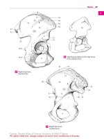
Posterior-Anterior View of the Skull
3
1
2
3
4
6
5
7
9
8
10
11
12
13
15
14
16
18
17
19
20
21
22
23
1
2
3
4
5
6
7
8
9
10
11
12
Sagittal suture
Pacchionian granulations
Lambdoid suture
Frontal sinus
Roof of the orbit
Sphenoid
Ethmoid sinus
Frontozygomatic suture
Petrous ridge
Innominate line
Internal auditory canal
Zygomatic arch
13 Condylar process of the mandible
14 Nasal septum and inferior nasal
bones
15 Maxillary sinus
16 Mastoid process
17 Occiput
18 Dens of the axis
19 Maxilla
20 Mandibular canal
21 Angle of the mandible
22 Mandible
23 Mental protuberance
Moeller, Pocket Atlas of Radiographic Anatomy © 2000 Thieme
All rights reserved. Usage subject to terms and conditions of license.
4
Skull
Frontal bone
Occipital bone
Temporal bone
Zygomatic bone
Parietal bone
Sphenoid bone
Moeller, Pocket Atlas of Radiographic Anatomy © 2000 Thieme
All rights reserved. Usage subject to terms and conditions of license.
Lateral View of the Skull
5
1
2
3
4
5
6
7
9
12
14
16
18
20
22
8
10
11
13
15
17
19
21
23
24
25
26
27
1
2
3
4
5
6
7
8
9
10
11
12
13
14
Outer table of the parietal bone
Diploe
Inner table of the parietal bone
Coronal suture
Groove of the middle meningeal
artery
Frontal sinus
Pituitary fossa
Greater wing of the sphenoid
Lambdoid suture
Cribriform plate
Anterior clinoid process
Posterior clinoid process
Nasal bone
Sphenoid sinus
15 Zygomatic bone (lateral wall of the
orbit)
16 Clivus
17 Ethmoid sinus
18 Petrous portion of the temporal
bone
19 Maxillary sinus
20 Opening of the external auditory
canal
21 Coronoid process of the mandible
22 Foramen magnum
23 Zygomatic process
24 Hard palate
25 Nasopharynx
26 Soft palate
27 Mandible
Moeller, Pocket Atlas of Radiographic Anatomy © 2000 Thieme
All rights reserved. Usage subject to terms and conditions of license.
6
Skull
Moeller, Pocket Atlas of Radiographic Anatomy © 2000 Thieme
All rights reserved. Usage subject to terms and conditions of license.
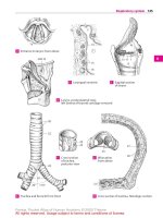
Towne View of the Posterior Skull
1 Lambdoid suture
2 Occipital protuberances (external
and internal)
3 Occipital bone
4 Sulcus of the transverse sinus
5 Occipital crest
6 Foramen magnum
7 Petrous bone
8 Arcuate eminence
7
9
10
11
12
13
14
15
16
Semicircular canals
Mastoid air cells
Internal auditory canal
Posterior arch of the atlas
Jugular foramen
Dens
Temporomandibular joint
Condyle of the mandible
Moeller, Pocket Atlas of Radiographic Anatomy © 2000 Thieme
All rights reserved. Usage subject to terms and conditions of license.
8
Skull
Moeller, Pocket Atlas of Radiographic Anatomy © 2000 Thieme
All rights reserved. Usage subject to terms and conditions of license.
Waters View of the Nasal Sinuses
1
2
3
4
5
6
7
8
9
10
Frontal sinus
Nasal bone
Anterior ethmoid sinus
Orbit
Nasal septum
Greater wing of the sphenoid
Infraorbital foramen
Foramen rotundum
Posterior ethmoid sinus
Zygomatic bone
9
11
12
13
14
15
16
17
18
19
Maxillary sinus
Alveolar recess of the maxilla
Sphenoid sinus
Foramen ovale
Alveolar process of the maxilla
Condyle of the mandible
Petrous ridge
Tongue
Mandible
Moeller, Pocket Atlas of Radiographic Anatomy © 2000 Thieme
All rights reserved. Usage subject to terms and conditions of license.
10
Skull
Moeller, Pocket Atlas of Radiographic Anatomy © 2000 Thieme
All rights reserved. Usage subject to terms and conditions of license.
Occipitofrontal View of the Nasal Sinuses
1
2
3
4
5
6
7
8
Frontal sinus
Roof of the orbit
Crista galli
Innominate line
Sphenoid
Petrous ridge
Lateral wall of the orbit
Zygomatic bone
9
10
11
12
13
14
15
Anterior ethmoid sinus
Internal auditory canal
Nasal septum
Maxillary sinus
Nasal cavity
Dens
Atlantoaxial joint
Moeller, Pocket Atlas of Radiographic Anatomy © 2000 Thieme
All rights reserved. Usage subject to terms and conditions of license.
11
12
Skull
Moeller, Pocket Atlas of Radiographic Anatomy © 2000 Thieme
All rights reserved. Usage subject to terms and conditions of license.
Posterior-Anterior View of the Orbit
1
2
3
4
5
6
7
8
9
10
11
Frontal sinus
Roof of the orbit
Sphenoid
Orbit (gray background)
Ethmoid sinus
Lateral wall of the orbit
Lesser wing of the sphenoid
Innominate line
Orbital plate
Greater wing of the sphenoid
Superior orbital fissure
12 Frontal process of the zygomatic
bone
13 Foramen rotundum
14 Floor of the orbit
15 Petrous ridge
16 Nasal septum
17 Zygomatic arch
18 Inferior turbinate
19 Maxillary sinus
20 Hard palate
Moeller, Pocket Atlas of Radiographic Anatomy © 2000 Thieme
All rights reserved. Usage subject to terms and conditions of license.
13
14
Skull
Moeller, Pocket Atlas of Radiographic Anatomy © 2000 Thieme
All rights reserved. Usage subject to terms and conditions of license.