100 Cases in Orthopaedics and Rheumatology - Singh, Parminder J, Swales, Catherine
Bạn đang xem bản rút gọn của tài liệu. Xem và tải ngay bản đầy đủ của tài liệu tại đây (3.33 MB, 291 trang )
100 CASES
in Orthopaedics and
Rheumatology
This page intentionally left blank
100 CASES
in Orthopaedics and
Rheumatology
Parminder J. Singh MBBS MRCS FRCS(Tr&Orth) MS
Consultant Orthopaedic & Trauma Surgeon and Senior Lecturer, Maroondah Hospital,
Monash and Deakin University, Melbourne, Australia
Catherine Swales MRCP PhD
Arthritis Research UK Clinical Research Fellow and Clinical Lecturer in
Rheumatology, Nuffield Orthopaedic Centre, Oxford, UK
100 Cases Series Editor:
Professor P John Rees MD FRCP
Professor of Medical Education, King’s College London School of Medicine at Guy’s,
King’s and St Thomas’ Hospitals, London, UK
First published in Great Britain in 2012 by
Hodder Arnold, an imprint of Hodder Education, Hodder and Stoughton Ltd,
a division of Hachette UK
338 Euston Road, London NW1 3BH
© 2012 Parminder J. Singh and Catherine Swales
All rights reserved. Apart from any use permitted under UK copyright law, this publication may only
be reproduced, stored or transmitted, in any form, or by any means with prior permission in writing
of the publishers or in the case of reprographic production in accordance with the terms of licences
issued by the Copyright Licensing Agency. In the United Kingdom such licences are issued by the
Copyright Licensing Agency: Saffron House, 6–10 Kirby Street, London EC1N 8TS.
Whilst the advice and information in this book are believed to be true and accurate at the date of
going to press, neither the author[s] nor the publisher can accept any legal responsibility or liability
for any errors or omissions that may be made. In particular, (but without limiting the generality of the
preceding disclaimer) every effort has been made to check drug dosages; however it is still possible
that errors have been missed. Furthermore, dosage schedules are constantly being revised and new
side-effects recognized. For these reasons the reader is strongly urged to consult the drug companies’
printed instructions, and their websites, before administering any of the drugs recommended in this
book.
British Library Cataloguing in Publication Data
A catalogue record for this book is available from the British Library
Library of Congress Cataloging-in-Publication Data
A catalog record for this book is available from the Library of Congress
ISBN-13 978-1-444-11794-3
1 2 3 4 5 6 7 8 9 10
Commissioning Editor:
Project Editor:
Production Controller:
Cover Design:
Index:
Joanna Koster
Jenny Wright
Francesca Wardell
Amina Dudhia
Lisa Footitt
Typeset in 10/12pt RotisSerif by Phoenix Photosetting, Chatham, Kent
Printed and bound in India
What do you think about this book? Or any other Hodder Arnold title?
Please visit our website: www.hodderarnold.com
CONTENTS
ORTHOPAEDICS
Case 1 A painful knee in a neonate
Case 2 Atraumatic painful joints in a boy
Case 3 An atraumatic painful hip
Case 4 An expanding mass in the leg of an adolescent
Case 5 A boy with a swollen mass in his thigh
Case 6 A painful collarbone
Case 7 A painful proximal humerus in an elderly woman
Case 8 A boy with a painful elbow
Case 9 A painful distal radius following a fall
Case 10 A painful thumb following a fall
Case 11 A painful hip following a fall
Case 12 Knee pain following a traffic accident
Case 13 A painful swollen elbow in a young girl
Case 14 A painful thigh following a violent fall
Case 15 Groin pain following a fall
Case 16 A swollen painful leg after a sporting incident
Case 17 A painful and swollen knee following an accident
Case 18 A crushed leg
Case 19 A painful foot following a traffic incident
Case 20 A hindfoot injury following a fall from height
Case 21 Curvature of the spine
Case 22 Severe low back pain
Case 23 A clicky hip in a toddler
Case 24 Hip and thigh pain in a 7-year-old boy
Case 25 A painful hip in an adolescent
Case 26 Groin and buttock pain in an elderly man
Case 27 Lateral hip pain
Case 28 A painful red swollen knee
Case 29 Swelling in the back of the knee
Case 30 A mildly swollen knee in a young man
Case 31 An atraumatic painful knee in an elderly woman
Case 32 An unstable knee in a young man
Case 33 An injured ankle
Case 34 Deformed feet in a newborn child
Case 35 High-arched feet in a young girl
Case 36 Flat feet in a young girl
Case 37 A painful and swollen ankle
Case 38 Deformed feet in an elderly woman
Case 39 A stiff great toe
Case 40 Misshapen toe in a middle-aged woman (1)
1
5
7
11
13
17
21
23
25
27
29
31
35
39
41
45
49
53
57
59
61
63
65
69
71
73
77
79
81
83
87
91
95
97
99
101
105
107
109
111
v
Contents
Case
Case
Case
Case
Case
Case
Case
Case
Case
Case
41
42
43
44
45
46
47
48
49
50
Misshapen toe in a middle-aged woman (2)
Foot pain following a riding fall
A deformed, swollen and ulcerated foot
A stiff shoulder
Elbow pain (1)
Elbow pain (2)
A painful wrist
A deformed finger
A painless contracture deformity of a hand
A painful wrist following a fall
RHEUMATOLOGY
Case 51 A swollen joint and fever
Case 52 Painful hands in a young woman
Case 53 Back pain in a young man
Case 54 Clumsiness
Case 55 Back pain and thirst in an elderly man
Case 56 Acute back pain
Case 57 Painful bones and a waddling gait
Case 58 Hip pain and abnormal blood tests
Case 59 Knee pain in an athlete
Case 60 Painful hands and a rash
Case 61 Hypermobility and a pneumothorax
Case 62 Pins and needles in a young woman
Case 63 Joint pains, skin changes and muscle weakness
Case 64 An acutely painful knee
Case 65 Recurrent abdominal pain and fevers
Case 66 Painful hands and oral ulcers
Case 67 Painful hands and breathlessness
Case 68 Persistent ulceration in an elderly woman
Case 69 A sore throat, painful knees and facial movements
Case 70 A lumpy rash and a swollen knee
Case 71 Rash and swollen ankles
Case 72 A breathless rheumatoid patient
Case 73 Pleuritic chest pain
Case 74 Fever, rash and joint pains
Case 75 Pain and stiffness in shoulders and hips
Case 76 Painful ears and nose
Case 77 Weakness and a rash
Case 78 Diarrhoea, rash and a stiff back
Case 79 Headache and weight loss
Case 80 A painful swollen hand following a fracture
Case 81 Diarrhoea and painful joints
Case 82 Chronic extensive muscle pain
Case 83 Intractable synovitis
Case 84 Haemoptysis and renal failure
Case 85 Diarrhoea and a swollen knee
Case 86 Swollen ankles and a rash
Case 87 Rash, testicular pain and arthralgia
Case 88 Painful hands and dry eyes
Case 89 Genital ulcers and a rash
vi
113
117
121
123
125
127
129
131
133
135
137
139
143
147
150
155
159
163
165
167
171
173
175
177
181
183
187
191
195
197
199
201
203
207
209
211
215
219
221
225
227
229
231
233
237
241
245
247
249
Contents
Case
Case
Case
Case
Case
Case
Case
Case
Case
Case
Case
Index
90 Rash, arthralgia and facial weakness
91 Rash and abdominal pain in an adolescent
92 Weight loss and claudication in a young woman
93 Swollen cheeks in an elderly man
94 A swollen knee
95 A child with a swollen knee
96 Acne, arthralgia and chest pains
97 A breathless patient with lupus
98 Anaemia and weight loss in a patient with rheumatoid arthritis
99 A breathless patient with dermatomyositis
100 Asthma, rhinitis, foot drop and a rash
253
255
257
259
261
263
265
267
269
271
273
275
vii
ACKNOWLEDGEMENTS
CS would like to thank colleagues in the Rheumatology, Radiology and Respiratory
departments for their expertise, advice and invaluable contributions.
PJS: I would like to dedicate this book to my wife Rowena and children Kieran and
Angelina Singh who have been as committed as I have in completing this book.
Without their support and abundance of love none of this would be possible. I thank
them enormously for their support. I would like to acknowledge my mother Gurbaksh
Kaur for her lifetime support and love in making this all possible. I am very grateful
for the opportunity to write this book and thank Professor Christopher Bulstrode in
Oxford for this. Finally I would like to acknowledge two of my dearest and closest
friends Richard and Lisa Field for their expert support and guidance throughout my
professional life.
Orthopaedics
CASE 1:
A PAINFUL KNEE IN A NEONATE
History
A young primigravida mother has become concerned about her newborn child. She is
accompanied in the clinic by her aunt who recognized that something was not quite
right but was not sure what to advise. The baby has general symptoms of fever, fatigue,
irritability and malaise. There is no history of trauma.
Examination
Close inspection of the left leg reveals some localized oedema and erythema. On palpation
the baby appears to have pain overlying the proximal tibia. Passive manipulation shows
a full range of movement of the leg without any obvious indications of pain.
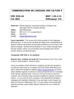
Investigations
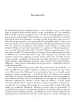
Initial investigations show a markedly elevated C-reactive protein (CRP). Imaging studies
of the knee show periosteal elevation of the proximal tibial metaphysis (Fig. 1.1).
Figure 1.1
Questions
• What is the diagnosis?
• What are the radiological signs?
• What blood tests would be most useful?
1
100 Cases in Orthopaedics and Rheumatology
ANSWER 1
The diagnosis is acute haematogenous osteomyelitis. Septic arthritis is less likely in view
of the excellent range of movement. There are two principal types of acute osteomyelitis:
• haematogenous osteomyelitis
• direct or contiguous inoculation osteomyelitis.
Acute haematogenous osteomyelitis is characterized by an acute infection of the bone
caused by the seeding of the bacteria within the bone from a remote source. This condition occurs primarily in children. The most common site is the rapidly growing and
highly vascular metaphysis of growing bones. Direct or contiguous inoculation osteomyelitis is caused by direct contact of the tissue and bacteria during trauma or surgery.
Clinical manifestations are more localized and tend to involve multiple organisms.
Predisposing comorbidities include diabetes mellitus, sickle cell disease, acquired immune
deficiency syndrome (AIDS), intravenous drug abuse, alcoholism, chronic steroid use,
immunosuppression, and chronic joint disease. Other possibilities are the presence of a
prosthetic orthopaedic device, recent orthopaedic surgery or an open fracture.
In general, osteomyelitis has a bimodal age distribution. Acute haematogenous osteomyelitis is primarily a disease in children. Direct trauma and contiguous focus osteomyelitis
are more common among adults and adolescents than in children. Spinal osteomyelitis is
more common in individuals older than 45 years.
The bacterial pathogen varies on the basis of the patient’s age and the mechanism of
infection:
• in neonates (<4 months) – Staphylococcus aureus, Enterobacter spp, and group A and
B Streptococcus spp
• in children (4 months to 4 years) – S. aureus, group A Streptococcus spp, Haemophilus
influenzae and Enterobacter spp
• in children and adolescents (4 years to adult) – S. aureus (80 per cent), group A
Streptococcus spp, H. influenzae and Enterobacter spp
• in adults – S. aureus and occasionally Enterobacter or Streptococcus spp.
Responsible pathogens may be isolated in only 35–40 per cent of infections.
With direct osteomyelitis the organisms include S. aureus, Enterobacter spp and
Pseudomonas spp. In the presence of puncture wounds there may be S. aureus and
Pseudomonas spp; and in the presence of sickle cell disease, S. aureus and Salmonella
spp.
Appropriate antibiotics are selected using direct culture results. Empirical therapy is often
initiated on the basis of the patient’s age and the clinical presentation. Further surgical
management may involve removal of the nidus of infection and implantation of antibiotic beads until resolution of the infection.
Plain radiographs may show evidence of soft-tissue swelling after 3–5 days. Bony changes
are usually present at 14–21 days. The earliest bony changes are periosteal elevation followed by cortical or medullary lucencies. At 28 days, 90 per cent of patients demonstrate
some abnormality. Magnetic resonance imaging (MRI) is effective in the early detection
of osteomyelitis with a sensitivity ranging from 90 to 100 per cent. Radionuclide bone
scanning using a 3-phase bone scan with technetium-99m may show up increased tracer
uptake in the affected region. Additional information can be obtained from scanning
with leucocytes labelled with gallium-67 and/or indium-111. Computed tomography (CT)
2
Orthopaedics
scanning can demonstrate calcification, ossification, and intracortical abnormalities. CT
is particularly helpful in the evaluation of spinal vertebral lesions. Ultrasonography is
useful in children with acute osteomyelitis. This modality can detect abnormalities as
early as 1–2 days after onset of symptoms. The abnormalities include soft-tissue abscess
or fluid collection and periosteal elevation.
The white cell count may be elevated, but it is frequently normal. The C-reactive protein
level is usually elevated, but this is non-specific. The erythrocyte sedimentation rate (ESR)
is elevated in 90 per cent, but this finding is clinically non-specific. Blood culture results
are positive in only 50 per cent of patients with haematogenous osteomyelitis. Culture
or aspiration findings in samples of the infected site are normal in 25 per cent of cases.
KEY POINTS
•
•
•
•
Osteomyelitis can be a result of haematogenous or direct spread.
The earliest radiographic change is periosteal elevation.
MRI is effective in the early detection of osteomyelitis.
The bacterial pathogen varies on the basis of the patient’s age and mechanism of
infection.
3
This page intentionally left blank
Orthopaedics
CASE 2:
ATRAUMATIC PAINFUL JOINTS IN A BOY
History
A 10-year-old boy has been brought to the emergency department by his father. The
youngster was in a playground when he developed a swollen and painful right knee. The
boy describes recurrent episodes of pain and swelling in his knees and shoulders over
the last year or so. He cannot remember any history of trauma. He does, however, have a
history of multiple blood transfusions but he is unsure of the reason for these.
Examination
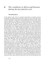
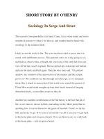
The boy has painful, tensely swollen, warm and diffusely tender knee and shoulder joints
(Fig. 2.1). He also has multiple bruises over his legs and arms.
Figure 2.1
Questions
• What is the diagnosis?
• What are the causes of this condition?
• What is the pathophysiology of joint involvement in this condition?
5
100 Cases in Orthopaedics and Rheumatology
ANSWER 2
This youngster suffers from recurrent episodes of atraumatic painful effusions to his knee
joints. He also has a history of multiple blood transfusions. He suffers from a blood dyscrasia leading to haemarthrosis. The most common condition seen in orthopaedic practice
is haemophilia and it is most likely in this patient.
Haemophilic arthropathy is a condition associated with a clotting disorder leading to
recurrent bleeding into the joints. Over time this can lead to joint destruction. Individuals
with haemophilias A and B most commonly have haemophilic arthropathy. Haemophilia
A (classic haemophilia) is associated with a factor VIII deficiency and is a sex-linked
recessive trait. This occurs in approximately 1 per 5000 live male births, with 25 per
cent of cases being sporadic (no family history). Other clotting disorders may also lead
to haemarthrosis, examples being haemophilia B (Christmas disease) and factor IX
deficiency.
Haemosiderin deposition within the joint leads to synovial hypertrophy, bone erosions,
recurrent bleeding, and eventual destruction of the articular surfaces and arthrofibrosis.
The stages of degenerative changes in the joint are summarized below.
!
Stages of joint changes with haemarthrosis
•
•
•
•
Grade 1: Soft-tissue swelling (effusions, synovial thickening)
Grade 2: Widened epiphysis, small erosions (normal cartilage interval)
Grade 3: Large erosions, bone cysts, cartilage loss
Grade 4: Joint destruction and subluxation
KEY POINTS
• Haemosiderin deposition within the joint leads to synovial hypertrophy, bone erosions
and recurrent bleeding.
• Recurrent haemarthrosis can lead to joint destruction of the affected joints.
• Synovectomy should be considered, or replace or fuse the joint.
6
Orthopaedics
CASE 3:
AN ATRAUMATIC PAINFUL HIP
History
A 32-year-old Afro-Caribbean man was intending to visit his mother in Africa. While
packing he noticed a progressively worsening pain in the left groin. The pain radiated
down the upper thigh and was present at rest. There were no aggravating or relieving
factors and no associated symptoms of numbness or tingling. The pain was severe and
constant in nature for several days. He had also noticed some mild discomfort in his left
hip. He recalls no previous episodes. He obtained temporary relief of his pain with simple
analgesia. He has a history of asthma and has been taking regular steroids over the last
15 years. He drinks 20 units of alcohol per week and smokes 20 cigarettes a day. There
is no history of trauma and he is systemically well.
Examination
The man walks with a severe limp. Assessment of his hip abductors reveals a positive
Trendelenburg sign on the left side. Measurement of the true leg lengths reveals a 1 cm
shortening in the left leg compared to the right. Movement of the hip is painful but not
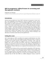
significantly restricted. He has no obvious neurological or vascular deficit of the legs. A
radiograph of this patient is shown in Fig. 3.1.
Figure 3.1
Questions
• What is the diagnosis?
• Describe the blood supply to the femoral head.
• What factors predispose to this condition?
• How would you investigate and classify this condition?
• What are the management options?
7
100 Cases in Orthopaedics and Rheumatology
ANSWER 3
This man has insidious and persistent left groin pain that is exacerbated by weight-bearing. This pain arises from his hip joint. While he retains a good range of movement, he
has pain. He also has true leg shortening. He has mild symptoms in the right hip. Finally,
he has a history of being on steroids and is a heavy smoker and consumes alcohol. All
these signs indicate that the most likely diagnosis is avascular necrosis of the femoral
head. This is the most common site to undergo avascular necrosis. The condition is
bilateral in 50 per cent of patients. Around 10 per cent are asymptomatic and diagnosed
incidentally.
The blood supply to the femoral head is derived from an arterial ring around the neck of
femur. The ring anastomosis is mainly from the medial femoral circumflex artery posteriorly and minor branches of the lateral femoral circumflex artery anteriorly. These vessels
traverse the femoral neck to perforate the head close to the articular cartilage. Ten per
cent of the blood supply comes from the vessels in the ligamentum teres.
An injury or ischaemia can predispose to arterial cut-off, venous stasis, intravascular
thrombosis, intraosseus sinusoidal compression or a combination of these. The decreased
blood flow to the femoral head leads to increased intraosseus pressure, osteonecrosis and
finally collapse of the femoral head.
Traumatic causes of avascular necrosis of the femoral head are fracture of neck of femur
and dislocation of the femoral head. Non-traumatic causes include steroid use, alcoholism, marrow-replacing diseases like Gaucher’s disease, high-dose radiotherapy, hypercoagulable states, sickle cell disease, hyperfibrinolysis, thrombophilia, protein C and S
deficiency, and Legg–Calvé–Perthes disease (LCPD).
Imaging the hip with anteroposterior and lateral views is the initial investigation. In
early cases the radiographs may not reveal any signs of avascular necrosis. In late
cases, subchondral sclerosis (increased density of the affected area, crescent sign), a thin
subchondral fracture line in the necrotic segment, flattening of the femoral head, and collapse of the femoral head can be seen. The important differentiating point from advanced
osteoarthritis is the preservation of joint space during the early stages of the disease.
MRI scans are the investigation of choice in patients with normal radiographs. They can
show early changes in the bone marrow long before the appearance of X-ray features.
There are a number of classifications for avascular necrosis of the femoral head. The most
commonly used classification was described by Ficat and associates in 1960, based on
radiological findings and bone scans.
!
The Ficat classification
Pre-collapse phase
• Ficat I: No X-ray changes
• Ficat II: Early X-ray changes, no distortion of femoral head
Post-collapse phase
• Ficat III: Increased bone destruction, femoral head deformed on X-ray
• Ficat IV: Complete collapse of femoral head with destruction of hip joint seen on
X-ray
8
Orthopaedics
Management depends on the stage of disease seen at first presentation. If early avascular
necrosis is left untreated it is likely to progress to the advanced stages. Management of
pre-collapse avascular necrosis comprises surgical intervention with core decompression
with or without bone grafts.
KEY POINTS
•
•
•
•
•
•
The femoral head is the most common site to undergo avascular necrosis.
The condition is bilateral in 50 per cent of cases.
Traumatic and atraumatic causes have been identified.
Radiographs may be normal in the early stages.
MRI is the investigation of choice.
Treatment options include core decompression and bone graft, osteotomy and
arthroplasty.
9
This page intentionally left blank
Orthopaedics
CASE 4:
AN EXPANDING MASS IN THE LEG OF AN ADOLESCENT
History
A 15-year-old boy was on holiday abroad with his parents. While sunbathing the boy
asked his father to apply suntan lotion to his legs. The father then noticed a lump over the
proximal aspect of his son’s tibia that was not present on the other leg. The boy explained
that the lump had been there for nearly 6 months but had been increasing in size over the
recent few weeks. The family immediately returned home to consult their doctor.
Examination
On inspection there is a firm, irregular and tender mass arising from the proximal tibia.
The boy describes no pain on palpation of the lump and his range of movement is full.
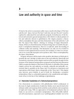
There are no other lumps. Radiographs of his knee are shown in Figs 4.1 and 4.2. His
erythrocyte sedimentation rate (ESR) and serum alkaline phosphatase are raised.
Figure 4.1
Figure 4.2
Questions
• What is the diagnosis?
• Describe the features of the X-rays.
• How would you confirm the diagnosis?
• What are the principles of management of this condition?
11
100 Cases in Orthopaedics and Rheumatology
ANSWER 4
This boy has an osteosarcoma of the proximal tibia. A firm, irregular mass fixed to underlying structures is more suspicious of a malignant lesion. This is a malignant tumour of
the proximal tibia. This is the most common primary malignant bone tumour of mesenchymal derivation. The tumour arises in adolescents and affects males more often than
females. The most common site affected is in the region of the knee in the metaphyseal
part of the bone. Other sites include the proximal humerus, proximal femur and pelvis,
and the spine is rarely involved.
Osteosarcoma can present as purely osteolytic or osteoblastic or a mixture of the two
types. Elevation of the periosteum may appear as a classic Codman’s triangle. Near the
junction of the healthy bone with the tumour there is reactive new bone formation
beneath the periosteum as seen in the proximal tibia in this case. Extension of the tumour
through the periosteum may produce a sunburst appearance which is also present in the
X-rays. Remember always to image the entire bone to assess for skip lesions or joint
involvement.
A biopsy of the lesion will help confirm the diagnosis. The biopsy should be undertaken
ideally by the same surgeon who will be responsible for the definitive tumour resection
in a dedicated bone and soft-tissue sarcoma unit. Biopsies performed without communication with the dedicated sarcoma unit’s input can lead to amputation of a salvageable
limb.
The Enneking staging system is the most widely used. The key components in staging
are histological grade (low-grade vs high-grade), the anatomical location of the tumour
(intracompartmental vs extracompartmental), and the presence or absence of metastatic
disease.
Management comprises staging of the tumour, neo-adjuvant treatment, and surgery in a
specialist bone and soft-tissue sarcoma unit with a multidisciplinary team.
Most patients have micrometastases at the time of presentation. All should be screened
for pulmonary metastasis. The principle of treatment is, therefore, to combine surgery
(limb salvage or amputation) with chemotherapy. Limb salvage is possible only if the
nerves can be preserved, adequate muscles and soft tissues can be left intact while a
reasonably wide margin of resection of the tumour can be achieved. Radiotherapy is
usually reserved for palliation and disease location in an inaccessible location. Pre- and
postoperative chemotherapy is used for osteosarcoma. Patients with a greater than 95 per
cent tumour cell kill or necrosis have a better prognosis than those whose tumours do
not respond as favourably. With adjuvant chemotherapy, the 5-year survival rate can be
greater than 50 per cent.
KEY POINTS
• Osteosarcoma is the most common primary malignant bone tumour of mesenchymal
•
•
•
•
12
derivation.
The most common site affected is in the region of the knee.
MRI of the primary lesion is the best method to use.
Biopsy of the lesion will help confirm the diagnosis.
Management comprises staging of the tumour, neo-adjuvant treatment, and surgery in
a specialist unit.
Orthopaedics
CASE 5:
A BOY WITH A SWOLLEN MASS IN HIS THIGH
History
A 14-year-old boy presents to his general practitioner with a swelling around the mid
shaft of his femur. His mother has brought him in following his complaints of tiredness
and intermittent fevers over the last few weeks, which has caused him to miss football
training and to be inactive.
Examination
Manual examination of the mid thigh reveals a tender mass. The mass is firm and appears
to be well fixed to the underlying muscle. There are no neurological or vascular deficits
to the leg. Initial investigations reveal elevated white blood cells (WBC), erythrocyte
sedimentation rate (ESR) and anaemia. A radiograph of the femur is shown in Fig. 5.1.
Figure 5.1
Questions
• What is the diagnosis?
• Describe the features of the X-ray.
• How would you manage this condition?
• What is the prognosis?
13
100 Cases in Orthopaedics and Rheumatology
ANSWER 5
The diagnosis is Ewing’s sarcoma. The features suspicious of a malignant lesion in this
age group are the firm, fixed mass, its location, and raised inflammatory markers associated with anaemia, fatigue and intermittent fevers. This condition typically occurs in
young patients and presents with pain and fever.
The tumour was first described by James Ewing in 1921 and is the second most common
primary malignant bone tumour (the first being osteosarcoma). The tumour is more common in males and affects children and young adults. The majority develop this between
the ages of 10 and 20 years. Rarely, the tumour develops in adults older than 30 years.
The earliest symptom is pain, which is initially intermittent but becomes intense. Rarely,
a patient may present with a pathological fracture. Eighty-five per cent of patients have
chromosomal translocations associated with the 11/22 chromosome. Ewing’s sarcoma is
potentially the most aggressive form of the primary bone tumours. The most common
sites affected are the femoral diaphysis, pelvis, tibia, humerus, fibula and ribs.
Radiological features seen in the femoral shaft include a typical onion-skin appearance
or sunburst pattern due to spread of the tumour via Haversian canals with periosteal
reaction which indicates an aggressive process. In some patients, Codman triangles may
be present at the margins of the lesion. These result from the elevation of the periosteum
and central destruction of the periosteal reaction caused by the tumour. Radiographs
may vary from highly lytic to predominantly sclerotic in appearance. Most commonly,
radiographs show a long, permeative lytic lesion in the meta-diaphysis and diaphysis of
the bone. Sclerotic lesions are less common. MRI provides a more accurate assessment of
the tumour size and relation to the surrounding structures.
Patients are usually assigned to one of two groups, the tumour being classified as either
localized or metastatic disease. Tumours in the pelvis typically present late and are
therefore larger with a poorer prognosis. Treatment comprises chemotherapy, surgical
resection and/or radiotherapy. With combined treatment, patient survival has improved:
with the use of adjuvant chemotherapy, the 5-year survival rate is more than 60 per
cent. With localized disease, wide surgical excision of the tumour is preferred over radiotherapy if the involved bone is expendable (e.g. fibular, rib), or if radiotherapy would
damage the growth plate. If there is a pathological fracture, limb salvage surgery with
the implantation of a long mega-prosthesis is the preferred option.
Non-metastatic disease survival rates are 55–70 per cent, compared to 22–33 per cent
for metastatic disease. Patients require careful follow-up owing to the risk of developing
osteosarcoma following radiotherapy, particularly in children in whom it can occur in up
to 20 per cent of cases. A European study of 359 patients with non-metastatic Ewing’s
sarcoma revealed that the following factors are associated with a poor prognosis:
•
•
•
•
•
•
14
male sex
age >12 years
anaemia
elevated lactate dehydrogenase (LDH)
radiation therapy only for local control
poor chemotherapeutic course.
Orthopaedics
KEY POINTS
• The majority of cases of Ewing’s tumour are seen between the ages of 10 and 20 years.
• Eighty-five per cent of patients have chromosomal translocations associated with
chromosome 11/22.
• Most common sites affected are the femoral diaphysis, pelvis, tibia, humerus, fibula
and ribs.
• Radiological features include an onion-skin appearance or sunburst.
• Treatment comprises chemotherapy, surgical resection and/or radiotherapy.
15
This page intentionally left blank