Entomofauna, ZEITSCHRIFT FÜR ENTOMOLOGIE VOL 0012-0033-0044
Bạn đang xem bản rút gọn của tài liệu. Xem và tải ngay bản đầy đủ của tài liệu tại đây (902.58 KB, 16 trang )
© Entomofauna Ansfelden/Austria; download unter www.biologiezentrum.at
Bntomojauna
ZEITSCHRIFT FÜR ENTOMOLOGIE
Band 12, Heft 3: 33-48
ISSN 0250-4413
Ansfelden, 15.März 1991
A Review of the Exenterine Genus Acrotomus Holmgren, 1855
(Hymenoptera, Ichneumonidae)1
Virendra Gupta
Abstract
The taxonomy of the exenterine genus Acrotomus HOLMGREN. 1855, is reviewed.
Four species are included in ihe genus: Acrotomus lucidulus (GRAVENHORST,1829)
and Acrotomus succinctus (GRAVENHORST, 1829) from Europe, Acrotomus
albidulus KASPARYAN,1986 from Primorskij kraj, USSR, and Acrotomus
aithogaster sp. nov. from Jordan, Acrotomus succinctus (GRAVENHORST, 1829) also
occurs in North America. It is recordcd herc from Jordan and Kargil, Ladakh, India.
Records from Jordan and India are new records for ihe genus.
Introduction
Acrotomus HOLMGREN, 1855, belongs to the subfamily Tryphoninae.tribe
Exenterini (=Cteniscini). It is a Palearctic genus, and until recently only two species
were known: Acrotomus lucidulus (GRAVEN'HORST,1829) and Acrotomus succinctus
(GRAVENHORST, 1829) from Europe. KASPARYAN (1986) described Acrotomus
albidulus from USSR. A new species, Acrotomus ailhogaster sp. nov. is described
here from Jordan. MASON (1962: 1287) rcportcd A. lucidulus from Japan.
1 Florida Agriculiural Experiment Journal Serips No. R-00682.
33
© Entomofauna Ansfelden/Austria; download unter www.biologiezentrum.at
He (1978: 605) reported Ihe accidental introduction of A. succinctus into Quebec,
Canada in 1962 and its spread lo Onlario, Canada and Michigan, U.S.A. during
1963-67. CARLSON (1979) reported iis occurrence in New York. It is reported here
from ihe high mountains of Ladakh, India and also from Jordan. MASON (1956,
1962) included Exenterus ornatus WALSII, 1873 in ihis genus but TOWNES (1969:
190) made it the type speciesof his new genus Scapnetes. S. ornatus is a different
looking species, with an unspecialized clypeus and normal vertex. The egg of ihis
species, however, resembles that of A. succinctus in having a large anchor, but the
subgenitalplate, ovipositor and ovipositor shcaths arc similar to those of A.
lucidulus.
Biology
Littlc is known about the biology of the species of the genus. They are apparently
parasites of sawfly larvae. A. lucidulus has been reported from the larvae of Cladius
difformis in England, France and Germany (KERRICII, 1952: 321).
Acrotomus HOLMGREN, 1855 (Figs. 1-3)
Acrotomus HOLMGREN, 1855. Svenska Vetcnsk. Akad. Handl. (N.F.), 1: 222. Type species:
Tryphon lucidulus GRAVENIIORST. 1829; designated by VIERECK, 1912.
Delotomus FOERSTER, 1869. Verh. Naturh. Ver. Rhcinlande, 25: 194, 213, 215. Unnecessary
new namc for Acrotomus.
Taxonomy: KERRICII, 1952: 319; MASON, 1962: 1285; TOWNES, 1969: 187.
Diagnosis: Clypeus appearing almost pentagonal, as long as wide, not transverse as
is normal for the group. Mediän 0.3 of clypeus projeeting and squarely truncate,
with sharp lateral corners (Figs. 2, 14). Mandible wide apically, somewhat twisted
medially so that leeth lie in ihe samc plane, Iower toolh larger than upper tooth.
Head swollen behind and above eyes. Vericx clevated. Notauli weak. Subtegular
ridge normal. Apex of hind tibia roundcd off ventrally, widest just before apex,
without a polished rim between the uirsal socket and the apical fringe of bristles.
Propodeum arcolated (Fig. 3). Tcrgiie 1 gradually widening from base to apex, its
dorsolatcral carina passing through spiracle or along the upper edge of spiracle.
Tergites 2-4 with fine punetures and shiny. Hairs on median part of tergites 4-6 not
slanted mesad. Female subgenital platc modcraicly long, partly or wholly folded
medially and ending in a rounded point. Ovipositor moderately slender, weakly
decurved. Egg ovate-reniform, with a Single short apicoventral stalk.
TOWNI-S (1969) provided a key to distinguish Acrotomus from other exenterine
genera.
34
© Entomofauna Ansfelden/Austria; download unter www.biologiezentrum.at
Key to the species
1. Apical margin of clypeus slightly concave (Fig. 14). Scape about 1.7x as long as
wide in front view (Fig. 7). Flagellum slcndcr. Tergite 1 without distinct
basolatcral projections. Fcmale subgcnital plate sharply folded medially (Fig. 5).
Ovipositor sheath broad apically, projecting bcyond tip of abdomen, and with
normal hairs. Egg brownish in color. Lateral margins of male abdomen yellow.
Face of male yellow. Europe, North America, India, Jordan.
1. A. succinetus (GRAVENHORST)
Apical margin of clypeus straight (Fig. 2). Scape Short, aboul 1.2-1.5x as long as
wide in front view (Fig. 8). Flagellum somewhat thickened medially. Tergite 1
with distinct basolateral projections (Fig. 3). Female subgcnital plate flal basally
and pointed apically (Fig. 9). Lateral margins of male abdomen black except
rarely.
2.
2. Abdomen reddish-brown bcyond tergite 1. Fcmale subgeniial plate wholly flat
and with a Sharp median apical point. Jordan.
4. A.aithogaster, sp. nov.
Abdomen black with yellow apical margins on tergites. Fcmale subgenital plate
partly folded medially.
3.
3. Scape about 1.2x as long as wide. Ovipositor thick and almost straight. Ovipositor
sheaths acute apically, not projecting beyond tip of abdomen, and densely hairy
ventrally (Fig. 4). Egg yellowish-brown in color. Face often with black marks or
line. Mesopleurum largely black or brown. Europe, Japan.
2. A. lucidutus (GRAVENHORST)
Scape about 1.5x as long as wide. Ovipositor slcndcr and decurved (Fig. 6).
Ovipositor sheaths flattcncd, not concave benealh (Fig. 6) and with a
desclerotized basal portion. Egg ycllowish-whiic, with a short central stalk. Face
yellow. Mesopleurum (and also Pronotum and mesosternum) apparently with
more yellowish-white spots. USSR.
3. A, albidulus KASPARYAN
1. Acrotomus succinetus
(GRAVENHORST,
1829) (Figs. 5,7,10,11,14-19)
Tryphon succinetus GRAVENHORST, 1829. Ichncumonologia Europaea, 2: 166. des. Lectotype
(selected by KERRICII 1952): Male, Gcrmany: Hannover: Baricrodc (Wroclaw).
Acrotomus succinetus: PPANKUOI, 1906. Zeiischr. System. Hymen. Dipt., 6: 94. comb.nov.
. Acrotomus succinetus: KKRRIOI, 1952. Bull. Brit. Mus. (Nai. Hist.) Eni., 2: 321. M,F., syn.,
des., fig. Several counlrics in Europe.
35
© Entomofauna Ansfelden/Austria; download unter www.biologiezentrum.at
Acrotomus succinctus: MASON, 1962. Canadian Ent., 94: 1285. des., figs. -- 1978. Canadian
Ent., 110: 605. North America: Quebec and Ontario, Canada; Michigan, LI.S.A.
Adventive.
Acrotomus succinclus: CARLSON, 1979. Catalog of Hymenop tcra in America Nonh of Mexico,
1: 379. N.Y., Ont., Mich. Adventive.
Acrotomus succinclus: KASPARYAN, 1981. Opred. Faune SSSR, No. 129: 154. key, fig. USSR.
Redescribed: Head: Head in front view from clypeus to Icvcl of lateral ocelli about
as long as wide at middle of cyes. Clypeus appcaring pcntagonal, as long as the
length of its apical emargination, sidcs clongale and concave (Fig. 14). Face and
clypeus shiny and wiih shallow minute puncturcs. Frons minutely punctate, shiny.
Vertex as wide as eye width in dorsal view, shiny and impunctate. Temples wide
dorsally and polishcd, narrowcd bclow. Malar space 0.5x thc basal width of mandible. Interocellar distance about 0.5 the occllocular distancc (Fig. 15). Scape in front
view about 1.7xas long as wide. First threc flagellar scgments in the ralio of
25:15:14 (Fig. 7).
Thorax subpolished, hairy. Mesoscutum convex. Notauli weakly impressed.
Mesopleurum and mctapleurum with minulc puncturcs, ihcse shallower in male.
Propodeum largely shiny, fully areolatcd (Fig. 18, 19). Areola rectangular, weakly
separated from basal arca or not distinctly scparatcd. Areolct prcscnt. Ncrvellus
interceptcd bclow the middle.
Abdomen: Tcrgitc 1 gradually widened postcriorly, ils dorsomedian and
dorsolatcral carinae distinet, with weak basolatcral projeetions (Fig. 16).
Dorsolateral carina narrowly inlcrruptcd ai spiraclc. Tcrgite 1 laterally impressed in
apical 0.3 and rugulosc. Abdomen subpolished, wiih tergites 2-3 punctate. Hairs on
tergites short; in the male from India absent along the middle of tergites. Ovipositor
sheaüis broadly roundcd and projeeting bcyond apcx of abdomen, wiih normal hairs
all over (Fig. 5). Male claspers also similarly shaped. Fcmale genital plate large and
sharply foldcd mcdially. Ovipositor siender and wcakly decurved.
Egg (Fig. 11): Egg with a large anchor ofiripodal shapc. Egg color brownish.
Color: Black. Face, clypeus, mandiblc, and malar space yellow. Usually area just
below antennal sockets wiih a V-shapcd black mark. Thorax black with front margin
of pronotum, tegula, wing bases, sublcgular ridge, hind corner of pronotum
narrowly, scutcllum except ccntrally, mctascutcllum, and fore and middle coxae
and trochanters, yellow. (Pronoial collar and prosternum broadly yellow in both
male and female, cf. lucidulus). Hind coxa, trochanters and femur, and fore and
middle femora and tibiac, ycllowishbro.wn. Hind tibia and all larsi blackish.
Sometimes legs including coxae, largely yellowish-brown except for hind tibia and
tarsus. Abdomen black with apical margins of tergites 2-6 usually narrowly yellow.
(In Indian speeimen yellow margins clcar). Lateral margins of apical abdominal
36
© Entomofauna Ansfelden/Austria; download unter www.biologiezentrum.at
tergiies yellow in bolh male and female (cf. lucidulus). Abdominal venter yellow in
male and brown in female.
Lenglh: 6.25-7.0 mm.; Fore wing 5.0-5.8 mm.; ovipositor shealh 0.4 mm.
Specimens examined: India: Kashmir: Ladakh: Kargil, 2590 m., 1 male,
14.VI.1973, Coll. Santosh GUPTA (GUPTA). Europe: Several males and females
examined from England, Ireland, Czechoslovakia, Sweden, Belgium, Denmark,
Austria, Germany and Italy (AEI, Gainesvillc; CNC, Ottawa). U.S.A: Michigan:
Ann Arbor, 3 males and 1 female, 12.VIII.1967 to 4.IX.1967, H. & M. TOWNES;
New Jersey: High Point State Park, 1 female, 13.VI.1973, R. REARDON (AEI,
Gainesville). Canada: Ontario: Siittsville, 1 female, 22.X.1963, W.R.M. MASON;
Quebec: Mt. Albert, 1500 ft., 1 male, 11.VIII.1975, H. & M. TOWNES (AEI,
Gainesville). Canada: Quebec: Mcach Lake, 28.V.1962, S.M. CLARK; Old Chelsea:
Summit King Mt., 1150 ft., 1 male, 23.VII.1965, Malaise trap; Hüll, 1 female,
6.VIII.1965, Malaise trap. Ontario: Aylmer West, 1 female, 7-15.IX.1972, Malaise
trap; Innisville, 1 female, 28.VII.1963, W.R.M. MASON (all CNC, Ottawa). Jordan:
Mansura b. Mafrak, 1 female, 22.X.1965, J. KLAPPURICH (CNC, Ottawa).
Distribution: Europe, North America, India: Kashmir, Jordan.
It is apparently a widcly distributed spccics in Europe, having bccn recorded from
Austria, Belgium, Czechoslovakia, Denmark, England, Finland, France, Germany,
Ireland, Italy, Netherlands, Swcdcn, Swit/.crland, and USSR. In North America, it
was apparently first collected in Chclsca, Quebec, in 1962, then in Ontario in 1963,
and later in Michigan in 1967 (MASON, 1978: 605). It is reported here from New
Jersey. Carlson listed it from New York also.
2. Acrotomus lucidulus
(GRAVENHORST,1829)
(Figs. 1-4,8,9,13)
Tryphon lucidulus GRAVENIIORST, 1829. Ichneumonologia Europaca, 2: 162. F. des. Lectotype
(selected by KERRJCH, 1952): F, Germany or Italy (Wroclaw).
Acrotomus lucidulus: HOLMGREN, 1855. Svcnska Vciensk. Akad. Handl. (N.F.), 1: 222. M, F.
n. comb., des. Norway, Sweden. Host: Lophyrus pini.
Acrotomus lucidulus MEYER, 1936. Tables systematiques des hymenopteres parasites (fam.
Ichneumonidae) de l'URSS, 5: 20. M, F. key, des. Russia: Pswowsk, Wjansk,
Leningradsk. Europe. Hosis: Cladius difformis, C. albipes, Dineura alni.
Acrotomus lucidulus: KF.RKICH, 1952. Bull. Bril. Mus. (Nat. Hist.) Ent., 2: 320. M, F.
Lectotype design., des., fig. England, scvcral countries in Europe.
Acrotomus lucidulus: MASON, 1962. Canadian Ent., 94: 1287. F. des. Japan: Hakone.
Acrotomus lucidulus: KASPARYAN, 1981. Opred. Faune SSSR, No. 129: 154. key, fig. USSR.
Diagnosis:Scape in front view about 1.2x as long as wide. First three flagellar
Segments in the ratio of 20:15:13 (Fig. 8). Flagcllum slightly thickened medially.
Apical margin of clypeus truncate, not coneave (Fig. 2). Notauli comparatively
deeply impressed anteriorly. Basolalcral conical projeetions on tergile 1 ralher
37
© Entomofauna Ansfelden/Austria; download unter www.biologiezentrum.at
prominent (Fig. 3). Female subgeniial plate broad and convex but not creased
medially, with an acute apical point (Fig. 9). Ovipositor short and thick. Ovipositor
shcaths short and triangulär, not projecting beyond lip of abdomen, excavated and
densely hairy ventrally (Figs. 4, 9). Male claspers also more hairy and triangulär as
in the female. Otherwise similar to A. succinclus in structure.
Egg (Fig. 13): Egg dorso-ventrally flattencd with a small anchor.
Color: Similar to that of A. succinclus exccpt lhat face of male black with two
large yellow spots, or yellow with a black central line, the V-shaped black mark on
the face of female touchingtheepistomalgroovc, and yellow marks onpronotum
and mesopleurum reduced to abscni. Thorax in side vicw black in male; prosternum
black when face with large black spots and yellow whcn face largely yellow. Female
with pronotal collar wilh a yellow line and prosternum largely yellow. Subtegular
ridge sometimes yellow. Male abdomen seldom pale yellow laterally. Male coxae
often yellowish-brown. Female abdominal tergiics reddish-brown laterally,
particularly the apical ones.
Similar to A. succinclus in measurements.
Specimcns examined: Scvcral malcs and fcmalcs from England, Denmark,
Belgium, Austria, Germany, and Swcdcn in CNC, Ottawa and AEI, Gainesville
Collections. Japan: Hakone, 1 female, August 19, 1900? W. H. HARRINGTON
(CNC).
Distribution: Europc, Japan.
KERRICH (1952) reported it from Belgium, Czcchoslovakia, England, Finland,
France, Germany, Nethcrlands, Norway, Swcdcn, Switzcrland, and Spain. It is
apparently widely distributed in Europc. Il has been reported from USSR also.
MASON (1962) reported this specics from Japan. MOMOI examined the Japanese
speeimen in CNC in 1964 and considered it a distincl specics (inacutus, nov. sp.
MSS, see also TOWNES, MOMOI & TOWNES, 1965: 113). I have examined this
speeimen. The distinetions and the matcrial arc not sufficient enough to consider it
different from succinetus.
According to KERRICH (1952: 321, 426), this specics has been misidentified in
European literature. He stated lhat the host records other than from Cladius
difformis as quoted by MORLEY and SeiIMIF.DI-KNECHT probably refer to Cteniscus
curtisii.
3. Acrotomus
albidulus
KASPARYAN, 1986 (Fig. 6)
Acrotomus albidulus KASPARYAN, 1986. Systcmatics and ccology of insecls from the Far
East, Akad. Nauk SSSR, Vladivostock, 1986: 54. F. des., fig. Type: F, USSR: Primorskij
kraj: Gornotaezhnaya Station, 20 km. SE Ussurijsk (LENINGRAD).
Specimens of this species were not available for examination. Dr. D. R.
KASPARYAN was kind enough to provide mc with an English translation of his
38
© Entomofauna Ansfelden/Austria; download unter www.biologiezentrum.at
original descriplion. The following diagnostic fcaiurcs have been taken from the
notes provided by him.
Diagnosis: A. albidulus is rclated to A. lucidulus in having a short scape, tergite 1
with basolateral angulation, and cpiplcurac largcly black dorsally. It differs from
both lucidulus and succinetus in the form of ovipositor sheaths, decurved ovipositor
(Fig. 6), and whitish egg wilh a short ccniral stalk. Body smooth and shiny, with
ralher fine body punetation and silvery pubcsccnce. Central arca of face, lower half
of mesopleurum, and basc of second tergile wilh morc definite punetation.
Propodeum and tergite 1 except apically shallowly rugulosc. Scape 1.5x as long as
wide. Vertex raised dorsally. Malar spacc 0.66x the basal width of mandible.
Epomia short. Notauli sharp. Propodeum areolatcd but median longitudinal carinae
weak on sides of basal area. Tergite 1 aboul 2.0x as long as wide. Tergite 2 as long
as wide. Ovipositor strongly and uniformly curved (Fig. 6). Ovipositor sheath
flattened, apically not coneave beneath, and its in lower 0.6 strongly desclerotized
and with acrease.
Egg: 0.4 x 0.2 mm; ycllowish-while; stalk short, arising from the middle of the
ventral surface of ihe egg.
Color: Black. Antenna blackish-brown, ventral aspect of apical segments reddishbrown. The following parts yellowish-whitc: Facc, elypeus, mandible except teeth,
malar space, lower 0.33 of lemple, proplcura partly, a wide band along upper margin
of pronotum, hind corncr of pronotum, a sport on upper margin of pronotum basad
of epomia, tegula, subtegular ridge, narrow bands along front and hind margins of
mesopleurum, prepectus largcly, a ling along mcsostcrnal suture, scutellum except
basally, metascutellum, coxae and trochanters of fore and middle legs, hind margin
of 2-7th tergites, sides of tergites 3 and on wards up to level of spiracles, and all
sternites. Fore and middle femora, tibiac and larsi yellow. Hind coxa whitish-yellow
wilh pale reddish spol vcntrally, trochanters yellowish-brown, femora reddish,
darker apically, tibiae brownish-grey, palcr mcdially, and tarsi brownish with
segments 1-2 yellow at basc.
Length: 5.6 mm.; fore wing 4.4 mm.
Distribution: USSR: South of Primorskij kraj.
4. Acrotomus aithogaster sp. nov.
Essentially similar to A. lucidulus and A. albidulus in the naturc of the elypeus and
tergite 1, butdistinguished from them by its flat subgenital plate and reddish-brown
abdomen.
Female: Face, elypeus, and frons with shallow minute punetures. Scape about 1.5x
as long as wide in front view. First ihree flagcllar segments in the ratio of 20:13:13.
Malar space about 0.5x as long as basal width of mandible. Thorax and propodeum
largely subpolished. Propodeum fully areolatcd, costula and median longitudinal
39
© Entomofauna Ansfelden/Austria; download unter www.biologiezentrum.at
carinae distinct and streng. Basal area wcakly separaied frorn areola. Areola
penlagonal, about 2.0x as long as wide. Pcüolc with small basolateral flanges.
Tergite 1 finely ruguloso punctate. Tergitc 2 with fine punctures. Apex of tergite 2
and tergiie 3 with scattercd minute punctures. Rest of the tergites subpolished.
Female subgcnital plate flat, its apical margin arcuate and with a sharp median
point. Ovipositor sheaths small, concave bcneath and hairy. Ovipositor and egg not
visible.
Color: Black. Abdomen rcddish-brown bcyond tcrgitc 1. Flagellum brownish,
lighter in color in middlc. Face, clypcus, mandiblcs, malar space, lower part of
temple, and palpi, yellow. Propleura, pronotal collar, tcgula, subtegular ridge, apex
of scutellum, metascutellum, and forc and middle coxae and trochanters, yellow.
Rest of the fore and middlc legs, and hind leg ycllowish-brown. All tarsi with a
fuscous tinge, particular the hinder oncs. Hind femora apically fuscous.
Lenglh: 6 mm.; fore wing 5 mm.
Holotypc: Fcmalc, Jordan: Mansura b. Mafrak, J. KI.APPF.RICH, 22.X.1965 (CNC,
Ottawa).
Distribution: Jordan.
The name is derived from the Grcck, aithos = reddish brown + gaster = abdomen.
Acknowledgments
I am thankful to Drs. Henry TOWNES and W. R. M. MASON for reviewing the
manuscript and offering valuablc suggestions. I also thank Dr. J. R. BARRON for
loan of the speeimens from the Canadian National Collcctions.
Figures
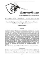
Figs. 1-3. Acrotomus lucidulus: 1, habilus; 2, facc; 3, propodeum. (After TOWNES,
1969).
Figs. 4-13. 4, ovipositor sheaths and subgcniial plate of Acrotomus lucidulus; 5,
same of Acrotomus succinetus; 6, samc of Acrotomus albidulus; 7, scape + basal
flagellar segmenls of A. succinclus; 8, samc of Acrotomus lucidulus; 9, ventral view
of female subgenilal plate and ovipositor sheaths of Acrotomus lucidulus; 10,
oviposilor of Acrotomus succinclus; 11, lateral and ventral view of egg of
Acrotomus succinetus; 12, samc of Scapnctcs ornatus; 13, samc of Acrotomus
lucidulus. (Figs. 4 and 9-13 after MASON, 1962; 5 aftcr KERRICII, 1952; 6 after
KASPAR YAN, 1986).
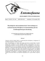
Figs. 14-19. Acrotomus succinetus (Ladakh speeimen): 14, face and clypeus; 15,
vertex; 16, tergites 1-2, lateral view; 17, tergites 2-4, dorsal view; 18, propodeum,
lateral view; 19, scutellum, metascutellum and propodeum, dorsal view.
40
© Entomofauna Ansfelden/Austria; download unter www.biologiezentrum.at
41
© Entomofauna Ansfelden/Austria; download unter www.biologiezentrum.at
11
A. succinctus
S. ornatus
42
A. lucidulus
© Entomofauna Ansfelden/Austria; download unter www.biologiezentrum.at
43
© Entomofauna Ansfelden/Austria; download unter www.biologiezentrum.at
References
CARLSON, R. W., 1979. Family Ichneumonidac. In KROMRP.IN, Hurd, et al.: Calalog of
Hymenoptera in America north of Mexico, 1: 315-741.
KASPARYAN, D. R. & TOLKANITZ, V. I., 1981. [A guide lo ihc insects of the Europcan part of Ihe
USSR. Vol. 3, Hymenoptera, Ichneumonidac. 2. Subfam. Tryphoninae]. Opred. Faune
SSSR.No. 129:98-166.
KASPARYAN, D. R., 1986. [Two new specics of ihc tribe Exentcrini (Hymenoptera,
Ichneumonidae) of the Far East]. In Lerr, P.A. (Ed.): [Systematics and ecology of insects
frorn the Far East]. Akad. Nauk SSSR, Vladivostock, 1986: 54-57.
KERRIOI, G. J., 1952. A review and a revision in grealer part, of the Ctcniscini of the Old
World (Hym., Ichneumonidae). Bull. Brii. Mus. (Nat. Hist.) Enl., 2: 3;.5^160.
MASON, W. R. M., 1956. A revision ofthe Ncarctic Cicniscini (Hymenoptera: Ichneumonidae)
II. Acrotomus Hlgr. and Smicroplccirus Thom. Canadian J. Zool., 34: 120-151.
MASON, W. R. M., 1962. Somc new Asialic specics of Exentcrini (Hymenoptera:
Ichneumonidac) with remarks on gencric limils. Canadian Ent., 94: 1273-1296.
MASON, W. R. M., 1978. Ichncumonoid parasites (Hymcnoplcra) accidentally introdueed into
Canada. Canadian Ent., 110: 603-608.
TOWNI-S, H., 1969. The genera of Ichneumonidac, Part 1. Mcm. American Enl. Inst., 11: 1300.
Aulhor'saddrcss:
Dr. Virenclra GUPTA
American Entomological Institute
3005 S.W. 56lh Avenue
Gainesville, Florida, 32608
U.S.A.
44
© Entomofauna Ansfelden/Austria; download unter www.biologiezentrum.at
Literaturbesprechung
HERRERA, L. & F.J. ARRICIBITA (1990): Los Caräbidos de Navarra Espana
(Coleoptcra, Carabidae). Dcscripciön, bionomfa, disiribueiön geogräfica y
clasificacion. - Entomograph 12. E.J. Brill / Scandinavian Science Press, Leiden,
New York, Kopenhavn, Köln. 241 S., zahlr. Abb. ISBN 90 04 08980 2.
Der vorliegende Band ist zugleich Katalog und Bestimmungsbuch für die
Laufkäfer der spanischen Provinz Navarra. Auf eine, allerdings sehr knappe,
Einführung in die geographischen und systematischen Grundlagen folgen
Bestimmungsschlüssel für die Unicrfamilicn, die Gattungen und die in der Provinz
Navarra nachgewiesenen Arten. Diese werden jeweils kurz beschrieben und zum
Teil in Ganz- oder Datailabbildungen vorgestellt. Kurze Angaben zur Biologie, zur
Verbreitung und zum Vorkommen in der Provinz Navarra vervollständigen die
Beschreibungen. Den zweiten Teil des Buches bildet ein Kartenteil, der für jede Art
die Gesamtverbreitung bzw. auf UTM-Gillcrkartcn die Funde in der Provinz
Navarra enthält. Diesem Kartenteil ist eine Tabelle vorangestellt, die das
Vorkommen jeder Art in allen anderen spanischen Provinzen zeigt. Leider sind aber
nur die in Navarra vorkommenden Arten aufgeführt, so daß diese Liste für die
anderen Provinzen und auch als Vergleichsbasis von wenig Wert ist. Ein recht
ausführliches Literaturverzeichnis schließt den Band.
Erfreulich ist, daß die Autoren eine recht konservative systematische Einteilung
benutzen, so daß der Leser nicht mit den zahlreichen auf Unterfamilien bzw. auf
Untergattungen beruhenden, inflationären Familien- und Gattungsnamen der
frankophonen Autoren belastet wird.
Man hätte sich vielleicht noch eine zoogeographische Würdigung der Fauna einer
Grenzprovinz, wie es Navarra ist, gewünscht, zumal hier relativ viele
mitteleuropäische Arten vorkommen. Aber dies lag vielleicht außerhalb der
Intentionen der Verfasser. Insgesamt eine ansprechende Studie, die nur leider eine
der faunenärmsten Provinzen der Iberischen Halbinsel behandelt. Ähnliche Arbeiten
würde man sich für faunistisch reichhaltigere und bedeutsamere Gebiete wünschen.
Ein wichtiger Nachschlagcband für alle, die mit faunistischen Fragen der Iberischen
Halbinsel, bzw. speziell mit ihrer Laufkäferfauna befaßt sind.
Martin BAEHR
D'VINCENT, C. (1990): Reisen mit den Walen. - 224 S., zahlreiche Farbfotos, Heyne
Verlag.
Einige Bücher wirken durch ihren Einband derart faszinierend, daß der flüchtige
Betrachter unversehens stehenbleibt, blättert und liest. Und dann existieren noch
45
© Entomofauna Ansfelden/Austria; download unter www.biologiezentrum.at
ganz wenige Bücher, deren Inhalt das hält, was der Einband verspricht. - So wie
dieses:
Der Autorin ist es gelungen, auf ihren Reisen mit Walen unglaubliches
Bildmaterial zusammenzutragen. Basierend auf diesen zugleich dokumentarisch wie
ästhetisch einmaligen Fotografien sowie ihren Beobachtungen entstand ein
faszinierendes Lebensbild der Buckelwale. Neben dem Drang, möglichst viel über
die Lebensweise dieser hochintelligenten Meeressäuger zu erfahren, stand für die
Autorin der Schutz der Wale vor bedenkenloser Dezimierung an vorderster Stelle
ihrer Forschungen. In Zusammenarbeit mit Fachkollcgcn glückte Cynthia d'Vincent
eine Synthese von Wort und Bild, die selten geworden ist. Das vorliegende Werk
bietet alle Voraussetzungen, um selbst dem stumpfsinnigsten linserer Zeitgenossen
die Augen für die großartige Welt der Buckelwale zu öffnen. Es wäre ein Verbrechen, diese Welt durch Ausrottung der letzten Wale unseren Kindern für immer
vorzuenthalten.
Michael CARL
Gow, G. (1989): Completc Guide to Auslralian Snakcs. - Angus & Robertson Publ.,
North Ryde, Australien. 171 S. Zu beziehen über: Gazelle Books Services, Falcon
House, Queen Square, Lancaster LA1 1 RN, England.
Abgesehen von einigen unterirdisch lebenden Blindschlangen (Typhlopidae) und
marinen Sccschlangen (Hydrophiidac), werden zum ersten Mal alle Schlangen
Australiens farbig abgebildet; auch Unterarten, Varietäten und Unterschiede zwischen jungen und erwachsenen Schlangen werden dokumentiert. Die einleitenden
Kapitel informieren über die allgemeinen Eigenschaften der Schlangen, das
Sammeln von Schlangen, Bestimmung, Haltung von Schlangen, Geschlechtsbestimmung, Fortpflanzung, Krankheiten und Parasiten sowie Giftapparat und
Schlangenbiß. Der systematische Teil beinhaltet Bestimmungsschlüssel der
Familien, Gattungen und Arten sowie Vcrbrcitungskarten. Im Text werden die
genauen Merkmale, Besonderheiten und Vergleiche mit ähnlichen Arten genau
beschrieben.
Der Autor gehört zusammen mit Worrcll und Cogger zu den großen
australischen Hcrpctologen. Seine über zwei Jahrzehnte lange Erfahrung bürgt für
Qualität. Zahlreiche der Farbfotos wurden in Gefangenschaft aufgenommen, was
ihren Wert aber keineswegs mindert. Der Laie wird troizdcm Schwierigkeiten bei
der Identifikation der Schlangen im Frciland haben, nicht weil die Fotos schlecht
wären, sondern weil sehr viele Schlangen sich sehr ähnlich sind und man die
scheuen Tiere ja meist nur kurz zu Gesicht bekommt. Für den Hcrpctologcn ist
dieses Buch ein unentbehrliches Nachschlagewerk.
Roland GERSTMEIER
46
© Entomofauna Ansfelden/Austria; download unter www.biologiezentrum.at
P., HAMILTON.C.J. (1990): Eniomology. A Guide to Information Sources.
- Mansell Publishing Ltd., London - New York. 2.Auflage, 259 S.
GILBERT,
Dieses Buch präsentiert eine Auswahl der wichtigsten Informationsquellen, die
für ein weltweites Studium der Entomologie wichtig sind. Natürlich wurde diese 2.
Auflage erweitert und aktualisiert: Die Anzahl der Zeitschriften beträgt nun über
330, dazu kommen über 60 "Spczial-Zeitschriftcn aus den Ordnungen Coleoptera,
Diptera, Lepidoptera und Odonata; mehr als 80 "ncwslelters" und neue Kapitel wie
"Angel-Entomologie" (z.B. "Insektenkunde für Fliegenfischer"), "SchmetterlingsGärten" und "Naturschutz" wurden aufgenommen. Die Quellen - seien es Bücher,
Zeilschriften, Personen, Institutionen oder Vereine - sind einfach durchnummeriert;
im Index wird dann auf diese Namen Bezug genommen. Beginnend mit der
"Geschichte der Entomologie" geht es über "Benennung und Bestimmung von
Insekten", "Zucht, Sammlungen, Sammclmcthodcn" zum umfangreichsten Kapitel
"Literatur", welches Zeilschriften und Bücher berücksichtigt. Kapitel 5 informiert
dann über Suche und Ausfindigmachen von Literatur. "Newsletters" wurden bereits
erwähnt; den Abschluß bilden Verzeichnisse von Entomologen und ihren
Organisationen sowie Übersetzungsdicnstc. Einige deutschsprachige Adressen (nur
die kann der Rezensent beurteilen) sind allerdings schon seit Jahren veraltet.
Eine überaus wichtige und verdienstvolle Zusammenstellung, auf die jeder
Entomologe gerne zurückgreifen wird.
Roland GERSTMEIER
R. (1990): Dynamic Biogcography. - Cambridge University Press,
Cambridge. 249 S.
HENGEVELD,
Die Einbeziehung biogeographischer Aspekte wird in den Wissenschaften
Ökologie, Taxonomie, Florislik und Faunistik immer bedeutender. "Dynamische
Biogeographie" betrifft dabei das Studium biologischer Muster und Entwicklungen
innerhalb eines breiten geographischen und zeitlichen Maßstabes. Das ultimative
Ziel, eine Interpretation evolutionärer Prozesse, wird über die Beschreibung und
Erklärung räumlicher Muster und Entwicklungen von Taxa erreicht. Im ersten Teil
des Buches werden qualilative und quanliiaiive Ansalze zu einer biogeographischen
Klassifizierung vorgestellt, wobei hinsichtlich der quantitativen Klassifizierung
einige statistische Modelle und tcsls diskutiert werden. Der 2. Teil betrachtet
geographische Trends bei Anenzahlcn über weile, sogar globale Flächen und Trends
bei biologischen Eigenschaften (z.B. Lebens- und Blaltformen bei Pflanzen). Teil 3
beinhaltet die quantitative intraspezifische geographische Variation und beschreibt
den Aktionsradius von Arten mil ihrer Struktur und Dynamik.
47
© Entomofauna Ansfelden/Austria; download unter www.biologiezentrum.at
Dieses Buch beinhaltet sehr viele theoretische Ansätze, steht damit auf einem
sehr hohen Niveau und kann fast ausschließlich nur Wissenschaftlern empfohlen
werden, die ökologisch und biogeographisch arbeiten.
Roland GERSTMEIER
J.A., LAMAR, W. W. (1989): The Vcnomous Rcptiles of Latin America.
- Comell University Press, New York. 425 S.
CAMPBELL,
Erstmalis werden in einem Buch die giftigen Reptilien Lateinamerikas (Amerika
südlich der USA) in solcher Ausführlichkeit behandelt: 145 Schlangenarten und die
beiden Krustenechsen (Heloderma horridus und H. suspectus). In den
Anfangskapiteln werden von zwei Ärzten medizinische Aspekte über
Schlangenbisse - Verhütung, Erste Hilfe und weitere medizinische (Antivenom-)
Behandlung -, eine sachdienliche Bibliographie und die Hersteller von Seren
vorgestellt. Danach wird ein Überblick über die einzelnen Länder gegeben, welcher
jeweils Beschreibungen von Landschaften, Klima und Vegetation sowie
Bestimmungslabellcn und Tabellen über die Verbreitung in den einzelnen
Landesteilcn enthält. Der Großteil des Buches ist der detaillierten Beschreibung der
Giftschlangen gewidmet: Charakterisierung der Gattung und der einzelnen Ar in,
exakte Beschreibung, Anmerkungen, genaue Verbreitungskarten und weiterführende
Literatur (über 1200 Titel). 504 in Tafeln arrangierte Farbfotos lassen das Buch zu
einem unentbehrlichen Nachschlagewerk für Ärtzc und Biologen werden.
Roland GERSTMEIER
Druck, Eigenlümcr, Herausgeber, Verleger und für den Inhalt verantwortlich: Maximilian Schwarz,
Konsulent für Wissenschaft der O.Ö. Landesregierung, P.ibcnwcg 6, A-4052 Ansfclden.
Redaktion: Erich Diller, Münchhausenstraßc. 21, D-8000 München 60.
Max Kühbandner, Marsstraße. 8, D-8081 Aschheim.
Wolfgang Schacht, Scherrcrstraßc 8, D-8081 Schöngeistig.
Thomas Witt, Tengslraße 33, D-8000 München 40.
Postadresse: Entomofauna, Münchhausenslr. 21, D 8000 München 60.
48