Bulletin of the California Lichen Society 11-1
Bạn đang xem bản rút gọn của tài liệu. Xem và tải ngay bản đầy đủ của tài liệu tại đây (1.46 MB, 36 trang )
Bulletin
of the
California Lichen Society
Volume 11
No.1
Summer 2004
The California Lichen Society seeks to promote the appreciation, conservation and study
of the lichens. The interests of the Society include the entire western part of the continent,
although the focus is on California. Dues categories (in $US per year): Student and fixed
income - $10, Regular - $18 ($20 for foreign members), Family - $25, Sponsor and Libraries
- $35, Donor - $50, Benefactor - $100 and Life Membership - $500 (one time) payable to the
California Lichen Society, P.O. Box 472, Fairfax, CA 94930. Members receive the Bulletin and
notices of meetings, field trips, lectures and workshops.
Board Members of the California Lichen Society:
President:
Bill Hill, P.O. Box 472, Fairfax, CA 94930,
email: <>
Vice President: Boyd Poulsen
Secretary:
Sara Blauman
Treasurer:
Kathy Faircloth
Editor:
Tom Carlberg
Committees of the California Lichen Society:
Data Base:
Charis Bratt, chairperson
Conservation:
Eric Peterson, chairperson
Education/Outreach: Lori Hubbart, chairperson
Poster/Mini Guides:
Janet Doell, chairperson
The Bulletin of the California Lichen Society (ISSN 1093-9148) is edited by Tom Carlberg,
<>. The Bulletin has a review committee including Larry St. Clair,
Shirley Tucker, William Sanders and Richard Moe, and is produced by Richard Doell. The
Bulletin welcomes manuscripts on technical topics in lichenology relating to western North
America and on conservation of the lichens, as well as news of lichenologists and their activities. The best way to submit manuscripts is by e-mail attachments or on 1.44 Mb diskette
or a CD in Word Perfect or Microsoft Word formats. Submit a file without paragraph formatting. Figures may be submitted as line drawings, unmounted black and white glossy photos
or 35mm negatives or slides (B&W or color). Contact the Production Editor, Richard Doell, at
<> for e-mail requirements in submitting illustrations electronically. A
review process is followed. Nomenclature follows Esslinger and Egan’s 7th Checklist on-line
at < The editors
may substitute abbreviations of author’s names, as appropriate, from R.K. Brummitt and
C.E. Powell, Authors of Plant Names, Royal Botanic Gardens, Kew, 1992. Style follows this issue. Reprints may be ordered and will be provided at a charge equal to the Society’s cost. The
Bulletin has a World Wide Web site at < />and meets at the group website < />Volume 11(1) of the Bulletin was issued June 11, 2004.
Front cover: Solorina spongiosa (Sm.) Anzi. Photo curtesy of Steve Sharnoff.
Bulletin of the California Lichen Society
Volume 11
No.1
Summer 2004
Solorina spongiosa: A new species record for Nevada
Cheryl Beyera and Larry St. Clairb
Forest Botanist, Modoc National Forest, Alturas, California 96101
Email: <>
b
Curator of Nonvascular Cryptogams, Brigham Young University, Provo, Utah 84602
Email: <>
a
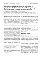
Abstract: Solorina spongiosa (“fringed chocolate chip lichen”) is reported new to Nevada from the Spring
Mountains where it grows over and among several species of mosses at elevations above 2900 m.
Solorina is a small genus of five species within the
Peltigeraceae. All but one species occur on moist
calcareous soil in cold regions. Solorina spongiosa
(Sm.) Anzi, with the most reduced thallus of the
group, is a bipolar arctic-alpine species, reported
from Europe, North America, South Island (New
Zealand), and James Ross Island (Antarctica), but
seldom collected. The map for Solorina spongiosa in
Lichens of North America (Brodo et al. 2001) indicates
that this lichen, within U.S. borders, is confined
to Alaska, Montana, Colorado and New Mexico.
Manierre (1999) notes that it is rare wherever it
appears and Geiser et al. (1994) lists it as rare in
western North America. Finding crustose lichens in
southern Nevada is expected, but the discovery of
Solorina spongiosa was a surprise to most (Bungartz,
pers. comm.; McCune, pers. comm.; Rosentreter,
pers. comm.). However, St.Clair (1999) lists it
as “Common…in upper montane throughout
northern Rocky Mountains south into Colorado
Rockies.” In this paper it is reported as new to
Nevada.
Solorina spongiosa is a rarely collected, brown,
grayish, or greenish squamulose, granulose to
Figure 1. Solorina spongiosa collected in the Spring
Mountains, Nevada. Urceolate apothecia are surrounded
by a ring of tissue containing a green alga, and imbedded
in squamules containing the cyanobacterium, Nostoc.
Photo by Bill Hill.
coralloid, spongiose lichen. The apparent thallus,
which is appressed to the soil or moss substrate,
forms a dark, warted to coralloid mass, gelatinous
when wet. It is composed of cephalodia containing
the cyanobacterium, Nostoc. The true thallus
contains a green alga and is reduced to a thin ring
or collar surrounding a large urceolate apothecium
(Figure 1). Its paraplectenchymatous upper cortex
1
Bulletin of the California Lichen Society 11(1), 2004
contains Coccomyxa in the algal layer. Brodo et al.
(2001) consider the green alga to be the primary
photobiont for the genus. The underside lacks a
cortex. The apothecia and squamules are attached
to the substratum by rhizines. The apothecia are
sunken in the upper surface of the thallus lobes,
the disk is dark brownish red to blackening.
Dobson (2000) describes the apothecia as up to 5
mm in diameter. The hymenium is hyaline, and
the paraphyses are unbranched with the tips redbrown, coherent, and little thickened. Ascospores
are brown, 1-septate (Figure 2), 4/ascus, 30-50 x
18-22 µm, with a
warted,
furrowed
surface.
Solorina
spongiosa occurs over
mosses in subalpine
and alpine calcareous
areas. An exception
is at Pictured Rocks
National Lakeshore,
Alger
County,
Michigan, where it
Figure 2. Solorina spongiosa spores,
has been reported,
40X, from collections at Three
surprisingly,
on
Springs, Spring Mountains, NV.
sandstone (Manierre
Photo by C. Beyer.
1999). Lichens are
hosts to many, often
specialized host-specific fungal parasites. A lichen
parasite is often found on Solorina spongiosa (F.
Bungartz, pers. comm.). The above description is
a compilation from Jahns et al. (1995), Martinez
and Burgaz (1999), McCune (2002), McCune
and Goward (1995), Nash (2002), Øvstedal and
Smith (2001), Thomson (1984) and Thomson and
Thomson (1984).
Site Location and Description
The Spring Mountains are located in southern
Nevada near the California border. Pahrump Valley
and the Amargosa River basin lie to the west and
Las Vegas Valley, draining into the Colorado River,
lies to the east (Charlet 2001). Las Vegas, with 1.5
million people, is 48 km to the southeast. The range
is a sedimentary escarpment 68 km long and up to
26 km wide, with elevations ranging from about 853
m to the highest point on Mt. Charleston at 3633 m.
This ‘sky island’ is among the most isolated ranges
in North America, its nearest neighbor being the
Panamint Range of California, 161 kilometers away
(Mohlenbrock 1992).
2
Geologically, the range is made up of many
sedimentary layers of limestone, dolomite,
sandstone, shale, and gypsum deposited by a
shallow sea that covered the region 590 to 250
million years ago (mya), during the Paleozoic era.
The mountains themselves were formed about 60
mya, close to the end of the Cretaceous Period,
when east-west pressure caused the sedimentary
layers to buckle and shear. During the Pleistocene
– 1.6 million to 12,000 ya – southern Nevada was
much cooler and wetter than it is today. As the
Pleistocene ended, the plants that had become
established in the Spring Mountains became
isolated (Mohlenbrock 1992).
Charlet (2001) notes it as the most biologically
diverse of all mountain ranges in Nevada, with
37 tree species and 17 endemic plants. On the
lower slopes, plants typical of the Great Basin
such as sagebrush and creosote merge into the
Mojave Desert flora where a variety of cacti and
other desert-dwelling plants live. Higher in the
range, pinyon pine and Utah juniper take over the
drier habitats while ponderosa pine and white fir
dominate the more mesic canyons. Bristlecone pine
range from as low as 2103 m to tree line at 3048 to
3353 m. At the higher elevations, limber pine joins
bristlecone pine. Charlet (2001) notes that there
are probably more than 1000 plant species in the
Spring Mountains, representing about one-third of
the entire Nevada flora. An additional 8 species are
endemic to southern Nevada and California and
another 3 are endemic to southern Nevada and
Utah. A high number of moonwort species of ferns
grow in limited habitat available within the Spring
Mountains, including some of the same habitats
where Solorina spongiosa is found. Several endemic
vascular species also occur in these mesic, upper
elevation sites.
The Spring Mountains are administered by two
federal agencies: the Bureau of Land Management
(BLM) manages some lower elevation areas,
including Red Rock Canyon National Conservation
Area; and the Humboldt-Toiyabe National Forest
manages the higher elevations of the range. In
August 1993, Congress established the Spring
Mountains National Recreation Area, administered
by the U.S. Forest Service.
Solorina spongiosa in Nevada
In July of 2002, Solorina spongiosa was collected
in the Spring Mountains, Clark County, Nevada,
at Three Springs (Figure 3) in upper Lee Canyon,
above the Lee Canyon Ski and Summer Resort
(Beyer 20020710.1 OSC). Specimens were found
growing on a vertical limestone surface over moss
between 2957 and 2987 m elevation (UTM 11,
618206E 4016990N), in open canopy. During spring
runoff this microhabitat is very wet to saturated.
Later in the summer and fall, the moss cover
provides a moist environment. Small specimens
were also found growing over moss on soil in the
vicinity of the limestone boulder. St. Clair (pers.
comm.) has seen Solorina spongiosa growing on
vertical surfaces of small frost heaves in alpine
habitats throughout the Rocky Mountain region. A
small, 1-2 meter diameter floating mat bog is found
a few meters from the Lee Canyon site. We do not
know of any other floating bogs in Nevada.
Figure 3. Three Springs area, August, 2002, habitat
picture of collection site. Endemic Clokey thistle (Cirsium
clokeyi) in foreground. Photo by C. Beyer.
Extensive vascular plant collections were made
in the mid-1900s, primarily by Ira Clokey (1951),
but the moss and lichen flora has remained
relatively unknown until fairly recently. Elva
Lawton collected bryophytes at a few locations
in the 1950s, and Lloyd Stark of the University of
Nevada Las Vegas has collected bryophytes over
the past eight years. Preliminary data show that
the moss flora of the Spring Mountains differs from
that in the surrounding desert, with species more
characteristic of cooler, wetter climates. However,
until recently, the lichens were unknown (St. Clair
2004). Larry St. Clair of Brigham Young University
(Utah) has, over the last five years, made extensive
collections from various locations in the Spring
Mountains, primarily to support the air quality
biomonitoring program established in cooperation
with the U.S. Forest Service (St. Clair, pers.
comm.). Beyer has augmented that collection with
several species. Currently, ninety-eight species of
lichens are known from the Spring Mountains,
primarily from U.S. Forest Service lands. Besides
Solorina spongiosa, other species found that may
be considered uncommon include Dermatocarpon
luridum, Stenocybe mccunei, and Cladonia cariosa.
A second site within the Spring Mountains was
later discovered approximately 5 km to the east at
Mummy Springs (Figure 4), where a small specimen
Figure 4. Mummy Springs site in November, 2003. Photo
by C. Beyer.
was found growing over moss on a limestone cliff
at 3048 m elevation. Mummy Springs is in the
Deer Creek drainage just south of Lee Canyon.
Population size is unknown; however, habitat for
this species is very limited at this location, as the
drainage is essentially dry except at the spring.
Although the Spring Mountains are a desert
mountain range, the upper elevations often receive
several feet of snow cover in the winter. Snowmelt
and occasional rainstorms provide water that
3
Bulletin of the California Lichen Society 11(1), 2004
percolates through cracks and fissures in the porous
limestone, coming to the surface as springs when it
meets an impermeable layer. Both collection sites in
the Spring Mountains are in spring areas, between
2957 and 3048 m, that are seepy to saturated during
spring runoff, drying out somewhat in the summer
months, and covered by a thick layer of snow/ice
during the winter. Over 200 springs of various sizes
have been documented in the range, and other
potential occurrences of Solorina spongiosa may
exist. However, most of the springs are too low in
elevation, or on an aspect that makes the site too
hot to support Solorina spongiosa.
Both documented sites of Solorina spongiosa are
on moss over calcareous substrata within the
bristlecone pine zone with quaking aspen nearby,
in east to northeast-facing canyons below the two
highest peaks in the range: Mt Charleston and
Mummy Mountain. Brodo et al. (2001) found that
the most significant property of a potential rock
substrate, in terms of lichen distribution, is its
calcium carbonate (CaCO3) content. Calcicoles,
those species that prefer alkaline rocks made of
CaCO3, such as limestone, often cannot tolerate
acidic conditions.
Apothecia
One apothecium from a specimen collected at the
Three Springs site had unusual width dimensions
between 9 and 10 mm. However, the diameters of
most of the apothecia seen fell within the normal
range according to the literature, equal to or less
than 5 mm.
Distribution
Knowledge concerning the regional distribution
of Solorina spongiosa has expanded from what
was known just a few years ago when Lichens of
North America (Brodo et al. 2001) was published.
Collections within the contiguous U.S. have
been located that report Solorina spongiosa from
Michigan, Montana, Idaho, California, Colorado,
Utah, Washington, Wyoming, and New Mexico.
In the Pacific Northwest, Oregon is the only state
where a collection has not been reported (Figure
5). This is likely related to the lack of calcareous
substrata along the Cascade Crest.
Ryan (pers. comm.) indicated that the occurrences
in Arizona and California would be reported as
4
Figure 5. Western states with collections of Solorina
spongiosa are shown in gray.
new records in the Sonoran Flora v. II and the new
California checklist, respectively. The California
collection is the occurrence closest to the Spring
Mountains site. Air distance between the two sites
is 274.4 km < />java/lat-long.htm>.
The term “bipolar” indicates occurrence in both
the arctic and Antarctic. Smith and Øvstedal
(1994) found that 41% of Antarctic lichens are
bipolar. The worldwide distribution of Solorina
spongiosa, a bipolar arctic-alpine species with a
strong affinity for calcareous substrates, indicates
one of two possible scenarios. Either this lichen
occurs as a relict from a time when continents were
connected and cold, moist habitats were prevalent,
or, following continent drift, it has been effectively
dispersed from its origin by means of spores to
suitable habitats that are extremely cold for part
of the year, and cool and moist for the remainder.
Smith and Øvstedal (1994) venture to say that
bipolarity probably represents many worldwide
distributions that became dissected with climate
change and continental movements. I.M. Brodo
(pers. comm.) suggests that there is probably a mix
of long distance dispersal on the one hand, and
mountain hopping on the other, as well as some
relict distributions. He states, “We know that many
lichen distributions are very ancient, and newly
available genetic techniques will undoubtedly be
used to sort out these phytogeographic puzzles,
with a variety of origins for bipolar distributions
emerging.”
Solorina spongiosa in Nevada
Threats
Both sites where Solorina spongiosa has been found
are within 48 kilometers of one and one-half million
people in the city of Las Vegas. Both sites are also
very accessible to day hikers. The main threat to
this species in the Spring Mountains is from local
recreationists. For example, the Three Springs site
is just above the Lee Canyon Ski and Summer
Resort, which is currently seeking a permit to
expand operations. This area also receives heavy
summer use from hikers, especially those who
wish to reach the top of Mt. Charleston by a route
that is shorter than the North Loop Trail. The usercreated path along the brook emanating from the
spring has eliminated plants in its treadline. This
sensitive area supports endemic vascular plants,
moonworts, and Solorina spongiosa.
Mummy Springs, also a site of high biodiversity,
including moonworts, receives high recreation use
as a popular day-use destination, and also as a rest
spot on the way to the upper elevations of Mummy
Mountain. In 2003 a bypass trail was constructed
to divert use from the spring area. A similar
mitigation may be available in the near future
for user trails along Three Springs. However, this
would not necessarily ameliorate possible impacts
from an expansion of the ski area.
Another potential threat is air pollution from
an expanding megalopolis, which is predicted
to have 2.6 million people by 2020. Over 5,000
people a month come to live in Las Vegas
traffic, services, and facilities will yield increasing
air pollution. Solorina spongiosa sensitivity to air
pollution is unknown, but locations where it is
found are historically in remote arctic/alpine
areas.
Conclusion
In this paper Solorina spongiosa, commonly known
as the “fringed chocolate chip lichen,” is reported
as new to Nevada from the Spring Mountains, near
Las Vegas, where it grows over and among several
species of moss. This remarkable occurrence was
unexpected as many were not aware that sites
for this lichen had already been discovered in the
southwest in New Mexico, Arizona, Utah and
California. Additionally, many were not aware
of the relatively restricted habitat in the Spring
Mountains, where appropriate geology, elevation,
moisture and aspect come together to provide a
suitable microsite for this species, in the middle of
the Mojave desert.
Acknowledgements
The first author has many people to thank,
including several members of CALS. I would like
to thank especially Bill Hill and Darrell Wright.
Additionally, I am grateful to a number of people
who answered the list server with specimens to
report, and to Bruce McCune for confirming the
identification, Lloyd Stark for helping me with
a related independent study, Tom Carlberg for
comments on a draft of this article, and Trista
Crook who sent copies of packet labels from the
University of Colorado at Boulder. Last, but not
least, I wish to thank Barbara Lachelt for helping
me get started with lichens in 1995 during her
CALS workshop at San Francisco State University.
Appendix I: Representative collections of Solorina spongiosa in western U.S.
Table 1. Western states from which collections of Solorina spongiosa have been reported.
STATE
HERBARIUM*
COLLECTOR
Arizona
ASU
Nash
California
CAS
Shevock (#12531)
Colorado
BRY #3462
Shushan
Idaho
Rosentreter
Rosentreter (#9385)
Michigan
herbarium not known
Re: Manierre (1999)
Montana
Rosentreter
Rosentreter (#2071)
New Mexico
BRY #9629
Egan
Utah
CU #407700
Flowers
Washington
herbarium not known
Re: Thomson (1984)
Wyoming
BRY #3417
Wirth
* ASU, Arizona State University; BRY, Brigham Young University; CAS, California Academy of
Sciences; CU, University of Colorado.
5
Bulletin of the California Lichen Society 11(1), 2004
References
Brodo,
I.M. 2004. Personal communication.
Canadian Museum of Nature, Ottawa.
Brodo, I.M., S. Duran Sharnoff, and S. Sharnoff.
2001. Lichens of North America, Yale
University Press.
Bungartz, F. 2002. Personal communication.
Arizona State University.
Charlet, D.A. 2001. < />mtn/html/springr.html> accessed 12/17/
2003
Clokey, I. 1951. Flora of the Charleston Mountains,
Clark County, Nevada. University of
California Press, Berkeley and Los
Angeles.
Dobson, F.S. 2000. Lichens An illustrated guide to
the British and Irish species. The Richmond
Publ. Co. Ltd., Slough, England.
Geiser, L.H., K.L. Dillman, C.C. Derr, M.C.
Stensvold. 1994. Lichens of Southeastern
Alaska, USDA-Forest Service, Petersburg,
AK.
Jahns, H.M., P. Klockner and S. Ott. 1995.
Development of thalli and ascocarps in
Solorina spongiosa (Sm.) Anzi and Solorina
saccata (L.) Ach. In: Studies in Lichenology
with Emphasis on Chemotaxonomy,
Geography and Phytochemistry, JG
Knoph, K Schrufer, HJM Sipman, ed., J
Cramer, Berlin, Stuttgart, 241-251.
Manierre, W.R. 1999. Bryophytes and lichens of
the Huron Mountain Club, Evansia 16(4):
153-166.
Martinez, I., and A.R. Burgaz. 1999. Revision of the
genus Solorina (lichenes) in Europe based
on spore size variation. Annales Botanici
Fennici, 35, 137-142.
McCune, B. and Goward, T.G. 1995. Macrolichens
of the Northern Rocky Mountains, Mad
River Press, Inc., Eureka, CA.
McCune, B. 2002. Key to the lichen genera of the
Pacific Northwest.
< />pnw.PDF>.
McCune, B. 2002. Personal communication. Oregon
State University.
Molhlenbrock, R.H. 1992. Charleston Mountains,
Nevada. Natural History 3/92.
6
Nash, T. 2002. The lichen flora of the greater
Sonoran Desert region. Thomas-Shore Inc.,
Dexter, MI.
Øvstedal, D.O. and R.I.L Smith. 2001. Lichens of
Antarctica and South Georgia, A guide to
their identification and ecology. Cambridge
University Press. Cambridge, England.
Rosentreter, R. 2003. Personal communication.
USDI Bureau of Land Management.
Ryan, B. 2003. Personal communication. Arizona
State University.
Smith, R.I.L., and D.O. Øvstedal. 1994. Solorina
spongiosa in Antarctica: an extremely
disjunct bipolar lichen. The Lichenologist,
26, 209-213.
St. Clair, L., S.B. St. Clair, and L.D. Porter. 2003.
Interim Report: Establishment of lichen
air quality biomonitoring program and
baseline for the Spring Mountains National
Recreation
Area,
Humboldt-Toiyabe
National Forest, Nevada.
St. Clair, L. 1999. A color guidebook to common
Rocky Mountain lichens. ML Bean Life
Science Museum of Brigham Young
University, Provo, UT.
St. Clair, L. 2003. Personal communication. Brigham
Young University.
Thomson, J.W. 1984. American Arctic lichens 1. The
macrolichens, Columbia University Press,
New York.
Thomson, N.F., and J.W. Thomson. 1984. Spore
ornamentation in the lichen genus Solorina.
The Bryologist, 87, 151-153.
Tucker, S.C. 2001. New reports or divergences in
range for lichens of California, based on
Lichens of North America by I. Brodo, S.D.
Sharnoff, and S. Sharnoff, 2001. Bulletin of
the California Lichen Society 8 (2): 59-71.
USDI,
BLM. 2000 < />s u r v e y a n d m a n a g e / M R /
Lichens/dermato.pdf>,
lvrj_home/1998/Jul-05-Sun-1998/
news/7505334.html>,
Scoliciosporum sarothamni (Vain.) Vezda, New to California.
Doris E. Baltzo
Pleasant Hill, CA 94523 <>
Scoliciosporum sarothamni (Vain.) Vezda has been
reported from the Pacific Northwest (for example
in Seattle, Washington and British Columbia), (see
Tonsberg, 1995; Brodo et al, 2001 with a mention
of S-shaped spores) and it also occurs in Europe.
However, there does not seem to be a published
report of its occurrence in California.
could be mistaken for green algae.
The Alexander collection had S-shaped colorless
ascospores measuring 28.8-31.4 x 2.0-3.6 µm
which had three to seven (or more) indistinct septa
A population of Scoliciosporum sarothamni (Vain.)
Vezda was found on the bark of Pinus radiata D.
Figure 2. Spores of Scoliciosporum sarothamni
showing septae and curvature. Photo by Bill
Hill.
Figure 1. Soredia and apothecia (arrows) of
Scoliciosporum sarothamni (Baltzo 13113-O). Photo
by Bill Hill.
Don (Montery pine) in the Oakland-Berkeley Hills
by Earl Alexander on October 20, 2002, while he
was making a survey of lichens on plants on or
near serpentine. The sorediate crust resembled a
Lepraria; however some inconspicuous, minute
apothecia were found to be present (Figure 1), and
the soredia were localized within circular soralia.
The apothecia were pale to yellowish-brown or
darker brown and lacked a visible exciple. The
yellow-green to green soredia covering the thallus
(Figure 2). The soredia were KC+ black, but the
KC test under the microscope showed that only
small groups of cells had turned black. This could
indicate scanty or scattered amounts of gyrophoric
acid. (Tonsberg, 1992, noted that microscope
preparations of the soredia reacted “C+ fugitive
faintly red” and stated that “gyrophoric acid was
present [trace].”) The apothecium had a mediumbrown epithecium and a hyaline hypothecium
(Figure 3). The thallus was UV-. Comparison
with several descriptions and keys from around
the world pointed to S. sarothamni. See pertinent
information below.
This lichen has been reported as toxitolerant, i.e., it
is a species which may occur in polluted areas. It is
7
Bulletin of the California Lichen Society 11(1), 2004
Bacidia umbrina (Ach.) Bausch. Hasse, (1913)
referred to the spores of his material as acicular,
bowed and doubly arcuate, whereas the spore
shape was not mentioned by Fink (1935). Sirois
(1988) reported S. umbrinum var. compacta (Koerber)
Vezda on serpentine in Quebec.
An attempt here has been made to gather pertinent
information about Scoliciosporum in the world from
the literature:
Figure 3. Squash mount of Scoliciosporum
sarothamni showing brown (darker) epithecium
and hyaline hypothecuim. Photo by Bill Hill.
not known whether it occurs only in polluted areas
throughout its range. (See Tonsberg’s discussion,
1995). Redwood Regional Park is partially
surrounded by freeways and a variable amount of
air pollution may be present.
Collection data: Corticolous on branch of Pinus
radiata D. Don, in Redwood Regional Park,
Oakland-Berkeley
Hills,
Alameda
County,
California. Latitude 37º, 80.5’ N, longitude 122º,
17.8’ W, 345 msm, collected by Earl Alexander on
October 20, 2002 (Baltzo 13113-O, UC 1751254).
Pyrrhospora quernea (Dickson) Koerber, another
soraliate crustose corticolous lichen looks somewhat
similar, but its soredia are a more pronounced
yellow color, its apothecia have a distinct lecideine
margin, i.e., with no algae, which is easy to see,
and its spores are ovoid and unicellular rather
than spirally curled and multiseptate. A K+
pinkish-purple reaction occurs in the apothecium
of P. quernea which is also said to be UV+ orange
(Tucker, pers. comm., 2004).
The rock lichen Scoliciosporum umbrinum (Ach.)
Arnold has been reported in California (Hasse,
1903), as Biatora umbrina (Ach.); Hasse, 1913, as
forma psotina (Fries) T. Fries of Bacidia umbrina; Fink,
1935, as Bacidia umbrina (Ach.) Branth & Rostr.,
mostly on rock, rarely on wood, with apothecia
light brown to black; Tucker & Jordan, 1979, as
8
S. schadeanum (Erichs.) Vezda
Apothecia white to whitish-flesh or whitish-pink
or in age turning brownish, 0.1-0.2 (0.3) mm diam.,
spores 1-2 µm wide (thick) x 24-30 µm. Corticolous.
Paraphyses frequently not close, apices sparingly
branched, epithecium not granulose (Vezda, 1978).
S. pruinosum ( P. James) Vezda
Apothecia white, whitish-flesh or in age turning
brownish, 0.1-0.2(0.3) mm diam., spores 1.2 µm
wide (thick) x 20-33 µm. Corticolous. Paraphyses
close together, apices abundantly branched,
epithecium filled with tiny granules (Vezda, 1978).
See photo of thickly pruinose white apothecia in
Wirth, 1995.
S. sarothamni (Vain.) Vezda
The only other sorediate Scoliciosporum is S. gallurae,
which has spores that are straight to slightly curved,
while S. sarothamni has distinctly curved spores and
discrete soralia (Vezda, 1978). Purvis et al, (1992)
mentions morphs on bark with pale apothecia and
irregular, pale green soralia (KC+red). Tonsberg
(1992), states that the spores are spirally curved.
Vezda, (1978) indicates that apothecia are brown
to black, the thallus is sorediate, the soredia are
yellowish, the spores are 3(-7)-septate, 22-40 x 2
µm, and the thallus is generally corticolous and
rarely on rock.
S. umbrinum (Ach.) Arnold (syn. S. homomelaenum
(Flk.) Massal.)
Thallus not sorediate, spores wider than 2 µm.
Apothecia brown to black. Spores spirally twisted,
always about 3 µm wide. Apothecia 0.3-0.8 mm
diam., spores 3(-7)-septate; on rock and rarely on
bark. Apothecia sessile (not stipitate) (Vezda, 1978).
See also Purvis et al (1992).
Scoliciosporum sarothamni in California
S. ophiosporum (Hellb.) Hav. (syn. Bacidia kuopioensis
(Vain.) Vain.)
Apothecia brown to black, thallus not sorediate,
spores wider than 2 µm, spirally contorted.
Apothecia 0.3-0.8 mm diam.; spores 3(-7)-septate;
on rock and rarely on bark. Apothecial base tightly
constricted and in part stipitate (Vezda, 1978).
S. perpusillum Lahm ex Koerb.
Apothecia brown to black, thallus not sorediate,
spores wider than 2 µm, spirally contorted.
Apothecia 0.1-0.3 mm diam., spores (3-)5-7septate;
on bark (Vezda, 1978). Also reported from the
coastal-fir dry subzone of British Columbia (Noble,
1982) with thallus commonly granular, abundant
apothecia, hyaline, acicular, curled spores, 2035(48) x 2.0-2.5 µm.
S. chlorococcum (Stenh.) Vezda
Apothecia brown to black, thallus not sorediate;
spores curved like a bow (arcuate) to sub-straight,
4-5 x 20-40 µm, 7-septate commonly. On bark,
rarely on rock or wood (Vezda, 1978). See photo
(Wirth, 1995).
S. gallurae Vezda & Poelt
Apothecia pale to dark brown, sessile, flat to
convex. Continuous mass of soredia and hyphae,
discrete soralia sparse or absent. Spores straight
to slightly curved, 15-22 x 2.5-3.5 µm, fusiform to
slightly curved (Tonsberg, 1992). Has a resemblance
to S. chlorococcum but Nimis & Poelt (1987) indicate
that ascospores are commonly 3-septate, 15-22 x 4-5
µm and rarely simple or 1-septate.
Grateful thanks and appreciation to Isabelle
Tavares, Shirley Tucker, Tom Carlberg, Richard
L. Moe and Bill Hill for their help, comments,
constructive criticism, photos, and encouragement.
Brodo, I.M., S.D. Sharnoff, & S. Sharnoff. 2001.
Lichens of North America. Yale University
Press, New Haven. 795 pp.
Fink, B. 1935. The lichen flora of the United States.
University of Michigan Press, Ann Arbor.
426 pp., 46 plates.
Hasse, H.E. 1903. Additions to the lichen flora of
southern California. Part II. Bulletin of the
Southern California Academy of Sciences
2: 58-60.
Hasse, H.E. 1913. The lichen flora of southern
California. Contributions from the United
States National Herbarium 17: 1-132.
Nimis, P.L. & J. Poelt. 1987. The lichens and
lichenicolous fungi of Sardinia (Italy):
An annotated list. Studia Geobotanica 7
(Supplement l): 1-269.
Noble, W.J. 1982. The lichens of the coastal
Douglas-fir dry subzone of British
Columbia. Unpublished Ph.D. dissertation,
University of British Columbia, Vancouver.
Part II reprinted and updated in 1997.
Almost 1000 pp.
Purvis, O.W., B.J. Coppins, E.L. Hawksworth, P.W.
James, & D.M. Moore (Eds.) 1992. The
lichen flora of Great Britain and Ireland.
Natural History Museum and Publications
& British Lichen Society, London. 710 pp.
Sirois, L., F. Lutzoni, & M.M. Grandmer. 1988. Les
lichens sur serpentine et amphibolite du
amphibolite du plateau du mont Albert,
Gaspesie, Quebec. Canadian Journal of
Botany 66: 851-862.
Tonsberg, T. 1992. The sorediate, isidiate,
corticolous, crustose lichens in Norway.
Sommerfeltia 14: 1-331.
Tonsberg, T. 1995. Additions to the lichen flora
of North America IV. Scoliciosporum
sarothamni. Evansia 12(1): 27-30.
Tucker, S.C., W.P. Jordan. 1979 (1978). A catalog
of California lichens. Wasmann Journal of
Biology 36: 1-105.
Vezda, A. 1978. Neue oder wenig bekannte
Flechten in der Tschechoslowakei II. Folia
Geobotanica Phytotaxonomica. Praha 13:
397-420.
Wirth, V. 1995. Die Flechten Baden-Wuertembergs,
Teil 2. 555 color photos, 1006 pp.
9
Bulletin of the California Lichen Society 11(1), 2004
A Study of Acarosporas in The Lichen Flora of the Santa Cruz Peninsula by A.W.C.T. Herre
Kerry Knudsen
University of Riverside Herbarium, University of California at Riverside 92521-1024
Email: <>
Acarospora is a crustose genus with global
distribution. Many species occur on several
continents and most wide-spread species of
Acarospora are extremely variable. Part of this
variability appears to be genetic. The other part
of the variability is phenotypic plasticity: the
variation of characters caused by the interaction
of the environment with the genotype. It is not
always possible to know the causes of a particular
variation.
The
two
most
significant
characteristics
distinguishing the genus are the large number of
spores per ascus (24-200) and the non-amyloid
(K/I-) apical cap of the ascus. The hymenium
is usually over 80 µm, though the beautiful A.
glaucocarpa averages a hymenium 60 µm in height.
The width of paraphyses, measured near the
base, is an important characteristic in delineating
species. Spore size is not always diagnostic,
though in some species it is decisive, such as A.
thelococcoides (Knudsen 2003) or A. oligospora. The
cortex is paraplectenchymatous (though this is
rather too general in practice as the hyphal walls
can be distinct, anticlinal to intricate, with cells
angular to globose). The cortical layer has two or
three layers: (1) sometimes an amorphous upper
layer of gelatinized hyphae or necral material; (2)
a pigmented layer; and (3) a lower non-pigmented
layer. This arrangement is sometimes diagnostic, as
are the size of the hyphal cells of the cortex. Overemphasis of this aspect was one factor that led
Magnusson to split species too narrowly.
ssp. lesdainii.
Another important characteristic of Acarospora is
the development of the thallus. Acarospora thalli
generally begin as areoles broadly attached to the
substrate but many species eventually develop
stipes. A few species have very slender stipes, but
many have thick short stipes called a gomphus.
These raise the thallus slightly off the surface of the
substrate. A definite lower surface is formed which
may be corticate or ecorticate. The color of the lower
surface may vary from white or brown to black.
Though not always a valuable character and much
abused in some keys, the color of the underside is
consistent in some species and diagnostic.
A modern revision of Acarospora in both California
and North America is badly needed. Our state
probably has more than twenty species including
at least two endemics. No comprehensive keys
for California Acarospora exist at this time and
taxonomic problems subvert the value of older
keys.
In his landmark flora, Herre (1910) listed eight
species of Acarospora as occurring in the Santa Cruz
Peninsula and named two new species: A. hassei
and A. arenosa. I will discuss each of the taxa. The
names and authorities used in the headings below
are those used in Herre’s flora and are sometimes
incorrect. They are corrected in discussions.
Acarospora chlorophana (Walhb.) Mass
Hyphal bands through the algal layer are important
characteristics in some species such as A. smaragdula
10
Recently
Acarospora chlorophana was transferred
Acarospora study
to the genus Pleopsidium because its ascus tip
stains Lecanora-type with an apical amyloid
ring (K/I+blue) and it has a cortex that is
prosoplectenchymatous (Hafellner, 1993). Due to
the current method of determining Pleopsidium by
the morphology of their thallus yellow Acarospora
are often misdetermined as Pleopsidium because the
ascus stain is not routinely checked. It is important
to stain the asci of all specimens. It takes a little
practice to get the right stain but using 5% KOH
with diluted IKI will make the task easier. The stain
is not as clear as the above technical description of
ascus structure suggests. One mainly has to see if
there is any blue reaction in the tholus.
In California there should be an effigurate species
of Acarospora named A. novomexicana H. Magnusson
occurring at both lower and higher elevations.
William Weber misdetermined it as A. chlorophana
in the Rockies and he said his picture of A.
chlorophana in the Rocky Mountain Lichen Primer
(Corbridge and Weber, 1998) is A. novomexicana
(Weber, pers. comm.). Weber also suspects the
picture of A. flavum in Lichens of North America may
be A. novomexicana (Weber, pers. comm.).
It should be noted that Herre’s concept of A.
chlorophana includes specimens that are now
identified as either Pleopsidium flavum (Bellardi)
Acharius or P. chlorophana. Both Pleopsidium and
yellow Acarospora known so far in California are
negative to all spot tests and UV+ a yellowishorange. One should do all spot tests as there are
yellow species which are C+ red or K+ red that
have not been found in California yet.
Acarospora bella (Nyl.) Herre
In Herre’s time, the bright yellow A. bella grew
abundantly “on rocks in the foothills and along
the seashore.” He noted that it sometimes formed
“very extensive and conspicuous patches on dry,
perpendicular rocks” usually associated with the
orange and effigurate Caloplaca saxicola (Herre
1910). Now it is often casually and incorrectly called
a Pleopsidium and it was first pointed out to me as
Pleopsidum on a lichen walk. You will be exasperated
trying to analyze the thallus morphology of A. bella
using Brodo’s key for Pleopsidiums!
Herre points out that A. bella is “somewhat”
variable; this is an understatement. The squamules
may be areolate or gomphate, bleached white
to greenish-yellow to bright yellow, sometimes
with irregular lobes (though not effigurate). The
apothecia are black to reddish-brown, sometimes
with prominent thalline margins or with umbos.
The thickness of hymenium and depths of the cortex
are quite variable in even a single population.
A. bella (Nyl.) Jatta is an acceptable name to use until
there is a full California or North America revision.
The species Herre described occurs in Morocco,
Asia, South and North America, and on Hawaii
(Clauzade and Roux 1981). Specimens from Santa
Cruz into cismontane Southern California and the
Channel Islands are all similar though variable.
Magnusson’s division of A. bella in California into
A. socialis, A. evoluta, and A. subalbida and other
species (Magnusson, 1929b) does not appear to
hold up. Neither does Weber’s belief that all yellow
species on rock are environmental modifications of
A. schleicheri hold up (Weber, 1967) (see Knudsen,
2004). My research finds that A. schleicheri should
only be applied to the yellow species on soil at this
time and not applied to yellow species on rock. The
terricolous species may be a complex containing
other species. This practice of naming everything
A. schleicheri has made it very hard to borrow
specimens from herbaria for study.
The current checklist of North America recognizes
six yellow species (Esslinger 1997). In Clauzade
and Roux’s excellent paper (1981) on Acarospora
fourteen yellow species are recognized. Eva
Berrano’s current work in progress on yellow
species for Volume Three of the Sonoran flora
(Hafellner, et al.) should give us a better idea of the
diversity of yellow species in Sonoran Mexico and
the southwestern United States including Southern
California.
Acarospora schleicheri (Ach.) Mass.
This is the yellow Acarospora that grows on soil.
In Herre’s day it was rare in central California. He
found it once “on a rocky clay bank near Stanford
University.” He stated that Bolander collected it in
the Mission Dolores area of San Francisco before it
was urbanized (1910). He also believed it grew on
rock sometimes but he probably confused it with
some variations of A. bella with equally blackish
11
Bulletin of the California Lichen Society 11(1), 2004
apothecia. Ron and Judy Robertson have collected
it in Marin County and in other counties north of
San Francisco (Robertson, pers. comm.). It was
once common in Southern California in the Santa
Monica Mountains, the Verdugo Mountains, and
in the Lake Elsinore area of Riverside County
(Hasse 1913) but I have rarely seen it myself. It
consists of a very fragile mound of squamules. It
must be carefully collected and handled (I use wax
paper) and the soil glued. It grows in full sun. This
lichen has suffered from the introduction of weed
species and human development. I have only seen
it on thin-soiled, weed-free sites that have not been
disturbed.
Acarospora fuscata (Schrad.) Arn.
This is the most common Acarospora species in
temperate North America and one of the most
variable. Herre collected both of its most common
forms on sandstone: an areolate crust and scattered
lobate squamules. It is always black underneath and
C+ red KC+ red (KC sometimes has the stronger
reaction). It can be dull brown but it is often a
beautiful creamy brown hue. There are other C+R
species in California but they are quite different
like A. bullata or A. obpallens. Herre collected one
specimen on Castle Rock ridge at 3000 feet in 1906
(Magnusson 1929a) where he collected A. hassei.
(The two species grow together in Santa Monica
Mountains on sandstone.)
Acarospora rufescens (Sm.) Th. Fr.
One group of Acarosporas is hard to classify. They
are mostly dark brown, with immersed apothecia
in flat or convex areoles and squamules, usually
0.5 mm or less across, growing on silicate rocks,
“forming inconspicuous indeterminate dark
blotches” (Herre, 1910). All spot tests are negative.
They are rarely noticed and even less often
collected. But Herre and Hasse collected them and
called them Acarospora rufescens or Hasse called
some of them A. squamulosum, a completely invalid
taxon with several species mixed in the type
(Magnusson 1929a).
A. rufescens, whose correct authority is (Acharius)
Krempth, is actually a species which grows in the
south of England, in France, Belgium, and Sweden,
but like Herre’s Acarospora rufescens it forms
“smooth, very even, dull or dark brown patches”
on silicate substrates (Purvis etc.,1992).
On April 23, 1904, in the foothills near Stanford,
Herre collected one of these brown blotches at 150
feet (A.C.T.W. Herre #450, CAS). It is Acarospora
veronensis Massal and is the most common
species you find determined as A. rufescens or A.
squamulosum in California collections by Herre and
Hasse.
A. veronensis is a cosmopolitan species. It is variable
in form but is distinguished by usually dispersed
dark brown areoles or squamules mostly 0.5 or
less in diameter with one or more apothecia,
paraphyses 1-2 µm in diameter near the base,
ellipsoid spores 3-5 x 1-2 µm, lack of fissures
between apothecia, negative spot tests, white or
brown lower surface, cortex ca. 30 µm thick, and
occurrence on acidic rocks. Magnusson described
many varieties, attesting to its variability, and it
is probable that some of his species he described
from single American specimens are varieties too.
As currently circumscribed, it is also possible that
species not yet known from California could be
determined as A. veronensis, just as other species
been have misdetermined as A. rufescens in the
20th century. For example, one collection from
Lava Beds National Monument, which Herre
determined as A. rufescens, is a very nice specimen
of A. badiofusca which was probably not reported
in the United States at the time of his diagnosis
(collected by Elmer T. Applegate, Siskiyou County,
California, 4000 feet, CAS).
A rimose-areolate crust that is closely related to
A. veronensis is Acarospora americana Magnusson,
first collected by Fink in Illinois in 1895. It has
been collected at least three times in California.
One collection is from Tulare County in Sequoia
National Park by Clifford Wetmore (#50513 MIN)
where it formed dull brown patches on boulders
along the North Fork of the Kaweah River. It has a
thicker cortex than A. veronensis and does not form
a stipe. The other two collections were by Herre
in Santa Cruz foothills in 1906 (FH) and Hasse in
Santa Monica Mountains (O) both annotated by
Magnusson (Magnusson, 1929a).
Acarospora obpallens (Nyl.) Zahlbr.
This is one of our endemic Acarospora. Herre
12
Acarospora study
collected it on “soft crumbly sandstone at Laguna
Creek, on the coast 9 miles north of Santa Cruz”
(Herre 1910). This is probably in the northern
limit of its range as are recent collections by Shelly
Benson at Pinnacles National Monument (Benson
#109, 110, 112, 113 pr. p.,115, 355B, SBBG). Once it
was common on soil in Southern California like A.
schleicheri but even in the Santa Monica Mountains,
where it is abundant and the type was collected
on soil, it is confined to sandstone outcrops. Only
on arid slopes on spike moss-formed terraces in
the San Jacinto Mountains can it still be observed
on soil. It is C+ red and KC+ red and has a welldeveloped black lower surface. On soil its form
is more reduced, epruinose, and it is actively
lichenicolous. The correct authority for A. obpallens
is (Nylander in Hasse) Zahlbruckner.
Acarospora hassei Herre
This is the first of two new Acarospora Herre
identified. The type specimens are at the Farlow
Herbarium at Harvard and were collected on
sandstone at Castle Rock at 3000 feet on June
16, 1906. Apparently Herre never collected any
more and no one else has ever collected A. hassei
again (Tucker, pers. comm.). The North American
checklist (Esslinger, 1997) still lists it as a valid
taxon. Magnusson (1929a) recognized that it was
synonymous with Acarospora smaragdula var.
lesdainii (Harmand in A.L. Smith) H. Magnusson.
Clauzade and Roux annotated the type as var.
lesdainii on May 15, 1979. I recently compared the
type with Magnusson exsiccati from Sweden (he
had seen the type) and my own collections of ssp.
lesdainii from the Santa Monica Mountains and
they are congruent (Knudsen, 2004). Herre (1910)
wrote: “It reminds me of Acarospora glaucocarpa, but
quite different in appearance from any Acarospora I
have been able to examine.” It is currently rare in
California and all collections are on sandstone at
665-1000 meters.
The following modern draft description is given
below to help facilitate determination as Herre’s
description is not exact enough by modern
standards. It is slightly edited from a fuller
description which includes European specimens.
European material seen so far differs with the cortex
more distinct and a paler yellowish-brownish
without a thin dark line of cells between the
amorphous layer and lower cortical layer. Verrucae
with a single apothecium are more common in
California collections. In well-developed specimens
from Santa Monica Mountains the constriction
of septation of the upper third of paraphyses is
pronounced. An environmentally-reduced form
from the San Bernardino Mountains was called
A. particularis by Magnusson and is lacking an
amorphous upper layer (Knudsen 2004).
Acarospora smaragdula ssp. lesdainii (Harm. ex A.L.
Smith) Clauz. et Roux.
Thallus: areoles or squamules with detached
edges sometimes upturned or lobate, dispersed
or contiguous to rimose-areolate, (0.5-)1.0-2.0 mm
across, irregular in shape, round to angular, subconcave to flat, swelling with development of
apothecia, becoming sub- to fully convex and often
verruca-like with one apothecium. Upper surface:
light or dirty yellow-brown, uneven, undulate,
rough, epruinose but often with embedded crystals
from substrate. Upper cortex: ca. 30-50 µm, the
whole cortex opaque: the upper layer amorphous
and ca. 10 µm, lower layer indistinct and yellowishbrown with a narrow upper border of darker cells.
Lateral cortex: continuous with upper cortex. Rim:
sometimes upturned. Attachment: broad. Lower
surface: corticate and dark or pale. Medulla: white
of intricate hyphae with irregular cells. Algal
layer: ±70 µm, penetrated by hyphal bands, upper
and lower surface uneven, algal cells to 15 µm.
Apothecia: immersed, 1-4 per areole or squamule,
0.1-0.9 mm across, round to uneven. Disc: reddish
to dark and blackish, very rough, concave to level.
Thalline margin: not usually prominent. Exciple:
ca. 10-30 µm. Hymenium: (110-)120-140 µm,
yellowish to hyaline, coherent. Epihymenium: ca.
10-20 µm, yellowish-brown or darker, coherent.
Paraphyses: ca. 1-1.5(-2) µm, septation short in
upper part (ca. 3-4 µm or less), ±constricted, apices
unexpanded. Hypothecium: indistinct ca. 20-30
µm. Ascus: cylindrical swelling to subclavate, ca.
100-110 x 10-30 µm. Ascospores: hundred-plus per
ascus, ellipsoid, ca. 3-4(-5) x 1.0-1.5(-2.0) µm. Spot
tests: negative. Subspecies smaragdula intergrades
with ssp. lesdainii but its medulla is K+ forming
abundant red crystals and in specimens I have seen
the apothecia are smooth.
13
Bulletin of the California Lichen Society 11(1), 2004
Acarospora arenosa Herre
Herre apparently collected A. arenosa once in the
hills four miles west of Stanford University at
four hundred feet on very hard sandstone on June
11, 1904. Since then no one is reported as having
collected it (Tucker, pers. comm.). It is listed as
a valid taxon in the North American checklist
(Esslinger 1997).
The type has a very thin rimose-areolate crust, a
dirty sandy brown. The apothecia develop one
per areole, emerging from the areole. They have a
true exciple which is black and lacking algae but is
not carbonized. The margin becomes reduced and
the disk convex. The disc is rough with a very thin
distribution of pruina. The apothecia are mostly
black (there are a few immature discs that are a
dark red), even at 40x power, but become red when
wetted.
The annotation on the holotype by Magnusson
states “Biatorellum pertineti fide Magnusson.” He
believed it to be a Biatorella. Magnusson treated A.
arenosa in the spurious species section at the end of
his monograph on Acarospora (1929a).
Biatorella is a genus which once contained
Polysporina and Sarcogyne. All these genera are
in the family Acarosporaceae and have as many as
a hundred spores per ascus. They also have no
thalline margin. Their thalli are generally endolithic
(but occasionally there is a small amount of
medullary tissue with algae beneath the apothecia
as in Sarcogyne similis H. Magnusson). Sometimes S.
regularis has a very thin areolate thallus. The only
species with a regular areolate thallus is S. bicolor H.
Magnusson, a rare species of Southern California
with gyrose apothecia, which is quite different
from A. arenosa and seems to belong in the genus
Polysporina.
To my understanding A. arenosa is a Sarcogyne with
apothecial characteristics closest to S. regularis. The
development of the apothecia from the thallus is
similar to the description of S. bicolor. The thallus
of A. arenosa is thin, the hyphae of the medulla
interlaced with algae through the substrate. The
cortex is poorly developed above the substrate. The
apothecia are much smaller than most Sarcogyne.
14
On the duplicate packet at FH Herre wrote that
the sandstone where he collected A. arenosa is four
miles west of Stanford. The site should be within
the Jasper Ridge Biological Preserve and more
specimens can possibly be collected. Because of its
tiny black apothecia and dirty brown crust it looks
a little like a small Lecidea to the eye. A drop of
water in the field will turn the apothecia red.
Sarcogyne in North America are badly in need of
revision and many specimens collected do not fall
easily into any of the accepted taxa.
Conclusion
Herre’s flora of the Santa Cruz Peninsula remains
an important historical and scientific document
for studying the lichen flora of California, despite
changes in the taxonomy of lichenology that makes
it obsolete as a field book.
A flora is based on the scientific collections
documenting the occurrence of lichens in the
study area. As can be seen in my study of the
Acarospora Herre reported from Santa Cruz
Peninsula, a researcher can borrow the specimens
Herre collected from herbaria and study those
species or genera in the flora one is interested
in. In comparison, checklists relying heavily on
literature searches are invaluable research tools but
often contain many inaccuracies and perpetuate
taxonomic errors, misdeterminations, and obsolete
synonyms.
As you can see in the discussion of A. bella and the
other species from the Santa Cruz Mountains the
problems associated with this genus are far from
settled. This is true of many other lichen genera.
Well-documented collections of good specimens
properly prepared are invaluable for solving
these taxonomic problems. Such CALS members
as Charis Bratt, Eric Petersen, Rick Riefner Jr.,
Judy Robertson, Ron Robertson, Shirley Tucker,
and many others have made collections that
have enriched our understanding of California’s
biodiversity, led to the recognition of new species,
and to the clarification of many taxonomic
problems. It is essential that all CALS members
adhere to minimum scientific standards in making
collections, including WAS-based GPS readings
and a field notebook. Lichens are slow-growing and
it is a shame to see poorly-documented collections
that cannot be cited in studies or cannot be donated
to public herbaria. This is far more important
than the specimens being accurately determined.
As seen with Acarospora accurate determinations
may not even be possible. Lichenology is poorly
funded and non-paid lichenologists, who work
as software engineers or biological consultants or
who are retired or students, can make important
contributions to the science. All can at least make
the valid collections necessary for an eventual state
flora.
Crustose genera are difficult and require
microscopic examination and measurements and
often careful staining. But they are not impossible
and their study has its own special pleasures.
All you need is a good microscope, some good
literature, and a lot of patience.
Acknowledgments
For their help I thank Tom Nash and Corinna Gries,
Frank Bungartz and Florke Ziemmeck at ASU, the
California Academy of Science, Scott LaGreca at FH,
Orvo Vitikainen at H, Andy Sanders at UCR, and
Clifford Wetmore at MIN. Special thanks to Charis
Bratt for keeping Herre’s flora in print (photocopies
are available from Charis Bratt <>
for 12 dollars plus postage as is Hasse’s Southern
California flora for the same price). Thanks to
Eva Berrano, Mikki McGee, Judy Robertson,
Shirley Tucker, and Bill Weber. This article is the
result of Tom Carlberg’s encouragement and Judy
Robertson’s curiosity in current research. Special
thanks to James Lendemer and Shirley Tucker for
their help in editing the manuscript.
Literature Cited
Clauzade, L. and Roux, C.L. 1981. Les Acarospora
de l’Europe Occidentale et la region
Mediterraneenne. Bulletin du Musee
d’Histoire Naturelle de Marseille 41:41-93.
Corbridge, J.N. and W.S. Weber. 1998. A Rocky
Mountain lichen primer. University Press
of Colorado, 47pp.
Esslinger, T.L. 1997. A cumulative checklist of
the lichen-forming, lichenicolous and
allied fungi of the continental United
States and Canada. North Dakota State
University: Fargo, North Dakota.
chcklst/chcklst7.htm> (First Posted 1
December 1997. )
Hafellner, J. 1993. Acarospora und Pleopsidium zwei lichenisierte Ascomycetengattungen
(Lecanorales)
mit
zahlreichen
Konvergenzen. - Nova Hedwigia 56(3-4):
281-305.
Hafellner, J. 2004. Sarcogyne. In Nash III, T.H., Ryan,
B.D., Gries, C., and Bungartz, F. (eds.)
Lichen flora of the Greater Sonoran Desert
Region. Vol.2, 2004 (in ed.) Tempe, Arizona:
Lichens Unlimited, Dept. of Plant Biology,
Arizona State University.
Hasse, Herman Edward. 1913. The lichen flora of
Southern California. Contributions from
The United States National Herbarium,
Vol. 17, Part 1: 1-132.
Herre, A.W.T.C. 1910. The lichen flora of the Santa
Cruz Mountains. Proceedings of the
Washington Academy of Sciences, XII(2):
27-269.
Knudsen, K. 2003. Type specimens: investigations
and observations. Bulletin of the California
Lichen Society 10(2):36-38.
Knudsen, K. 2004. A preliminary study of
Acarospora smaragdula var. lesdainii in
California. Opuscula Philolichenum, 1:
21-24. Downloadable PDF file at
Magnusson, A.H. 1929a. A monograph of the
genus Acarospora. Kungliga Svenska
Vetenskapsakademiens Handlingar, ser. 3,
8(4): 1-400.
Magnusson, A.H. 1929b. The yellow species of
Acarospora in North America. Mycologia
21:249-260.
Purvis, O.W., Coppins, B.J., Hawksworth, D.L.,
James, P.W., and Moore, D.M. 1992. The
lichen flora of Great Britain and Ireland.
Natural History Museum Publications,
Great Britain. 710 pp.
Weber, W.A. 1968. A revision of Acarospora subgenus
Xanthothallia. The Lichenologist 4:16-31.
15
Bulletin of the California Lichen Society 11(1), 2004
News and Notes
CALS Field Trip to Whiskeytown National
Recreation Area
October 4-5, 2003
After the hoped-for funding for a major survey
of the lichens at Whiskeytown NRA proved not
to be forthcoming, a small group of CALS lichen
devotees nevertheless went up for the weekend of
October 4-5 on a reconnaissance kind of mission.
They were: Tom Carlberg, Richard and Janet Doell,
Lawrence Glacy, Edie McAbier and Boyd Poulsen.
Those who arrived Friday evening met after dinner
at the campground where we were staying with
Jennifer Gibson, Ecologist for the NRA. She helped
us plan the next day’s activities, although she
regrettably was not able to join us then.
The seventy square mile Whiskeytown NRA
lies at the upper end of the Sacramento Valley.
Highway 299 traverses the northern portion of it,
following the shoreline of the lake. Whiskeytown
NRA consists of rolling to steep forested or brushy
hills, the highest point being Shasta Bally at 6189
ft. elevation. It also includes the five square mile
Whiskeytown Lake. The lake is part of a large
watershed formed by the seven major streams
which feed into it and thence into the Sacramento
River.
Saturday morning was spent at the mineral springs
near the highway close to where Crystal Creek and
Willow Creek converge. An alkali grass (Puccinella
howellii) which is only known globally to grow
at Whiskeytown NRA is found at these mineral
springs and we were interested in discovering
what lichens were there. On the greenstone we
found Candelariella concolor, Lecidea tessellata,
Rhizocarpon geographicum, Trapeliopsis wallrothii,
Umbilicaria phaea, Xanthoparmelia cumberlandia, and
X. mexicana.
On the abundant Oregon oak (Quercus garryana)
growing nearby we collected Evernia prunastri,
(surely the most ubiquitous lichen of all),
Flavopunctelia flaventior, Melanelia glabra, M.
subolivacea, Physcia adscendens, P. aipolia, P. tenella,
Physconia americana, P. isidiigera, Punctelia subrudecta
and Xanthoria polycarpa.
Towards the middle of the day Tom, who had been
to Whiskeytown before, led us down the Crystal
Creek Road until we found a suitable spot for
lunch. The group split up for parts of the afternoon.
Richard and Janet were taking photos and collecting
voucher specimens for an upcoming mini-guide
and were somewhat encumbered by equipment.
The others wandered a little further afield. We
all continued on down the western part of the
NRA, however, and found Alectoria sarmentosa
ssp. sarmentosa on Pinus ponderosa, Esslingeriana
idahoensis, Leptogium lichenoides and Physconia
perisidiosa on canyon live oak (Quercus chrysolepis),
and Lecanora fuscescens on an old stump. We found
Usnea filipendula and another as yet unidentified
Usnea with interesting white stripes on the
branches at Coggins Park, and shortly thereafter
called it a day. The Doells served a simple supper
for the group back at the campground.
Sunday’s outing was relatively short. We looked
around in the southeastern section of the NRA,
examining lichens in the parking lot of the
Environmental School and the N.E.E.D. camp
there. Kaernefeltia merrillii showed up there on
the canyon live oak; and Collema furfuraceum,
Parmelina quercina and Physcia adscendens on black
oak (Quercus kelloggii). In other areas in that general
part of the park Tom collected Collema furfuraceum,
C. nigrescens, Physcia aipolia, Physconia enteroxantha
and P. perisidiosa on canyon live oak, and Parmelina
quercina on black oak.
One group looked around below the dam and
16
News and Notes
found the tiny pale green Normandina pulchella and
the also very small Waynea californica which Tom
knew were there from a previous excursion.
We broke up and most of us headed home around
noon on Sunday. As a preliminary survey of
the lichens at Whiskeytown NRA the weekend
was a success. It would take a larger group and
considerably more time to do a definitive report
on the lichens of Whiskeytown, given the variety
of elevations, substrates, plant communities and
microclimates found there.
Many thanks are due Jennifer Gibson for making
camping and parking arrangements and being
supportive throughout.
A formal list of the lichen species collected will
become available later this summer.
Reported by Janet Doell
The 34th annual Fungus Fair
December 6-7, 2003
The 34th annual Fungus Fair was held at the
Oakland Museum on December 6th and 7th, 2003.
Visitors saw an incredible display of mushrooms
of different sizes, shapes, colors, and textures. The
mushroom displays, artfully arranged, transported
one to a forest floor of pine needles, leaves, mosses
and lichens, twigs and branches.
If you have not seen previous Fungus Fair exhibits,
make a point to see the next one, December, 2004.
In addition to the displays, one can attend lectures,
slide shows, cooking classes, and buy books, Tshirts, and fresh mushrooms.
The California Lichen Society had three display
tables. One table had a few microscopes set up to
show fruiting bodies, a live green alga on a rock
sample collected and stored for five years in a
drawer (the alga was still alive), and a cross section
of an Usnea sp. cord. A second table displayed
lichen books. The third table displayed a phylogeny
of life poster complete with a geological time line,
a flow chart with numerous live samples showing
the latest classification system arrangement of three
domains: Archaea, Bacteria, and Eukarya.
Single celled organisms with complex cell walls,
photo- or chemosynthetic nutrition, and special
biochemicals are grouped into the Bacteria domain.
Relatively simple, single celled organisms living
in extreme environments, e.g., high pressure, high
salinity, high acidity, etc. and having a primarily
absorptive mode of nutrition are grouped into the
Archaea domain. Organisms with a cytoskeleton,
ie., structural complexity with internal membranes,
mitochondria, and with nuclei having discrete
multiple linear chromosomes and a nuclear
membrane are grouped into the Eukarya domain.
The main focus of the poster was of one of the crown
eukaryotes (where all multicellular organisms are),
the fungi. Modern day fungi, dating from around
600 million years ago (MYA) derive from land
fungi (900 – 570 MYA) which in turn derive from
an ancestral fungal form about 1BYA. It is currently
thought that this ancestral fungal form had an
Archaean ancestor.
Cyanobacteria fossils have been found in rocks
dated as long ago as 3.5 BY. Some cyanobacteria
combined with land fungi to form lichens. The
fungi/algae symbiosis arose several times. These
lichenized fungi lost their ability to absorb their
own food and do not grow free.
Descendants of some bacteria became engulfed by
some protists (endosymbiosis) and formed green
algae in salt water (1 BYA). Free-living chlorophyllcontaining bacteria resembling chloroplasts are
thought to have been engulfed by protists, forming
single-celled green algae. That’s why they are green,
not blue-green. This is why green algae, along with
their chloroplasts, are thought to be the ancestors
of land plants. Cyanobacteria use entirely different
photosynthetic compounds (bacteriorhodopsins).
Five hundred million years later green algae lived
in fresh water. Some of the fresh water green algae
combined with land fungi to form lichens too.
This evolutionary line of freshwater green algae
also produced the first land plants (505-440 MYA),
the nonvascular (liverworts, hornworts, mosses),
and seedless vascular plants (lycopods, Selaginella,
horsetails, ferns).
17
Bulletin of the California Lichen Society 11(1), 2004
The liverworts or hornworts represent the first nonvascular non-seed plants. A number of evolutionary
lines followed the freshwater green algae ancestry:
mosses, still non-vascular land plants, then
tracheophytes, i.e., vascular plants, first seedless:
Lycopodium, Selaginella, then Sphenopsida, e.g.,
horsetails (Equisetum), ferns, conifers with naked
seeds (425MYA), and then flowering plants with
protected seeds (150MYA).
The first land plants still needed to be close to water
to allow for the reproductive cells to combine. As
the male and female reproducing structures became
more protected, plants were able to live away from
a wet environment.
Relatedness for organisms is shown by DNA or
RNA analysis which checks for highly-conserved
gene segments (i.e., having few mutations)
coding for complex different components for
protein sequences (translation and transcription).
Morphology at some stage of development, which
is used for taxonomic classification seems for the
most part to correlate well with the new groupings
or clades. Lichens, however, since they are not
composed of one organism but are symbiotic/
parasitic associations cannot be analyzed by these
same methods alone. When fungi and algae and
cyanobacteria associations form lichens, emergent
properties arise requiring other additional
analyses.
Reported by Irene Winston
CALS field trip to Howarth Park, Sonoma Co.
January 10, 2004
Howarth Park is a lovely 150 acre park in Sonoma
County on the East Side of Santa Rosa. Annadel
State Park adjoins the Park and can actually be
entered on foot or bike, from Howarth Park.
Picnickers, walkers, hikers and bikers can begin at
either Lake Ralphine on the west or Spring Lake
on the east and take the approximate 3 mile loop
through the park. Old oak woodlands mix with
California bay, madrone, manzanita and buckeye
trees. Douglas-fir dots the park. The two large lakes
are used for boating or fishing, there is a swimming
pond which is open in the summer, and another
18
small pond that has become marsh. On any day,
you can find people walking, running or biking the
trail that is lined with benches dedicated to loved
ones.
The lichens to be found are the typical species
occurring in Sonoma County oak woodlands and
chaparral. Ramalina menziesii Taylor festoons the
oaks and the twigs are covered with Parmotrema
chinense (Osbeck) Hale & Ahti, Ramalina farinacea
(L.) Ach., R. leptocarpha Tuck., Evernia prunastri (L.)
Ach., Usnea arizonica Mot., Physcia adscendens (Fr.) H.
Olivier, and Xanthoria species. Punctelia subrudecta
(Nyl.) Krog, Parmelia sulcata Taylor, Hypotrachyna
revoluta (Flörke) Hale can be found. Probably the
most common foliose lichens at Howarth park are
Flavoparmelia caperata (L.) Hale and Flavopunctelia
flaventior (Stirton) Hale. These species cover most
oak trunks. Also quite common in Sonoma County
are both Teloschistes chrysophthalma (L.) Th. Fr. and
T. exilis (Michaux) Vainio, and Howarth Park is no
exception to this phenomenon.
On the January day, we had overcast skies, but
no rain. The field trip was actually sponsored by
the Milo Baker chapter of the California Native
Plant Society. Twenty-Two people attended and
Judy Robertson was originally scheduled to lead
the trip, but a very bad case of laryngitis brought
CALS President, Bill Hill to the forefront. Bill did
an excellent job, taking over for the day.
We met at the Lake Ralphine parking lot. Close
to the parking lot is the official Howarth Park
Nature Trail. This is a short trail through oaks and
manzanita shrubs. Our first destination was the
picnic tables above the nature trail, where various
organizations hold summer and vacation camps
for the local kids. As we walked the Nature Trail,
we picked up fallen branches to take to the picnic
tables. There we spent the first hour using a simple
key to local Sonoma County lichens that Judy had
made to familiarize ourselves with lichen growth
forms and morphology, reproductive structures,
colors and common genera in the county. After
this mini-workshop, we were ready to retrace our
steps and see the specimens in their more natural
setting. We inspected the rocks and trees along the
paths. It is amazing that lichen field trips can travel
over such a short distance and still take all day. This
was the case for this day at Howarth Park, but after
News and Notes
we closed at 2 pm, all the participants had a good
foundation for recognizing the lichens to be found
throughout the park.
Reported by Judy Robertson
CALS field trip to McClellan Ranch Park,
Santa Clara Co., Saturday
January 17, 2004
We started with a slide presentation by Judy
Robertson. She talked about the variety and
diversity we find in lichens and then explained the
3 morphological divisions, identification techniques
including color differentiation, morphological and
reproductive characteristics. The room was very
crowded, but everyone had a good idea of what to
look for when we headed outside.
There were very few rocks for observing crustose
lichens except for some boulders transplanted to
make a stone wall directly outside of the center.
There we found small specimens of Xanthoparmelia
sp., Lecanora muralis (Schreber) Rabenh.,
Rhizocarpon geographicum (L.) DC., Caloplaca sp.,
Lecidea atrobrunnea (Ramond ex Lam DC.) Schaerer,
Verrucaria sp., Aspicilia sp., and Candelariella sp.
This 23.5 acre park is owned and maintained as a
nature preserve by the City of Cupertino. Stevens
Creek flows through the park, shaded by western
sycamores, black cottonwoods, willows and other
riparian trees. Steelhead, roach, stickleback and
crayfish are at home in these waters, and numerous
species of birds can be seen or heard from the
nature trail that parallels the creek’s path as it
curves around the old pasture. Many of the original
farm buildings have been restored and are open to
the public. In addition, the City of Cupertino has
set aside space for an award-winning 4H program.
You can see hogs, goats and lambs in season. The
Santa Clara Audubon Society offices are housed in
the original farmhouse and Cupertino Community
gardens occupy two acres of the park.
The Nature Trail parallels Stevens Creek and
after leaving the rock wall, we moved to the
old wooden fence along the trail. Exploring for
lichens on old fences is always a favorite pastime
for lichenologists. We found the common species:
Candelaria concolor (Dickson) Stein, Xanthoria sp.,
Flavopunctelia flaventior (Stirton) Hale, Ramalina
leptocarpha Tuck. and R. farinacea (L.) Ach., with
Punctelia subrudecta (Nyl.) Krog.
This fertile land was supporting a thriving
population of Native Americans when Juan
Bautista De Anza camped nearby in 1776. His
expedition named the creek Arroyo San Joseph
Cupertino. Today it is known as Stevens Creek,
after Captain Elisha Stephens who settled there
is 1859. By 1964 this area had become “too durn
civilized” for the Stephens family and they sold the
land to W. T. McClellan and George McCauley who
raised dairy cattle there. It was operating as a horse
ranch at the time the City of Cupertino purchased
it. It was designated a Nature and Rural Preserve
in 1975. Each year thousands of children and adults
participate in naturalist-led activities in McClellan
Ranch Park.
The many sycamore trees lining much of Stevens
creek were very sparse in lichens growth. A few
other species of trees were interspersed with
the sycamores and on a black walnut we found
Physconia sp., Xanthoria sp., Hyperphyscia adglutinata
(Flörke) H. Mayrh. & Poelt, and Physcia adscendens
(Fr.) H. Olivier. Some old orchard trees were home
to the fruticose lichens Ramalina leptocarpha Tuck.
and Teloschistes chrysophthalmus (L.) Th. Fr. On a
box elder tree we found Phaeophyscia orbicularis
(Necker) Moberg, Phaeophyscia cernohorskyi (Nadv.)
Essl. and Candelaria concolor (Dickson) Stein. The
smooth bark of young buckeye trees had many
colonies of Lecanora pacifica Tuck., Tephromela atra
(Hudson) Hafellner, and Caloplaca sp.
Barbara Banfield, naturalist at the McClellan Ranch
Park, organized this lichen field trip. It was well
attended; over 40 people Barbara had invited
from various local organizations came for a sunny
Saturday morning foray through the Park.
We even searched the cement surfaces for lichens
and with our KOH that Bill Hill always brings
along we differentiated the Caloplaca from
Candelaria species.
19
Bulletin of the California Lichen Society 11(1), 2004
Further along, the trail veers away from the creek
and enters an old walnut grove. Some of the orchard
trees were golden with Xanthoria species covering
the twigs. We also found Trapeliopsis granulosa
(Hoffm.) Lumbsch on some dead stumps. We
gathered around a fallen walnut tree and examined
the branches for lichens. The alga Trentepohlia, a
variety of crusts and the common lichens we had
seen earlier were growing on the tree branches .
Planning to finish by noon, we completed our
day by walking through the community gardens.
Old wooden fences in the garden were rich with
the species we had seen throughout the day with
Xanthoria predominating.
Barbara did a great job advertising the field trip
to so many groups in the area. We had children to
older folks in attendance. She has organized a wide
variety of naturalist activities in this small county
park. Bill Hill and other CALS members were a
great help on the trip; because of the large size, one
leader is just not enough. We were thankful for this
team effort to introduce lichens to such a large and
interested audience.
of Mount Diablo is underlain by a plug of broken
and jumbled Upper Jurassic sedimentary, igneous,
and metamorphic rock of the Franciscan formation,
which was thrust upward through surrounding
rocks and lubricated by serpentine veins present
on the north side. Exposures of greenstone,
chert, greywacke, shale, limestone, schist, and
conglomerate comprise most of the northern end of
the mountain, including the summit. Three ridges
on the north side of the mountain are North Peak to
the northeast (3563 ft.), of greenstone, pillow basalt
and fine-grained basalt; Eagle Peak to the northwest
(2369 ft.), of diabase, and Deer Ridge, just south of
Eagle peak, a grassy area supporting lichens on
soil. The southwest side of the mountain consists
mainly of fossiliferous clastic marine beds ranging
from late Jurassic to late Miocene. Sandstone is
abundant consisting of 33-50% feldspar, of granitic
origin.
Twenty lichen enthusiasts met at the picnic area
of Rock City, Mt Diablo State Park on Saturday,
January 31, 2004. Doris E. Baltzo, one of the
founding members of CALS, led the field trip.
Doris received her Masters Degree from San
Francisco State University with her thesis A Study of
the Lichens of Mount Diablo State Park, Contra Costa
California in 1970. In 1989, her findings appeared
in MYCOTAXON Vol. XXXIV, No. 1, pp. 37-46
with updated nomenclature and comments. She
reported 140 species at that time.
Rock City is an area of large, picturesque,
sandstone outcrops. We started out exploring the
oak woodland surrounding the parking/picnic
area. Sandstone outcrops intersperse with the old
oaks. Almost half of the group were California
Native Plant Society members and relatively new
to lichens, so this was a field trip of introducing
lichen morphology and names. The oak trees
were a good place to start with many typical
species: Flavopunctelia flaventior (Stirton) Hale,
Flavoparmelia caperata (L.) Hale, Evernia prunastri
(L.) Ach., Melanelia spp., Ochrolechia subpallescens
Vers., Parmelia sulcata Taylor, Parmelina quercina
(Willd.) Hale, Parmotrema spp., Physconia americana
Essl., Physconia isidiigera (Zahlbr.) Essl., Punctelia
subrudecta (Nyl.) Krog, Ramalina farinacea (L.)
Ach., and Xanthoria polycarpa (Hoffm.) Rieber.
We did KOH spot tests to differentiate Xanthoria
from Candelaria species and Physcia from Physconia
species. On nearby rocks we found Xanthoparmelia
cumberlandia (Gyelnik) Hale and X. mexicana
(Gyelnik) Hale. Cladonia species were on the soil
surrounding the rocks.
Mount Diablo rises to 3849 ft in Contra Costa
County about 40 miles ENE of San Francisco.
Because of its height and location along the western
edge of California’s large inland valley, United
States surveyors selected Mount Diablo as a base
meridian (37°53’) used today in legal descriptions
and maps. According to Pampeyan (1963), most
Above the picnic area were larger sandstone
outcrops and we spent the time after lunch with our
noses to the sandstone. Aspicilia spp., Rhizocarpon
bolanderi (Tuck.) Herre, Lecidea atrobrunnea
(Raymond in Lam. & DC.) Schaerer, Pleopsidium
chorophanum (Wahlenb.) Zopf., Neofuscelia sp.
Lecanora muralis (Schreber) Rabenh., L. mellea
Reported by Judy Robertson
CALS field trip to Mount Diablo State Park,
January 31, 2004
20
News and Notes
W. Weber, Caloplaca sp., Acarospora fuscata (Nyl.)
Arnold were some of the lichens on the rocks. On
the trail back to the parking area we found Caloplaca
chrysophthalma Degel on the lower part of many of
the oak trunks.
About 3 pm, some of the group headed to Pt.
Richmond for the CALS board meeting while the
remainder traveled to the Peak to look at the lichens
on greenstone. Please refer to the MYCOTAXON
article for a list of the lichens Doris found on her
research of Mt. Diablo. In addition, the following
species are recorded as new from the State Park by
Judith and Ronald Robertson.
Voucher specimens will be placed in the UC
Berkeley herbarium.
Buellia badia (Fr.) A. Massal: On moss - Trail to
North Peak - JRR 8364
Caloplaca variabilis (Pers.) Müll. Arg.: On sandstone
- W. Fork Sycamore Creek - JRR 8377
Candelariella terrigena Räsänen: On Fuscopannaria
sp. - Mt. Diablo Peak - JRR 8359
Catapyrenium psoromoides (Borrer) R. Sant.: On oak
- Rock City - JRR 8327
Cladonia furcata (Hudson) Schrader: On soil - W.
Fork Sycamore Creek - JRR 8427
Collema tenax (Sw.) Ach.: On soil - Trail to North
Peak - JRR 8360
Cyphelium tigillare (Ach.) Ach.: On dead manzanita
- Rock City - JRR 8312
Dimelaena radiata (Tuck.) Hale & Culb.: On chert
- Trail to North Peak - JRR 8485
Dimelaena thysanota (Tuck.) Hale & Culb.: On
greenstone - Devil’s Elbow - JRR 8487
Dermatocarpon intestiniforme (Körber) Hasse: N.
side of Mt. Diablo Summit - JRR 8350
Dermatocarpon luridum (With.) J.R. Laundon: On
chert - Trail to North Peak - JRR 8416
Endocarpon pusillum Hedwig: On soil - Sandstone
outcrops, Mt. Diablo Rd. - JRR 8304f
Hypogymnia physodes (L.) Nyl.: On dead manzanita
- W. Fork Sycamore Creek - JRR 8386
Lecania brunonis (Tuck.) Herre: On sandstone - Mt.
Diablo Rd. - JRR 8458
Lecanora demissa (Flotow) Zahlbr.: On sandstone N. side of Mt. Diablo Summit - JRR8504
Lecanora gangaleoides Nyl.: On sandstone - Rock
City - JRR 8316
Lecidella asema (Nyl.) Knoph & Hertel: On sandstone
- Diablo Rd. - JRR 8306
Lecidella elaeochroma (Ach.): On Pinus sabiniana - Mt.
Diablo Peak - JRR 8355
Letharia vulpina (L.) Hue: On dead juniper - Devil’s
Elbow - JRR 8491
Lichinella nigritella (Lettau) More: On chert - Trail to
North Peak - JRR 8409
Lobothallia alphoplaca (Wahlenb.) Hafellner: On
greenstone - N. side of Mt. Diablo Summit - JRR
8513
Melanelia panniformis (Nyl.) Essl.: On greenstone
- N. side of Mt. Diablo Summit - JRR8511
Ophioparma rubricosa (Müll. Arg.) S. Ekman - On
dead juniper - Devil’s Elbow - JRR 8488
Parmeliella cyanolepra (Tuck.) Herre: On soil Sandstone outcrops - Mt. Diablo Rd - JRR 8460
Placidium lacinulatum (Ach.) Bruess: On soil - N.
side of Mt. Diablo Summit - JRR 8456
Placynthium nigrum (Hudson) Gray: On chert - N.
side of Mt. Diablo Summit - JRR 8502
Peltigera canina (L.) Willd.: On moss - N. side of Mt.
Diablo Summit - JRR 8358
Peltigera membranacea (Ach.) Nyl.: On moss - W.
Fork of Sycamore Creek - JRR 8383
Peltula obscurans var. hassei (Zahlbr.) Wetmore: On
soil - Sandstone outcrops - Mt. Diablo Rd. - JRR
8459
Polychidium muscicola (Sw.) Gray: In moss - Devil’s
Elbow - JRR 8494
Psora decipiens (Hedwig) Hoffm.: Sandstone
outcrops - Mt. Diablo Rd - JRR 8462
Staurothele areolata (Ach.) Lettau: On chert - Trail to
North Peak - JRR 8417
Stereocaulon intermedium (Savicz) H. Magn.: On
chert - N. side of Mt. Diablo Summit - JRR 8440
Tephromela atra (Hudson) Hafellner: On manzanita
- Rock City - JRR 8313
Toninia ruginosa (Tuck) Herre: On chert - Mt. Diablo
Peak - RR 8346
Toninia sedifolia (Scop.) Timdal: On moss/soil - Trail
to North Peak - JRR 8400
Trapelia coarctata (Sm.) Choisy: On sandstone - Rock
City - JRR 8495
Trapeliopsis californica McCune & Camacho: On
moss - Rock City - JRR 8322
Trapeliopsis steppica McCune & Comacho: On
sandstone - Rock City - JRR 8329
Trapeliopsis flexuosa (Fr.) Coppins: On old stump Rock City - JRR 8331
Trapeliopsis granulosa (Hoffm.) Lumbsch: On
Adenostoma - Rock City - JRR 8339
21
Bulletin of the California Lichen Society 11(1), 2004
Tremolecia atrata (Ach.) Hertel: On chert - N. side of
Mt. Diablo Summit - JRR 8445
Waynea stoechadiana (Abassi Mauf & Roux) Roux &
Clerc: On oak - Trail to North Peak - JRR 8403.
Wirth,
Volkmar. 1995. Die Flechten BadenWurttembergs, Teil 1. Eugen Ulmer GmbH
& Co.
Reported by Judy Robertson
Resources:
Baltzo, Doris E. 1970. A study of the lichens of
Mount Diablo State Park. Master’s Thesis,
San Francisco State College.
Baltzo, Doris E. 1989. Lichens of Mount Diablo State
Park, Contra Costa County, California.
Mycotaxon Vol. XXXIV, No. 1 pp. 37-46.
Brodo, I.M., S.D. Sharnoff, and S. Sharnoff. 2001.
Lichens of North America. Yale University
Press, New Haven.
Dobson, Frank S. 2000. Lichens An illustrated
guide to the British and Irish species. The
Richmond Publishing Co. Ltd.
Esslinger,T.L. 1997. A cumulative checklist for the
lichen-forming, lichenicolous and allied
fungi of the continental United States and
Canada. North Dakota State University.
< />esslinge/chklst/chcklst7.html>,
Fargo,
North Dakota.
Hale, M.E., Jr. and M. Cole. 1988. Lichens of
California. University of California Press,
Berkeley.
Jorgensen, P.M. 2000. Survey of the lichen family
Pannariaceae on the American continent,
north of Mexico. The Bryologist Vol. 103
No. 4, pp.670-705.
McCune, B., F. Camacho, and J. Ponzetti. 2002.
Three new species of Trapeliopsis on soil
in Western North America. The Bryologist
Vol. 105. No. 1.
McCune, B. and L. Geiser. 1997. Macrolichens of the
Pacific Northwest. Oregon State University
Press, Corvallis.
Nash, T.H.III, B.D. Ryan, C. Gries, and F. Bungartz.
2002. Lichen flora of the Greater Sonoran
Desert Region, Volume 1. Lichens
Unlimited, Dept. of Plant Biology, Arizona
State University.
Pampeyan, E.H. 1963. Geology and mineral
deposits of Mount Diablo. Special Report
80, Calif. Div. Mines & Geology. San
Francisco.
Ryan, B.D. Unpublished CD Lichens of the Sonoran
region and of North America.
22
CALS Potluck/Birthday Celebration/
General Meeting
January 31, 2004
The CALS Birthday Celebration, Pot Luck and
General meeting was another success this year.
After an inspiring day at Mt. Diablo, with long time
member Doris Baltzo leading the trip, 12 persons
returned to the Brickyard Landing Clubhouse
for the evening. Bill brought his usual layout of
tasty appetizers from Trader Joe’s, and the Board
snacked while holding their Board meeting. Those
on the Board had returned early from the field trip
for the meeting. The remainder of the group had
driven to the summit to look for more lichens. After
they arrived at the Clubhouse we continued the
evening with a pot luck. There were many tasty
treats including a red beet and cranberry salad,
tabouli salad, bean salad, tomato and parsley
salad, chicken enchiladas, fried chicken, a variety
of vegetarian dishes, corn bread, garlic bread and
wine and juice. Janet had made the CALS Birthday
cake, decorated with real lichens. After singing
“Happy Birthday” we held our Annual General
meeting. The highlight was the introduction of the
new Officers for 2004-2006. The minutes follow on
p. 29.
After the General Meeting Janet Doell gave a brief
slide show about the preparation of the new Mini
Guide to Some Southern California Lichens which she
and her husband Richard had recently completed.
They had traveled to 25 sites over a period of two
years, photographing (Richard) and collecting
voucher specimens (Janet). They visited several
National and State Parks, Forests, and Recreation
Areas in all but 2 of the Southern counties in an
attempt to record lichens which the general public
would be apt to see on a weekend outing. This
new mini guide is a beginner’s field guide, similar
to the first CALS Mini Guide to Some Common
California Lichens compiled by the Doells. Finally,
Janet showed a number of good lichen slides which
for one reason or another could not be included in
the book.
News and Notes
Attending the evening events were: Janet and
Richard Doell, Bill Hill, Boyd Poulsen, Tom
Carlberg, Sara Blauman, Stephen Buckhout,
Barbara Lachelt, Judy Robertson, Irene Winston,
Doris Baltzo, Jim Waters, Kathy Faircloth and her
son Steve, and Lee and Rick Ellis.
Reported by Judy Robertson
Field Trip to Pierce Pt. Ranch, Pt. Reyes
National Seashore
March 13, 2004
On a sunny March 13, 2004 a large contingent of
CALS members showed up for a field trip to Pierce
Pt. Ranch at the Pt. Reyes National Seashore. The
ranch is one of the older historic ranches which
dot Pt. Reyes Seashore, and this one is no longer
inhabited. Tall wooden fences were constructed
around the center of the property as windbreaks in
this windy spot. They are covered with what has to
be one of the most remarkable collections of lichens
in their natural habitat in the Bay Area.
This was a “looking only” kind of field trip.
No collecting. In an effort to make it more
interesting and informative, Janet Doell brought
her collections from a previous photography trip
there, gave everyone a list of these lichens, with
brief descriptions of some, and challenged the
participants to find as many of these species as
possible on the fences. This kept everyone busy for
a couple hours.
Among other things, we learned that Cetraria
chlorophylla differs from the similar C. platyphylla
in that the first has white or gray soredia along the
edge of the lobes, while the second has the dark
brown pycnidia more typical for this genus. Also,
we could compare Buellia oidalea and Pyrrhospora
quernea, two crustose lichens with black apothecia.
We looked for Chrysothrix candelaris and found that
bright yellow lichen on a stretch of fence where the
sun did not shine. We compared three Hypogymnia
spp.: large flattish H. enteromorpha, H. heterophylla
with lobes of various widths, and H. occidentalis, a
small lichen with narrow lobes, growing close to
the substrate.
The bluish-gray Parmelia sulcata, with white
markings on the thallus, was well represented on
these fences. Usnea lapponica stood out with its
many small soredia along the short branches. The
red color of U. rubicunda was hard to overlook.
Pretty orange Xanthorias were clinging to the fence
as well.
All these and more kept us all busy until midday
when we broke for lunch and gathered around the
wooden tables near the fence. In the afternoon we
proceeded to Mt. Vision and drove to the summit.
This was a more natural environment for lichens, in
this case chaparral. Brown Nephroma laevigatum, the
lichen with a yellow medulla and apothecia on the
lower surface, was found here, along with Collema
furfuraceum, a lichen with a cyanobacterium for a
photobiont instead of an alga. Our last stop on Mt.
Vision was a Bishop pine forest where we found
a Bryoria, rare in the Bay Area. This one was B.
fuscescens, hairy, short and greenish. Hypogymnia
physodes was clinging to the pine bark here as well.
Finally, we also found some Cladonia macilenta, the
one with red topknots, to complete our day.
But there was one more order of business for some.
It was time for a meeting of the CALS Board of
Directors, most of whom were on the field trip.
So this illustrious group chose a scenic spot at
the entrance to a gated side road to hold their
meeting. Proceedings were briefly interrupted as a
truck drove up, the driver unlocked the gate, and
they drove away right through the middle of the
meeting.
Those of us that were still left returned home from
there.
Present on this field trip were :
Tony Alexander, Shelly Benson, Sara Blauman,
Don Brittingham, Tom Carlberg, Sidney Crocker,
Janet and Richard Doell, Kathy Faircloth, Janet
Gawthrop, Daniel George, Bill Hill, Lori Hubbart,
Greg Jirak, Steve Korman, Barbara Lachelt, Russell
Lachelt, Michele Lee, Judy and Ron Robertson, and
David Strain.
Reported by Janet Doell
23