Transactions of the Linnean Society of London 34
Bạn đang xem bản rút gọn của tài liệu. Xem và tải ngay bản đầy đủ của tài liệu tại đây (28.71 MB, 586 trang )
THE
T
RANS ACTIONS
OP
THE LINNEAN SOCIETY
OF
LONDON.
VOLUME XX
LONDON:
PRINTED BV RICHARD TAYLOR, RED LION COURT, FLEET STREET:
SOLD AT THE SOCIETY'S HOUSE, SOHO-SQUARE
;
AND BY LONGMAN, BROWN, GREEN, AND LONGMANS, PATERNOSTER-ROW
AND WILLIAM WOOD, TAVISTOCK-STREET, COVENT-GARDEN.
/SUXc>
— MDCCCLI.
;
CONTENTS.
PART I.— 1846.
I.
On
the Development of the
Griffith, Esq., F.L.S.
F.R.S., F.L.S. 8^c
II.
Some Observations upon
parasitic upon
Ovulum
in Avicennia.
By
the late
William
Communicated by R. H. Solly, Esq.,
Sgc.
page
1
new Species of Hectocotyle,
D. Ch., and Argonauta Argo,
the Structure of two
Tremoctopus violaceus,
with an Exposition of the Hypothesis that these Hectocotylae are
the Males of the Cephalopoda upon which they are found.
By A. Kol-
Linn.
;
liker, Professor of Physiology and Comparative
versity
of Zurich.
Anatomy in the UniCommunicated by Robert Brown, Esq., V.P.L.S.
9
Sgc
III.
Descriptions of some unpublished Species of Plants from North-PVestern
India.
By M. Pakenham Edgeworth,
Esq., F.L.S.,
Bengal Civil Ser23
vice
IV.
On
the Indian Species of Balanophora,
Balanophoreae.
V.
On
By
and on a new Genus of
the
the lateWiLhiAM Griffith, Esq., F.L.S. S^c.
Agaricus crinitus, Linn., and some allied Species.
Berkeley, M.J., F.L.S. 8fc
By
Family
93
the Rev,
.
M.
J.
109
VI. Caricis Species novce, vel minus cognitce. Auctore Francisco Boott, M.D.,
S.L.S. 8sc
115
VII. Remarks on the Examination of some Fossil JVoods, which tend to elucidate the Structure of certain Tissues in the recent Plant.
John Quekett,
Esq., F.L.S. 5fc
VIII. Descriptions of ChB\c\6\{Q&.
By
By Edwin
149
Francis Walker, Esq., F.L.S. ^c. 153
CONTENTS.
vi
PART
An Enumeration
IX.
of the Plants of the Galapagos Archipelago ; with Descriptions of those which are new.
By Joseph Dalton Hooker, Esq.,
M.D., F.L.S.
X.
II.— 1847.
On
page 163
S^c
the Vegetation of the
Galapagos Archipelago, as compared with that
of some other Tropical Islands and of the Continent of America.
By
Joseph Dalton Hooker, Esq., M.D., F.R.S., F.L.S. <^c.
235
.
XI.
On
the Ainbrosinia ciliata of Roxburgh.
Esq., F.L.S.
Use.
XII.
On
the
8sc.
By
the late
.
.
William Griffith,
Communicated hy R. H. Solly, Esq., F.R.S., F.L.S.
263
Aqueous Vapour expelled from Bee-hives.
By George Newport,
Esq., F.R.S., Fellow of the Royal College of Surgeons, Sfc.
Commu-
nicated by the Secretary
XIII. Note on the Generation of Aphides.
277
By George Newport, Esq., F.R.S.,
Fellow of the Royal College of Surgeons,
8gc.
Communicated by
281
Secretary
XIV. Description of the Asafoetida Plant of Central Asia.
coner, M.D., F.L.S. 8fc
XV. Account
Hugh
XVI.
On
q/"
the
By Hugh
Fal285
Gamoplexis, an undescribed Genus of Orchideous Plants.
Falconer, M.D., F.L.S. S^c
By
293
Natural History, Anatomy and Development of the Oil Beetle,
Meloe, more especially of Meloe cicatricosus. Leach.
By George
the
Newport, F.R.S., F.L.S., Fellow of the Royal College of Surgeons, S^c.
First Memoir. The Natural History of Meloe
297
—
XVII. The Natural History, Anatomy, and Development q/" Meloe {continued).
By George Newport, Esq., F.R.S., F.L.S. 8(c. Second Memoir. The
History and General Anatomy of Meloe, and its Affinities, compared
—
with those of the Strepsiptera and Anoplura, with reference
nexion which exists between Structure, Function, and Instinct
to the con.
.321
CONTENTS.
PART
XVIII. Note on Samara
F.L.S.
8gc.,
vii
III.— 1851.
G. A. Walker-Arnott, Esq., LL.D.,
Regius Professor of Botany in the University of GlasIseta,
Linn.
By
P^g^ 359
gow
XIX. On a new Genus of Plants of
MiERs, Esq., F.R.S., F.L.S.
XX. On
the Family of Burmanniaceae.
373
&!c
Jansonia, a new Genus of Leguminosse,
By Mr. Richard
By John
from
fVestern Australia.
383
Kippist, Lihr. L.S. 8^c
XXI. On
the Structure of the Ascidia
XXII. On
the Impregnation of Dischidia.
and Stomata of Dischidia Rafflesiana,
Wall. By the /«^e William Griffith, Esq., F.L.S.
by R. H. Solly, Esq., F.R.S., F.L.S. 8fc
F.L.S.
8fc.
Communicated
By
the late
William Griffith,
by R.BROwfi, Esq., P^.P.L.S.
XXIII. On Athalaraia, a new Genus of Marcliantiese.
M.D.,
Esq.,
XXIV. On
.
.391
By Hugh
F.R.S., F.L.S., Superintendent of the
Company s Botanic Garden
Sfc.
Esq.,
Falconer,
Hon. East India
at Calcutta, 8fc
397
the early Stages of Development of Lenianea fluviatilis, Agardh.
G. H. K. Thwaites, Esq., Lecturer on Botany and Vegetable PhyCommunicated by the Rev. M. J.
siology at the Bristol Medical School.
By
399
Berkeley, F.L.S.
XXV. On
Meliantheae, a new Natural Order, proposed and defined by J. E.
Planchon, Docteur-es-Sciences. Communicated by the Secretary 403
.
XXVI. On
the
Insects.
XXVII. On
Formation and Use of the Air-sacs and Dilated Trachece
By George Newport,
Esq., F.R.S., F.L.S. 8^c.
.
.
in
.419
of Pteronarcys regalis, Newm. : with
a Postscript, containing Descriptions of some American Perlidse, together'
with Notes on their Habits. By George Newport, Esq., F.R.S., F.L.S.
8fc
the
Anatomy and
Affinities
425
CONTENTS.
viii
XX VII J.
Descriptions of some
new Species of Athyreiis, a Genus of Lamellicorri
J.
O. Westwood, Esq., F.L.S. 8^c
XXIX. Some Account
of an undescribed Fossil Fruit.
Beetles.
By
Esq., D.C.L., F.R.S.,
XXX.
F.P.L.S
pag;e
45H
By Robert Brown,
469
Extracts from the Minute-Book of the Linnean Society of London 477
Catalogue of the Library of the Linnean Society
483
List of Donors to the Library of the Linnean Society
499
Donations
to the
Museum of the Linnean
Society
505
\
THE
TRANSACTIONS
OF
THE LINNEAN SOCIETY
OF
LONDON.
VOLUME
PART THE
XX.
FIRST.
LONDON:
PRINTED BY RICHARD AND JOHN
E.
TAYLOR, RE» LION COURT, FLEET STREET:
SOLD AT THE SOCIETY'S HOUSE, SOHO-SQUARE;
AND BY LONGMAN, BROWN, GREEN, AND LONGMANS, PATERNOSTER-ROW;
AND WILLIAM WOOD, TAVISTOCK-STREET, COVENT-GARDEN.
MDCCCXLVI.
X
I
CONTENTS.
I.
On
Griffith, Esq., F.L.S.
F.R.S., L.S. Ssc.Ssc
Some Observations upon
II.
Ovulum
the Development of the
S^c. 8gc.
in Avicennia.
By
the late
William
Communicated by R. H. Solly,
Esq.,
1
page
new Species of Hectocotyle,
D. Ch., and Argonauta Argo,
the Structure of two
parasitic upon Tretnoctopiis violaceus,
with an Exposition of the Hypothesis that these Hectocotylae are
the Males of the Cephalopoda upon which they are found.
By A. KolLinn.
;
LiKER, Professor of Physiology and Comparative
sity
of Zurich.
Anatomy in the UniverCommunicated by Robert Brown, Esq., f-^.P.L.S. 8^c.
9
^c
Descriptions of some unpublished Species of Plants
III.
India.
By M. Pakenham Edgeworth,
vice
On
IV.
On
,
Bengal Civil Ser23
and on a new Genus of the Family
the lateWihLiAM Griffith, Esq., F.L.S. (^c.8fc.
93
the Indian Species of Balanophora,
By
Agai'icus crinitus, Linn., and some allied Species.
Berkeley, M.Jl., F.L.S.
S.L.S.^c,
Remarks on
8fc.
By
the Rev.
M.
J.
109
8fc. bfc
VI. Caricis Species novce, vel minus cognitae.
VII.
Esq., F.L.S.
,
Balanophorese.
V.
from North- Western
Auctore Francisco Boott, M.D.,
J15
.
the Examination of some Fossil Woods, which tend to eluci-
date the Structure of certain Tissues in the recent Plant.
John Quekett,
By Edwin
Esq., F.L.S. 8^c
VIII. Descriptions of Cha\c\dites.
By
149
Frat^cis
Walker,
Esq., F.L.S. ^c.
153
/c/i^^
^:
MJiig-"
I
—
I
ii_
TRANSACTIONS
OF
THE LINNEAN SOCIETY.
I.
On
Ovulum
the Development of the
Griffith, Esq., F.L.S.
F.R.S., L.S.
8gc.
in Avicennia.
By
the late
William
Communicated hy R. H. Solly, Esq.,
Sgc.
8sc. 8sc.
Read November
19th, 1844.
In
connexion with the development of the seed and embryo in Santalum
and Osyris, the following account of the development of the same parts in
Avicennia may not be altogether misplaced for the placentation is almost
;
precisely the
place
;
and
cleus or
same
the
in all the
same
embryo
posterior elongation of the embryo-sac takes
is,
at least
when matured,
external to the nu-
body of the ovulum.
The ovula
oi Avicennia appear to
in appearance the
&c. (Tab.
The
;
I.
first
same bodies
in
me
to
be nucleary: they closely resemble
Santalum, Osyris,
Schcejrfia,
Olax, Congea,
figs. 1,2.)
change observed takes place
in the central tissue of the
which appears to become of a denser nature than the
rest,
ovulum,
the density gradually
extending to near the apex of the ovulum, in which, at a period antecedent to
fecundation, the ernbryo-sac will be found. This etnbryo-sac appeared in most
instances to be a
membranous
sac with an enlarged apex or head, contained
within the apex of the nucleus, and a subcylindrical body, extending backwards
a short way to the termination of the dense central tissue, into which at this
period a vascular fascicle
VOL. XX.
is
seen to be extended (Tab.
B
I.
figs. 3, 4.).
2
Mr. Griffith on
The
first
the Development of the Ovitlum in Avicennia.
change, subsequent to the application of the pollen-tubes to the
apex of the sac, appeared to consist of the usual preparatory steps in the
mation of cellular tissue (Tab. I. fig. 5.).
The next change observed was one
which now exhibited, as
it
;
affecting the figure of the sac itself,
were, a short prolongation posteriorly in the direc-
tion of the axis of the ovulum,
dense central tissue
for-
or, in
and consequently
in exact relation with the
other words, instead of being straight,
it
now
appeared curved at its anterior extremity. The subcylindrical body of the
sac was also observed to have become prolonged posteriorly within the inner
side of the
ovulum (Tab.
I. figs. 6, 7-)-
That half of the dilated head of the embryo-sac next the short central prolongation was at this period observed to be filled with rudimentary cellular
young albumen. As this albuminous tissue increases, it first occupies
the whole of the original head of the sac, which then appears to become
tissue or
enlarged, and then to pass out of the apex of the
to
which direction
its
At the same time the
The albuminous
towards
subsequent enlargement
is
posterior prolongation of the
tissue having attained
its centre,
ovulum (Tab.
some
figs. 8, 9.),
almost entirely confined.
body of the sac continues.
size, will
and corresponding with the
I.
be found to present
axis of the
ovulum and that
of the application of the pollen-tubes, the rudiments of the future
(Tab.
embryo
I. fig. 9.).
At a subsequent period the albuminous mass, being considerably increased
in size, presented
was found
on
anterior surface a curved furrow or groove, which
its
young emAt this
12.).
to correspond with the points of the cotyledons of the
now
considerably increased in size (Tab. I. figs. 11,
little change,
period the part of the sac within the ovulum has undergone
time passed
except the posterior (lateral) prolongation, which has by this
back into the placenta, within which it is divided in a digitate irregular
bryo,
manner (Tab. I. fig. 10.).
The next stage presented
the points of the cotyledons quite
naked
(/. e.
external to any part of the seed), they having protruded through the groove
above mentioned. As the embryo increases in size the cotyledons become
-more and more exposed the part of the albumen below the line of exsertion
of the cotyledons does not undergo much change; but that part above the
:
Mr. Griffith on
same
line,
the Development of the
Ovulum
3
in Avicennia.
or rather between the inner cotyledon and body of the ovulum,
becomes enlarged and
flattened almost into a
membrane
and even when the
;
cotyledons are as long as the placenta, this part of the albuminous tissue
equals them in length (Tab.
The mature embryo, with
bedded
of the
in the
albuminous
albumen
I. fig. 13.).-; ^J^\^
-i
the exception of
is
always im-
may be said to be naked. The upper
much dilated, and almost membranous
tissue,
at this period
is
edges are very irregular (Tab.
;
I. figs.
13
&
part
the
14.).
l^he conduplication of the cotyledons takes place at
much
which
its radicle,
an early period
;
their
even before the protrusion of their points.
The central prolongation of the sac was not observed later than the period
represented by fig. 12. Tab. I., but it is probable from appearances that it is
inequality at a
at length filled with
The
earlier,
albuminous
tissue.
exact distance to which the vascular fascicle at length reaches was not
observed
:
probably
it
extends,
when complete,
prolongation of the sac.
The above observations were
made very
to the apex of the short central
shortly before
my
departure from
they are deficient in several respects but of the mode by which the
embryo becomes external to the seed to so great a degree I can speak with
Malacca
:
;
the requisite confidence.
\
r-^«
''•.
- ",
proceed to offer my remarks on the circumstances detailed above.
elongation of the posterior end of the embryo-sac, occurring as it does
I novi^
The
a plant so different in general organization from those in which it has
hitherto been observed, appears to me remarkable.
It is curious that this
in
prolongation has only been observed in association with a particular form of
the free central placenta, and thus the exact observation of the corresponding
developments in Olax and Congea becomes more desirable than ever.
The shape
is
of the embryo-sac in that stage, represented by fig. 7- Tab. I.,
also worthy of notice
so far as I know, it is the only instance of an
:
embryo-sac prolonged posteriorly, it may be said, from two points of its
surface, or which may not be considered to be in itself a rectilinear body.
The general analogy
lead
me
of the relations of the embryo-sac with the nucleus would
to suppose that the
embryo-sac of Avicennia consisted originally of
that part in the axis of the ovulum, viz. the head or dilated end,
B 2
and what
I
Mr. Griffith on
4
the
Development of the Ovulum
in
Avicennia.
have called the short central prolongation. But what has been recorded of
Santalacece (and the whole of my observations on Avicennia) is opposed to
this
;
for in all the instances observed, the posterior prolongation
is
a pro-
longation of the posterior end of the sac itself, which obviously would not be
the case if the ordinary relations of embryo-sacs to their nuclei existed in
j4vicennia.
Another non-analogous instance may be observed in the gradual protrusion
outwards of the young albumen, which is assumable as being at one period
entirely interior to the nucleus or ovulum.
stances in which the
albumen
is
In
all
the really analogous in-
exterior to the ovulum,
that part of the embryo-sac in which
it is
always exterior,
it is
developed being protruded long
before any albuminous tissue has been developed, which indeed is almost
always subsequent to fecundation properly speaking, viz. the completion of
certain relations between the anterior end of the pollen-tube and the embryosac.
A
third non-analogous instance seems to
protrusion of the cotyledons.
is
to
not, perhaps,
uncommon
;
me
presented by the exsertion or
Protrusion of the radicular end of the embryo
but in these cases it may be difficult to ascertain
what extent the protrusion may be due
to germination.
In Cryptocoryne ciliata {Ambrosinia ciliata, Roxb.) however the protrusion
takes place long before the cotyledon has acquired its full growth, up to
which period moreover it retains its firm fleshy substance. In a Malacca
subgeneric form of Cryptocoryne, in which the margins of the spatha cohere
into a tube to a great extent, although the plumula is still of considerable size,
no protrusion whatever takes place. By the peculiar way in which this is per-
formed the embryo becomes almost entirely naked, without however changing
the direction it would have had, had it been developed, as it so generally is,
within the body of the seed.
of the
young embryo, which
early period, for
it
It is curious that the obliquity in the direction
is still
more extraordinary, takes place
at a very
forms an obtuse angle with the line of the axis of the
ovulum and application of the pollen-tubes before there is any indication of
cotyledons. For this I do not see any appreciable reason, mechanical or otherwise,
would perhaps be amiss to overlook the comparative density
of the ovulum in endeavouring to account for the protrusion of
though
of the axis
it
Mr. Griffith on
Ovulum
the Development of the
in Avicennia.
6
the albumen, and perhaps for the production of the lateral posterior prolongation.
The extension
of the vascular fascicle so far into what has been considered
the ovulum, leads
me
to
doubt the
real extent of this organ.
I
cannot recall
which the vascular supply of the ovulum is prolonged
into the substance of the nucleus. A similar doubt is suggested by the extent
to
mind any instance
in
of the head of the embryo-sac inside the
ovulum
the development of the albumen and embryo,
;
is
for this sac in general,
made gradually
during
to encroach
by which this originally solid cellular body becomes geneor possibly to be
rally reduced to a mere cellular membranous covering,
But whatever may be the real extent of the ovulum, the
entirely obliterated.
the nucleus,
upon
only physiologically distinguishable from the placenta, the co-existence of a vascular fascicle with the posterior prolongation
in Avicennia seems to me to be against the opinion of these curious extensions
nucleary form of which
is
being of a chalazal nature.
I was not able to ascertain clearly the absolute relations with the embryosac established by the pollen-tube after it had reached the sac, still less the
absolute relations which the end of the pollen-tube bore to the nascent embryo.
All the indications however furnished by my sketches are in favour of the
penetration of the pollen-tube into the sac, as far as the spot in which the
embryo makes
its first
appearance.
Attention to a peculiarity between the direction of the unimpregnated ovulum and that of the seed in Avicennia was first pointed out by Mr. Brown in
his
'
Prodromus*,' in which
it is
ascribed to the fecundated ovulum becoming
This would manifestly make the radicle superior but if the ovulum
were of the same nature as in Myoporince, to which Mr. Brown's remarks
erect.
;
would as obviously make the radicle inferior. In a subsequent account given by Mr. Brown through Dr. Wallichf, the erection of
the seed is attributed to an elongation upwards of the body of the seed, the
seem
to refer,
it
apex maintaining its original (inferior) situation.
The most important difference between this last account and that which I
(true)
have attempted to give,
is,
that I find the embryo only to be erect
of the ovulum (the nucleus), from which
*
Op.
cit.,
ed. Nees, p. 374.
it is
f
;
one part
assumable the seed-coat might
PI- Asiat.
Rar.
iii.
pp. 44, 45.
6
Mr. Griffith on
Ovulum
the Development of the
in Avicennia.
have been, partly at least, derived, suffering no change in direction whatever,
and the other, from which the albuminous covering might have equally resulted,
only a partial one. The embryo also, in its earlier stages of development, undergoes a degree of change of direction, but only sufficient to enable it to pass
up outside the ovulum, in the same direction it would have maintained had it
been ordinarily developed.
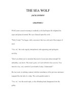
EXPLANATION OF THE PLATE.
Tab.
I.
Avicennia resinifera, Forst. fide Jack, and Av. intermedia, GrifF.
Fig.
1.
MSS.*
Placenta and ovula, at an early period before expansion of flower, and before the
corolla exceeds the calyx in length (species not noted).
Fig. 2. Longitudinal section of one of the ovula of the
tissue appears to be
Fig. 3.
same
;
the subsequent dense central
commenced.
an ovulum, more advanced ; the apex of the embryo-sac is
to the apex of the ovulum, and its body cylindrical,
reaching to the central
Longitudinal section of
close
dense tissue {A. resinifera).
Fig. 4.
Fig. 5.
Embryo-sac of the same, separated.
Embryo-sac of an ovulum at the period
its
apex:
alter the application of the
pollen-tubes to
—magnified about 500 times {A.
Fig. 6. Longitudinal section of an
style, the
fall
is
pollen-tube
posteriorly
;
resinifera).
ovulum of the same
after
blackening of the apex of the
of the corolla, and evident enlargement of the ovarium
The embryo-sac
seen attached.
otherwise there
is little
Fig. 7« Longitudinal section of an ovulum, a
change in
little
;
part of a
enlarged, and extends further
the ovulum.
is
more advanced. The embryo-sac
is
more
prolonged posteriorly, and also presents a short prolongation corresponding with
It is
still
interior to the
ovulum ; the
and ovula {A. intermedia)
at a
more advanced
stage
the axis of the ovulum.
commenced
Fig. 8. Placenta
aborted ; the fertilized one
its
dilated
apex has
to be cellular.
is
:
three of the ovula have
seen laterally, and a protuberance
(a) is visible
from
apex.
* A. intermedia
is
A. tomentosa and A.
founded on a Malacca plant altogether intermediate between what appears to be
resinifera.
the Development of the
Mr. Griffith on
of an
Fig. 9. Longitudinal section
protuberance of
tube
is still
ovulum of this period
fig. 8.) is
now
in attachment.
the end, which
:
Avicennia.
in
7
young albuminous mass
the
disc represents the
(the
A pollen-
seen to be partly exterior to the ovulum.
The
the whole
Fig. 10. Part of the ovulum,
Ovulum
rudimentary embryo.
now
of the posterior lateral elongation,
digitate at
confined in the placenta, and once-branched also within the
is
ovulum, the central or
axile prolongation, the
now almost
entirely exserted albu-
This figure does not represent a section of the albumi-
men, and the embryo.
nous mass, but of the body of the ovulum
alone, one side of
to expose the albumen.
Placenta
(entire) of an ovarium some time after fecundation,
Fig. 11.
centa,
b, b.
Barren ovula.
c.
showing the furrow or chink ?
Fecundated ovulum.
d.
which was
a.
Apex
sliced off
of the pla-
Exserted albuminous mass,
by which the points of the cotyledons will pass out
{A. intermedia).
Fig.
1 2.
Fecundated ovulum ; longitudinal section through the body of the nucleus, but not
through the albuminous mass the tips of the cotyledons reach the furrow or
:
chink.
Fig. 13.
An
entire placenta of ^. resinifera at a
same references ;
d.
Fig. 14.
the large inner
Young
e.
more advanced period: the
letters
have the
shows the lower edge of the former furrow, now an opening ;
Up or edge with
seed and embryo
irregular margins overlapping the cotyledons.
about the same period of development
which
:
the embryo
is
viewed obliquely, a. Body of the ovulum or
b. Fleshy part of the exserted albuminous mass.
nucleus,
c. Lower or outer
the
the
fissure
which
have
d. Inner or upper,
of
by
cotyledons
protruded,
edge
removed from the
seed,
now membranous,
cellular
is
edge of the same.
Tro}i».
Zimt Soc Jh/'.
.
XX tah. Jjb.
ff.
a
n.
WOriiTuAJflm.
14.
EmcUclh,
••
'm
•ft;-.;:-:
%
9
L
II.
Some Observations upon
parasitic upon
]
new Species of Hectocotyle,
D. Ch., and Argonauta Argo, Linn.
the Structure of two
Tremoctopus violaceus,
;
with an Exposition of the Hypothesis that these Hectocotylae are the Males
of the Cephalopoda upon which they are found.
fessor of Physiology and Comparative
Anatomy
A. Kolltker, Pro-
in the University
Communicated by Robert Brown, Esq., V.P.L.S.
Read April 15th and May
By
of Zurich.
8^c. 8^c.
6th, 1845.
Whilst visiting Messina and Naples during the summer of
1842, 1 found
two worms resembling the Hectocotyle Octopodis, described by Cuvier as found
upon the Octopus granulosus, Lam.
;
the one upon the Tremoctopus violaceus,
Delle Chiaje (Octopus velifer, F6r.), the other upon the Argonauta Argo, L. At
first sight I took them for epizootic worms, to which, from their white colour
and numerous suckers, they bore a great resemblance but when I examined
them more accurately, I met with so many peculiarities, a few of which I will
;
here mention, namely the existence of a heart, arteries and veins, branchiae, and
coloured contractile pigment-cells, that at length I was compelled to abandon
Proceeding with my examinations, I soon found that the animals
were all males and remembering that that sex of the Argonauta and Tremoctopus was not as yet known, I supposed that I had discovered the males of
that idea.
;
those Cephalopoda.
upon
I
must confess
that at first I did not place
this conjecture, for the great dissimilarity
surprised
me
However, as
so
I
to strengthen
much
my
my
it
to be well-founded.
researches, I found
more and more
supposition, so that finally I was led to regard
But before proceeding
reliance
between the supposed sexes
that I scarcely dared to believe
proceeded further with
much
it
as very
arguments on which I formed
my opinion, I propose to give a slight sketch of my anatomical researches
with respect to the above-mentioned animals.
probable.
VOL. XX.
to submit the
c
Prof. KoLLiKER on the Structure of
10
Description of the Hectocotyle q/'Tremoctopus violaceus.
External Form.
1.
This animal very
has described in the
much
'
resembles the Hectocotyle Octopodis, which Cuvier
Annales des Sciences Naturelles' for the year 1829*, the
body being long, worm-like, and provided with two rows
ventral surface, the posterior short and of an oval shape ;
anterior part of the
of suckers on
its
but independently of
its
being
much
smaller,
it differs
also at first sight
from
Octopodis in the back being fringed with a great number of little branchial
appendages, and the posterior extremity having a long process.
H.
The
body varies in length from 1^1 f of an inch, is
Without
thickest in the middle, and diminishes towards both extremities.
the branchiae and suckers it would be nearly cylindrical, but in the natural
anterior part of the
almost quadrangular, while that immediately
behind is fiat, and has sharp margins. Its colour is almost entirely pure white
it exhibits however at the branchiae some azure tints, and here and there some
state the cephalic portion
is
;
yellowish hues
;
there are also
some remarkable spots on the back, of which
The upper surface of this
portion the branchial appendages and the
I
shall speak presently.
anterior part bears in its
cephalic
spots just mentioned
;
the
upper surface of the posterior portion, on the contrary, is perfectly smooth ;
the spots are arranged in two slightly-irregular rows between the branchiae
along the middle of the back they are circular, about -aV^ of a line in
diameter, of a brown or violet colour, and about 50 in number. The branchial
;
appendages are placed on a low, mantle-like margin, which takes its origin
from the side of the animal between the suckers and the coloured spots the
;
appendages have a conical form, are 1 line long and ^th of a line broad
during life those next to the middle of the back stand vertically erect, the
:
others successively assume a
nearly transversely.
more inclined
The appendages
direction,
and the outermost
lie
are disposed in irregular longitudinal
amounting to about 250 on each side.
The ventral surface of the anterior part is quite smooth in the middle, and
on each of its margins are situated 40 or 4 1
slightly convex from side to side
lines
;
their
number
is
considerable,
;
* Ire S6rie,
t. xviii.
p. 147.
two new Species
suckers, which are disposed alternately,
those of the Tremoctopus
The
o/"
11
Hectocotyle.
and bear the strongest resemblance
to
itself.
posterior part of the
body
is
a large oval sac, of 5 lines in length, to
which the penis is appended. The sac is quite as large as the largest part of
the body, and incloses a great many convolutions of a small canal and one
The
large duct.
penis
is
situated on the abdominal surface,
origin from the foremost part of the oval sac
partly confined in a delicate
contracted or extended
it is
thick,
and curved
;
it is
conical
and partly
its
free,
sheath, which is attached to the
suckers its form and length vary accord-
membranous
ventral aspect between the last six
ing as
:
and takes
;
;
in the first case
in the latter, 7 lines long,
:^
it is
^ a line
and nearly
4 lines long,
of a line thick,
straight.
2. Structure.
a. Skin.
The
skin of the animal consists of two layers
;
the exterior
is
the epidermis,
formed by delicate, polygonal cells, of a diameter of 0'018-0"036"', which
contain round nuclei the interior, the corium composed of cellular tissue,
;
the outer fibres of which are disposed longitudinally, and the inner transversely.
The coloured
form a very remarkable
each of them is a cell, containing a
spots, already mentioned,
object in the description of the skin
;
nucleus and a great many coloured granules, and exhibits during life the
same curious phaenomenon which R. Wagner has discovered in the pigmentcells of Cephalopoda, viz. a regularly alternate expansion and contraction. As
example just quoted, the colour of the cells varies according as the
granules are assembled together in a mass or scattered about in the expanded
in the
cell.
With regard
proved whether
it
to the
power which
lies in
the contractile
effects these contractions, it is
membrane
of the
cells, as
not yet
Wagner
supposes, or in the contractile cellular tissue which surrounds them, an opinion which I have lately advocated.
have yet to mention some peculiar minute pores which are found on the
anterior part of the ventral surface.
They are arranged in rows of four or
I
five
on each
continuous
side
series.
between the mesial
Each opening
line
and the suckers, thus forming a
is elliptical
in figure, the longest diameter
being 0'024-00I2 P. L., and conducts to a canal of the same
c2
size,
which