Modifiled nucleaic acids in biology and medicince
Bạn đang xem bản rút gọn của tài liệu. Xem và tải ngay bản đầy đủ của tài liệu tại đây (10.23 MB, 456 trang )
Free ebooks ==> www.Ebook777.com
RNA Technologies
Stefan Jurga
Volker A. Erdmann
Jan Barciszewski Editors
Modified
Nucleic Acids
in Biology
and Medicine
www.Ebook777.com
Free ebooks ==> www.Ebook777.com
RNA Technologies
www.Ebook777.com
More information about this series at />
Stefan Jurga • Volker A. Erdmann •
Jan Barciszewski
Editors
Modified Nucleic Acids
in Biology and Medicine
Free ebooks ==> www.Ebook777.com
Editors
Stefan Jurga
Nanobiomedical Center
Adam Mickiewicz University
Poznan´
Poland
Volker A. Erdmann (Deceased)
Formerly at Institute of Chemistry and
Biochemistry
Free University Berlin
Berlin
Germany
Jan Barciszewski
Institute of the Bioorganic
Chemistry of the Polish Academy
of Sciences
Poznan´
Poland
ISSN 2197-9731
ISSN 2197-9758 (electronic)
RNA Technologies
ISBN 978-3-319-34173-6
ISBN 978-3-319-34175-0 (eBook)
DOI 10.1007/978-3-319-34175-0
Library of Congress Control Number: 2016944798
© Springer International Publishing Switzerland 2016
This work is subject to copyright. All rights are reserved by the Publisher, whether the whole or part of
the material is concerned, specifically the rights of translation, reprinting, reuse of illustrations,
recitation, broadcasting, reproduction on microfilms or in any other physical way, and transmission
or information storage and retrieval, electronic adaptation, computer software, or by similar or
dissimilar methodology now known or hereafter developed.
The use of general descriptive names, registered names, trademarks, service marks, etc. in this
publication does not imply, even in the absence of a specific statement, that such names are exempt
from the relevant protective laws and regulations and therefore free for general use.
The publisher, the authors and the editors are safe to assume that the advice and information in this
book are believed to be true and accurate at the date of publication. Neither the publisher nor the
authors or the editors give a warranty, express or implied, with respect to the material contained
herein or for any errors or omissions that may have been made.
Printed on acid-free paper
This Springer imprint is published by Springer Nature
The registered company is Springer International Publishing AG Switzerland
www.Ebook777.com
Preface
The Power of Modified Nucleic Acids
With this volume of the series RNA technologies, we aim to cover various aspects
of nucleic acid modifications. This is an interesting issue in the study of macromolecular components of cells.
DNA and RNA are key molecules of the cell. The structure, function, and
reactivity of DNA and RNA are central to molecular biology and are crucial for
the understanding of complex biological processes. For a long time DNA was
considered as the most important molecule of all biology and the key of life.
However DNA is not the be all and end all of the living cell, but it appears as an
important by-product of the RNA evolution. Deoxyribonucleotides as DNA precursors are synthesized by specific enzymatic modification of ribonucleotides in
which the 20 -hydroxyl group of the ribose moiety is replaced by 20 -hydrogen with
ribonucleotide reductase. One of these DNA bases, thymine, is produced by
methylation of uracil. There are a significant number of adenosines and cytidines
in genomic DNA converted by spontaneous or enzymatic deamination to hypoxanthine and uracil, respectively. Cytosine can be methylated to 5-methylcytosine
derivative with not only a coding capacity but also a regulatory potential. These
data suggest that DNA looks similar to a modified RNA molecule although chemically more stable than RNA. In the eukaryotic cell, all postreplicative modifications of DNA showed a few percent of all bases. Cellular DNAs and RNAs can be
chemically modified in more than 100 different ways. Some of these modifications
to nucleic acids are random or spontaneous and their formation requires significant
energy from the cell. The broad range of chemical modifications to nucleic acids is
not restricted to simple nucleophilic substitution but extends to oxidative reactions
and C–H activation by various agents. The modifications might occur on DNA as
well as different types of RNA such as transfer ribonucleic acids (tRNAs), ribosomal RNA (rRNA), messenger RNA (mRNA), and other noncodings (ncRNAs).
Among them, tRNAs represent ca 15 % of the total cellular RNAs and are highly
v
vi
Preface
stable. The primary role of tRNA is to deliver amino acids to the polypeptide chain
during protein translation. tRNA molecules 73–93 nucleotides long are heavily
modified types of ribonucleic acid. tRNA modifications (up to 25 %) are dynamic
and adaptive to different environmental changes. Modified nucleosides of tRNAs
play an important role in the translation of the genetic code.
The modified nucleosides are utilized to fine tune nucleic acids structure and
function. These modifications are dynamic and participate in regulating diverse
biological pathways. They can also be used as specific markers of different states of
cells and diseases or pathologies. The process of RNAs turnover is directly correlated to their presence in the human. Evaluation of modified nucleosides might
become novel markers to facilitate early clinical diagnosis of cancer to improve
human cancer risk assessment.
Nucleoside methylation and other nucleic acid modifications are of a great
interest, prompted by the discovery of methylation and active demethylation of
DNA and RNA. In eukaryotic genomic DNA, 5-methylcytosine is a well-know
epigenetic modification and is also known to exist in both rRNA and tRNA.
In response to oxidative stress caused by reactive oxidative species (ROS) as
well as nutrient depletion and other growth arrest conditions, modified nucleotides
are synthesized in the cell to serve various purposes. Accidental non-enzymatic
methylation or oxidation of a base in a DNA and RNA, in addition to the normal
enzymatic methylation processes, induces serious problems for living cells, especially for DNA, for which abnormal alkylation can be mutagenic. To remove this
type of modification, the cell has developed oxidative mechanisms in an
indirect way.
The recent development of high-throughput sequencing technologies has
enabled us to identify tRNA-derived RNA fragments. It seems that they are not
by-products from random degradation but rather functional molecules that can
regulate translation and gene expression.
It takes a large effect to map RNA modifications globally as well as to identify
the cellular function as writers, readers, and erasers for each modification. Basic
cellular pathways use ubiquitous metabolites and coenzymes to transfer methyl and
amino acid groups, isoprenoids, sugars, phosphates, and various metabolite nucleic
acid conjugates have been found that affect a functionality of their specific targets.
The turnover of nucleic acids increases when cell proliferation takes place. Any
disease or metabolic alteration affecting RNA turnover consequently results in
altered nucleoside excretion patterns, leading to the hypothesis that RNA metabolites may be used as early indicators of disease. In addition, increased RNA
metabolism with altered nucleoside excretion patterns related to metabolic disorders such as cancer may be suitable markers to facilitate the monitoring of therapeutic intervention.
In this book we have collected work describing modified nucleosides, naturally
occurring or chemically synthesized nucleic acids. Their role in cell biology has
huge potential for application in medicine. Further research frontiers and new
developments are also discussed.
Preface
vii
In total there are 18 chapters. Five of them deal with tRNAs and their modifications in relation to biomedical applications. Three discuss modified nucleosides
including N6-methyladenosine and 8-hydroxyguanosine as well as 20 -O-methylated
ribonucleotides. A very interesting chapter describes the role of diadenosine
tetraphosphate in health and disease. Similar properties are described for circular
RNAs and for modified therapeutic oligonucleotides. Other chapters describe the
properties of modified oligonucleotides.
Poznan´
Berlin
Poznan´
January 2016
Stefan Jurga
Volker A. Erdmann
Jan Barciszewski
ThiS is a FM Blank Page
Free ebooks ==> www.Ebook777.com
RNA Around the Clock: Volker A. Erdmann
in Memoriam
On September 11, 2015 we lost our colleague and dear friend Professor Volker
A. Erdmann from the Institute of Chemistry–Biochemistry, Freie Universita¨t Berlin, Germany. He was born on February 8, 1941 in Stettin (Germany, now Poland)
and later became a U.S. citizen. In 1963 he earned his B.A. in Chemistry and in
1966 an M.Sc. in Biochemistry from the University of New Hampshire, Durham, N.
H., USA (advisor: Prof. Dr. E.J. Herbst). From 1966 to 1969 at the Max-PlanckInstitut f€
ur experimentelle Medizin, G€ottingen, Germany, and Technische
Universita¨t Braunschweig, Germany, he obtained a Dr. rer. nat. degree in Biochemistry with minors in Chemistry and Microbiology (advisor: Prof. Dr. F. Cramer).
After an NIH postdoctoral fellowship with Prof. Dr. M. Nomura at University of
Wisconsin, Madison, Wisc., USA, in 1971 he became a research group leader at the
Max-Planck-Institut f€ur Molekulare Genetik in Berlin at the Department led by
Prof. Dr. H.G. Wittmann. In 1978 Volker did Habilitation in Biochemistry and
Molecular Biology at the Freie Universita¨t at Berlin, Germany.
ix
www.Ebook777.com
x
RNA Around the Clock: Volker A. Erdmann in Memoriam
From 1980 he was full professor of Biochemistry in the Department of Chemistry
at the Institute of Biochemistry, and from 2009 he was a guest Professor at the Free
University of Berlin. In 1987 he received an award for scientific excellence from the
German Research Council (DFG), the highest scientific award given in Germany
(F€
orderpreis f€
ur deutsche Wissenschaftler im Gottfried Wilhelm Leibniz Programm
der Deutschen Forschungsgemeinschaft). He was a member of the BerlinBrandenburgische Akademie der Wissenschaften (Berlin Brandenburg Academy of
Science, former Prussian Academy of Science) and the Polish Academy of Science.
His research interests have always been in the area of gene expression with
special emphasis in the structure and function of ribosomes and RNA technologies.
His RNA research includes studies on the structure and function of ribozymes,
antisense oligonucleotides, siRNAs, micro RNAs, DNAzymes, high affinity RNA
molecules (aptamers), enantiomeric catalytic nucleic acids (ribozymes and
DNAzymes), and large noncoding RNAs such as the H19 RNA. With his group
at Free University he developed methods for the chemical synthesis of RNA
molecules, including a large number of modified nucleotides. He has also concentrated on the crystallization of RNA molecules and their protein complexes by
X-ray analysis. These crystallization experiments include microgravity experiments (participation in 17 space missions). The results of Volker’s research have
appeared in more than 450 publications. He created data bases on 5S ribosomal
RNA and non-coding RNAs and obtained 14 patents in the area of RNA technologies. In 1998 he and his colleagues founded the Berlin Network for RNA Technologies, with the goal to pursue further the structural and functional potentials of
RNA molecules.
Volker’s most important discoveries include the first total reconstitution of
bacterial 50S and 70S ribosomes, first identification of ribosomal 5S RNA binding
proteins and 5S RNA protein complexes, first crystallization of ribosomes, first
crystallization of RNA molecules under microgravity conditions, first crystallization and X-ray structural determination of a mirror image RNA structure, first to
discover mirror image aptamers and first to discover mirror image L-catalytic
nucleic acid as alternatives to siRNAs and microRNAs to cure cancer and viral
infections.
These L-form aptamers have a number of advantages when compared with Daptamers. They are very stable in human sera and cells. Because nature does not
make L-nucleic acids, there was no need to develop any enzymes hydrolyzing the Lform of nucleic acids. L-Aptamers can be compared with protein antibodies, and
indeed aptamers can assume very similar functions to antibodies. Aptamers are
considerably smaller than antibodies and they are easily synthesized by nucleic acid
synthesizers. They are not toxic or immunogenic and are therefore most likely
ideally suited for the development of new types of pharmaceutical drugs. The
development of mirror image catalytic RNA opened new possibilities in basic
research and in the area of molecular evolution. Volker A. Erdmann was one of
the first Editors-in-Chief and founders of RNA Biology journal published by
Landes Bioscience, Georgetown, Texas (USA). He was co-editor of several
RNA Around the Clock: Volker A. Erdmann in Memoriam
xi
books in the series “RNA Technologies” published by Springer. In 2013, he
established a private-biotech company called Erdmann Technologies based in
Berlin.
Married to Hannelore Erdmann, he had two children (J€orn, and Gabriele).
Volker was a very quiet and kind person and supportive of young scientists. He
directed his students and fellow researchers carefully and with pride. He derived
much pleasure from the successes of his scientific offspring—his former students.
His influence extended well beyond his scientific contributions to shaping policy on
important issues at the interface between science and society. He was a source of
inspiration to all around him and will be greatly missed.
Poznan´, Poland
Jan Barciszewski
ThiS is a FM Blank Page
Contents
Transfer RNA Modifications: From Biological Functions to Biomedical
Applications . . . . . . . . . . . . . . . . . . . . . . . . . . . . . . . . . . . . . . . . . . . . . .
Adrian Gabriel Torres and Lluı´s Ribas de Pouplana
1
Regulated tRNA Cleavage in Biology and Medicine: Roles
of tRNA Modifications . . . . . . . . . . . . . . . . . . . . . . . . . . . . . . . . . . . . . .
Shawn M. Lyons, Marta M. Fay, and Pavel Ivanov
27
Sulfur Modifications in tRNA: Function and Implications for
Human Disease . . . . . . . . . . . . . . . . . . . . . . . . . . . . . . . . . . . . . . . . . . . .
Naoki Shigi
55
Regulation of Protein Synthesis via the Network Between Modified
Nucleotides in tRNA and tRNA Modification Enzymes in Thermus
thermophilus, a Thermophilic Eubacterium . . . . . . . . . . . . . . . . . . . . . .
Hiroyuki Hori, Ryota Yamagami, and Chie Tomikawa
Post-Transcriptional Modifications of RNA: Impact on RNA
Function and Human Health . . . . . . . . . . . . . . . . . . . . . . . . . . . . . . . . .
Kyla M. Frohlich, Kathryn L. Sarachan, Gabrielle C. Todd,
Maria Basanta-Sanchez, Ville Y.P. Va¨re, and Paul F. Agris
73
91
RNA Modification N6-Methyladenosine in Post-transcriptional
Regulation . . . . . . . . . . . . . . . . . . . . . . . . . . . . . . . . . . . . . . . . . . . . . . . 131
Guifang Jia
8-Hydroxyguanine, an Oxidative DNA and RNA Modification . . . . . . . 147
Hiroshi Kasai and Kazuaki Kawai
Methods for Determination of 20 -O-Me in RNA . . . . . . . . . . . . . . . . . . . 187
Ulf Birkedal, Nicolai Krogh, Kasper Langebjerg Andersen,
and Henrik Nielsen
xiii
xiv
Contents
Diadenosine Tetraphosphate (Ap4A) in Health and Disease . . . . . . . . . . 207
Suliman Boulos, Ehud Razin, Hovav Nechushtan, and Inbal Rachmin
Thinking Small: Circulating microRNAs as Novel Biomarkers
for Diagnosis, Prognosis, and Treatment Monitoring in
Breast Cancer . . . . . . . . . . . . . . . . . . . . . . . . . . . . . . . . . . . . . . . . . . . . 221
Yin-Long Yang
Modified Antisense Oligonucleotides and Their Analogs
in Therapy of Neuromuscular Diseases . . . . . . . . . . . . . . . . . . . . . . . . . 243
Patryk Konieczny, Ewa Stepniak-Konieczna, and Krzysztof Sobczak
Effect of Depurination on Cellular and Viral RNA . . . . . . . . . . . . . . . . 273
Kass A. Jobst, Alexander Klenov, Kira C.M. Neller,
and Katalin A. Hudak
Recognition of RNA Sequence and Structure by Duplex
and Triplex Formation: Targeting miRNA and Pre-miRNA . . . . . . . . . 299
Kiran M. Patil and Gang Chen
Modifications in Therapeutic Oligonucleotides Improving
the Delivery . . . . . . . . . . . . . . . . . . . . . . . . . . . . . . . . . . . . . . . . . . . . . . 319
Ilya Dovydenko, Alya Venyaminova, Dmitrii Pyshnyi, Ivan Tarassov,
and Nina Entelis
Interstrand Cross-Linking of Nucleic Acids: From History
to Recent and Future Applications . . . . . . . . . . . . . . . . . . . . . . . . . . . . . 339
Ellen Gyssels, Nathalie De Laet, Emily Lumley, and Annemieke Madder
Chemical Synthesis of Lesion-Containing Oligonucleotides
for DNA Repair Studies . . . . . . . . . . . . . . . . . . . . . . . . . . . . . . . . . . . . . 371
Re´my Lartia
Single-Molecule Visualization of Biomolecules in the Designed
DNA Origami Nanostructures Using High-Speed Atomic Force
Microscopy . . . . . . . . . . . . . . . . . . . . . . . . . . . . . . . . . . . . . . . . . . . . . . . 403
Masayuki Endo
Polymerase Reactions that Involve Modified Nucleotides . . . . . . . . . . . . 429
Masayasu Kuwahara, Kenta Hagiwara, and Hiroaki Ozaki
Transfer RNA Modifications: From
Biological Functions to Biomedical
Applications
Adrian Gabriel Torres and Lluı´s Ribas de Pouplana
Contents
1 Transfer RNAs Are Post-Transcriptionally Modified . . . . . . . . . . . . . . . . . . . . . . . . . . . . . . . . . . . . . . .
2 Links Between tRNA Modifications and Human Diseases . . . . . . . . . . . . . . . . . . . . . . . . . . . . . . . . .
2.1 Neurological Disorders . . . . . . . . . . . . . . . . . . . . . . . . . . . . . . . . . . . . . . . . . . . . . . . . . . . . . . . . . . . . . . . . .
2.2 Cancer . . . . . . . . . . . . . . . . . . . . . . . . . . . . . . . . . . . . . . . . . . . . . . . . . . . . . . . . . . . . . . . . . . . . . . . . . . . . . . . . . .
2.3 Metabolic Dysregulations . . . . . . . . . . . . . . . . . . . . . . . . . . . . . . . . . . . . . . . . . . . . . . . . . . . . . . . . . . . . . .
2.4 Mitochondrial-Linked Dysfunctions . . . . . . . . . . . . . . . . . . . . . . . . . . . . . . . . . . . . . . . . . . . . . . . . . . .
3 Biomedical Strategies Based on tRNA Modifications . . . . . . . . . . . . . . . . . . . . . . . . . . . . . . . . . . . . . .
3.1 Diagnosis and Prognosis . . . . . . . . . . . . . . . . . . . . . . . . . . . . . . . . . . . . . . . . . . . . . . . . . . . . . . . . . . . . . . .
3.2 Potential Therapeutic Treatments . . . . . . . . . . . . . . . . . . . . . . . . . . . . . . . . . . . . . . . . . . . . . . . . . . . . . .
4 Conclusions and Perspectives . . . . . . . . . . . . . . . . . . . . . . . . . . . . . . . . . . . . . . . . . . . . . . . . . . . . . . . . . . . . . . . .
References . . . . . . . . . . . . . . . . . . . . . . . . . . . . . . . . . . . . . . . . . . . . . . . . . . . . . . . . . . . . . . . . . . . . . . . . . . . . . . . . . . . . . . . .
2
4
8
11
12
13
15
15
16
19
21
Abstract Transfer RNAs (tRNAs) are essential components of the protein translation machinery. In order to become fully active, they need to be heavily modified
post-transcriptionally. Such modifications affect the structure, stability and functionality of tRNAs; however, their exact roles at the molecular level remain largely
elusive. Here we focus on the biological functions of tRNA modifications associated to human diseases and how such information can be used for biomedical
applications. We put an emphasis on mitochondrial-linked dysfunctions, metabolic
disorders, neurological defects and cancer. We also present methods and
approaches currently used in the clinic to detect and monitor different human
A.G. Torres
Institute for Research in Biomedicine (IRB Barcelona), Barcelona Institute for Science and
Technology, C/Baldiri Reixac 10, Barcelona 08028, Catalonia, Spain
e-mail:
L. Ribas de Pouplana (*)
Institute for Research in Biomedicine (IRB Barcelona), Barcelona Institute for Science and
Technology, C/Baldiri Reixac 10, Barcelona 08028, Catalonia, Spain
Catalan Institution for Research and Advanced Studies (ICREA), P/Lluis Companys 23,
Barcelona 08010, Catalonia, Spain
e-mail:
© Springer International Publishing Switzerland 2016
S. Jurga et al. (eds.), Modified Nucleic Acids in Biology and Medicine, RNA
Technologies, DOI 10.1007/978-3-319-34175-0_1
1
2
A.G. Torres and L. Ribas de Pouplana
pathologies involving tRNA modifications or tRNA modification enzymes, and,
additionally, we propose novel tRNA modification-based strategies that could be
used for diagnosis, prognosis or treatment of human diseases.
Keywords tRNA • tRNA modification • Protein translation • Human disease •
Biomedicine
1 Transfer RNAs Are Post-Transcriptionally Modified
Transfer RNAs (tRNAs) play a key role in the protein translation machinery. They
are transcribed as long primary tRNAs that are processed during their biogenesis to
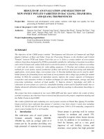
yield 70–100 nucleotides long RNA species, that fold into a cloverleaf-shape (2ry
structure) (Fig. 1) and L-shape (3ry structure) arrangement (Pi~neyro et al. 2014).
Following maturation, they are charged at their 30 -end with their cognate amino
acid, which will be incorporated into the growing polypeptide chain during protein
synthesis. Residues 34, 35 and 36 of the tRNA form the tRNA ‘anticodon’ (Fig. 1)
that pairs specifically with nucleotide triplets on the messenger RNA (mRNA)
called ‘codons’. Each mRNA codon codifies for a specific amino acid; hence,
tRNAs are adaptor molecules that translate specific mRNA codons into specific
amino acids during translation (Pi~neyro et al. 2014).
During maturation, tRNAs are required to go through a series of posttranscriptional chemical modifications. In general, for a given tRNA, about
10–15 % of the tRNA residues are found modified (Phizicky and Alfonzo 2010).
There are more than 50 different chemical modifications described for eukaryotic
tRNAs, that include methylations, thiolations, deaminations, acetylations, isomerizations and hydroxylations, among others (Machnicka et al. 2013). Such modifications affect the structure, processing, stability and overall functionality of tRNAs.
Modifications in particular regions of the tRNA affect different aspects of tRNA
functionality. In general, modifications in the main body of the tRNA affect the
rigidity/flexibility of the molecule. For example, pseudouridines increase the binding affinity of tRNA residues by inducing a C30 -endo sugar conformation, and
dihydrouridines make these interactions more flexible by retaining the sugar pucker
into a C20 -endo conformation (El Yacoubi et al. 2012). Modifications at the
anticodon region of the tRNA have a direct role on codon recognition and prevent
frameshifting during protein translation. On the one hand, modifications at position
34 of the tRNA increase (or restrict) the number of codons a tRNA can recognize,
by promoting or inhibiting tRNA ‘wobbling’ (non-Watson-Crick nucleotide
pairing) (Crick 1966). Some examples include A34-to-I34 editing (deamination)
that allows the modified tRNAs to decode codons ending not only in uridine (U) but
also in adenine (A) and cytosine (C) (Torres et al. 2014b) or U34 modifications,
such as the one on tRNALys(UUU), which allows the tRNA to decode its cognate
codons AAA and AAG but restricts the recognition of the near-cognate asparagine
Transfer RNA Modifications: From Biological Functions to Biomedical Applications
3
Acceptor
stem
m1G
9
Gm
54
49 50
17
48
26
m5U
46
m22G
42
m7G
m5C
m5U
Cm, Gm
32
38
37
ncm5U, ncm5s2U, ncm5Um,
mcm5U, mcm5s2U, ncm5Um,
I, m5C, Q, τm5U, τm5s2U
34
m5C
yW, ms 2 t 6A, m1G
Anticodon
Fig. 1 Representation of the tRNA secondary structure (‘cloverleaf’). Post-transcriptionally
modified tRNA residues associated to human diseases are shown (black boxes). Abbreviations:
m1G, 1-methylguanosine; Gm, 20 -O-methylguanosine; m22G, N2,N2-dimethyl guanosine; Cm,
20 -O-methylcytidine; ncm5U, 5-carbamoylmethyluridine; ncm5s2U, 5-carbamoylmethyl-25-carbamoylmethyl-20 -O-methyluridine;
mcm5U,
thiouridine;
ncm5Um,
5-methoxycarbonylmethyluridine; mcm5s2U, 5-methoxycarbonylmethyl-2-thiouridine; mcm5
Um, 5-methoxycarbonylmethyl-20 -O-methyluridine; I, inosine; m5C: 5-methylcytosine; Q,
queuosine; τm5U, 5-taurinomethyluridine; τm5s2U, 5-taurinomethyl-2-thiouridine; yW,
wybutosine; ms2t6A, 2-methylthio-N6-threonyl carbamoyladenosine; m5U, 5-methyluridine; m5
U, 5-methyluridine; m7G, 7-methylguanosine
codons AAU and AAC (Yarian et al. 2002). On the other hand, modifications at
position 37 (adjacent to the anticodon) are usually associated to keeping in-frame
translation. These are in general bulk modifications that stabilize codon/anticodon
pairing by generating base-stacking interactions. Examples include the wybutosine
37 (yW37) modification that prevents À1 frameshifts (Waas et al. 2007) or the
1-methylguanosine 37 (m1G37) modification that impedes +1 frameshifting
(Urbonavicius et al. 2003). Finally, modifications in the acceptor stem of the
tRNA (the stem formed by the 50 - and 30 -ends of the tRNA; Fig. 1) usually serve
as identity elements for aminoacyl tRNA synthetases, the enzymes that charge the
tRNA with its cognate amino acid. A representative example is the post-
4
A.G. Torres and L. Ribas de Pouplana
transcriptional addition of a G residue at the 50 -end of tRNAHis (GÀ1 modification)
that is essential for correct charging by histidine-tRNA synthetase (Rudinger
et al. 1994). For detailed information on the biological functions of tRNA modifications, several comprehensive reviews are available (Phizicky and Alfonzo 2010;
El Yacoubi et al. 2012; Jackman and Alfonzo 2013; Pi~neyro et al. 2014).
Interestingly, while tRNA modifications seem very important for tRNA function, the vast majority of them are not essential for cell viability (Pi~neyro
et al. 2014). In fact, in many cases, the modulation of tRNA modifications does
not affect significantly the tRNA function and usually results in subtle phenotypes.
However, it is not possible to generalize on this matter, and known observations
need not apply to different cellular systems. Indeed, tRNA modification-based
phenotypes can be associated to specific tissues. It is well documented that the
tRNA pool and the expression of proteins carrying a particular codon bias may vary
in a tissue-dependent manner (Kirchner and Ignatova 2015). Additionally, the
‘penetrance’ of a phenotype may be linked to the degree of the tRNA modification
misregulation. This has been shown for mutations on mitochondrial tRNA genes
that prevent the tRNA to be modified. A cell has a variable number of mitochondria,
each of which carries their own mitochondrial genome. Only when a significant
amount of mitochondria carrying the mutated mitochondrial genome variant accumulate, clear phenotypes can be observed (Abbott et al. 2014). Finally, sometimes
tRNA modifications need to be considered not as individual modifications but as a
part of a whole set of modifications leading to a significant phenotype. In yeast,
overall tRNA modification patterns change in the tRNA pool when cells are
subjected to stress conditions (Chan et al. 2012), suggesting a coordinated regulation of tRNA modifications as a mechanism for stress response.
In this chapter, we will describe the known connections that have recently been
established between post-transcriptional tRNA modifications and human diseases.
It will become evident that the roles of such modifications on those diseases are
complex and that the molecular mechanisms behind the observed phenotypes
remain poorly understood. Importantly, we will not address human diseases caused
by mutations on tRNA genes or by defects on tRNA processing and maturation,
which have been recently reviewed in depth (Abbott et al. 2014; Kirchner and
Ignatova 2015), unless a clear direct effect on the tRNA modification pattern has
been observed. Finally, we will present different strategies that are being pursued in
the clinic for diagnosis, prognosis and treatment of this type of diseases and will
propose potentially novel tRNA modification-based therapeutic approaches to be
pushed forward in the future.
2 Links Between tRNA Modifications and Human Diseases
While tRNA modifications and the enzymes that catalyse such modifications have
not been studied in depth in metazoans, an association between tRNA modifications
and human diseases is starting to emerge (Torres et al. 2014a) (Fig. 1 and Table 1).
Several human genetic studies have shown links between mutations in genes that
mcm5U,
ncm5U, and
derivatives
Elongator complex
(IKAP, ELP2,
ELP3, ELP4)
www.Ebook777.com
HABH8
(ALKBH8)
TARBP1
(TRMT3)
TRMT12
TRMT2A
(HTF9C)
Bladder
Breast
Liver
Elongator complex
(IKAP)
yW
m5U
Gm
Phe
Several
Ser
Arg, Glu
Several
Several
Ala, Pro, Thr,
Val, Ser, Arg,
Leu, Ile
Several
m22G
I
TRMT1
ADAT3
mcm5U,
ncm5U and
derivatives
mcm5U
Affected
tRNAs
Leu, Phe, Trp
tRNA
modification
Cm, Gm,
ncm5Um
Gene
FTSJ1
Lung
Tissue
Brain
37
42, 54
17
34
34
34
26
34
tRNA
residues
32, 34
Breast cancer
Morris hepatoma
Bladder cancer
Intellectual disability
Amyotrophic lateral sclerosis
Rolandic epilepsy
Bronchial asthma in children
Familial dysautonomia
Intellectual disability
Intellectual disability and
strabismus
Associated human diseases
and phenotypes
Non-syndromic X-linked
mental retardation
Rodriguez et al. (2007)
Bartlett et al. (2010)
Randerath et al. (1981)
Shimada et al. (2009)
(continued)
Anderson et al. (2001), Slaugenhaupt
et al. (2001), Leyne et al. (2003),
Karlsborn et al. (2014)
Najmabadi et al. (2011)
Simpson et al. (2009)
Strug et al. (2009)
Takeoka et al. (2001)
References
Hamel et al. (1999), Freude
et al. (2004), Ramser et al. (2004),
Bonnet et al. (2006), Froyen
et al. (2007), Dai et al. (2008),
Takano et al. (2008), Giorda
et al. (2009)
Najmabadi et al. (2011)
Alazami et al. (2013)
Table 1 Post-transcriptional tRNA modifications discussed in this chapter, including their role in different tissues and associated human diseases
Free ebooks ==> www.Ebook777.com
Transfer RNA Modifications: From Biological Functions to Biomedical Applications
5
Tissue
Multiple
tissues
Several
Several
m1G
mcm5U,
mcm5s2U
Q
ms2t6A
m7G
TRMT10A
(HRG9MTD2)
HTRM9L
CDKAL1
WDR4
*
Several
Asn, Asp, His,
Tyr
Lys
Affected
tRNAs
Several
Gene
NSUN2
tRNA
modification
m5C
Table 1 (continued)
46
37
34
34
9
tRNA
residues
34,
48, 49,
50
Psoriasis
Down’s syndrome
Microcephalic primordial
dwarfism
Cardiovascular diseases
Crohn’s disease
Type 2 diabetes
Different types of cancer
Dubowitz-like syndrome
Noonan-like syndrome
Skin, breast and colorectal
cancer
Intellectual disability, microcephaly, short stature
Colorectal cancer
Hyperinsulinaemic
hypoglycaemia
Different types of cancer
Associated human diseases
and phenotypes
Autosomal-recessive intellectual disability
Randerath et al. (1984), Vinayak and
Pathak (2010)
Kirchhoff et al. (2008), Stancakova
et al. (2008), Quaranta et al. (2009),
Wei et al. (2011), Wei and Tomizawa
(2011), Xie et al. (2013)
Saade et al. (2011)
Barrett et al. (2008), Quaranta
et al. (2009)
Quaranta et al. (2009)
Michaud et al. (2000)
Shaheen et al. (2015)
References
Abbasi-Moheb et al. (2012), Khan
et al. (2012), Martinez et al. (2012),
Fahiminiya et al. (2014), Blanco
et al. (2014)
Martinez et al. (2012)
Fahiminiya et al. (2014)
Frye and Watt (2006), Pierga
et al. (2007), Frye et al. (2010)
Igoillo-Esteve et al. (2013), Gillis
et al. (2014)
Berg et al. (2010)
Igoillo-Esteve et al. (2013), Gillis
et al. (2014)
Begley et al. (2013)
6
A.G. Torres and L. Ribas de Pouplana
mt-Leu
mt-Lys
mt-Lys,
mt-Glu,
mt-Gln
mt-Leu
τm5U
τm5s2U
s2U (τm5s2
U)
m1G
**
**
MTU1 (TRMU)
TRMT5
37
34
34
34
38
MERRF
Acute liver failure in infancy
accompanied by lactic
acidaemia
Deafness associated with
mutations in mitochondrial
12S ribosomal RNA
Lactic acidosis and multiple
mitochondrial respiratory
complex deficiencies
MERRF
Increased red blood cell folate
levels
Different types of cancer
MELAS
Powell et al. (2015)
Guan et al. (2006)
Schaefer et al. (2009)
Hayashi et al. (1993), Kirino
et al. (2004), Suzuki and Nagao
(2011)
Yasukawa et al. (2001), Suzuki and
Nagao (2011)
Umeda et al. (2005)
Zeharia et al. (2009)
Franke et al. (2009)
For complex tRNA modifications, the underlined modification refers to the modification step at which the indicated gene is involved
Cm, 20 -O-methylcytidine; Gm, 20 -O-methylguanosine; ncm5U, 5-carbamoylmethyluridine; ncm5Um, 5-carbamoylmethyl-20 -O-methyluridine; m5C,
5-methylcytosine; mcm5U, 5-methoxycarbonylmethyluridine; yW, wybutosine; m5U, 5-methyluridine; m1G, 1-mehtylguanosine; m7G, 7-methylguanosine;
m22G, N2,N2-dimethylguanosine; Q, queuosine; ms2t6A, 2-methylthio-N6-threonyl carbamoyladenosine; I, inosine; τm5U, 5-taurinomethyluridine; τm5s2U,
5-taurinomethyl-2-thiouridine
*No associated gene
**Unknown enzyme
Mitochondrial
defects
Asp
m5C
DNMT2
(TRDMT1)
Transfer RNA Modifications: From Biological Functions to Biomedical Applications
7
8
A.G. Torres and L. Ribas de Pouplana
encode (or are expected to encode) for enzymes that catalyse tRNA modifications
and a wide range of complex human pathologies, including neurological disorders,
cardiac and respiratory defects, cancer, metabolic dysregulations and
mitochondrial-linked dysfunctions (see below). In most of these conditions, an
in-depth understanding of the molecular mechanisms of pathology is lacking. It
will become clear in this section that unravelling the details of such molecular
mechanisms will be very challenging given that some of the observed phenotypes
associated to tRNA modifications are tissue-specific and are dependent on the
degree to which the tRNA is modified or not (Kirchner and Ignatova 2015).
2.1
Neurological Disorders
The brain is perhaps the most sensitive tissue to defects in tRNA modifications. The
FtsJ RNA methyltransferase homolog 1 (FTSJ1) gene is likely the human homolog
of the yeast tRNA methyltransferase 7 (TRM7) gene that encodes for the enzyme
that methylates positions 32 and 34 on tRNALeu, tRNATrp and tRNAPhe (Towns and
Begley 2012). Mutations in FTSJ1 or a complete deletion of this gene has been
linked to non-syndromic X-linked mental retardation (Hamel et al. 1999; Freude
et al. 2004; Ramser et al. 2004; Bonnet et al. 2006; Froyen et al. 2007; Dai
et al. 2008; Takano et al. 2008) and reported to affect cognitive functions in
young males of the Han Chinese population (Gong et al. 2008). The tRNA modification state of these patients was not evaluated, but a mutant FTSJ1 transcript was
shown to be very unstable and likely degraded by nonsense-mediated mRNA decay
(Freude et al. 2004; Takano et al. 2008). Interestingly, the expression of wild-type
FTSJ1 in human tissues was reported to be high in foetal brain (Freude et al. 2004)
but low in adult brain (Ramser et al. 2004), consistent with a key role for this protein
in the developing brain. Notably, patients bearing a chromosomal duplication of
regions involving FTSJ1 and other genes presented mild/moderate mental retardation (Bonnet et al. 2006; Giorda et al. 2009), suggesting that overexpression of
wild-type FTSJ1 might also be detrimental. However, a patient with mild mental
retardation that presented a smaller chromosomal duplication, also involving
FTSJ1, did not show increased levels of FTSJ1 mRNA as measured by quantitative
PCR in blood; and instead the phenotype was attributed to the overexpression of
three other genes (also located within the duplicated chromosomal region): EBP,
WDR13 and ZNF81 (El-Hattab et al. 2011).
Mental retardation has also been reported in human patients with mutations in
other genes encoding for tRNA modification enzymes. The tRNA
methyltransferase 10A (TRMT10A), also known as human RNA (guanine-9)
methyltransferase domain containing 2 (HRG9MTD2), is the human ortholog of
the yeast enzyme that catalyses the methylation of guanosine at position 9 of several
tRNAs (Towns and Begley 2012). Mutations in the human TRMT10A gene have
been associated to microcephaly, short stature and intellectual disability (IgoilloEsteve et al. 2013; Gillis et al. 2014). TRMT10A was shown to be expressed in
Transfer RNA Modifications: From Biological Functions to Biomedical Applications
9
human embryonic and foetal brain (Igoillo-Esteve et al. 2013). Furthermore, Gillis
and colleagues showed in vitro that the mutant TRMT10A protein could effectively
bind to a tRNA substrate but showed a dramatic reduction in methylation activity as
compared to the wild-type protein, probably due to impaired ability to bind the
methyl donor S-adenosylmethionine (Gillis et al. 2014). As it will be discussed
later, defects on this enzyme not only affect brain tissues but also the liver and
colon.
Other examples of genes encoding for tRNA modification enzymes and mental
retardation include the gene encoding for tRNA methyltransferase 1 (TRMT1), an
enzyme that dimethylates guanosines at position 26 of several tRNAs (Liu and
Straby 2000), and the gene encoding for ELP2 (a component of the elongator
complex; see below). Both enzymes were reported as novel markers for recessive
cognitive disorders (Najmabadi et al. 2011). Notably, in mice, a potential homolog
of the human TRMT1 named ‘TRM1-like’ was shown to have a role in motor
coordination and exploratory behaviour (Vauti et al. 2007), suggesting a conserved
role for this protein in the brain. Finally, the human WD repeat domain 4 (WDR4)
gene was found as a potential candidate marker for phenotypes observed in Down’s
syndrome (Michaud et al. 2000); and mutations in this gene have recently been
associated to a distinct form of microcephalic primordial dwarfism (Shaheen
et al. 2015). This gene is the human homolog of the yeast TRM82, one of the
subunits of the heterodimeric enzyme responsible for the 7-methylguanosine modification at position 46 in several tRNAs (Towns and Begley 2012). Two transcript
variants for this gene were described in humans, where the shorter one (of about
1.5 kb) was highly expressed in foetal tissues and the longer one (of about 2.5 kb)
showed faint expression in most tissues (Michaud et al. 2000), suggesting a key
function for the shorter transcript in developmental processes. Interestingly, the
chromosomal region where WDR4 maps has already been associated to several
genetic disorders such as maniac-depressive psychosis, autosomal-recessive deafness, Knobloch syndrome and holoprosencephaly (Michaud et al. 2000).
Recently, the human adenosine deaminase acting on tRNA 3 (ADAT3) was
validated as one of the subunits of the heterodimeric enzyme responsible for
adenosine-to-inosine editing at position 34 of 8 different tRNAs (Torres
et al. 2015). A single missense mutation in the human ADAT3 gene was reported
to cause intellectual disability and strabismus (Alazami et al. 2013). Interestingly,
this protein and its catalytic partner ADAT2 were reported essential in yeast,
Trypanosoma brucei, Arabidopsis thaliana and likely also in human cell lines
(Gerber and Keller 1999; Rubio et al. 2007; Zhou et al. 2014; Torres et al. 2015).
This suggests that patients carrying mutations in the ADAT3 gene probably still
have some residual ADAT activity. However, a more detailed analysis of the
functional importance for this enzyme in mammals remains to be addressed (Torres
et al. 2014b).
NOP2/Sun RNA methyltransferase family member 2 (NSUN2) is an enzyme
that methylates cytosine at positions 34, 48, 49 and 50 on different tRNAs
(Brzezicha et al. 2006; Hussain et al. 2013) . Mutations in the NSUN2 gene have
been associated to autosomal-recessive intellectual disability (Abbasi-Moheb
10
A.G. Torres and L. Ribas de Pouplana
et al. 2012; Khan et al. 2012; Martinez et al. 2012; Fahiminiya et al. 2014). Khan
and colleagues investigated the subcellular localization of wild-type and mutant
NSUN2 and found that mutant NSUN2 failed to localize to nucleoli (Khan
et al. 2012). Abbasi-Moheb and colleagues further showed that deletion of the fly
NSUN2 ortholog resulted in a short-term memory phenotype (Abbasi-Moheb
et al. 2012), suggesting an evolutionary conserved function for tRNA methylation
in the brain. A recent study has addressed some mechanistic aspects on how
NSUN2 defects would be contributing to human disease. The authors showed that
lack of 5-methylcytosine (m5C) on tRNAs results in increased tRNA endonucleolytic cleavage mediated by angiogenin, leading to the accumulation of 50 -tRNA
halves that reduce protein translation rates and activate stress pathways in human
and mouse cells. Moreover, NSUN2-deficient brains were more sensitive to oxidative stress, and this phenotype could be rescued by inhibiting angiogenin during
embryogenesis (Blanco et al. 2014).
Two other reports have associated mutations in NSUN2 to different degrees of
mental retardation. Patients with these mutations showed overlapping phenotypes
to that of the Dubowitz syndrome (Martinez et al. 2012) and to that of the Noonan
syndrome (Fahiminiya et al. 2014). In the first case, the authors showed that patients
suffering from this Dubowitz-like syndrome were lacking the m5C modification at
positions 47 and 48 on tRNAAsp(GTC), one of the substrates of NSUN2 (Martinez
et al. 2012). In the second case, the link to a Noonan-like syndrome suggests a role
for NSUN2 beyond the brain, as this is a pathology that affects mainly the cardiac
tissue and only about 25 % of affected patients also suffer mental retardation. As it
will be discussed later, NSUN2 has also been linked to different forms of cancer
(see below) suggesting that this enzyme has key roles not only in the brain and heart
but also in different tissue types.
A set of very well studied tRNA modifications associated to neurological
disorders are those involving the formation of 5-methoxycarbonylmethyluridine
(mcm5U) and 5-carbamoylmethyluridine (ncm5U) at position 34 of several tRNAs.
These complex tRNA modifications (and derivatives of them) require a methylation
step catalysed by the elongator complex. This complex is highly conserved from
yeast to humans, where it was shown to be composed of six subunits: IκB kinase
complex-associated protein (IKAP/yeast ELP1), Stat3-interacting protein (StIP1/
yeast ELP2), elongator protein homolog 3 (ELP3), ELP4 and two unidentified
polypeptides (Hawkes et al. 2002).
Mutations in the gene encoding for IKAP (IKBKAP) have been linked to familial
dysautonomia (FD) (Anderson et al. 2001; Slaugenhaupt et al. 2001; Leyne
et al. 2003; Karlsborn et al. 2014). Although some of these mutations are missense
mutations, the most prevalent mutation (>99.5 %) was found in homozygosity and
involved a point mutation resulting in exon-skipping and aberrant protein truncation (Anderson et al. 2001; Slaugenhaupt et al. 2001). Moreover, levels of mcm5s2
U34 were shown to be reduced in brain tissue and fibroblast cell lines derived from
FD patients (Karlsborn et al. 2014). Strikingly, even though patients were homozygous for the exon-skipping mutation, they presented variable levels of wild-type
IKAP in a tissue-specific manner, where brain cells primarily expressed the mutant