Receptoer biology
Bạn đang xem bản rút gọn của tài liệu. Xem và tải ngay bản đầy đủ của tài liệu tại đây (10.47 MB, 267 trang )
Michael F. Roberts
Anne E. Kruchten
Receptor Biology
Michael F. Roberts and Anne E. Kruchten
Receptor Biology
Authors
Michael F. Roberts
Linfield College
Biology Department
McMinnville
97128 Murdock 216 OR
United States
Anne E. Kruchten
Linfield College
Biology Department
900 SE Baker Street
97128 McMinnville OR
United States
All books published by Wiley-VCH are
carefully produced. Nevertheless, authors,
editors, and publisher do not warrant the
information contained in these books,
including this book, to be free of errors.
Readers are advised to keep in mind that
statements, data, illustrations, procedural
details or other items may inadvertently
be inaccurate.
Library of Congress Card No.: applied for
British Library Cataloguing-in-Publication
Data
A catalogue record for this book is available from the British Library.
Cover
Frontcover picture: © Getty Images, ID
470751895
Bibliographic information published by the
Deutsche Nationalbibliothek
The Deutsche Nationalbibliothek
lists this publication in the Deutsche
Nationalbibliografie; detailed
bibliographic data are available on the
Internet at <>.
© 2016 Wiley-VCH Verlag GmbH & Co.
KGaA, Boschstr. 12, 69469 Weinheim,
Germany
All rights reserved (including those of
translation into other languages). No part
of this book may be reproduced in any
form – by photoprinting,
microfilm, or any other means – nor
transmitted or translated into a machine
language without written permission
from the publishers. Registered names,
trademarks, etc. used in this book, even
when not specifically marked as such, are
not to be considered unprotected
by law.
Print ISBN: 978-3-527-33726-2
ePDF ISBN: 978-3-527-80015-5
ePub ISBN: 978-3-527-80017-9
Mobi ISBN: 978-3-527-80016-2
Typesetting SPi Global, Chennai, India
Printing and Binding
Printed on acid-free paper
To our mentors: Warren Porter, University of Wisconsin – Madison and David Bernlohr,
University of Minnesota.
To our families:
Mike Roberts
Christopher
Rosemary
Yarrow
Sherill
Amelia
Mike Kruchten
John Paul
Luis
Anne
VII
Contents
Acknowledgment XIII
Part I
1
1.1
1.1.1
1.1.2
1.1.3
1.1.4
1.1.5
1.1.6
1.2
1.2.1
1.3
2
2.1
2.1.1
2.1.2
2.1.3
2.2
2.2.1
2.2.2
2.3
2.3.1
3.1
3.1.1
3.1.2
3.1.2.1
3.1.2.2
Introduction 1
Introduction 3
Receptors and Signaling 3
General Aspects of Signaling 3
Verbal and Physiological Signals 3
Criteria for Recognizing Transmitters
and Receptors 4
Agonists 4
Receptors 4
Receptor–Enzyme Similarities 4
Types of Receptors and Hormones 5
Receptor Superfamilies 5
Receptors Are the Chemical Expression
of Reality 6
The Origins of Chemical Thinking 9
Overview of Early Pharmacological
History 9
The Development of a Chemical
Hypothesis 9
Chemical Structure and Drug Action 10
The Site of Drug Action 10
Modern Pharmacology 10
Langley and Ehrlich: the Origins of the
Receptor Concept 10
Maturation of the Receptor Concept 13
Phylogenetics of Signaling 13
The First Communicators 13
Part II
3
3.1.2.3
3.1.2.4
3.1.3.3
3.2
3.2.1
3.2.2
3.2.3
3.2.4
3.2.5
3.2.6
4
4.1
4.1.1
4.2
4.2.1
4.2.2
4.2.3
4.2.3.1
4.2.3.2
4.2.3.3
4.2.3.4
4.3
Fundamentals 15
Membranes and Proteins 17
Membranes 17
The Cytoplasmic Membrane – the
Importance of Cell Membranes 17
History of Membrane Models 17
The Roles of Proteins in Membranes
Challenges to the Danielli–Davson
Model 19
3.1.3
3.1.3.1
3.1.3.2
18
4.3.1
4.3.1.1
4.3.1.2
4.3.2
4.3.2.1
A New View of Membrane Proteins 19
The Modern Concept of
Membranes – the Fluid Mosaic
Model 19
Membrane Components 19
Membrane Lipids 19
Asymmetry and Heterogeneity in
Membrane Lipids 20
Membrane Construction and Insertion of
Proteins 20
The Nature and Function of Proteins 21
Linear and Three-Dimensional
Structures 22
Primary Structure 22
Secondary Structure 23
Tertiary Structure 24
Protein Domains 25
Proteomics 25
27
Hormones and Cellular
Communication 27
Discovery of Hormones 27
Types of Hormones 27
Pheromones for Signaling between
Individuals 28
Archaea and Bacteria 28
Eukaryotes 29
Chromalveolates 29
Unikonts – Amoebozoa, Fungi,
Animals 29
Invertebrate Pheromones 31
Vertebrate Pheromones 31
Vertebrate Hormones and
Transmitters 31
Peptide and Non-Peptide Agonists 31
Peptides 31
Non-peptides 31
Peptide Hormones of the
G-Protein-Coupled Receptors 32
Hypothalamic-Pituitary Axis 32
Hormones as First Messengers
VIII
Contents
4.3.2.2
4.3.3
4.3.3.1
4.3.3.2
4.3.4
4.3.4.1
4.3.4.2
4.3.4.3
4.3.4.4
4.3.4.5
4.3.5
4.3.5.1
4.3.5.2
4.3.5.3
4.3.6
4.3.7
4.3.7.1
4.3.7.2
4.3.7.3
4.3.7.4
4.3.7.5
4.3.8
4.3.8.1
4.3.8.2
4.4
The Anterior Pituitary Trophic
Hormones 34
Other Neural Peptides 35
Opioids 35
Non-Opioid Transmitter Peptides 36
Peptides from Non-Neural Sources 36
Digestive Tract Hormones 36
Hormones from Vascular Tissue 38
Hormones from the Blood 38
Peptide Hormones from Reproductive
Tissues 39
Hormones from Other Tissues 39
Non-Peptides Acting on
G-Protein-Coupled Receptors 39
Transmitters Derived from Amino
Acids 39
Transmitters Derived from
Nucleotides 40
Transmitters Derived from Membrane
Lipids – Prostaglandins and
Cannabinoids 41
Transmitters of the Ion Channels 41
Hormones of the Receptor
Kinases – Growth Factor Receptors 43
Insulin 43
Insulin-Like Growth Factors 43
Natriuretic Peptides 43
Peptide Signal Molecules Important in
Embryogenesis 43
Pituitary Gland
Hormones – Somatotropin and
Prolactin 43
Hormones of the Nuclear Receptors 44
Steroids 44
Non-Steroid Nuclear Hormones 46
Analgesics and Venoms as Receptor
Ligands 46
5
Receptor Theory 47
5.1
5.2
5.2.1
5.2.1.1
5.3
5.3.1
The Materialization of Receptors 47
Receptor Mechanisms 47
Binding of Agonist to Receptor 48
Bonds 48
Binding Theory 49
Early Approaches to Understanding
Receptor Action 49
The Occupancy Model 49
Processes That Follow Receptor
Activation 52
Efficacy and Spare Receptors 52
Modern Approaches to Receptor
Theory 52
The Two-State Model 52
5.3.1.1
5.3.1.2
5.3.1.3
5.3.2
5.3.2.1
5.3.2.2
5.3.2.3
5.3.2.4
5.3.3
5.4
5.4.1
5.4.1.1
5.4.2
5.4.2.1
5.4.2.2
5.4.2.3
5.4.3
5.4.4
5.5
5.6
5.6.1
5.6.2
The Ternary Complex Model 53
Protean Agonism 54
Cubic Ternary Complex (CTC)
Model 55
Summary of Model States 55
Visualizing Receptor Structure and
Function 55
Determination of Receptor K d 55
Schild Analysis 56
Visualizing Ligand Binding 57
Receptor Preparation 58
Equilibrium Binding Studies 58
Competition Studies 58
X-ray Crystallography of Native and
Agonist-Bound Receptors 59
Probe Tagging (Fluorescent and
Photoaffinity) 60
Proteomics Approaches to Receptor
Efficacy 60
Physical Factors Affecting Receptor
Binding 61
Temperature 61
Relation of Agonist Affinity and Efficacy
to Distance Traveled Following
Release 61
Part III
Receptor Types and
Function 63
6
Transduction I: Ion Channels and
Transporters 65
6.1
6.1.1
6.2
6.2.1
6.2.2
6.2.2.1
Introduction 65
Family Relationships 65
Small Molecule Channels 66
Osmotic and Stretch Detectors 66
Voltage-Gated Cation Channels 66
History of Studies on Voltage-Gated
Channels 66
Structure and Physiology of Ion
Channels 68
Potassium Channels 68
Sodium Channels 70
Bacterial Na+ Channels 70
Vertebrate Na+ Channels 70
Calcium Channels 71
Non-Voltage-Gated Cation
Channels – Transient Receptor Potential
(TRP) Channels 72
Transporters 73
Pumps and Facilitated Diffusion 73
The SLC Proteins 73
The Pumps 74
The Chloride Channel 76
6.2.2.2
6.2.3
6.2.4
6.2.4.1
6.2.4.2
6.2.5
6.2.6
6.3
6.3.1
6.3.1.1
6.3.1.2
6.3.2
Contents
6.4
6.4.1
6.4.2
6.5
6.5.1
Major Intrinsic Proteins 76
Water Channels 76
Glycerol Transporters 77
Ligand-Gated Ion Channels 77
Four-TM Domains – the Cys-Loop
Receptors 77
The Four-TM Channels for Cations 78
The Four-TM Channels for Anions 80
Three-TM Domains – Ionotropic
Glutamate Receptors 82
Glutamate-Gated Channels 82
N-Methyl-D-aspartate (NMDA)
Receptor 82
Non-NMDA Receptors 82
Two-TM Domains – ATP-Gated
Receptors (P2X) 82
7.4.1.1
7.4.1.2
7.4.1.3
7.4.1.4
7.4.2
7.4.3
7
Transduction II: G-Protein-Coupled
Receptors 85
7.4.7.2
7.4.7.3
7.1
7.1.1
7.1.2
7.1.2.1
7.1.2.2
7.2
Introduction 85
Receptor Function 86
Sensory Transduction 87
Chemoreception in Non-Mammals 87
Chemoreception in Mammals 87
Families of G-Protein-Coupled
Receptors 89
Transduction Mechanisms 89
Discovery of Receptor Control of
Metabolism – Cyclic AMP and G
Proteins 89
Components of the Process of Metabolic
Activation 89
Discovery of Cyclic AMP 90
Discovery of G Proteins 90
Actions of G Proteins 91
G-Alpha Proteins 92
Roles of the Beta and Gamma
Subunits 95
Proteins That Enhance (GEF) or Inhibit
(GAP) GTP Binding 96
GEF Protein 96
GAP Protein 96
Signal Amplification 97
Signal Cessation – Several Processes
Decrease Receptor Activity 97
Interactions between Receptors and G
Proteins 97
Summary of Actions of GPCRs: Agonists,
Receptors, G Proteins, and Signaling
Cascades 98
The Major Families of G Protein-Coupled
Receptors 99
Family A – Rhodopsin-Like 99
6.5.1.1
6.5.1.2
6.5.2
6.5.2.1
6.5.2.2
6.5.2.3
6.5.3
7.3
7.3.1
7.3.1.1
7.3.1.2
7.3.1.3
7.3.2
7.3.2.1
7.3.2.2
7.3.3
7.3.3.1
7.3.3.2
7.3.4
7.3.5
7.3.6
7.3.7
7.4
7.4.1
7.4.3.1
7.4.3.2
7.4.4
7.4.5
7.4.6
7.4.7
7.4.7.1
IX
The α Subfamily 99
The β Subfamily 102
The γ Subfamily 102
The δ Subfamily 104
Family B – Secretin-Like 104
Family C – Metabotropic Glutamate and
Sweet/Umami Taste Receptors 104
Taste 1 Receptors (T1Rs) 105
Calcium-Sensing Receptors 106
Family D – Adhesion Receptors 106
Family F – Frizzled-Smoothened
Receptors 106
Family E – Cyclic AMP Receptors 106
Other G-Protein-Coupled Receptor
Types in Eukaryotes 106
Yeast Mating Pheromone
Receptors 106
Insect Taste Receptors 106
Nematode Chemoreceptors 106
8
Transduction III: Receptor Kinases and
Immunoglobulins 107
8.1
8.2
Protein Kinases 107
Receptors for Cell Division and
Metabolism 108
Overview of Family Members 108
Overall Functions of RTK 108
Extracellular Domains 108
Intracellular Domains 109
Receptor Tyrosine Kinase
Subfamilies 110
EGF Receptor Subfamily 111
Insulin Receptor Subfamily 111
FGF and PDGF Receptor
Subfamilies 111
NGF Receptor Subfamily 111
Receptor Serine/Threonine Kinases 112
Transforming Growth Factor-Beta
(TGF-β) Receptor 112
The Guanylyl Cyclase Receptor
Subfamily – Natriuretic Peptide
Receptors 112
Non-Kinase Molecules – LDL
Receptors 113
Cholesterol Transport 113
The Low-Density Lipoprotein (LDL)
Receptor 114
Clathrin-Coated Pits 114
Cell–Cell Contact Signaling 115
Notch–Delta Signaling 115
Immune System Receptors, Antibodies,
and Cytokines 115
The Innate Immune Responses 115
8.2.1
8.2.2
8.2.2.1
8.2.2.2
8.2.3
8.2.3.1
8.2.3.2
8.2.3.3
8.2.3.4
8.3
8.3.1
8.4
8.5
8.5.1
8.5.2
8.5.2.1
8.6
8.6.1
8.7
8.7.1
X
Contents
8.7.2
8.7.3
8.7.4
8.7.4.1
8.7.4.2
9
9.1
9.2
9.2.1
9.2.2
9.2.3
9.2.4
9.2.5
9.2.6
9.2.7
9.2.8
9.2.9
9.2.9.1
9.2.9.2
9.2.9.3
9.3
9.4
9.4.1
9.4.2
9.4.3
9.4.4
The Cells and Molecules of the Adaptive
Immune System 116
T-Cell Receptors and
Immunoglobulins 116
Cell-Surface Molecules 117
The MHC Proteins 117
Receptors of the B and T Cells 118
Transduction IV: Nuclear Receptors 121
Introduction 121
Genomic Actions of Nuclear
Receptors 122
Families of Nuclear Receptors 122
Transcription Control 122
Constitutively Active Nuclear
Receptors 122
Liganded Receptors 122
History of Steroid Receptor Studies 123
Receptor Structure 123
The Ligand-Binding Module 124
The DNA-Binding Module 125
Specific Nuclear Actions 125
Family 1 – Thyroid Hormone and
Vitamins A and D Receptors 125
Family 2 – Fatty Acid (HNF4) and
Retinoic X Receptors (RXR) 127
Family 3 – Steroid Receptors for
Estrogens, Androgens, Progestogens,
Mineralocorticoids, and
Glucocorticoids 128
Actions of Receptor Antagonists 129
Non-Traditional Actions of Steroid-Like
Hormones and Their Receptors 130
Cell-Membrane Progesterone
Receptors 131
Cell-Membrane Mineralocorticoid and
Glucocorticoid Receptors 131
Cell-Membrane Thyroid Hormone and
Vitamin A/D Receptors 131
Ligand-Independent Activation of
Transcription 131
Part IV
Applications 133
10
Signaling Complexity 135
10.1
10.2
Introduction 135
Experimental Determination of Signaling
Cascades 135
Glycolysis 135
MAPK: a Phosphorylation Cascade 136
Transduction across the Membrane 138
Ion Channels 138
G-Protein-Coupled Receptors 138
10.2.1
10.2.2
10.3
10.3.1
10.3.2
10.3.2.1
10.3.2.2
10.3.3
10.3.3.1
10.3.3.2
10.3.3.3
10.4
10.4.1
10.4.2
10.4.3
10.4.4
10.4.5
10.5
10.5.1
10.5.2
10.6
10.6.1
10.6.2
10.6.3
Other G-Protein-Like
Transducers – Ras 139
Other G-Protein-Like
Transducers – Ran 139
Cell Aggregation and Development 140
Coaggregation in Bacteria 140
Aggregation in Eukaryotes 140
The Molecules of Cell Adhesion 141
Complexity in Cross Talk – Roles of
PIP3, Akt, and PDK1 141
Signaling Cascades Using PIP3 142
Integrins 144
Receptor Tyrosine Kinases 144
Cytokine Receptors and the JAK/STAT
Proteins 144
Combined Cellular Signaling – GPCR
and RTK Actions 144
Role in Cancer 144
Constitutive versus Inducible
Activation 144
Cancer Pathways 146
Signaling Mediated by Gas
Molecules 146
Carbon Monoxide 147
Nitric Oxide 147
Hydrogen Sulfide 148
11
Cellular Interactions in
Development 149
11.1
11.2
11.2.1
Introduction 149
The Origins of Multicellularity 150
Multicellular Lineages in
Prokaryotes 150
Multicellular Lineages in
Eukaryotes 150
Chromalveolates – Generally Unicellular
but with One Multicellular Clade 151
Archaeplastida – Algae and Plants 151
Amoebozoans, Fungi, Choanoflagellates,
and Animals 151
The Origin of Symmetry and Axes 152
The Multicellular Body Plan 152
The Porifera – Asymmetric with a Single
Cell Layer 152
Cnidaria – Radial Symmetry, Two Cell
Layers, Tissues 153
Mesoderm 154
Fertilization and Organization of the
Multicellular Body Plan 154
Sperm–Egg Recognition 154
Sea Urchin Fertilization 154
Mammalian Fertilization 157
11.2.2
11.2.2.1
11.2.2.2
11.2.2.3
11.3
11.3.1
11.3.2
11.3.3
11.3.4
11.4
11.4.1
11.4.1.1
11.4.1.2
Contents
11.5
11.5.1
11.5.2
11.5.3
11.5.3.1
11.5.3.2
11.5.4
11.5.4.1
11.5.4.2
11.6
11.6.1
11.6.2
Differentiation of Triploblastic
Embryos – Organogenesis 158
Introduction 158
The Origin of Triploblastic Animals 158
Development in Protostomes 159
Segmentation and Organ Formation in
Drosophila 159
Cellular Interactions in Later Drosophila
Development 161
Development in Deuterostomes 162
Early Frog Development 162
Nerve Growth 164
Programmed Cell Death
(Apoptosis) 165
Apoptosis During Development 166
Apoptosis During Adult Life 166
12
Receptor Mechanisms in Disease
Processes 169
12.1
Genetic Basis for Receptor
Function 169
Genotype and Phenotype 169
Classical Dominance Mechanisms 169
Other Levels of Gene Expression 170
Pre-receptor Mutations 170
Receptor Mutations 171
Post-receptor Mutations 171
Receptor Pathologies 171
Ion Channel Superfamily 171
Calcium Channels 172
Transient Receptor Protein (TRP)
Channels 172
Voltage-Gated Na+ Channels 172
Ligand-Gated Na+ Channels 172
Chloride Transporter – Cystic
Fibrosis 172
G-Protein-Coupled Receptor
Superfamily 172
Cholera 172
Thyroid Diseases 173
Cardiovascular Disease 173
Obesity 174
Depression 175
Schizophrenia 175
Immunoglobulin Superfamily 176
Diabetes Mellitus 176
Atherosclerosis 176
Nuclear Receptor Superfamily – Steroid
Receptors 176
Alterations in Transcription 176
Additional Effects 177
Signaling Mutations Leading to
Cancer 177
12.1.1
12.1.2
12.1.3
12.1.4
12.1.5
12.1.6
12.2
12.2.1
12.2.1.1
12.2.1.2
12.2.1.3
12.2.1.4
12.2.1.5
12.2.2
12.2.2.1
12.2.2.2
12.2.2.3
12.2.2.4
12.2.2.5
12.2.2.6
12.2.3
12.2.3.1
12.2.3.2
12.2.4
12.2.4.1
12.2.4.2
12.3
12.3.1
12.3.2
12.3.2.1
12.3.2.2
12.3.2.3
12.3.2.4
13
13.1
13.1.1
13.1.2
13.1.3
13.1.4
13.2
13.2.1
13.2.2
13.2.2.1
13.2.2.2
13.2.3
13.2.3.1
13.2.3.2
13.3
13.3.1
13.3.2
13.3.2.1
13.3.2.2
13.3.2.3
13.3.2.4
13.4
13.4.1
13.5
13.5.1
13.5.1.1
XI
Pathogenesis of Cancer 177
Cancer as a Disease of Signaling
Molecules 178
Oncogenes that Encode Mutated
Transmitters 178
Oncogenes that Encode Mutated
RTKs 178
Oncogenes that Encode Mutated G
Proteins 179
Oncogenes that Encode Mutated
Transcription Factors – Steroid
Receptors 180
Receptors and the Mind 181
Origins of Behavior 181
Bacterial Short-Term Memory 181
Animals Without True Neural
Organization: The Porifera 182
Animals with Neural Networks: The
Cnidaria 182
Bilaterally Symmetrical Animals: The
Acoela 183
Nervous Systems 183
Organization 183
Neurons 183
Cell Structure 183
Mechanisms 184
Transmitters 184
Synthesis and Release of Brain
Transmitters 185
Converting Short-Term Memory to Long
Term 186
Animal Memory: Invertebrates 186
Discovery of the Signaling Contribution
to Memory 186
Receptor Mechanisms of Nerve Cell
Interactions 186
The Gill Withdrawal Reflex of
Aplysia 186
Mechanisms Underlying Sensitization
and Short-Term Memory 187
Ion Flows in Nerve Action
Potentials 187
Consolidation into Long-Term Memory
(LTP) 188
Animal Memory: Vertebrates 188
Intracellular Mechanisms of
Potentiation 188
Receptors and Behavior: Addiction,
Tolerance, and Dependence 190
Opioid Receptors 190
Opioid Neuron Pathways in the
Brain 191
XII
Contents
13.5.1.2
13.5.1.3
13.5.1.4
13.5.2
13.5.2.1
13.5.2.2
13.5.2.3
13.5.2.4
13.5.2.5
13.5.2.6
The Opioid Peptides and Receptors 192
Mechanisms of Transduction 192
Characteristics of Responses to
Continued Drug Presence 192
Individual and Cultural Distributions of
Depression 193
Depression 193
Polymorphisms in Neurotransmitter
Transporters 194
Polymorphisms in Opioid Receptor
Subtypes 194
Polymorphisms in Enzymes for
Transmitter Disposition 194
Society-Level Actions 194
Possible Mechanisms 195
14
Evolution of Receptors, Transmitters, and
Hormones 197
14.1
14.1.1
Introduction 197
Phylogeny of Communication: General
Ideas 197
The Receptors 197
Origins of Transmitters and
Receptors 197
Evolution of Signaling Processes 197
Homologous Sequences 198
Orthologous and Paralogous
Sequences 198
Phylogenetic Inference 199
Phylogenetic Illustration of Protein
Relationships 199
Whole-Genome Duplication
(WGD) 200
Origins of Novel Domains 201
Adaptation of Receptor Systems 201
Coevolution of Components of Signaling
Pathways 202
Peptide Hormones and Their
Receptors 202
Receptors and Their Non-Peptide
Hormones 202
Evolution of Hormones 202
Peptide Hormones for G
Protein-Coupled Receptors 202
The Yeast Mating Pheromones 203
14.1.2
14.2
14.2.1
14.2.2
14.2.2.1
14.2.3
14.2.4
14.2.5
14.2.6
14.2.7
14.2.8
14.2.9
14.2.10
14.3
14.3.1
14.3.1.1
14.3.1.2
14.3.1.3
14.3.1.4
14.3.1.5
14.3.2
14.3.2.1
14.3.2.2
14.3.2.3
14.4
14.4.1
14.4.1.1
14.4.1.2
14.4.2
14.4.2.1
14.4.2.2
14.4.2.3
14.4.2.4
14.4.2.5
14.4.3
14.4.3.1
14.4.3.2
14.4.4
14.4.4.1
14.4.4.2
14.4.4.3
14.5
14.6
The Anterior Pituitary Trophic
Hormones 203
The Hypothalamic Releasing
Hormones 203
The Posterior Pituitary Hormones 203
Miscellaneous Peptide Hormones 204
Hormones of the Receptor Tyrosine
Kinases 204
The Insulin Family 204
The Neurotrophins 204
The Growth Hormone Family 204
Evolution of Receptor
Superfamilies 205
Ion Channels 205
Voltage-Gated Channels 205
Ligand-Gated Channels 205
G Protein-Coupled Receptors 206
G-Protein-Coupled Receptor Types 206
Family A Receptors – Rhodopsin
Family 206
Family B – Secretin and Adhesion
Receptors 207
Family F – Frizzled and Smoothened
Receptors 208
Elements of the GPCR Transduction
Pathway 208
The Immunoglobulin Superfamily 210
The Receptor Tyrosine Kinases 210
Molecules of the Adaptive Immune
System 211
Steroid, Vitamin A/D, and Thyroid
Hormone Receptors 211
Origin of Nuclear Receptors: The Role of
Ligands 211
The Nuclear Receptor Families 211
Later Evolution of Nuclear
Receptors – Ligand Exploitation 212
Evolution of Receptor Antagonism 213
A Final Note 213
Glossary 215
References
Index 241
227
XIII
Acknowledgments
Drs Kent Thornburg and George Olsen (Oregon
Health and Sciences University) devoted much time
and thought to an early version of the manuscript,
and made valuable comments at all levels. Dr Paul
Kolenbrander (National Institutes of Health) provided valuable insights regarding bacterial signaling.
Drs Christian Burvenich and Eddy Roets (University
of Ghent) and Erik Raman (University of Antwerp)
were valued colleagues and mentors in receptor
pharmacology during MR’s sabbatical research in
Belgium. Numerous colleagues at the Mayo Clinic
were helpful mentors in cancer biology and receptor
signaling during AK’s postdoctoral fellowship.
We also thank our Linfield students and colleagues as sources of assistance and stimulation. John
Syring gave a valuable critical reading of the chapter
on receptor evolution. Linfield students Chelsey
Nieman, John Frank, Bonnie Hastings, Eric Lemieux,
Chelan Guischer, Jacob Priester, Christine Lewis,
and Henry Simons gave valuable suggestions and
editorial assistance. Lige Armstrong of the Linfield
Library Faculty Development Laboratory, provided
assistance with illustrations. In addition, Dr Miranda
Byse (Linfield graduate) read parts of the manuscript
and worked with MR on signaling experiments.
We are also pleased to acknowledge the scientific
and editorial assistance of the editors at WileyBlackwell, especially Dr Gregor Cicchetti, Ms Anne
DuGuerny, and Ms Stefanie Volk.
Finally, other members of the Biology Department
at Linfield College made the thinking and writing process especially enjoyable, and we thank them for their
collegiality and conversations concerning the book.
“Drinking coffee with people cleverer than oneself is
not a waste of time, but one of the best ways of
expanding horizons.”
David Colquhoun, 2006
Part I
Introduction
3
1
Introduction
The beauty of reductionism is that it gives you
something to do next.
Steve Jones [1]
Biological processes require communication between
cells and between individuals. In all kinds of living
organisms, this communication begins at the molecular level. Small signaling molecules (proteins,
amino acids, steroids, and other substances) are the
messages that pass from one cell to the next; large
protein receptors are the receivers of the message.
Receptors bind the smaller molecules much as a
lock receives a key or a glove receives a hand [2].
Other proteins in the cell membrane associated with
the receptors convey the message to the interior of
the cell.
Very few biochemical or physiological functions
in our bodies are not somehow touched by these
molecules or by the process of cellular communication. Here are some examples of how receptors are
involved in a variety of biological processes:
• Sperm and egg meet, recognize each other, and bind
This introductory chapter covers general concepts
of communication and how chemical communication
compares with human communication; how evolution applies to receptor molecules; and how a pure
chemical entity such as a receptor can initiate such
large-scale functions as thought.
1.1
Receptors and Signaling
1.1.1
General Aspects of Signaling
Signaling is the means by which a cell knows what is
happening in its surroundings, and is also the method
by which one cell instructs nearby cells to alter their
behavior. Organismal cell signaling involves molecular interactions, but the biological mechanisms of signaling are analogous to the ones humans use for verbal
communication.
1.1.2
Verbal and Physiological Signals
by a receptor mechanism.
• Embryos develop by cell communication: one
•
•
•
•
cell releases a hormone that binds to a receptor
on another cell, and the second cell changes
its shape and function, initiating the process of
differentiation.
Hormone-like neurotransmitters are released
from one cell (a nerve) and bind to receptors on the
surface of a nearby cell (another nerve or a muscle)
to cause thought or movement.
The digestive system propels food and releases
enzymes according to the binding of hormones to
cells lining the digestive tract.
Immune system cells contain on their surfaces
receptors that are able to recognize foreign
proteins and attack invading cells.
Diseases often act by subverting normal receptor
function.
Any sort of signaling requires that the sender and
receiver are capable of interpreting the signals in the
same way [3]:
• The sender must relay a characteristic signal, and it
must be received by a characteristic device;
• The signal is arbitrary: it bears no real relation to
the process it starts but is simply a way of obtaining
a response in the receiver;
• The signal is simpler than the process it sets in
motion.
These rules are easily understood in terms of human
communication:
• The signals are the words of the language, and the
receiver is the hearing/thinking/acting apparatus of
another person;
Receptor Biology, First Edition. Michael F. Roberts and Anne E. Kruchten.
© 2016 Wiley-VCH Verlag GmbH & Co. KGaA. Published 2016 by Wiley-VCH Verlag GmbH & Co. KGaA.
4
1 Introduction
• Each language uses different words, yet all people
can express the same thoughts.
• Any word (e.g., HELP) evokes in its hearer a set of
thoughts or behaviors that are much more complex
than the word itself.
The units of cellular communication abide by these
same rules:
• The correct signal is the drug or hormone, the correct receiver is the cell surface receptor or nuclear
receptor.
• It is arbitrary that one amino acid (e.g., glutamic
acid) is an excitatory transmitter in the nervous
system, whereas another amino acid (e.g., glycine)
is an inhibitory transmitter.
• The binding of a single transmitter molecule to its
receptor is adequate to start a cascade of intracellular events that amplifies the signal into a complex
biochemical response.
In addition to these constraints, three more generally apply to biological communication:
• The receptor must be present on the correct tissue,
it must be selective or specific to the hormone, and
the receptor must not be present in tissues where
the response is not desired; thus, the timing of the
message must be coordinated with the presence of
the receptor for that message.
• The signal must always mean the same thing to a
particular receptor–effector mechanism.
• Some transmitters act on more than one type
of receptor, often activating antagonistic cellular
processes.
The analogies drawn between human communication and chemical communication are symbolic, yet
the correspondence between the two systems is being
strengthened as we find more instances where human
interactions are being found to be at least partly
chemical (e.g., the importance of pheromone-like
substances in human behavior [4]).
1.1.3
Criteria for Recognizing Transmitters and Receptors
This book refers to signaling molecules in several
ways. The most general term is ligand, which means
any molecule that binds to a receptor. A ligand
that activates its receptor is called an agonist.
Hormones, transmitters, and pheromones are all
agonists, and are naturally produced by organisms for
signaling.
1.1.4
Agonists
The substances that serve as agonists are often also
important as metabolic molecules within the cell.
Thus, simply showing that a cell produces acetylcholine, for example, does not demonstrate its role
as a transmitter. For a substance to be accepted
as a specific transmitter or hormone, it must be
shown to: [5]
• be synthesized, stored, and released from the
proper type of cell (e.g., neuron or endocrine cell);
• have a specific mechanism for removal from the
extracellular space near the target cell;
• be effective as an agonist if added to the target cell
by experimenters.
1.1.5
Receptors
Cells can be activated by processes other than receptor mechanisms. To be accepted as a receptor mechanism, a process must be shown to [6]
• be activated by one or only a few substances;
• bind these substances with high affinity;
• be able to transmit the binding event to the cell
interior.
These criteria for identifying receptors are not
just for convenience; each has its basis in receptor
structure, and later chapters show how these criteria
are derived from, and actually define, the molecular
mechanisms by which receptors operate.
1.1.6
Receptor–Enzyme Similarities
Enzymes are familiar proteins: they have active sites
at which small substrate molecules bind and are converted to products. The relation between a receptor
and its agonist is quite similar, at least at the binding step, to the action of enzymes: the receptor binds
the agonist with high affinity because of the match
between the shape and electric charge distribution of
both molecules. The act of binding alters the shape of
the receptor at another location; this change in shape
is transmitted to other cellular proteins, thus stimulating further cellular processes.
As useful as the enzyme analogy is, however,
enzyme action is unlike the receptor mechanism in
some ways:
1.2
Types of Receptors and Hormones
5
Table 1.1 Locations and properties of the four receptor superfamilies.
Location
Effector
Time scale
Examples
Ion channel receptors
G-protein-coupled receptors
Receptor tyrosine kinases
Nuclear receptors
Plasma membrane
Ion channel
Milliseconds–seconds
Nicotinic receptors
Plasma membrane
Enzyme or ion channel
Seconds–minutes
Adrenoceptors
Plasma membrane
Enzyme
Minutes–hours
Insulin receptors
Nucleus
Regulation of gene action
Hours–days
Steroid receptors
• A receptor-binding event has no “product” because
the agonist is unaltered by its interaction with the
receptor.
• The receptor–agonist complex has an additional
role after binding: the conversion of the binding
signal to an intracellular event, such as enzyme
activation or gene transcription.
Enzymes are important intracellular biochemical
regulators; receptors are important regulators at the
interface of the cell. Because of this location, they
have a crucial role as molecular guardians, controlling
the initial encounters between cells and chemicals in
their environments.
1.2
Types of Receptors and Hormones
1.2.1
Receptor Superfamilies
A protein superfamily is a group of proteins that
share structure, sequence, and functional features
suggesting they are derived from the same common
ancestral protein [5]. At present, researchers recognize four large superfamilies1 of receptors: three
reside in the cell membrane and one remains within
the cytoplasm of the cell. Almost a thousand types of
cell surface receptors belong to a single superfamily,
the G-protein-coupled receptors. Their DNA thus
comprises about 5% of all human genes. Another large
superfamily of receptors is the fast ion channels that
mediate neurotransmission in the central nervous
system and skeletal muscles. A small superfamily, the
receptor kinases, mediates metabolic, developmental,
1 Several terms are used in receptor literature to denote classes of
receptors: superfamily, family, class, group, and clan, often inconsistently. We use the term superfamily to refer only to the four
major groups of receptors, and use the other terms in order: within
each superfamily are found families (e.g., the several types of ion
channels); within each family are found classes (e.g., the types of
Ca2+ channel). Group will be used informally and sparingly, clan
not at all.
and immunological processes (Chapter 8). Also
present in small quantities in the cytoplasm of the cell
are the nuclear receptors that control transcription of
new proteins. Table 1.1 summarizes the properties of
the four superfamilies or receptors [7].
These four types are easily distinguished by shape,
they each have characteristic agonists, and each
causes characteristic intracellular changes. Figure 1.1
shows general structures of the four superfamilies.
The first superfamily (Chapter 6) consists of ion
channels such as the nerve Na+ channel that is
activated by acetylcholine. These receptors consist of
several protein chains held together in a ring. Each
protein has four transmembrane regions. Together
the separate chains form the pore through which the
Na+ ion moves when the agonist binds. The inward
flux of Na+ depolarizes the cell, causing it to generate
an action potential.
The second superfamily (Chapter 7) consists of
receptors such as the one for the neurotransmitter
norepinephrine (NE) on heart muscle cells. This
receptor has seven transmembrane regions, meaning
the single receptor molecule passes through the cell
membrane seven times and has both intracellular
and extracellular regions. When a transmitter such
as NE binds, it causes the receptor to activate a multiprotein assemblage in the membrane that produces
an intracellular second messenger (such as cyclic
adenosine monophosphate (cAMP)) that activates
the cell by altering the level of phosphorylation of
cellular enzymes. In the heart muscle, one effect of
NE is to increase the strength of the heartbeat.
The third superfamily (Chapter 8) consists of the
receptor kinases, growth factor receptors for substances such as the proteins insulin and epidermal
growth factor. These receptors have a single transmembrane region, and their cytoplasmic end is an
enzyme – a kinase. The binding of the hormone to the
outer portion activates the kinase to phosphorylate
cellular enzymes that regulate nutrient transport and
cell division.
The fourth superfamily (Chapter 9) consists of
the intracellular receptors, the proteins that bind
6
1 Introduction
Out
G-proteincoupled
receptor
In
Ion
channel
Cell
membrane
Receptor
tyrosine
kinase
Nuclear
receptor
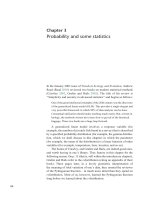
Figure 1.1 Overview of the four major receptor types. (A) Ion channel with extracellular domain labeled in red and transmembrane
chains labeled in green; (B) G-protein-coupled receptor with extracellular domains labeled in red, seven transmembrane domains
labeled in green; (C) receptor tyrosine kinase with transmembrane domain labeled in green, extracellular regions in red, and intracellular regions in blue. Black lines represent lipid bilayer; and (D) nucleus (dashed line) with nuclear receptors labeled in blue dimerizing
on a DNA template labeled in black. Images were created using Rasmol [8] from PDB ID [9], PDB ID 1F88 [10], PDB ID [11], and PDB
ID [12].
steroids, thyroid hormone, and certain vitamins.
These fat-soluble ligands diffuse through the cell
membrane to the interior of the cell, where they
bind to and activate receptor proteins that enhance
synthesis of new proteins within the cell.
Receptors are involved in cellular processes, such as
metabolism and ionic changes, as well as cell division,
growth, and protein synthesis. This book also covers
receptor actions at a higher, organismal level. Embryonic development (Chapter 11), disease (Chapter
12), and the activities of the mind (Chapter 13) all
involve integration of many physiological systems,
all bound by the same receptor process as cell–cell
communication.
All classes of receptors are encoded by genes
within each cell. Genes for receptors are also subject
to mutation and evolve by natural selection. As
a consequence, receptors will change over time,
allowing us to draw evolutionary inferences from
the present phylogenetic distribution of genes for
families of receptor molecules. The “fossil record”
of proteins is thus found not in the rocks of the
world but in the diversity of present-day organisms.
The four superfamilies of receptors described are all
widespread in eukaryotes. Some superfamilies are
also present in prokaryotes, and the study of their
distribution among all organisms (Chapter 14) gives
researchers an understanding of their functions and
role in organismal adaptations.
The relationships among protein families suggest
that their genes have mutated, changed location,
and duplicated many times, each time allowing the
production of new protein molecules with similar
functions. These similarities indicate further that
protein function can change over time, and that new
proteins with completely different functions can arise
from gene mutations. This seems to be how some
receptors arose, and how the families of receptors
have changed.
1.3
Receptors Are the Chemical Expression of Reality
Because receptors are at the interface between cells
and their environments, they are the first cellular
units to receive environmental information and
provide crucial information about the surroundings.
For example, animals know that nighttime is the
time to sleep, even though their brains have no
way of directly sensing the light or dark. Visual
information from the eyes goes to the pineal gland,
which produces the hormone melatonin in inverse
proportion to the amount of ambient light. Melatonin
is therefore the chemical expression of darkness [13].
1.3
In an analogous manner, other hormone and receptor
systems give information about the food taken in
by organisms. Insulin is produced in the pancreas
following a meal when blood glucose levels rise.
Insulin is therefore the chemical expression of plenty.
When food is scarce, the adrenal gland produces
the steroid cortisol as a means of liberating glucose
from storage forms in cells. Cortisol can be seen as
the chemical expression of starvation. As melatonin,
insulin, and cortisol all act on cellular receptors, we
view receptor mechanisms as an important way that
organisms have of knowing what reality is.
As the foregoing suggests, receptors are complex,
as are their interactions with cellular processes.
However, we hope that this complexity will be made
comprehensible by the approach we are taking:
the thousands of different receptors fall into only
four fundamental superfamilies; each has a unique
structure and a unique way of activating the cell,
so it is possible to identify an unfamiliar receptor if
one knows only a few things about it. Knowledge of
receptor function illuminates the many interactions
among proteins in the body and gives researchers
important information on higher level functions
of cell physiology (e.g., the normal workings of the
mind or the aberrant interactions involved in disease
states).
The book is divided into three parts: first is a
general discussion of cell membranes, proteins, hormone types, and receptor theory.2 Next follows one
chapter on each of the four receptor types. Finally,
several chapters outline receptor-mediated biological
processes such as embryonic development, disease,
mechanisms of the mind, and the evolution of these
remarkable molecules.
Pharmacology texts generally focus on hormones
and the kinetics of drug actions. We have written
this book for students who wish to become more
familiar with receptors themselves: the mechanisms
by which they act, the sorts of processes they direct,
and their evolution as molecules. It is meant for
students at the advanced undergraduate and early
2 The term theory is often used by mistake in place of hypothesis. In
proper usage, a theory is a hypothesis that has been tested and promoted to the level of widespread acceptance as a major concept in
science. It is unfortunate that scientists themselves often misuse
theory to mean hypothesis, as in “I have a theory about that” and
non-scientists often pounce on this misuse to denigrate science,
as in “evolution is only a theory.” In this book, we restrict the use
of the term theory to major scientific concepts, such as the theory
of evolution, or cell theory, or receptor theory. All three of these
ideas have been rigorously tested; they are no longer hypothetical, but have become key concepts in biological thinking. Other
concepts, still provisional, are called hypotheses.
Receptors Are the Chemical Expression of Reality
7
graduate levels and requires an understanding of
fundamental chemical and biological principles, a
general knowledge of evolutionary thought, and a
grasp of physiological interactions – all concepts that
are part of any good general biology course. The text
builds on these ideas to help students form a more
complex understanding of pharmacology and cellular
biochemistry.
Evolutionary inferences provide information that
allows the study of receptors to be not only exciting
and useful but also conceptually possible: despite the
bewildering array of cell surface receptor types, they
fall into just four major categories and interact with
only a few dozen other membrane effector proteins
that transmit the binding event into a biochemical
process. Thus, genetic relationships among receptors are relatively simple, and their use of similar
biochemical mechanisms shows that the important
problems of cell-to-cell signaling have needed to be
solved only a few times in evolution.
We wrote the book because in our teaching and
research we see the importance of receptor mechanisms and intracellular signaling across all kingdoms
of organisms and in many types of cellular processes.
Even so, it is difficult to find a book that gives them
complete coverage (structure, mechanism of action,
evolutionary history) without being written specifically for professionals. The two unifying themes of
the book are (i) the receptor concept itself: the idea
that biological communication is involved in nearly
all the activities of living things, and that receptor
function is the mechanism of that communication
and (ii) the role of natural selection and evolution in
shaping receptor structure and function. We hope
that this book will give a clear idea of the roles that
hormones and their receptors play in our lives, from
the reactions of individual cells to the behavior of
whole organisms.
9
2
The Origins of Chemical Thinking
A mystery is a phenomenon that people don’t know
how to think about – yet.
Daniel Dennett [14]
2.1
Overview of Early Pharmacological History
2.1.1
The Development of a Chemical Hypothesis
The earliest Greek thinkers such as Thales (sixth
century BCE) and Democritus (fourth century BCE)
taught that life was material and that physical components of the environment were responsible for the
organization of matter into living things. Thales also
initiated an experimental approach to studying natural phenomena [15]. However, these early thinkers
were unusual – the scholars who followed them had
a non-material, non-experimental, non-molecular
concept of the world. The non-material worldview
promoted the idea that life processes were fundamentally different from processes in non-living systems.
The non-experimental worldview encouraged the
use of logic rather than the use of experiment to test
ideas about natural phenomena. The non-molecular
worldview is best seen by its two main hypotheses
concerning the physical and biological spheres: the
four “elements” (earth, air, fire, and water) and the
“humoral” hypothesis (yellow bile, black bile, blood,
and phlegm).
Plato (fourth century BCE) exemplified the nonmaterial view, as he deemphasized observation and
experiment, and claimed that our perceptions of
matter were transitory and only what the mind
perceived (via logic) was permanent [15]. Under
his system, mind and body were considered to be
separate entities – the senses give an inaccurate
version of the world; only the mind provides “purity”
of perception [16]. Aristotle’s (fourth century BCE)
and Galen’s (second century CE) thinking opposed
this attitude, as they appreciated the role of matter
in life and advocated experimental approaches to the
study of nature.
Descartes (seventeenth century CE), although a
proponent of reason and experimentation, maintained that the body is a dual being, both mind and
matter, and the workings of the mind are outside
nature. He said that because mechanism describes
non-human workings, then other laws must apply to
human workings [17]. The concept of mind–body
dualism of Descartes and others thus furthered
non-material approaches to the study of life, and
inhibited development of a systematic approach to
the study of, among other things, the function of
the brain.
However, chemical thinking did arise among some
medieval thinkers: the physician Paracelsus in the
sixteenth century was the first to take up the concepts of the earliest Greeks, teaching that the body
was composed of chemicals and that illnesses were
the result of chemical imbalances. He anticipated
modern thinking in two of his teachings: that within
a natural product the curative agent could be a
single substance, and that the curative or poisonous
functions of a drug were directly proportional to its
concentration [18].
Felix Fontana in the late eighteenth century experimentally confirmed Paracelsus’ view that a crude drug
exerted its effect through a specific active principle
that acted on a discrete tissue in the organism. His
contemporary Peter Daries showed further that the
effect was proportional to the concentration of drug
applied. Setürner in the early nineteenth century
was the first actually to isolate a pure drug when he
obtained morphine from opium. This achievement
initiated a period of rapid change: before many
decades had passed, the chemical natures of many
pharmacological substances were determined, and
the new drug manuals, or pharmacopoeias, were
based on pure substances rather than on crude
plant extracts. Organic chemistry was emerging as
Receptor Biology, First Edition. Michael F. Roberts and Anne E. Kruchten.
© 2016 Wiley-VCH Verlag GmbH & Co. KGaA. Published 2016 by Wiley-VCH Verlag GmbH & Co. KGaA.