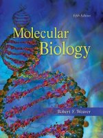
Molecular biology 5th ed r weaver (mcgraw hill, 2012)
Bạn đang xem bản rút gọn của tài liệu. Xem và tải ngay bản đầy đủ của tài liệu tại đây (40.91 MB, 915 trang )
This page intentionally left blank
This page intentionally left blank
wea25324_fm_i-xx.indd Page i 12/22/10 10:16 PM user-f468
/Volume/208/MHCE016/san74946_disk1of1/0073374946/san74946_pagefiles
Molecular Biology
Fifth Edition
R o b e r t F. We a v e r
University of Kansas
TM
wea25324_fm_i-xx.indd Page ii
30/12/10
5:25 PM user-f467
/Volume/208/MHCE016/san74946_disk1of1/0073374946/san75292_pagefile
TM
MOLECULAR BIOLOGY, FIFTH EDITION
Published by McGraw-Hill, a business unit of The McGraw-Hill Companies, Inc., 1221 Avenue of the
Americas, New York, NY 10020. Copyright © 2012 by The McGraw-Hill Companies, Inc. All rights reserved.
Previous editions © 2008, 2005, and 2002. No part of this publication may be reproduced or distributed in
any form or by any means, or stored in a database or retrieval system, without the prior written consent of
The McGraw-Hill Companies, Inc., including, but not limited to, in any network or other electronic storage or
transmission, or broadcast for distance learning.
Some ancillaries, including electronic and print components, may not be available to customers outside the
United States.
This book is printed on acid-free paper.
1 2 3 4 5 6 7 8 9 0 QDB /QDB 1 0 9 8 7 6 5 4 3 2 1
ISBN 978-0-07-352532-7
MHID 0-07-352532-4
Vice President & Editor-in-Chief: Marty Lange
Vice President EDP/Central Publishing Services: Kimberly Meriwether David
Publisher: Janice Roerig-Blong
Executive Marketing Manager: Patrick E. Reidy
Project Manager: Robin A. Reed
Design Coordinator: Brenda A. Rolwes
Cover Designer: Studio Montage, St. Louis, Missouri
Lead Photo Research Coordinator: Carrie K. Burger
Cover Image: © Getty Images RF
Buyer: Sandy Ludovissy
Media Project Manager: Balaji Sundararaman
Compositor: Aptara®, Inc.
Typeface: 10/12 Sabon
Printer: Quad/Graphics
All credits appearing on page or at the end of the book are considered to be an extension of the
copyright page.
Library of Congress Cataloging-in-Publication Data
Weaver, Robert Franklin, 1942Molecular biology / Robert F. Weaver.—5th ed.
p. cm.
ISBN 978–0–07–352532–7 (hardcover : alk. paper)
1. Molecular biology. I. Title.
QH506.W43 2011
572.8—dc22
2010051759
www.mhhe.com
wea25324_fm_i-xx.indd Page iii 12/22/10 10:16 PM user-f468
/Volume/208/MHCE016/san74946_disk1of1/0073374946/san74946_pagefiles
To Camilla and Nora
wea25324_fm_i-xx.indd Page iv 12/22/10 10:16 PM user-f468
A B O U T
T H E
/Volume/208/MHCE016/san74946_disk1of1/0073374946/san74946_pagefiles
A U T H O R
Rob Weaver was born in Topeka, Kansas, and
grew up in Arlington, Virginia. He received his
bachelor’s degree in chemistry from the College
of Wooster in Wooster, Ohio, in 1964. He
earned his Ph.D. in biochemistry at Duke
University in 1969, then spent two years doing
postdoctoral research at the University of
California, San Francisco, where he studied the
structure of eukaryotic RNA polymerases with
William J. Rutter.
He joined the faculty of the University of
Kansas as an assistant professor of biochemistry
in 1971, was promoted to associate professor,
and then to full professor in 1981. In 1984, he
became chair of the Department of Biochemistry,
(Source: Ashvini C. Ganesh)
and served in that capacity until he was named
Associate Dean of the College of Liberal Arts
and Sciences in 1995.
Prof. Weaver is the divisional dean for the science and mathematics departments
within the College, which includes supervising 10 different departments and
programs. As a professor of molecular biosciences, he teaches courses in introductory
molecular biology and the molecular biology of cancer. In his research laboratory,
undergraduates and graduate students have participated in research on the molecular
biology of a baculovirus that infects caterpillars.
Prof. Weaver is the author of many scientific papers resulting from research
funded by the National Institutes of Health, the National Science Foundation, and
the American Cancer Society. He has also coauthored two genetics textbooks and
has written two articles on molecular biology in the National Geographic
Magazine. He has spent two years performing research in European laboratories
as an American Cancer Society Research Scholar, one year in Zurich, Switzerland,
and one year in Oxford, England.
iv
wea25324_fm_i-xx.indd Page v 12/22/10 10:16 PM user-f468
/Volume/208/MHCE016/san74946_disk1of1/0073374946/san74946_pagefiles
B R I E F
C O N T E N T S
About the Author iv
Preface xiii
Acknowledgments xvii
Guide to Experimental Techniques in
Molecular Biology xix
PA R T V
PA R T I
Post-Transcriptional Events
Introduction
14 RNA Processing I: Splicing 394
15 RNA Processing II: Capping and Polyadenylation 436
16 Other RNA Processing Events and Post-Transcriptional
Control of Gene Expression 471
1 A Brief History 1
2 The Molecular Nature of Genes 12
3 An Introduction to Gene Function 30
PA R T I I
Methods in Molecular Biology
4 Molecular Cloning Methods 49
5 Molecular Tools for Studying Genes and Gene
Activity 75
PA R T I I I
Transcription in Bacteria
Translation
17 The Mechanism of Translation I: Initiation 522
18 The Mechanism of Translation II: Elongation
and Termination 560
19 Ribosomes and Transfer RNA 601
PA R T V I I
6 The Mechanism of Transcription in Bacteria 121
7 Operons: Fine Control of Bacterial
Transcription 167
8 Major Shifts in Bacterial Transcription 196
9 DNA–Protein Interactions in Bacteria 222
PA R T I V
Transcription in Eukaryotes
10 Eukaryotic RNA Polymerases and
Their Promoters 244
11 General Transcription Factors
in Eukaryotes 273
12 Transcription Activators in Eukaryotes
13 Chromatin Structure and Its Effects
on Transcription 355
PA R T V I
DNA Replication, Recombination,
and Transposition
20
21
22
23
DNA Replication, Damage, and Repair 636
DNA Replication II: Detailed Mechanism 677
Homologous Recombination 709
Transposition 732
PA R T V I I I
Genomes
314
24 Introduction to Genomics: DNA Sequencing on
a Genomic Scale 759
25 Genomics II: Functional Genomics, Proteomics,
and Bioinformatics 789
Glossary 827
Index 856
v
wea25324_fm_i-xx.indd Page vi 12/22/10 10:16 PM user-f468
C
O
N
T
/Volume/208/MHCE016/san74946_disk1of1/0073374946/san74946_pagefiles
E
N
T
S
About the Author iv
Preface xiii
Acknowledgments xvii
Guide to Experimental Techniques
in Molecular Biology xix
CHAPTER 3
An Introduction to Gene Function 30
3.1
3.2
3.3
1
35
39
Replication 45
Mutations 45
45
2
Genetic Recombination and Mapping
4
Physical Evidence for Recombination
5
The Discovery of DNA
37
2
The Chromosome Theory of Inheritance
Molecular Genetics
31
40
Sickle Cell Disease
Mendel’s Laws of Inheritance
3
PA R T I I
Methods of Molecular Biology
5
5
CHAPTER 4
The Relationship Between Genes and Proteins
1.3
Protein Function
Translation
Transmission Genetics
Activities of Genes
31
Transcription
CHAPTER 1
1.2
Protein Structure
Discovery of Messenger RNA
Introduction
1.1
31
Overview of Gene Expression
PA R T I
A Brief History
Storing Information
6
7
The Three Domains of Life
Molecular Cloning Methods
4.1
9
Gene Cloning
50
The Role of Restriction Endonucleases
Vectors
49
50
53
Identifying a Specific Clone with a
Specific Probe 58
CHAPTER 2
cDNA Cloning
The Molecular Nature of Genes
2.1
The Nature of Genetic Material
Transformation in Bacteria
DNA Structure
2.3
2.4
13
The Polymerase Chain Reaction
62
62
Using Reverse Transcriptase PCR (RT-PCR)
in cDNA Cloning 64
19
Real-Time PCR
Genes Made of RNA 22
Physical Chemistry of Nucleic Acids
23
DNAs of Various Sizes and Shapes
vi
61
Box 4.1 Jurassic Park: More than a Fantasy?
15
19
A Variety of DNA Structures
Rapid Amplification of cDNA Ends
Standard PCR
18
Experimental Background
The Double Helix
4.2
13
The Chemical Nature of Polynucleotides
2.2
12
60
27
4.3
23
64
Methods of Expressing Cloned Genes
Expression Vectors
65
Other Eukaryotic Vectors
71
Using the Ti Plasmid to Transfer Genes
to Plants 71
65
63
wea25324_fm_i-xx.indd Page vii 12/22/10 10:16 PM user-f468
/Volume/208/MHCE016/san74946_disk1of1/0073374946/san74946_pagefiles
Contents
CHAPTER 5
Molecular Tools for Studying Genes
and Gene Activity 75
5.1
Molecular Separations
Gel Electrophoresis
76
80
Gel Filtration Chromatography
80
82
Autoradiography
82
Phosphorimaging
83
79
PA R T I I I
CHAPTER 6
84
The Mechanism of Transcription
in Bacteria 121
84
Using Nucleic Acid Hybridization
85
Southern Blots: Identifying Specific DNA Fragments 85
DNA Fingerprinting and DNA Typing
6.2
122
123
89
126
Sigma Stimulates Transcription Initiation
89
Reuse of s
129
Automated DNA Sequencing
91
High-Throughput Sequencing
93
Promoter Clearance
S1 Mapping
6.4
6.5
102
Nuclear Run-On Transcription
Reporter Gene Transcription
Rho-Dependent Termination
104
Assaying DNA–Protein Interactions
106
156
159
108
Operons: Fine Control of Bacterial
Transcription 167
7.1
The lac Operon
168
Negative Control of the lac Operon
109
Discovery of the Operon
109
The Mechanism of Repression
112
112
169
169
Repressor–Operator Interactions
DMS Footprinting and Other Footprinting
Methods 109
Assaying Protein–Protein Interactions
146
156
CHAPTER 7
108
Chromatin Immunoprecipitation (ChIP)
144
104
105
Measuring Protein Accumulation in Vivo
Termination of Transcription
Rho-Independent Termination
Measuring Transcription Rates in Vivo
DNase Footprinting
144
Structure of the Elongation Complex
100
Gel Mobility Shift
Elongation
Core Polymerase Functions in Elongation
Run-Off Transcription and G-Less
Cassette Transcription 103
Filter Binding
139
The Role of the a-Subunit in UP Element
Recognition 142
99
Primer Extension
132
134
Structure and Function of s
Protein Engineering with Cloned Genes:
Site-Directed Mutagenesis 97
Mapping and Quantifying Transcripts 99
127
128
The Stochastic s-Cycle Model
95
123
125
Transcription Initiation
Local DNA Melting at the Promoter
Northern Blots
5.9
Promoters
Promoter Structure
6.3
DNA Sequencing and Physical Mapping
Restriction Mapping
5.8
122
Binding of RNA Polymerase to Promoters
The Sanger Chain-Termination
Sequencing Method 90
5.7
RNA Polymerase Structure
Sigma (s) as a Specificity Factor
In Situ Hybridization: Locating Genes in
Chromosomes 88
Immunoblots (Western Blots)
6.1
86
Forensic Uses of DNA Fingerprinting and
DNA Typing 87
5.6
115
Transcription in Bacteria
Nonradioactive Tracers
5.5
115
115
Transgenic Mice
81
Liquid Scintillation Counting
5.4
114
5.11 Knockouts and Transgenics
Ion-Exchange Chromatography
Labeled Tracers
114
Functional SELEX
Knockout Mice
Affinity Chromatography
5.3
SELEX
76
Two-Dimensional Gel Electrophoresis
5.2
5.10 Finding RNA Sequences That Interact
with Other Molecules 114
173
174
Positive Control of the lac Operon
The Mechanism of CAP Action
177
178
vii
wea25324_fm_i-xx.indd Page viii 12/22/10 10:16 PM user-f468
viii
7.2
/Volume/208/MHCE016/san74946_disk1of1/0073374946/san74946_pagefiles
Contents
The ara Operon
182
9.4
The ara Operon Repression Loop
183
Evidence for the ara Operon Repression Loop
7.3
DNA-Binding Proteins: Action at
a Distance 237
The gal Operon
183
237
Autoregulation of araC 185
Duplicated l Operators
The trp Operon
Enhancers
186
237
238
Tryptophan’s Role in Negative Control of the
trp Operon 186
Control of the trp Operon by Attenuation
Defeating Attenuation
7.4
Riboswitches
187
PA R T I V
188
Transcription in Eukaryotes
190
CHAPTER 8
CHAPTER 10
Major Shifts in Bacterial
Transcription 196
8.1
Sigma Factor Switching
Phage Infection
Sporulation
Eukaryotic RNA Polymerases
and Their Promoters 244
197
10.1
197
199
Separation of the Three Nuclear Polymerases
Genes with Multiple Promoters 201
Other s Switches
Anti-s-Factors
8.2
8.3
The Roles of the Three RNA Polymerases
201
RNA Polymerase Subunit Structures
202
The RNA Polymerase Encoded in
Phage T7 202
Infection of E. coli by Phage l 203
Lytic Reproduction of Phage l
Establishing Lysogeny
204
211
10.2
Determining the Fate of a l Infection: Lysis or
Lysogeny 217
Lysogen Induction
218
DNA–Protein Interactions
in Bacteria 222
263
Class III Promoters
264
Enhancers and Silencers
Enhancers
Silencers
267
267
269
CHAPTER 11
11.1
Class II Factors
274
The Class II Preinitiation Complex
274
Structure and Function of TFIID
276
Probing Binding Specificity by SiteDirected Mutagenesis 223
Structure and Function of TFIIB
286
Structure and Function of TFIIH
288
Box 9.1 X-Ray Crystallography
The Mediator Complex and the
RNA Polymerase II Holoenzyme
295
The l Family of Repressors
223
224
High-Resolution Analysis of l Repressor–Operator
Interactions 229
High-Resolution Analysis of Phage 434
Repressor–Operator Interactions 232
The trp Repressor
Elongation Factors
11.2
234
The Role of Tryptophan
9.3
259
Class I Promoters
10.3
248
259
General Transcription Factors
in Eukaryotes 273
CHAPTER 9
9.2
Promoters
Class II Promoters
Autoregulation of the cI Gene During
Lysogeny 212
9.1
Multiple Forms of Eukaryotic
RNA Polymerase 245
234
General Considerations on
Protein–DNA Interactions 235
Class I Factors
296
299
The Core-Binding Factor
299
The UPE-Binding Factor
300
Structure and Function of SL1
11.3
Class III Factors
Hydrogen Bonding Capabilities of the Four
Different Base Pairs 235
TFIIIA
The Importance of Multimeric DNA-Binding Proteins 236
The Role of TBP
303
303
TFIIIB and C
304
307
301
245
246
wea25324_fm_i-xx.indd Page ix 12/22/10 10:16 PM user-f468
/Volume/208/MHCE016/san74946_disk1of1/0073374946/san74946_pagefiles
Contents
CHAPTER 12
Nucleosome Positioning
Transcription Activators
in Eukaryotes 314
Histone Acetylation
12.1
Categories of Activators
DNA-Binding Domains
Chromatin Remodeling
315
315
Homeodomains
Nucleosomes and Transcription
Elongation 387
315
Post-Transcriptional Events
318
319
CHAPTER 14
320
The bZIP and bHLH Domains
RNA Processing I: Splicing
321
Independence of the Domains
of Activators 323
Functions of Activators 324
Recruitment of TFIID
14.1
RNA Splicing
329
Box 12.1 Genomic Imprinting
Transcription Factories
Complex Enhancers
Enhanceosomes
334
Control of Splicing
343
14.3
344
Activator Acetylation
346
348
RNA Processing II: Capping and
Polyadenylation 436
Chromatin Structure and Its Effects
on Transcription 355
356
15.2
356
Capping
437
Cap Structure
437
Cap Synthesis
438
Functions of Caps
440
Polyadenylation
442
Poly(A)
357
442
Functions of Poly(A)
360
Higher-Order Chromatin Folding
13.2
430
CHAPTER 15
348
15.1
The 30-nm Fiber
427
Group II Introns
CHAPTER 13
Nucleosomes
427
347
Signal Transduction Pathways
Chromatin Structure
411
425
Self-Splicing RNAs
Group I Introns
Activator Sumoylation
Histones
402
Commitment, Splice Site Selection, and Alternative
Splicing 415
338
Regulation of Transcription Factors
399
401
Spliceosome Assembly and Function
337
398
The Mechanism of Splicing of Nuclear
mRNA Precursors 399
Spliceosomes
336
Activator Ubiquitylation
13.1
397
A Signal at the Branch
339
Coactivators
395
A Branched Intermediate
332
Architectural Transcription Factors
Insulators
14.2
328
Action at a Distance
395
Effect of Splicing on Gene Expression
328
394
396
Splicing Signals
325
Interaction Among Activators
Dimerization
Genes in Pieces
Evidence for Split Genes
324
Recruitment of the Holoenzyme
12.6
383
PA R T V
The Nuclear Receptors
12.5
376
Heterochromatin and Silencing
316
The GAL4 Protein
12.4
373
Structures of the DNA-Binding
Motifs of Activators 316
Zinc Fingers
12.3
372
Histone Deacetylation
Transcription-Activating Domains
12.2
367
362
Chromatin Structure and Gene
Activity 364
The Effects of Histones on Transcription
of Class II Genes 365
443
Basic Mechanism of Polyadenylation
Polyadenylation Signals
445
446
Cleavage and Polyadenylation of a Pre-mRNA
Poly(A) Polymerase
454
Turnover of Poly(A)
454
448
ix
wea25324_fm_i-xx.indd Page x 12/22/10 10:16 PM user-f468
x
/Volume/208/MHCE016/san74946_disk1of1/0073374946/san74946_pagefiles
Contents
15.3
Coordination of mRNA Processing Events
456
PA R T V I
Binding of the CTD of Rpb1 to mRNA-Processing
Proteins 457
Translation
Changes in Association of RNA-Processing Proteins
with the CTD Correlate with Changes in CTD
Phosphorylation 458
CHAPTER 17
A CTD Code?
460
Coupling Transcription Termination with mRNA
39-End Processing 461
Mechanism of Termination
The Mechanism of Translation I:
Initiation 522
17.1
462
tRNA Charging
Role of Polyadenylation in mRNA Transport
466
Other RNA Processing Events and
Post-Transcriptional Control of Gene
Expression 471
Ribosomal RNA Processing
Eukaryotic rRNA Processing
Bacterial rRNA Processing
16.2
17.3
472
475
537
545
545
548
475
CHAPTER 18
Forming Mature 39-Ends
476
The Mechanism of Translation II:
Elongation and Termination 560
Trans-Splicing 477
RNA Editing
477
18.1
479
479
18.2
482
Mechanism of RNAi
The Triplet Code
The (Almost) Universal Code
18.3
Processing Bodies
Other Small RNAs
517
569
Elongation Step 3: Translocation
G Proteins and Translation
511
514
570
18.4
Termination
580
583
584
Termination Codons
Release Factors
577
582
The Structures of EF-Tu and EF-G
507
584
Stop Codon Suppression
Degradation of mRNAs in P-Bodies
Relief of Repression in P-Bodies
569
Overview of Elongation
Elongation Step 2: Peptide Bond Formation
502
510
The Elongation Cycle
Elongation Step 1: Binding an Aminoacyl-tRNA to the
A Site of the Ribosome 573
Piwi-Interacting RNAs and Transposon Control 501
Post-Transcriptional Control of Gene
Expression: MicroRNAs 502
Translation Repression, mRNA Degradation,
and P-Bodies 510
567
A Three-Site Model of the Ribosome
Role of the RNAi Machinery in Heterochromatin
Formation and Gene Silencing 495
Stimulation of Translation by miRNAs
564
Unusual Base Pairs Between Codon and Anticodon 566
494
Silencing of Translation by miRNAs
562
563
563
Breaking the Code
484
489
Amplification of siRNA
No Gaps in the Code
484
Post-Transcriptional Control of Gene Expression:
RNA Interference 488
The Direction of Polypeptide Synthesis and
of mRNA Translation 561
The Genetic Code 562
Nonoverlapping Codons
Post-Transcriptional Control of Gene
Expression: mRNA Stability 483
Transferrin Receptor mRNA Stability
16.9
Control of Initiation
533
475
Casein mRNA Stability
16.7
16.8
533
Eukaryotic Translational Control
Editing by Nucleotide Deamination
16.6
Initiation in Eukaryotes
533
Forming Mature 59-Ends
Mechanism of Editing
16.5
531
Bacterial Translational Control
The Mechanism of Trans-Splicing
16.4
525
Formation of the 70S Initiation Complex
Eukaryotic Initiation Factors
472
Cutting Apart Polycistronic Precursors
16.3
Formation of the 30S Initiation Complex
The Scanning Model of Initiation
474
Transfer RNA Processing
523
Summary of Initiation in Bacteria
17.2
523
523
Dissociation of Ribosomes
CHAPTER 16
16.1
Initiation of Translation in Bacteria
586
586
Dealing with Aberrant Termination
588
Use of Stop Codons to Insert Unusual Amino Acids 593
wea25324_fm_i-xx.indd Page xi 12/22/10 10:16 PM user-f468
/Volume/208/MHCE016/san74946_disk1of1/0073374946/san74946_pagefiles
xi
Contents
18.5
Posttranslation
593
Excision Repair
Folding Nascent Proteins
660
Double-Strand Break Repair in Eukaryotes
594
Release of Ribosomes from mRNA
Mismatch Repair
595
Failure of Mismatch Repair in Humans
CHAPTER 19
Ribosomes
CHAPTER 21
Ribosome Composition
DNA Replication II:
Detailed Mechanism
602
605
Fine Structure of the 30S Subunit
606
Fine Structure of the 50S Subunit
612
21.1
621
Transfer RNA
The Discovery of tRNA
tRNA Structure
678
Priming in Eukaryotes
21.2
623
Initiation
677
Priming in E. coli 678
Ribosome Structure and the Mechanism of Translation 616
19.2
668
601
602
Fine Structure of the 70S Ribosome
Polysomes
668
Coping with DNA Damage Without Repairing It
Ribosomes and Transfer RNA
19.1
665
667
Elongation
683
Speed of Replication
623
679
683
The Pol III Holoenzyme and Processivity
of Replication 683
623
Recognition of tRNAs by Aminoacyl-tRNA Synthetase:
The Second Genetic Code 626
21.3
Termination
694
Decatenation: Disentangling Daughter DNAs
Proofreading and Editing by Aminoacyl-tRNA
Synthetases 630
Termination in Eukaryotes
694
695
Box 21.1 Telomeres, the Hayflick Limit, and
Cancer 699
PA R T V I I
Telomere Structure and Telomere-Binding Proteins in
Lower Eukaryotes 702
DNA Replication, Recombination,
and Transposition
CHAPTER 22
CHAPTER 20
Homologous Recombination
22.1
DNA Replication, Damage,
and Repair 636
20.1
22.2
General Features of DNA Replication
Semiconservative Replication
Bidirectional Replication
Rolling Circle Replication
20.2
RuvC
642
22.3
645
646
721
722
Creation of Single-Stranded Ends at DSBs
650
22.4
651
Gene Conversion
728
651
CHAPTER 23
653
DNA Damage and Repair
719
Meiotic Recombination
The Double-Stranded DNA Break
Single-Strand DNA-Binding Proteins
20.3
717
The Mechanism of Meiotic Recombination:
Overview 721
649
Multiple Eukaryotic DNA Polymerases
Topoisomerases
715
RuvA and RuvB
Three DNA Polymerases in E. coli 646
Strand Separation
712
RecBCD
639
641
Enzymology of DNA Replication
Fidelity of Replication
The RecBCD Pathway for Homologous
Recombination 710
Experimental Support for the RecBCD
Pathway 712
RecA
637
At Least Semidiscontinuous Replication
Priming of DNA Synthesis
637
709
Transposition
656
Damage Caused by Alkylation of Bases
Damage Caused by Ultraviolet Radiation
Damage Caused by Gamma and X-Rays
Directly Undoing DNA Damage
659
657
658
658
23.1
732
Bacterial Transposons 733
Discovery of Bacterial Transposons
733
Insertion Sequences: The Simplest Bacterial
Transposons 733
728
wea25324_fm_i-xx.indd Page xii 12/22/10 10:16 PM user-f468
xii
Contents
More Complex Transposons
734
Mechanisms of Transposition
23.2
23.3
/Volume/208/MHCE016/san74946_disk1of1/0073374946/san74946_pagefiles
Eukaryotic Transposons
734
Sequencing Standards
737
24.3
Studying and Comparing Genomic Sequences 774
P Elements
Other Vertebrate Genomes
Rearrangement of Immunoglobulin Genes
Retrotransposons
Retroviruses
Personal Genomics
739
740
The Barcode of Life
779
779
782
784
743
CHAPTER 25
743
Genomics II: Functional Genomics,
Proteomics, and Bioinformatics 789
745
745
Retrotransposons
774
The Minimal Genome
742
Mechanism of V(D)J Recombination
749
25.1
799
Single-Nucleotide Polymorphisms:
Pharmacogenomics 810
CHAPTER 24
25.2
Introduction to Genomics: DNA
Sequencing on a Genomic Scale 759
Positional Cloning: An Introduction
to Genomics 760
Classical Tools of Positional Cloning
765
767
Vectors for Large-Scale Genome Projects
770
812
Protein Separations
Protein Analysis
769
812
813
Quantitative Proteomics
25.3
760
Techniques in Genomic Sequencing
Proteomics
Protein Interactions
Identifying the Gene Mutated in a Human Disease 762
The Clone-by-Clone Strategy
790
Genomic Functional Profiling
Genomes
The Human Genome Project
Functional Genomics: Gene Expression
on a Genomic Scale 790
Transcriptomics
PA R T V I I I
24.2
774
The Human Genome
The Recombinase
24.1
773
The First Examples of Transposable Elements:
Ds and Ac of Maize 737
Recombination Signals
23.4
Shotgun Sequencing
Bioinformatics
814
816
820
Finding Regulatory Motifs in Mammalian
Genomes 820
Using the Databases Yourself
Glossary 827
Index 856
822
wea25324_fm_i-xx.indd Page xiii 12/22/10 10:16 PM user-f468
/Volume/208/MHCE016/san74946_disk1of1/0073374946/san74946_pagefiles
P
One of my most exciting educational experiences was my
introductory molecular biology course in graduate school.
My professor used no textbook, but assigned us readings
directly from the scientific literature. It was challenging,
but I found it immensely satisfying to meet the challenge
and understand, not only the conclusions, but how the
evidence supported those conclusions.
When I started teaching my own molecular biology
course, I adopted this same approach, but tried to reduce
the challenge to a level more appropriate for undergraduate students. I did this by narrowing the focus to the most
important experiments in each article, and explaining
those carefully in class. I used hand-drawn cartoons and
photocopies of the figures as illustrations.
This approach worked well, and the students enjoyed
it, but I really wanted a textbook that presented the concepts of molecular biology, along with experiments that
led to those concepts. I wanted clear explanations that
showed students the relationship between the experiments
and the concepts. So, I finally decided that the best way to
get such a book would be to write it myself. I had already
coauthored a successful introductory genetics text in
which I took an experimental approach—as much as possible with a book at that level. That gave me the courage
to try writing an entire book by myself and to treat the
subject as an adventure in discovery.
Organization
The book begins with a four-chapter sequence that should
be a review for most students. Chapter 1 is a brief history
of genetics. Chapter 2 discusses the structure and chemical
properties of DNA. Chapter 3 is an overview of gene expression, and Chapter 4 deals with the nuts and bolts of
gene cloning. All these are topics that the great majority
of molecular biology students have already learned in an
introductory genetics course. Still, students of molecular
biology need to have a grasp of these concepts and may
need to refresh their understanding of them. I do not deal
specifically with these chapters in class; instead, I suggest
students consult them if they need more work on these topics.
These chapters are written at a more basic level than the
rest of the book.
Chapter 5 describes a number of common techniques
used by molecular biologists. It would not have been possible to include all the techniques described in this book in
one chapter, so I tried to include the most common or, in a
R
E
F
A
C
E
few cases, valuable techniques that are not mentioned elsewhere in the book. When I teach this course, I do not present Chapter 5 as such. Instead, I refer students to it when
we first encounter a technique in a later chapter. I do it that
way to avoid boring my students with technique after technique. I also realize that the concepts behind some of these
techniques are rather sophisticated, and the students’ appreciation of them is much deeper after they’ve acquired
more experience in molecular biology.
Chapters 6–9 describe transcription in bacteria. Chapter 6 introduces the basic transcription apparatus, including promoters, terminators, and RNA polymerase, and
shows how transcripts are initiated, elongated, and terminated. Chapter 7 describes the control of transcription in
three different operons, then Chapter 8 shows how bacteria and their phages control transcription of many genes at
a time, often by providing alternative sigma factors. Chapter 9 discusses the interaction between bacterial DNAbinding proteins, mostly helix-turn-helix proteins, and
their DNA targets.
Chapters 10–13 present control of transcription in eukaryotes. Chapter 10 deals with the three eukaryotic RNA
polymerases and the promoters they recognize. Chapter 11
introduces the general transcription factors that collaborate with the three RNA polymerases and points out the
unifying theme of the TATA-box-binding protein, which
participates in transcription by all three polymerases.
Chapter 12 explains the functions of gene-specific transcription factors, or activators. This chapter also illustrates
the structures of several representative activators and
shows how they interact with their DNA targets. Chapter 13
describes the structure of eukaryotic chromatin and shows
how activators and silencers can interact with coactivators
and corepressors to modify histones, and thereby to activate
or repress transcription.
Chapters 14–16 introduce some of the posttranscriptional events that occur in eukaryotes. Chapter 14 deals
with RNA splicing. Chapter 15 describes capping and
polyadenylation, and Chapter 16 introduces a collection of
fascinating “other posttranscriptional events,” including
rRNA and tRNA processing, trans-splicing, and RNA editing. This chapter also discusses four kinds of posttranscriptional control of gene expression: (1) RNA interference;
(2) modulating mRNA stability (using the transferrin receptor
mRNA as the prime example); (3) control by microRNAs,
and (4) control of transposons in germ cells by Piwi-interacting
RNAs (piRNAs).
xiii
wea25324_fm_i-xx.indd Page xiv 12/22/10 10:16 PM user-f468
xiv
/Volume/208/MHCE016/san74946_disk1of1/0073374946/san74946_pagefiles
Preface
Chapters 17–19 describe the translation process in both
bacteria and eukaryotes. Chapter 17 deals with initiation
of translation, including the control of translation at the
initiation step. Chapter 18 shows how polypeptides are
elongated, with the emphasis on elongation in bacteria.
Chapter 19 provides details on the structure and function of
two of the key players in translation: ribosomes and tRNA.
Chapters 20–23 describe the mechanisms of DNA replication, recombination, and translocation. Chapter 20 introduces the basic mechanisms of DNA replication and
repair, and some of the proteins (including the DNA polymerases) involved in replication. Chapter 21 provides details of the initiation, elongation, and termination steps in
DNA replication in bacteria and eukaryotes. Chapters 22
and 23 describe DNA rearrangements that occur naturally
in cells. Chapter 22 discusses homologous recombination
and Chapter 23 deals with translocation.
Chapters 24 and 25 present concepts of genomics, proteomics, and bioinformatics. Chapter 24 begins with an oldfashioned positional cloning story involving the Huntington
disease gene and contrasts this lengthy and heroic quest
with the relative ease of performing positional cloning with
the human genome (and other genomes). Chapter 25 deals
with functional genomics (transcriptomics), proteomics,
and bioinformatics.
New to the Fifth Edition
The most obvious change in the fifth edition is the splitting
of old Chapter 24 (Genomics, Proteomics, and Bioinformatics) in two. This chapter was already the longest in the book,
and the field it represents is growing explosively, so a split
was inevitable. The new Chapter 24 deals with classical genomics: the sequencing and comparison of genomes. New
material in Chapter 24 includes an analysis of the similarity
between the human and chimpanzee genomes, and a look at
the even closer similarity between the human and Neanderthal genomes, including recent evidence for interbreeding
between humans and Neanderthals. It also includes an update on the new field of synthetic biology, made possible by
genomic work on microorganisms, and contains a report of
the recent success by Craig Venter and colleagues in creating
a living Mycoplasma cell with a synthetic genome.
Chapter 25 deals with fields allied with Genomics:
Functional Genomics, Proteomics, and Bioinformatics.
New material in Chapter 25 includes new applications of
the ChIP-chip and ChIP-seq techniques—the latter using
next-generation DNA sequencing; collision-induced dissociation mass spectrometry, which can be used to sequence
proteins; and the use of isotope-coded affinity tags (ICATs)
and stable isotope labeling by amino acids (SILAC) to
make mass spectrometry (MS) quantitative. Quantitative
MS in turn enables comparative proteomics, in which the
concentrations of large numbers of proteins can be compared between species.
All but the introductory chapters of this fifth edition
have been updated. Here are a few highlights:
• Chapter 5: Introduces high-throughput (next
generation) DNA sequencing techniques. These have
revolutionized the field of genomics. Chromatin
immunoprecipitation (ChIP) and the yeast two-hybrid
assay have been moved to Chapter 5, in light of their
broad applicabilities. A treatment of the energies of
the b-electrons from 3H, 14C, 35S, and 32P has been
added, and the fluorography technique, which captures information from the lower-energy emissions,
is discussed.
• Chapter 6: Adds a discussion of FRET-ALEX (FRET
with alternating laser excitation), along with a description of how this technique has been used to
support (1) the stochastic release model of the
s-cycle and (2) the scrunching hypothesis to explain
abortive transcription. This chapter also updates
the structure of the bacterial elongation complex,
including a discussion of a two-state model for
nucleotide addition.
• Chapter 7: Introduces the riboswitch in the mRNA
from the glmS gene of B. subtilis, in which the end
product of the gene turns expression of the gene off
by stimulating the mRNA to destroy itself. This
chapter also introduces a hammerhead ribozyme as
a possible mammalian riboswitch that may operate
by a similar mechanism.
• Chapter 8: Introduces the concepts of anti-s-factors
and anti-anti-s-factors as controllers of transcription
during sporulation in B. subtilis.
• Chapter 9: Emphasizes the dynamic nature of protein structure, and points out that a given crystal
structure represents just one of a range of different
possible protein conformations.
• Chapter 10: Presents a new study by Roger Kornberg’s group that identifies the RNA polymerase II
trigger loop as a key determinant in transcription
specificity, along with a discussion of how the
enzyme distinguishes between ribonuncleotides and
deoxyribonucleotides. This chapter also introduces
the concepts of core promoter and proximal promoter, where the core promoter contains any combination of TFIIB recognition element, TATA box,
initiator, downstream promoter element, downstream core element, and motif ten element, and
the proximal promoter contains upstream
promoter elements.
• Chapter 11: Introduces the concept of core TAFs—
those associated with class II preinitiation complexes
from a wide variety of eukaryotes, and introduces
the new nomenclature (TAF1–TAF13), which
replaces the old, confusing nomenclature that was
wea25324_fm_i-xx.indd Page xv 12/22/10 10:16 PM user-f468
/Volume/208/MHCE016/san74946_disk1of1/0073374946/san74946_pagefiles
Preface
based on molecular masses (e.g., TAFII250). This
chapter also describes an experiment that shows the
importance of TFIIB in setting the start site of transcription. It also shows that a similar mechanism
applies in the archaea, which use a TFIIB homolog
known as transcription factor B.
• Chapter 12: Introduces the technique of chromosome
conformation capture (3C) and shows how it can be
used to detect DNA looping between an enhancer
and a promoter. This chapter also introduces the concept of imprinting during gametogenesis, and explains
the role of methylation in imprinting, particularly
methylation of the imprinting control region of the
mouse Igf2/H19 locus. It also introduces the concept
of transcription factories, where transcription of multiple genes occurs. Finally, this chapter refines and
updates the concept of the enhanceosome.
• Chapter 13: Presents a new table showing all the
ways histones can be modified in vivo; brings back
the solenoid, alongside the two-start helix, as a
candidate for the 30-nm fiber structure; and presents
evidence that chromatin adopts one or the other
structure, depending on its nucleosome repeat
length. This chapter also introduces the concept of
specific histone methylations as markers for transcription initiation and elongation, and shows how
this information can be used to infer that RNA polymerase II is poised between initiation and elongation
on many human protein-encoding genes. It also
emphasizes the importance of histone modifications
in affecting not only histone–DNA interactions, but
also nucleosome–nucleosome interactions and
recruitment of histone-modifying and chromatinremodeling proteins. Finally, this chapter shows how
PARP1 (poly[ADP-ribose] polymerase-1) can
facilitate nucleosome loss from chromatin by
poly(ADP-ribosyl)ating itself.
• Chapter 14: Introduces the exon junction complex
(EJC), which is added to mRNAs during splicing in
the nucleus, and shows how the EJC can stimulate
transcription by facilitating the association of
mRNAs with ribosomes. This chapter also introduces exon and intron definition modes of splicing
and shows how they can be distinguished experimentally. This test has revealed that higher eukaryotes primarily use exon definition and lower
eukaryotes primarily use intron definition.
• Chapter 15: Demonstrates that a subunit of CPSF
(CPSF-73) is responsible for cutting a pre-mRNA at
a polyadenylation signal. It also shows that serine 7,
in addition to serines 2 and 5 in the repeating
heptad in the CTD of the largest RNA polymerase
subunit, can be phosphorylated, and shows that this
serine 7 phosphorylation controls the expression of
xv
certain genes (e.g., the U2 snRNA gene) by controlling the 39-end processing of their mRNAs.
• Chapter 16: Identifies a single enzyme, tRNA 39 processing endoribonuclease, as the agent that cleaves
excess nucleotides from the 39-end of a eukaryotic
tRNA precursor; points out the overwhelming prevalence of trans-splicing in C. elegans; presents a new
model for removal of the passenger strand of a
double-stranded siRNA—cleavage of the passenger
strand by Ago2; introduces Piwi-interacting RNAs
(piRNAs) and presents the ping-pong model by
which they are assumed to amplify themselves and
inactivate transposons in germ cells; introduces
plant RNA polymerases IV and V, and describes
their roles in gene silencing. This chapter also greatly
expands the coverage of miRNAs, and points out
that hundreds of miRNAs control thousands of
plant and animal genes, and that mutations in
miRNA genes typically have very deleterious effects.
Chapter 16 also updates the biogenesis of miRNAs,
introducing two pathways to miRNA production:
the Drosha and mirtron pathways. Finally, this
chapter introduces P-bodies, which are involved in
mRNA decay and translational repression.
• Chapter 17: Updates the section on eukaryotic
viral internal ribosome entry sequences (IRESs).
Some viruses cleave eIF4G, leaving a remnant
called p100. Poliovirus IRESs bind to p100 and
thereby gain access to ribosomes, but hepatitis C
virus IRESs bind directly to eIF3, while hepatitis A
virus IRESs bind even more directly to ribosomes.
This chapter also refines the model describing how
the cleavage of eIF4G affects mammalian host
mRNA translation. Different cell types respond
differently to this cleavage. Finally, this chapter
introduces the concept of the pioneer round of
translation, and points out that different initiation
factors are used in the pioneer round than in all
subsequent rounds.
• Chapter 18: Introduces the concept of superwobble,
which holds that a single tRNA with a U in its wobble position can recognize codons ending in any of
the four bases, and presents evidence that superwobble works. This chapter also introduces the hybrid P/I
state as the initial ribosomal binding state for fMettRNA Met
f . In this state, the anticodon is in the P site,
but the fMet and acceptor stem are in an “initiator”
site between the P site and the E site. This chapter
also describes no-go decay, which degrades mRNA
containing a stalled ribosome, and introduces the
concept of codon bias to explain inefficiency of
translation. Finally, this chapter explains how the
slowing of translation by rare codons can influence
protein folding both negatively and positively.
wea25324_fm_i-xx.indd Page xvi 12/22/10 10:16 PM user-f468
xvi
/Volume/208/MHCE016/san74946_disk1of1/0073374946/san74946_pagefiles
Preface
• Chapter 19: Includes a new section based on recent
crystal structures of the ribosome in complex with
various elongation factors. One of these structures
involves aminoacyl-tRNA and EF-Tu, and has
shown that the tRNA is bent by about 30 degrees in
forming an A/T complex. This bend is important in
fidelity of translation, and also facilitates the GTP
hydrolysis that permits EF-Tu to leave the ribosome.
Another crystal structure involves EF-G–GDP and
shows the ribosome in the post-translocation E/E,
P/P state, as opposed to the spontaneously achieved
pre-translocation P/E, A/P hybrid state. This chapter
also provides links to two excellent new movies describing the elongation process and an overview of
translation initiation, elongation, and termination.
Finally, this chapter describes crystal structures that
illustrate the functions of two critical parts of RF1
and RF2 in stop codon recognition and cleavage of
polypeptides from their tRNAs.
• Chapter 20: Introduces the controversial proposal,
with evidence, that DNA replication in E. coli is discontinuous on both strands. This chapter also introduces ACL1, a chromatin remodeler recruited via its
macrodomain to sites of double-strand breaks by
poly(ADP-ribose) formed at these sites by poly(ADPribose) polymerase 1 (PARP-1).
• Chapter 21: Presents a co-crystal structure of a b dimer bound to a primed DNA template, showing that
the b clamp really does encircle the DNA, but that
the DNA runs through the circle at an angle of
20 degrees with respect to the horizontal. This chapter
also includes a corrected and updated Figure 21.17
(model of the polIII* subassembly) to show a single
g-subunit and the two t-subunits joined to the core
polymerases through their flexible C-terminal domains. This section also clarifies that the g- and
t-subunits are products of the same gene, but the
former lacks the C-terminal domain of the latter.
This chapter also introduces the complex of telomerebinding proteins known as shelterin, and focuses on
the six shelterin proteins of mammals and their roles
in protecting telomeres, and in preventing inappropriate repair and cell cycle arrest in response to
normal chromosome ends.
• Chapter 22: Adds a new figure (Figure 22.3) to
show how different nicking patterns to resolve the
Holliday junction in the RecBCD pathway lead to
different recombination products (crossover or
noncrossover recombinants).
• Chapter 23: Reports that piRNAs targeting P element
transposons are likely to be the transposition
suppressors in the P-M system. Similarly, piRNAs
appear to play the suppressor role in the I-R transposon system.
Supplements
For the Student www.mhhe.com/weaver5e
The text website for Molecular Biology is a great place to
review chapter material and to enhance your study routine.
Here you will have access to:
• digital image files
• questions
• animation quizzes
• web links.
• PowerPoint lecture outlines
• answers to end-of-chapter
For the Instructor www.mhhe.com/weaver5e
The Molecular Biology website offers a wealth of teaching
and learning aids for instructors and students. Instructors
will appreciate:
• Test bank questions and software
options with EZ Test Online, desktop
version or Word docs.
• Answers to end-of-chapter questions
• Lecture outline PowerPoint files
• Image PowerPoint files
• McGraw-Hill Presentation Center
McGraw-Hill Presentation Center
Build instructional materials
wherever, whenever, and however you want! Presentation
Center is an online digital library containing assets such as
photos, artwork, PowerPoints, animations, and other media
types that can be used to create customized lectures, visually
enhanced tests and quizzes, compelling course websites, or
attractive printed support materials.
Options
You’re in charge of your course, so
why not be in control of the content of
your textbook? At McGraw-Hill Custom Publishing, we can help you create the ideal text—the
one you’ve always imagined. Quickly. Easily. With more
than 20 years of experience in custom publishing, we’re
experts. But at McGraw Hill, we’re also innovators,
leading the way with new methods and means for creating
simplified value added custom textbooks.
eBooks
Going green . . . it’s on everybody’s minds these days. It’s
not only about saving trees; it’s also about saving money.
Available for a greatly reduced price, McGraw-Hill eBooks
are an eco-friendly and cost-savings alternative to the
traditional print textbook. So, you do some good for the
environment . . . and you do some good for your wallet.
Visit www.mhhe.com/ebooks for details.
wea25324_fm_i-xx.indd Page xvii 12/22/10 10:16 PM user-f468
/Volume/208/MHCE016/san74946_disk1of1/0073374946/san74946_pagefiles
A C K N O W L E D G M E N T S
In writing this book, I have been aided immeasurably by the advice of many editors and reviewers. They have contributed
greatly to the accuracy and readability of the book, but they cannot be held accountable for any remaining errors or
ambiguities. For those, I take full responsibility. I would like to thank the following people for their help.
Fifth Edition Reviewers
Aimee Bernard
University of Colorado–
Denver
Brian Freeman
University of Illinois,
Urbana–Champaign
Dennis Bogyo
Valdosta State University
Donna Hazelwood
Dakota State University
Margaret Ritchey
Centre College
Nemat Kayhani
University of Florida
Nicole BourniasBardiabasis
California State University,
San Bernardino
Ruhul Kuddus
Utah Valley University
Tao Weitao
University of Texas at
San Antonio
Fourth Edition Reviewers
Dr. David Asch
Youngstown State University
Christine E. Bezotte
Elmira College
Mark Bolyard
Southern Illinois University,
Edwardsville
Diane Caporale
University of Wisconsin,
Stevens Point
Jianguo Chen
Claflin University
Chi-Lien Cheng
Department of Biological
Sciences, University of Iowa
Mary Ellard-Ivey
Pacific Lutheran University
Olukemi Fadayomi
Ferris State University, Big
Rapids, Michigan
Charles Giardina
University of Connecticut
Eli V. Hestermann
Furman University
Dr. Dorothy Hutter
Monmouth University
Cheryl Ingram-Smith
Clemson University
Dr. Cynthia Keler
Delaware Valley College
Jack Kennell
Saint Louis University
Charles H. Mallery
College of Arts and Sciences,
University of Miami
Jon L. Milhon
Azusa Pacific University
Hao Nguyen
California State University,
Sacramento
Thomas Peterson
Iowa State University
Ed Stellwag
East Carolina University
Katherine M. Walstrom
New College of Florida
Cornelius A. Watson
Roosevelt University
Fadi Zaher
Gateway Technical College
Third Edition Reviewers
David Asch
Youngstown State
University
Gerard Barcak
University of Maryland
School of Medicine
Bonnie Baxter
Hobart & William Smith
Colleges
André Bédard
McMaster University
Felix Breden
Simon Fraser University
Laura Bull
UCSF Liver Center
Laboratory
James Ellis
Developmental Biology
Program, Hospital for Sick
Children, Toronto, Ontario
Robert Helling
The University of
Michigan
David Hinkle
University of Rochester
Robert Leamnson
University of Massachusetts
at Dartmouth
David Mullin
Tulane University
Marie Pizzorno
Bucknell University
Michael Reagan
College of St. Benedict/
St. John’s University
Rodney Scott
Wheaton College
Second Edition Reviewers
Mark Bolyard
Southern Illinois University
M. Suzanne Bradshaw
University of Cincinnati
Anne Britt
University of California, Davis
Robert Brunner
University of California,
Berkeley
Caroline J. Decker
Washington State University
Jeffery DeJong
University of Texas, Dallas
Stephen J. D’Surney
University of Mississippi
John S. Graham
Bowling Green State
University
Ann Grens
Indiana University
Ulla M. Hansen
Boston University
Laszlo Hanzely
Northern Illinois University
Robert B. Helling
University of Michigan
Martinez J. Hewlett
University of Arizona
David C. Hinkle
University of Rochester
Barbara C. Hoopes
Colgate University
Richard B. Imberski
University of Maryland
Cheryl Ingram-Smith
Pennsylvania State
University
xvii
wea25324_fm_i-xx.indd Page xviii 12/22/10 10:16 PM user-f468
xviii
/Volume/208/MHCE016/san74946_disk1of1/0073374946/san74946_pagefiles
Acknowledgments
Alan Kelly
University of Oregon
Robert N. Leamnson
University of Massachusetts,
Dartmouth
Karen A. Malatesta
Princeton University
Robert P. Metzger
San Diego State University
David A. Mullin
Tulane University
Brian K. Murray
Brigham Young University
Michael A. Palladino
Monmouth University
James G. Patton
Vanderbilt University
Martha Peterson
University of Kentucky
Marie Pizzorno
Bucknell University
Florence Schmieg
University of Delaware
Zhaomin Yang
Auburn University
First Edition Reviewers
Kevin L. Anderson
Mississippi State University
Rodney P. Anderson
Ohio Northern University
Prakash H. Bhuta
Eastern Washington
University
Dennis Bogyo
Valdosta State University
Richard Crawford
Trinity College
Christopher A. Cullis
Case Western Reserve
University
Beth De Stasio
Lawrence University
R. Paul Evans
Brigham Young University
Edward R. Fliss
Missouri Baptist College
Michael A. Goldman
San Francisco State
University
Robert Gregerson
Lyon College
Eileen Gregory
Rollins College
Barbara A. Hamkalo
University of California,
Irvine
Mark L. Hammond
Campbell University
Terry L. Helser
State University of New
York, Oneonta
Carolyn Herman
Southwestern College
Andrew S. Hopkins
Alverno College
Carolyn Jones
Vincennes University
Teh-Hui Kao
Pennsylvania State
University
Mary Evelyn B. Kelley
Wayne State University
Harry van Keulen
Cleveland State University
Leo Kretzner
University of South Dakota
Charles J. Kunert
Concordia University
Robert N. Leamnson
University of Massachusetts,
Dartmouth
James D. Liberatos
Louisiana Tech University
Cran Lucas
Louisiana State University
James J. McGivern
Gannon University
James E. Miller
Delaware Valley College
Robert V. Miller
Oklahoma State
University
George S. Mourad
Indiana University-Purdue
University
David A. Mullin
Tulane University
James R. Pierce
Texas A&M University,
Kingsville
Joel B. Piperberg
Millersville University
John E. Rebers
Northern Michigan
University
Florence Schmieg
University of Delaware
Brian R. Shmaefsky
Kingwood College
Paul Keith Small
Eureka College
David J. Stanton
Saginaw Valley State
University
Francis X. Steiner
Hillsdale College
Amy Cheng Vollmer
Swarthmore College
Dan Weeks
University of Iowa
David B. Wing
New Mexico Institute of
Mining & Technology
wea25324_fm_i-xx.indd Page xix 12/22/10 10:16 PM user-f468
/Volume/208/MHCE016/san74946_disk1of1/0073374946/san74946_pagefiles
GUIDE TO EXPERIMENTAL TECHNIQUES
IN MOLECULAR BIOLOGY
Technique
Activity gel assay
Affinity chromatography
Affinity labeling
Allele-specific RNAi
Autoradiography
Baculovirus expression vectors
Cap analysis of gene expression (CAGE)
cDNA cloning
ChIP-chip analysis
ChIP-seq analysis
Chromatin immunoprecipitation
(ChIP)
Chromosome conformation capture (3C)
Colony hybridization
CsCl gradient ultracentrifugation
Designing a probe by protein
microsequencing
Detecting DNA bending by
electrophoresis
DMS footprinting
DNA fingerprinting
DNA helicase assay
DNA microarrays
DNA microchips
DNA sequencing (automated)
DNA Sequencing (next generation,
high throughput)
DNA sequencing (Sanger chain
termination)
DNA typing
DNase footprinting
Dot blotting
End-filling
Epitope tagging
Exon trapping
Expressed sequence tags (ESTs)
Expression vectors
Far Western blotting
Filter-binding assay (DNA–protein
interaction)
Chapter
Page
Technique
Chapter
13
4
6
18
5
4
25
4
25
25
372
68
145
592
82
70
796
60
803
804
5
12
4
20
112
331
61
637
18
588
7
5
5
20
25
25
5
181
109
86
651
790
790
91
5
93
5
5
5
5
5
10
24
24
4
15
90
87
109
105
101
249
761
773
65
457
5
108
Fingerprinting (protein)
3
Fluorescence in situ hybridization (FISH)
5
Fluorescence resonance energy transfer (FRET) 6
Fluorography
5
FRET-ALEX
6
Footprinting (protein)
21
Functional SELEX
5
Gel electrophoresis (DNA)
5
Gel electrophoresis (protein)
5
Gel filtration chromatography
5
Gel mobility shift assay
5
Gene cloning with BACs
24
Gene cloning with cosmid vectors
4
Gene cloning with l phage vectors
4
Gene cloning with M13 phage vectors
4
Gene cloning with phagemid vectors
4
Gene cloning with plant vectors
4
Gene cloning with plasmid vectors
4
Gene cloning with YACs
24
G-less cassette transcription
5
Hybridization
2
Hydroxyl radical probing
18
Immunoblotting (Western blotting)
5
Immunoprecipitation
5
In situ expression analysis
25
In situ hybridization
5
Ion-exchange chromatography
5
Isoelectric focusing
5
Isotope coded affinity tags (ICAT)
25
Knockout mice
5
Linker scanning mutagenesis
10
Liquid scintillation counting
5
Mass spectrometry
25
Microsatellites
24
Nick translation
4
Northern blotting
5
Oligonucleotide-directed RNA
degradation
14
Oligonucleotide probe design
4
Page
46
88
129
83
135
691
114
76
78
80
109
769
57
55
57
58
71
53
769
104
26
596
89
106
808
88
80
79
814
115
261
84
813
771
60
99
407
59
xix
wea25324_fm_i-xx.indd Page xx 12/22/10 10:16 PM user-f468
xx
/Volume/208/MHCE016/san74946_disk1of1/0073374946/san74946_pagefiles
Guide to Experimental Techniques in Molecular Biology
Technique
Phage display
Phosphorimaging
Plaque hybridization
Polymerase chain reaction (PCR)
Positional cloning
Primer extension
Protein fingerprinting
Protein footprinting
Protein sequencing by mass spectrometry
Pulse-chase labeling
Pulsed-field gel electrophoresis
(PFGE)
Radiation hybrid mapping
Rapid amplification of cDNA
ends (RACE)
Real-time PCR
Reporter gene transcription assay
Restriction fragment length
polymorphisms (RFLPs)
Restriction mapping
Reverse transcriptase PCR
(RT-PCR)
R-looping
RNA helicase assay
RNA interference (RNAi)
RNA–RNA cross-linking (with
4-thioU)
RNase mapping (RNase
protection assay)
Chapter
Page
25
5
4
4
24
5
3
21
25
16
818
83
55
62
760
102
46
691
813
484
5
24
78
772
4
4
5
61
64
105
24
5
760
95
4
14
17
16
64
395
540
488
14
404
5
102
Technique
Run-off transcription
Run-on transcription
S1 mapping
SDS-PAGE (protein)
SELEX (systematic evolution of
ligands by exponential enrichment)
Sequence-tagged sites (STSs)
Serial analysis of gene
expression (SAGE)
Shotgun sequencing
Single molecule DNA nanomanipulation
Single molecule force spectroscopy
Site-directed mutagenesis
Southern blotting
Stopped-flow kinetic assay
Synthetic lethal screen
Toeprinting
Topoisomerase assay
Transformation
Transgenic mice
Two-dimensional gel
electrophoresis
Ultracentrifugation
Variable number tandem
repeats (VNTRs)
X-ray crystallography
Yeast two-hybrid assay
Yeast two-hybrid screen
Chapter
Page
5
5
5
5
103
104
100
78
5
24
114
771
25
24
6
13
5
5
18
14
17
20
2
5
794
773
136
362
97
85
581
418
542
654
13
115
5
2
79
13–14
24
9
5
5
770
224
113
113
wea25324_ch01_001-011.indd Page 1 10/21/10 10:13 AM user-f494
/Volume/204/MHDQ268/wea25324_disk1of1/0073525324/wea25324_pagefiles
C
H
A
P T
E
R
1
A Brief History
W
Garden pea flowers. Flower color (purple or white) was one of the
traits Mendel studied in his classic examination of inheritance in
the pea plant. © Shape‘n’colour/Alamy, RF.
hat is molecular biology? The
term has more than one definition. Some
define it very broadly as the attempt to
understand biological phenomena in
molecular terms. But this definition makes
molecular biology difficult to distinguish
from another well-known discipline, biochemistry. Another definition is more
restrictive and therefore more useful: the
study of gene structure and function at
the molecular level. This attempt to explain
genes and their activities in molecular
terms is the subject matter of this book.
Molecular biology grew out of the disciplines of genetics and biochemistry. In
this chapter we will review the major early
developments in the history of this hybrid
discipline, beginning with the earliest
genetic experiments performed by Gregor
Mendel in the mid-nineteenth century.
wea25324_ch01_001-011.indd Page 2 10/21/10 11:03 AM user-f494
2
/Volume/204/MHDQ268/wea25324_disk1of1/0073525324/wea25324_pagefiles
Chapter 1 / A Brief History
In Chapters 2 and 3 we will add more substance to this
brief outline. By definition, the early work on genes cannot be considered molecular biology, or even molecular
genetics, because early geneticists did not know the
molecular nature of genes. Instead, we call it transmission
genetics because it deals with the transmission of traits
from parental organisms to their offspring. In fact, the
chemical composition of genes was not known until 1944.
At that point, it became possible to study genes as molecules, and the discipline of molecular biology began.
1.1
Transmission Genetics
In 1865, Gregor Mendel (Figure 1.1) published his findings
on the inheritance of seven different traits in the garden
pea. Before Mendel’s research, scientists thought inheritance occurred through a blending of each trait of the
parents in the offspring. Mendel concluded instead that
inheritance is particulate. That is, each parent contributes
particles, or genetic units, to the offspring. We now call
these particles genes. Furthermore, by carefully counting
the number of progeny plants having a given phenotype, or
observable characteristic (e.g., yellow seeds, white flowers),
Mendel was able to make some important generalizations.
The word phenotype, by the way, comes from the same
Greek root as phenomenon, meaning appearance. Thus, a
tall pea plant exhibits the tall phenotype, or appearance.
Phenotype can also refer to the whole set of observable
characteristics of an organism.
Mendel’s Laws of Inheritance
Mendel saw that a gene can exist in different forms called
alleles. For example, the pea can have either yellow or
green seeds. One allele of the gene for seed color gives rise
to yellow seeds, the other to green. Moreover, one allele can
be dominant over the other, recessive, allele. Mendel demonstrated that the allele for yellow seeds was dominant
when he mated a green-seeded pea with a yellow-seeded
pea. All of the progeny in the first filial generation (F1) had
yellow seeds. However, when these F1 yellow peas were allowed to self-fertilize, some green-seeded peas reappeared.
The ratio of yellow to green seeds in the second filial generation (F2) was very close to 3:1.
The term filial comes from the Latin: filius, meaning
son; filia, meaning daughter. Therefore, the first filial generation (F1) contains the offspring (sons and daughters) of
the original parents. The second filial generation (F2) is the
offspring of the F1 individuals.
Mendel concluded that the allele for green seeds must
have been preserved in the F1 generation, even though it
did not affect the seed color of those peas. His explanation
Figure 1.1 Gregor Mendel. (Source: © Pixtal/age Fotostock RF.)
was that each parent plant carried two copies of the gene;
that is, the parents were diploid, at least for the characteristics he was studying. According to this concept,
homozygotes have two copies of the same allele, either two
alleles for yellow seeds or two alleles for green seeds.
Heterozygotes have one copy of each allele. The two parents in the first mating were homozygotes; the resulting F1
peas were all heterozygotes. Further, Mendel reasoned that
sex cells contain only one copy of the gene; that is, they
are haploid. Homozygotes can therefore produce sex cells,
or gametes, that have only one allele, but heterozygotes can
produce gametes having either allele.
This is what happened in the matings of yellow with
green peas: The yellow parent contributed a gamete with a
gene for yellow seeds; the green parent, a gamete with
a gene for green seeds. Therefore, all the F1 peas got one
allele for yellow seeds and one allele for green seeds. They
had not lost the allele for green seeds at all, but because
yellow is dominant, all the seeds were yellow. However,
when these heterozygous peas were self-fertilized, they produced gametes containing alleles for yellow and green color
in equal numbers, and this allowed the green phenotype to
reappear.
Here is how that happened. Assume that we have two
sacks, each containing equal numbers of green and yellow
marbles. If we take one marble at a time out of one sack
and pair it with a marble from the other sack, we will
wind up with the following results: one-quarter of the
pairs will be yellow/yellow; one-quarter will be green/green;
and the remaining one-half will be yellow/green. The
alleles for yellow and green peas work the same way.
